HAL Id: tel-01318370
https://tel.archives-ouvertes.fr/tel-01318370
Submitted on 19 May 2016
HAL is a multi-disciplinary open access
archive for the deposit and dissemination of
sci-entific research documents, whether they are
pub-lished or not. The documents may come from
teaching and research institutions in France or
abroad, or from public or private research centers.
L’archive ouverte pluridisciplinaire HAL, est
destinée au dépôt et à la diffusion de documents
scientifiques de niveau recherche, publiés ou non,
émanant des établissements d’enseignement et de
recherche français ou étrangers, des laboratoires
publics ou privés.
cérébral : Nouvelles données, outils d’évaluation et
perspectives : deuxième partie : retour d’expérience de
recherche
Constance Flamand
To cite this version:
Constance Flamand. L’aphasie en phase aiguë de l’accident vasculaire cérébral : Nouvelles données,
outils d’évaluation et perspectives : deuxième partie : retour d’expérience de recherche. Neurosciences
[q-bio.NC]. Université Pierre et Marie Curie - Paris VI, 2015. Français. �NNT : 2015PA066530�.
�tel-01318370�
Université Pierre et Marie Curie
Ecole doctorale 3C (Cerveau Cognition Comportement) 158
Dossier de Validation des acquis et de l’expérience
L’aphasie en phase aiguë de l’accident vasculaire cérébral :
Nouvelles données, outils d’évaluation et perspectives.
Deuxième partie : Retour d’expérience de recherche
Par Constance Flamand-Roze
Thèse de Doctorat
Dirigée par Alexandra Cristia
Présentée et soutenue publiquement le 15 juillet 2015
Devant un jury composé de : Alain Trembleau – Président du jury
Charlotte Jacquemot – Examinateur
Sandrine Deltour- Neurologue- Examinateur
Thierry Moulin – Neurologue - Examinateur
Mathieu Zuber- Neurologue- Rapporteur
Didier Smadja- Neurologue- Rapporteur
Pour m’avoir été indispensable tout au long de ces années, je tiens à remercier mon cerveau, de
tout mon cœur :
Mon hémisphère gauche :
Alex Cristia : siège de l’analyse, du langage écrit, du raisonnement, de la précision, du sens de
l’effort et celui du détail.
Professeur Christian Denier : siège du jugement, de la réflexion, de la compréhension des
sciences.
Mme France Uebersfeld : siège de l’esprit critique et de la concision.
Mesdames et Messieurs les membres du jury : l’excellence, la rigueur et le contrôle.
Mon hémisphère droit :
Maxence et Philippine : mon système limbique, mes plus belles émotions et tous mes rires.
Karine : mon hippocampe, ma mémoire et mes meilleurs souvenirs
Tristan : siège des dons artistiques
Michèle et Jimmy : siège du goût, des émotions et de la confiance.
Mon tronc cérébral :
Papa, Maman, pour m’avoir apporté, au delà de la respiration, chacune de mes fonctions vitales,
pour avoir été ce qui me maintient debout.
Et Emmanuel, mon corps calleux pour avoir permis la communication entre tout cela, mon
cervelet pour coordonner chacun de mes mouvements et me maintenir en équilibre…
Sommaire
Remerciements
1
Sommaire
2
Introduction
3
Evolution des aphasies en phase très aigue des AVC
5
Méthodes
6
Résultats
6
Article
7
Exploitation, valorisation et diffusion des résultats
12
Evaluation précoce de l’aphasie en phase aiguë des AVC
13
Méthodes
15
Elaboration et conceptualisation de l’outil
15
Description de l’outil
15
Validation de l’outil
15
Résultats et répercussion
16
Article
17
Exploitation, valorisation et diffusion des résultats
28
Evaluation précoce des aphasies en phase aiguë des AVC – Extensions
29
Exemples d’utilisation en pratique clinique
29
Extension à d’autres conditions
29
Extension à d’autres langues
30
Article
31
Exploitation des résultats
54
Aphasie et troubles moteurs dans les AVC ; quelle évolution précoce en
suite de thrombolyse ?
55
Méthodes
55
Résultats
55
Article
56
Conclusion
71
Exploitation, valorisation et diffusion des résultats
71
Conclusion
72
L’accident vasculaire cérébral (AVC) constitue un enjeu de santé publique majeur. En
effet, environ 150 000 AVC sont recensés en France chaque année. Ils représentent la troisième
cause de mortalité chez l’homme, la deuxième chez la femme et la première cause de handicap
acquis de l’adulte. Sachant que la tendance actuelle est à l’augmentation en raison du
vieillissement de la population, la prévention et la prise en charge des AVC est une thématique
nationale pour les années 2010 à 2014 (Plan d’actions national ministériel « AVC 2010 – 2014 » ;
http://www.has-sante.fr) (1, 2, 3).
La prise en charge immédiate est la plus efficace sur le plan de la survie et de la limitation
des séquelles. La création des unités de soins intensifs neuro- vasculaires (USINV) a permis de
diminuer la durée de séjour hospitalier, la gravité des séquelles ainsi que la mortalité. Le bénéfice
de ces unités sur la morbidité, la mortalité et la récupération est établi sur la base de nombreuses
études randomisées (4, 5, 6).
L’utilisation de la fibrinolyse a également modifié la prise en charge très précoce des
patients avec AVC ischémique : la thrombolyse peut être pratiquée jusqu’à 4 h 30 après le début
des symptômes, et permet de désagréger le thrombus à l’origine de l’accident vasculaire. Les
fibrinolytiques permettent une diminution de 20 % du nombre des patients handicapés dans les
suites d'un AVC ischémique, une baisse de 30 % de la mortalité et un taux de guérison de 40 %
(vs 25 % sans fibrinolyse).
L’aphasie est une des séquelles particulièrement invalidantes des AVC. L’aphasie est un
trouble du langage qui affecte l’expression et/ou la compréhension, dans la modalité orale comme
écrite. Le patient est alors incapable d’utiliser le langage pour communiquer. L’aphasie est liée à
une atteinte de l’hémisphère dominant, c’est-à-dire l’hémisphère gauche pour les droitiers ainsi
que pour 70 % des gauchers. Elle concerne entre 15 et 55 % des patients en phase aiguë (7, 8, 9,
10) et leur fréquence est probablement plus élevée à la phase très précoce (premiers jours suivant
l’AVC). Ces troubles du langage semblent d’autant plus fréquents que les patients sont âgés, de
sexe féminin, et que l’accident est d’origine cardio- embolique (11).
Si les troubles phasiques tendent à régresser naturellement dans la première année, 50%
des patients gardent des séquelles 18 mois après l’AVC (10,12,13,14). Leur persistance à distance
de l’AVC constitue un facteur indépendant d’altération de la qualité de vie qui va au- delà du
déficit de langage proprement dit (15), s’associant à des symptômes dépressifs, un retrait social et
une moindre probabilité de reprendre une activité professionnelle (12,15,16, 17) A ce jour, seule
la gravité initiale est prédictive du devenir des troubles du langage à long terme; aucun autre
facteur pronostic de récupération n’est identifié (7, 10)
Pendant les premières semaines suivant l’AVC, la plasticité cérébrale permet de
nombreux changements dans l’organisation du cerveau. La récupération des fonctions du langage
est liée à différents facteurs : la taille de la lésion, sa localisation, sa nature ischémique ou
hémorragique, le niveau antérieur du patient, l’implication de l’entourage. La prise en charge
précoce et intensive de l’aphasie permet de diminuer les séquelles langagières et d’améliorer
considérablement la communication des patients par la mise en jeu synergique de la
réorganisation neuronale précoce (17).
Orthophoniste depuis 20 ans dans le service de neurologie du CHU de Bicêtre, et depuis
2007 dans son USINV, je me suis particulièrement intéressée aux aspects suivants :
- Quelle est l’évolution et la typologie des aphasies en phase très aigue des AVC, c’est à dire
entre J0 et J3 ? Se présentent-elles différemment de ce qui est connu et déjà publié en phase
moins aiguë?
- Comment évaluer très précocement les déficits de langage, en particulier lors d’une alerte
thrombolyse, afin d’apporter des précisions capitales sur l’importance du déficit du patient, et son
éligibilité au traitement fibrinolytique ?
- L’évolution de l’aphasie pendant l’hospitalisation en USINV suit-elle la même courbe, grâce à
la prise en charge très précoce, que les autres déficits, tels que les troubles moteurs ?
A travers plusieurs travaux de recherche, j’ai pu tenter de répondre à ces questionnements.
Dans ce document, je présente plus en détail ces trois axes d’investigation, mais il doit être
signalé que cela ne recouvre pas tous mes champs de recherche.
En effet, je me suis intéressée par ailleurs à d’autres sujets, tels que la dysarthrie dans les
pathologies du mouvement, le langage dans l’épilepsie, et la déglutition en phase aiguë de
l’AVC. Au total, sur mes 20 ans de carrière hospitalière, j’ai produit 20 publications dans des
revues avec comité de lecture ; 18 en anglais et 2 en français. Cinq de ces publications ont été
acceptées dans des revues avec facteur d’impact (impact factor, IF) au-dessus de 5, 11 dans des
revues avec IF entre 3 et 5. Quant à mon rôle, j’ai été le premier auteur de 5 publications et je
suis le dernier auteur d’un article en cours d’écriture. Mon facteur d’index cumulé est 43,85, et
mon h-index Google scholar est 9.
La suite de ce document se centre sur les trois lignes de recherche évoquées
précédemment.
Les études concernant la typologie des aphasies dans les AVC sont peu nombreuses, et
leurs résultats sont hétérogènes, notamment en raison des disparités dans la méthodologie et le
moment de l’évaluation du langage.
Sur la base d’une classification en trois groupes de 1500 aphasiques en suite d’AVC, il a
été observé 38 % d’aphasies de type mixte, 37 % d’aphasies à prédominance expressive et 25 % à
prédominance sensorielle, incluant un nombre conséquent d’aphasies ne s’expliquant pas par les
corrélations clinico-radiologiques classiques (26 %) (11). D’autres études ont rapporté des
constatations plus fines : à partir de 207 patients, Godefroy et al. (2002) (9) décrivaient une
prédominance d’aphasies sévères et de type global ou « inclassables » (50 %), alors que les autres
types d’aphasies « classiques » (Wernicke, Broca, transcorticales, sous-corticales) étaient
étonnamment minoritaires lorsque les patients étaient explorés en phase aigue ̈ (en moyenne à J10
dans cette étude) (9). A partir d’une étude prospective de 270 patients aphasiques consécutifs,
examinés lors du premier mois après l’AVC, Pedersen et al. (2004) (10) rapportaient environ 32
% d’aphasies globales, 25 % d’anomiques, 16 % de type Wernicke, 12 % de type Broca, 7 % de
transcorticales sensorielles et 2 % de transcorticales motrices, et enfin 5 % de type conduction.
L’hétérogénéité des résultats de ces études est en partie expliquée par l’ amélioration rapides des
symptômes, caractéristique de l’histoire naturelle de ces aphasies, et observée chez près d’un
patient sur deux dès J10 (7).
Dans ce projet, je me suis concentrée sur un type d’aphasie particulier, les aphasies
transcorticales dans le cadre des infarctus jonctionnels. Au cours de ma pratique clinique j’ai
constaté que les patients avec un accident vasculaire cérébral dans le même territoire jonctionnel,
présentaient le même type d’aphasie avec des profils évolutifs similaires. Nous avons donc
effectué une recherche exhaustive dans la littérature, et constaté que ce pattern précis n’avait
jamais été décrit.
Fiche de recherche :
Rôle : investigateur principal. Rédaction du protocole pour obtention de l’accord du
comité d’éthique de Bicêtre, administration du protocole, rédaction et soumission de
l’article.
Equipe de recherche : Dr C. Denier (INSERM U788), Dr E. Roze (INSERM U952, CNRS
UMR7224 UPMC Paris 6), Pr D. Adams, Pr D. Ducreux, Dr C. Cauquil-Michon .
Nombre de patients : 8
Type de recherche : Etude de cas cliniques.
Méthodes
:
Nous avons recruté des patients consécutivement sur un an, ceux-ci présentant un accident
vasculaire cérébral de la jonction de deux artères. Le but de l’étude était de déterminer un pattern
aphasiologique commun à ces patients et lié à leurs localisations lésionnelles. Nous avons évalué
le langage le jour de l’accident, à 8 jours de l’accident et à 3 mois lors de la consultation
systématique de suivi.
Résultats
:
Tous les patients présentent le même type d’aphasie à l’entrée, et évoluent favorablement
selon un schéma précis en fonction des artères concernées. Cette recherche a permis de
déterminer un profil aphasiologique et un type d’évolution dans le cadre des AVC jonctionnels
gauches. Ce profil typique de trouble du langage peut donc s’inscrire dans une démarche
diagnostique en phase aigue : face à un patient présentant ce profil, il faut rechercher ce type de
localisation lors de l’imagerie. De plus, tous les patients ayant retrouvé leur niveau de langage
antérieur à 3 mois, nous avons également apporté des éléments en faveur d’un bon pronostic dans
ce type d’accident.
Aphasia in border-zone infarcts has a specific initial pattern and
good long-term prognosis
C. Flamand-Roze, C. Cauquil-Michon
a, E. Roze
b, R. Souillard-Scemama
c, L. Maintigneux
a,
D. Ducreux
c, D. Adams
aand C. Denier
aaService de neurologie, CHU Biceˆtre, AP-HP, Le Kremlin Biceˆtre;bService de neurologie, hoˆpital Salpeˆtrie`re, AP-HP, Paris; andcService de
neuroradiologie, CHU Biceˆtre, AP-HP Le Kremlin Biceˆtre, France
Keywords: aphasia, border-zone, prognosis, stroke, watershed infarct Received 9 November 2010 Accepted 31 March 2011
Background: While border-zone infarcts (BZI) account for about 10% of strokes, studies on related aphasia are infrequent. The aim of this work was to redefine spe-cifically their early clinical pattern and evolution.
Methods: We prospectively studied consecutive patients referred to our stroke unit within a 2-year period. Cases of aphasia in right-handed patients associated with a MRI confirmed left-sided hemispheric BZI were included. These patients had a standardized language examination in the first 48 h, at discharge from stroke unit and between 6 and 18 months later.
Results: Eight patients were included. Three had anterior (MCA/ACA), two pos-terior (MCA/PCA), two both anpos-terior and pospos-terior, and one bilateral BZI. All our patients initially presented transcortical mixed aphasia, characterized by comprehen-sion and naming difficulties associated with preserved repetition. In all patients, aphasia rapidly improved. It fully recovered within a few days in three patients. Initial improvement was marked, although incomplete in the five remaining patients: their aphasias specifically evolved according to the stroke location toward transcortical motor aphasia for the three patients with anterior BZI and transcortical sensory aphasia for the two patients with posterior BZI. All patients made a full language recovery within 18 months after stroke.
Conclusions: We report a specific aphasic pattern associated with hemispheric BZI, including an excellent long-term outcome. These findings appear relevant to (i) clini-cally suspect BZI and (ii) plan rehabilitation and inform the patient and his family of likelihood of full language recovery.
Introduction
Aphasia is one of the most common symptoms in acute cerebral infarction, occurring in 16–38% of cases [1,2]. Impairing communication, aphasia is associated with deleterious effects on social activities and can induce depression [1,2]. Although aphasia usually improves during the first year after stroke, the outcome of lan-guage function cannot be predicted, with 32–50% of patients still suffering from aphasia 6 months after stroke [1–3]. Moreover, the recovery of aphasia may for unknown reasons differ amongst patients despite
simi-lar age, clinical presentation and MRI findings. This wide variability in recovery makes individual prognosis difficult to predict [1,2]. To date, the severity of aphasia and initial NIHSS score stroke are the only independent strong predictors of long-term dependence[1–6]. No other prognosis factor has been identified: gender, age, as well as aphasia subtype were non-significant in pre-viously published studies [1–3].
Border-zone hemispheric cerebral infarcts (BZI) account for about 10% of ischaemic strokes [7]. Little attention has so far been paid to aphasia-related BZI. The few series devoted to this topic found that most aphasic patients with anterior cerebral artery (ACA)/ middle cerebral artery (MCA) (!anterior") BZI have transcortical motor aphasia (TMoA) and those with MCA/posterior cerebral artery (PCA) (!posterior") BZI have transcortical sensory aphasia (TSeA) [8,9]. Besides these classical patterns, patients have been also reported
Correspondence: C. Flamand-Roze, Service de Neurologie, Centre Hospitalo-Universitaire de Biceˆtre, Universite´ Paris 11, Assistance Publique – Hoˆpitaux de Paris, 78 rue du Ge´ne´ral Leclerc, 94270 Le Kremlin Biceˆtre, France (tel.: +33145212008; fax: + 33145213866; e-mail: constance.flamand-roze@bct.aphp.fr).
with other types of aphasia including Wernicke aphasia in posterior BZI, anomia, mutism or transcortical mixed aphasia in anterior or posterior BZI [8,10,11]. These studies were often based on CT imaging and often lacked details on speech evaluation, on patterns of recovery and on long-term outcome [8,9,12].
In order to describe the initial clinical pattern, early course and final outcome of aphasia in border-zone infarctions (BZI), we prospectively studied consecutive right-handed patients with a left-sided hemispheric MRI confirmed BZI using a standardized language examination.
Patients and methods
Patients
Our prospective stroke cohort includes 944 consecutive patients admitted in our stroke unit for suspected stroke or TIA between January 2008 and December 2009. Initial diagnostic studies systematically included routine laboratory work-up and cerebral MRI within the first 48 h after admission (on 1.5 Tesla Siemens MRI, according to a !MRI stroke protocol" including: T1-, diffusion-weighted (DW), FLAIR, gradient-echo sequences, time of flight (TOF) and magnetic resonance angiography of the supraaortic trunk (MRA SAT). Patient"s characteristics were routinely entered in our database. Amongst this cohort, stroke mimics were diagnosed in 284 patients (including mostly seizures/ postictal paresis, hypoglycemias, complicated migraines, conversion disorders, and various
myelopa-thies or brain tumors). Others, 660 patients presented acute cerebro-vascular diseases including 414 MRI confirmed cerebral infarctions; the 246 remaining patients presented transient ischaemic attacks and intracerebral hematomas. The study was approved by the ethics committee of Pitie´-Salpeˆtrie`re Hospital, Paris, and all patients gave their informed consent.
Methods
Patients were included in this study if they were right-handed French native speakers and presented an aphasia associated with a DWI MRI-confirmed acute hemispheric infarction located at the border zone between two adjacent vascular territories according to the mapping guidelines described by Damasio [8,13]. All brain MRI were independently reread by two of us (R.S-S and D.D), who were unaware of the clinical features and angiographic results, for the identification of the involved vascular territories. Three types of hemispheric BZI were herein considered: !anterior" when the infarct occurred between MCA and ACA, !posterior" between MCA and PCA (i.e. !letzte Wiese" between the anterior and posterior circulation), and !internal" between deep and superficial MCA perfora-tors. Previous dementia was an exclusion criterion.
For all patients, language and swallowing distur-bances were systematically assessed within the first 48 h. Including this initial screening, the patients had three standardized language evaluations performed by the same speech-language therapist (C-F R): (i) initially, i.e. within the first 48 h after admission (ii) at discharge
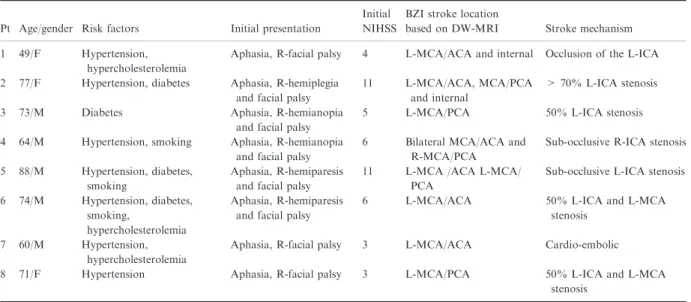
Table 1 General characteristics of our patients with aphasia because of BZ infarcts Pt Age/gender Risk factors Initial presentation
Initial NIHSS
BZI stroke location
based on DW-MRI Stroke mechanism 1 49/F Hypertension,
hypercholesterolemia
Aphasia, R-facial palsy 4 L-MCA/ACA and internal Occlusion of the L-ICA 2 77/F Hypertension, diabetes Aphasia, R-hemiplegia
and facial palsy
11 L-MCA/ACA, MCA/PCA and internal
> 70% L-ICA stenosis 3 73/M Diabetes Aphasia, R-hemianopia
and facial palsy
5 L-MCA/PCA 50% L-ICA stenosis 4 64/M Hypertension, smoking Aphasia, R-hemianopia
and facial palsy
6 Bilateral MCA/ACA and R-MCA/PCA
Sub-occlusive R-ICA stenosis 5 88/M Hypertension, diabetes,
smoking
Aphasia, R-hemiparesis and facial palsy
11 L-MCA /ACA L-MCA/ PCA
Sub-occlusive L-ICA stenosis 6 74/M Hypertension, diabetes,
smoking,
hypercholesterolemia
Aphasia, R-hemiparesis and facial palsy
6 L-MCA/ACA 50% L-ICA and L-MCA stenosis
7 60/M Hypertension, hypercholesterolemia
Aphasia, R-facial palsy 3 L-MCA/ACA Cardio-embolic 8 71/F Hypertension Aphasia, R-facial palsy 3 L-MCA/PCA 50% L-ICA and L-MCA
stenosis
BZI, border-zone infarcts; Pt, patient number; F, female; M, male; L, left; R, right; MCA, ACA and PCA, respectively middle, anterior cerebral and posterior cerebral artery; ICA, internal carotid artery.
1398 C. Flamand-Roze et al.
! 2011 The Author(s) European Journal of Neurology! 2011 EFNS European Journal of Neurology 18, 1397–1401
9
from stroke unit, and (iii) at long term, i.e. between 6and 18 months after stroke. The first two language evaluations consisted in five oral subtests: three for examination of verbal production (naming pictures, word and sentence repetition, and automatic language) and two in order to evaluate the comprehension level (auditory comprehension and comprehension of simple, semi-complex, and complex orders). Between these two evaluations, patients had standard language rehabili-tation. Between 6 and 18 months after stroke, the patient had a comprehensive language evaluation using the Boston Diagnostic Aphasia Evaluation (BDAE) scale (evaluating oral comprehension, oral agility, rep-etition, naming, oral reading, reading comprehension and writing, with 28 subtests) [14].
Results
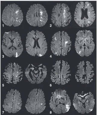
Amongst 26 individuals with hemispheric BZI, eight patients satisfied our inclusion and exclusion criteria. The 18 others patients were excluded because (i) the BZI affected the non-dominant hemisphere (right BZI in right-handed patient: n = 11), (ii) the BZI was left but in ambidextrous (n = 1) or in left-handed patient (n = 1), (iii) because the patient was not French native speaker (n = 4) or (iv) because of associated acute encephalopathy (by alcohol abuse; n = 1). Their baseline characteristics are shown in Table 1. Initial cerebral MRIs are shown in Fig. 1.
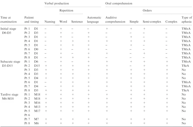
Within 48 h after admission, all patients presented with a transcortical mixed aphasia, i.e. altered com-prehension and naming difficulties including parapha-sias, while repetition was preserved. Detailed results of language examination are shown in Table 2. None presented swallowing dysfunction.
During first days, aphasia rapidly evolved. At dis-charge from stroke unit, between day 3 and 15, three patients had totally recovered (patients 3, 4, and 5). In remaining patients, aphasia changed from transcortical mixed (TMxA) toward transcortical motor (TMoA) or sensory (TSeA) depending on stroke location (respec-tively, in anterior and posterior BZI). Indeed, three patients, all with anterior BZI, had transcortical motor aphasia (paraphasia, reduced speech with short sen-tences, associated with preserved comprehension and repetition) (patients 1, 6, and 7), whereas two had transcortical sensory aphasia (altered lexical and syn-tactic comprehension, associated with preserved nam-ing and repetition) (patient 2 with both posterior and anterior BZI and patient 8 with an isolated posterior BZI).
Long-term language evaluation was performed between 6 and 18 months in seven patients (patient 6 was lost to follow-up). Amongst these seven patients,
none kept residual aphasia. To note, despite no aphasia, they had features of mild executive/attention dysfunc-tions: low verbal fluency, attention deficit (essentially highlighted by the BDAE!s subtest named "logic and reasoning!). and/or alteration of working memory (verbal span) in 5/7, and/or hemianopsia [disclosed in picture description (n = 2/7)].
Discussion
Our study indicates evidence that aphasia associated with hemispheric BZI is initially of transcortical mixed type (TMxA) and later changes to TMoA or TSeA. This is in contrast with previous reports in which most BZI patients had transcortical sensory aphasia (TSeA) or transcortical motor aphasia (TMoA) [8,9]. This dis-crepancy might be because of the fact that our patients were all evaluated in the first 48 h. In previous series, the delay of language examination was not reported, and it may be that initial TMxA may have been over-looked because of a later evaluation. There are only two
1 2
3 4
5 6
7 8
Figure 1 Cerebral diffusion-weighted MRI of the border-zone infarctions (BZI) associated with aphasia. Left BZI observed in patient 1 (MCA/ACA and internal); patient 2 (MCA/ACA, MCA/PCA and internal); patient 3 (MCA/PCA); patient 4 (bilateral MCA/ACA and right MCA/PCA); patient 5 (MCA/ ACA and MCA/PCA); patient 6 (MCA/ACA); patient 7 (MCA/ ACA) and patient 8 (MCA/PCA BZ infarction). MCA, middle cerebral artery; ACA, anterior cerebral artery; PCA, posterior cerebral artery.
reports on a total of four patients with TMxA associ-ated with left hemispheric BZI [10,11]. Interestingly, preservation of the repetition is a common feature of all types of transcortical aphasia and could thus be a good clinical clue to suspect a left BZI even before MR imaging. Normal repetition could reflect the integrity of the arcuate fasciculus, the pre motor cortex and the temporal lobe, as lesions of these structures are usually associated with a repetition disorder as observed in conduction, Broca or Wernicke aphasia [15,16]. These findings appear useful in practice to suspect BZI because hemodynamic factors are often implicated in these infarctions and may justify specific urgent man-agement such as surgery in tight internal carotid stenosis [with carotid endarteriectomy performed in 11 of our 26 patients with BZI (42%; data not shown)]. Inversely, to demonstrate that transcortical aphasia is predictive of BZI infarct, further studies are needed that will compare aphasic stroke patient with and without preserved repetition with respect to stroke location.
While all our patients with left BZI presented initially with a transcortical mixed aphasia, the subsequent pattern of aphasia depends on stroke location (TMoA in anterior BZI, and TSeA in posterior BZI,
respec-tively). These short-term modifications of the aphasic pattern may be because of a transient initial hypoper-fusion of apparently preserved territories (penumbra) and/or to an early reorganization of the neuronal net-works [12,17,18]. Indeed, TMxA, called by Geschwind !syndrome of isolation (of the speech area)" [19] because the perisylvian speech areas appear to be disconnected from the dominant hemisphere, can evolve toward TMoA or TSeA by re-connecting the supplementary motor area to Broca"s area in one hand and parietal to occipital areas on the other hand [11]. Finally, we cannot exclude that long-term presence of hemody-namic compromise in our patients with carotid stenosis might also have induced some degree of cerebral reor-ganization prior to the stroke that may have favoured rapid recovery following stroke. While anatomical basis of transcortical aphasia remains unclear, its specific initial pattern in BZ infarcts which rapidly improved with good long-term prognosis underlines the need for additional complementary explorations. In such patients, functional MR imaging, including perfusion study and fiber tracking, with parallel language testing at acute phase and during language recovery could be informative [20]. Several explanations for impaired
Table 2 Initial, short-, and long-term language examinations
Time at examination
Patient and timing
Verbal production Oral comprehension
Naming Repetition Automatic language Auditive comprehension Orders
Word Sentence Simple Semi-complex Complex Type of aphasia Initial stage D0-D3 Pt 1 D1 ) + ) ) + + + ) TMxA Pt 2 D3 ) + ) + ) ) ) ) TMxA Pt 3 D1 ) + ) + ) + ) ) TMxA Pt 4 D1 ) + + + + + + ) TMxA Pt 5 D1 ) + + + ) + ) ) TMxA Pt 6 D0 ) + ) + ) ) ) ) TMxA Pt 7 D1 ) + ) + + ) + ) TMxA Pt 8 D1 ) + + + ) ) ) ) TMxA Subacute stage D3-D15 Pt 1 D6 ) + ) + + + + + TMoA Pt 2 D15 + + + + + ) + + TSeA Pt 3 D3 + + + + + + + + No Pt 4 D3 + + + + + + + + No Pt 5 D4 + + + + + + + + No Pt 6 D1 ) + + + + + + + TMoA Pt 7 D4 ) + + + + + + + TMoA Pt 8 D3 + + + + ) + + + TSeA Tardive stage M6-M18 Pt 1 M18 + + + + + + + + No Pt 2 M18 + + + + + + + + No Pt 3 M16 + + + + + + + + No Pt 4 M13 + + + + + + + + No Pt 5 M17 + + + + + + + + No Pt 6 Pt 7 M7 + + + + + + + + No Pt 8 M6 + + + + + + + + No
Pt no, patient number; D, day after stroke; M, month; TMxA, Transcortical Mixed Aphasia; TMoA, Transcortical Motor Aphasia; TSeA, Transcortical Sensory Aphasia; No, No aphasia. Patient 6: No long-term evaluation (lost to follow-up).
1400 C. Flamand-Roze et al.
! 2011 The Author(s) European Journal of Neurology! 2011 EFNS European Journal of Neurology 18, 1397–1401
language and evolution in patients with BZI can be proposed including (i) loss of function, (ii) focal and transitory hypoperfusion (prolonged penumbra sur-rounding ischaemia), (iii) disruption of the language network owing to the infarction, resulting in a dys-function in remote areas in terms of diaschisis and (iv) preserved repetition in BZI related to language reor-ganization involving the contralateral language area [21]. Knowing that all these hypothesis should be studied in functional MR imaging, further studies are needed in order to demonstrate activation, or up- or down-regulation of the left or the right homologue language network regions [20].
Our data, although based on a small number of patients, indicate that the long-term outcome of aphasia associated with BZI is excellent. All patients made a full language recovery within the 18 months following stroke. This aphasic pattern we described in patients with BZI, i.e. initial TMxA followed in a few days by TMoA or TSeA, with excellent long-term recovery, needs to be confirmed in larger series to evaluate its specificity, its usefulness in clinical practice as well as for speech therapy.
Acknowledgements
We thank Pr M-G. Bousser for helpful discussions and excellent critical reading of this manuscript.
Disclosure of Conflict of Interest
The authors declare no financial or other conflict of interests.
References
1. Godefroy O, Dubois C, Debachy B, et al. Vascular aphasias: main characteristics of patients hospitalized in acute stroke units. Stroke 2002; 33: 702–705.
2. Inatomi Y, Yonehara T, Omiya S, et al. Aphasia during the acute phase in ischemic stroke. Cerebrovasc Dis 2008; 25: 316–323.
3. Pedersen PM, Vinter K, Olsen TS. Aphasia after stroke: type, severity and prognosis. The Copenhagen aphasia study. Cerebrovasc Dis 2004; 17: 35–43.
4. Kertesz A, McCabe P. Recovery patterns and prognosis in aphasia. Brain 1977; 100: 1–18.
5. Lendrem W, Lincoln NB. Spontaneous recovery of lan-guage in patients with aphasia between 4 and 34 weeks after stroke. J Neurol Neurosurg Psychiatry 1985; 48: 743– 748.
6. Demeurisse G, Demol O, Derouck M, et al. Quantitative study of the rate of recovery from aphasia due to ischemic stroke. Stroke 1980; 11: 455–458.
7. Jo¨rgensen L, Torvik A. Ischaemic cerebrovascular dis-eases in an autopsy series. 2. Prevalence, location, path-ogenesis, and clinical course of cerebral infarcts. J Neurol Sci 1969; 9: 285–320.
8. Bogousslavsky J, Regli F. Unilateral watershed cerebral infarcts. Neurology 1986; 36: 373–377.
9. Evrard S, Woimant F, Le Coz P, et al. Watershed cerebral infarcts: retrospective study of 24 cases. Neurol Res 1992; 14: 97–99.
10. Bogousslavsky J, Regli F, Assal G. Isolation of speech area from focal brain ischemia. Stroke 1985; 16: 441–443. 11. Bogousslavsky J, Regli F, Assal G. Acute transcortical mixed aphasia. A carotid occlusion syndrome with pial and watershed infarcts. Brain 1988; 111: 631–641. 12. Chaves CJ, Silver B, Schlaug G, et al. Diffusion- and
perfusion-weighted mri patterns in borderzone infarcts. Stroke 2000; 31: 1090–1096.
13. Damasio H. A computed tomographic guide to the identification of cerebral vascular territories. Arch Neurol 1983; 40: 138–142.
14. Goodglass H, Kaplan E. Assessment of Aphasia and Related Disorders. Philadelphia, USA: Lea and Fabiger, 1983.
15. Kertesz A, Sheppard A, MacKenzie R. Localisation in transcortical sensory aphasia. Arch Neurol 1982; 39: 475– 478.
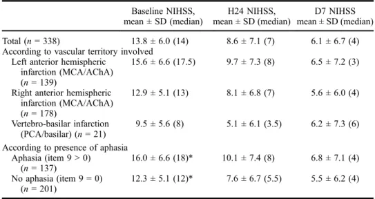
16. Bernal B, Ardila A. The role of the arcuate fasciculus in conduction aphasia. Brain 2009; 132: 2309–2316. 17. Hillis AE, Barker PB. MR Perfusion imaging reveals
regions of hypoperfusion associated with aphasia and neglect. Neurology 2000; 55: 782–788.
18. Reineck LA, Agarwal S, Hillis AE. Diffusion-clinical mismatch is associated with potential for recovery of aphasia. Neurology 2005; 64: 828–833.
19. Geschwind N, Quadfasel FA, Segarra JM. Isolation of speech area. Neuropsychologia 1968; 6: 327–340. 20. Saur D, Lange R, Baumgaertner A, et al. Dynamics of
language reorganization after stroke. Brain 2006; 129: 1371–1384.
21. Bando M, Ugawa Y, Sugishita M. Mechanism of repeti-tion in transcortical sensory aphasia. J Neurol Neurosurg Psychiatry 1986; 49: 200–202.
Exploitation, valorisation et diffusion des résultats
:
Etude publiée dans European Journal of Neurology, (Impact Factor : 3.9).
Ce travail a été présenté dans plusieurs congrès nationaux et internationaux de neurologie :
Initial presentation and course of aphasia due to watershed cerebral infarct. C.
Flamand-Rouviere, C. Cauquil
‐Michon, R. Souillard-Scemama, C. Denier, D. Adams. (Communication
affichée; European Federation of Neurological Society ; Florence 2009)
Initial presentation and course of aphasia due to watershed cerebral infarct. C.
Flamand-Rouviere, C. Cauquil
‐Michon, R. Souillard‐Scemama, C. Denier, D. Adams. (Communication
affichée ; European Neurological Society ; Milan 2009)
Présentation initiale et évolution des aphasies liées à un infarctus jonctionnel. C.
Flamand-Rouviere, C. Cauquil-Michon, R. Souillard
‐Scemama, C. Denier, D. Adams. (Communication
affichée ; Journées de Neurologie de Langue Française; Lille 2009)
Présentation initiale et évolution des aphasies liées à un infarctus jonctionnel. C.
Flamand-Rouviere, C. Cauquil
‐Michon, R. Souillard‐Scemama, C. Denier, D. Adams. (Communication
L’incidence de l’aphasie après un accident vasculaire cérébral (AVC) diffère en fonction
des étiologies, mais est globalement retrouvée dans 15 à 55% des cas (7,8,9,10). Sa fréquence est
probablement plus élevée à la phase très précoce, c’est à dire dans les premiers jours qui suivent
l’AVC. Les facteurs favorisants l’existence de troubles du langage lors d’AVC sont l’antécédent
d’AVC, l’âge, le sexe féminin, et l’origine cardio-embolique (en particulier la fibrillation atriale)
(11).
L’évolution des troubles phasiques peut être rapide : dans les 10 jours suivant l’AVC, on
considère que 46% des aphasies auront notablement régressé et 21% auront spontanément
disparu. Toutefois, les troubles du langage persistent chez environ la moitié des patients
aphasiques 18 mois après l’AVC (10,13,14). Cela constitue une altération majeure de la qualité
de vie, à l’origine de syndromes dépressifs fréquents. La persistance d’une aphasie s’associe à
une moins bonne participation à toute forme de rééducation (kinésithérapie, ergothérapie...) par
difficulté de communication, à un retrait social, une moindre probabilité de reprendre une vie
professionnelle (12,16,17). Si il est convenu que l’aphasie résultant d’une hémorragie aura un
meilleur pronostic que l’aphasie après une ischémie, il est difficile de prédire des évolutions au
cas par cas. Actuellement, seul le score initial de sévérité de l’AVC (au NIHSS) semble prédictif
: l’amélioration serait proportionnelle au déficit initial
avec, comme pour la récupération
motrice, une amélioration évaluable à 70 % environ du déficit initial à 3mois. (7,10).
Dans le cadre d’un AVC ischémique, le traitement le plus efficace, si il est administré
dans les 4h30 suivant l’apparition des premiers signes, est le traitement par thrombolyse intra
veineuse. Il comporte toutefois des risques de transformation hémorragique, il faut donc évaluer
au plus juste les bénéfices qu’il apporterait, compte tenu de ce risque. Effectuer le bilan des
lésions et de leur retentissement est primordial : cela permet d’envisager au mieux et au plus vite
la prise en charge thérapeutique active médicamenteuse comme la rééducation. Pour cela, les
neurologues disposent d’échelles validées et utilisées dans le monde entier. Le NIHSS (18) est
une échelle globale qui donne un score avec un seuil en dessous duquel la balance bénéfice/risque
n’est pas en faveur de l’utilisation du traitement. Ainsi, lors de l’arrivée en urgence d’un patient
potentiellement candidat à une thrombolyse (« alerte thrombolyse »), le langage, comme les
autres fonctions motrices et cognitives, doit être évalué de façon précise et reproductible, afin de
mesurer au mieux la gravité de l’AVC. De même, détecter rapidement l’aphasie permet la mise
en place d’une prise en charge précoce, et donc plus efficace en conjuguant la thérapie
orthophonique intensive à la réorganisation neuronale post AVC de manière optimale. Dans le
score du NIHSS, le langage est sous représenté, et son évaluation consiste en quelques questions
sommaires, l’altération reposant alors sur l’impression clinique du médecin. Il est donc fréquent
qu’un patient présentant un trouble du langage isolé ou associé à un trouble moteur léger ne
bénéficie pas de la thrombolyse, le score à l’échelle NIHSS étant un critère d’inclusion ou
d’exclusion : le score du NIHSS serait trop faible, le handicap trop léger, pour prendre le risque
d’ une transformation hémorragique.
Jusqu’à récemment, les tests validés pour évaluer les troubles du langage de façon fiable,
qualitative et quantitative, étaient soit longs et fastidieux et donc inutilisables en phase aiguë des
AVC, soit au contraire rapides, mais trop grossiers pour être pertinents et reproductibles. Les tests
de langage de référence, administrés par les orthophonistes, tels le Boston Diagnosis Aphasia
Examination (BDAE), la Western Battery for Aphasia ou le Montréal Toulouse 86 sont robustes
et complets, mais inadaptés en phase aiguë des AVC (19,20,21). En effet, le temps de passation
de ces échelles de langage est de 30 minutes à deux heures de sorte qu’elles ne peuvent pas être
proposées en phase aiguë. En effet, l’alerte thrombolyse est une situation d’urgence. , Il faut
également prendre en compte la fatigabilité et d’éventuels troubles de la vigilance du patient lors
de cette phase aiguë. De plus, les modifications rapides et les fluctuations du langage dans ce
contexte rendent difficile l’évaluation qui risque, en fonction de l’outil utilisé, d’être biaisée par
l’existence de troubles moteurs, praxiques, neuro-visuels, exécutifs ou de l’attention associée.
Les échelles plus générales conçues pour la phase aiguë de l’AVC, telles que le National Institute
of Health Stroke Score (NIHSS), comprennent des items langage, mais ne sont pas assez précises
pour appréhender efficacement l’aphasie qualitativement et quantitativement de façon
reproductible d’un médecin à l’autre.
S’il existe des échelles d’évaluation de l’aphasie destinées à la phase aiguë, peu sont
traduites en français ni même validées, et elles comportent souvent des items qui ne sont pas
adaptés à cette situation (items de langage écrit, non-utilisables chez des patients avec hémiplégie
ou chez les patients illettrés, utilisation de matériel visuel complexe qui biaisent les résultats des
patients présentant une héminégligence ou une hémianopsie, temps d’administration trop long)
(22).
Pourtant, le diagnostic précis et précoce des troubles du langage est nécessaire pour
affiner la connaissance du déficit du patient dés son arrivée, pour distinguer l’aphasie d’autres
atteintes cognitives ou fonctionnelles, et pour pouvoir mettre en place rapidement une
rééducation adaptée. Il peut également permettre d’identifier plus précisément des phénotypes
précoces spécifiques comme nous avons pu le montrer dans le cas particulier des aphasies liées
aux infarctus jonctionnels (23).
Dans le cadre des AVC, chaque minute compte, et correspond à la destruction de 2
millions de neurones et de 14 milliards de synapses (24). Il manquait donc un outil fiable, rapide
d’administration et validé pour évaluer l’aphasie dans ce contexte.
Fiche de recherche :
Rôle : investigateur principal. Conceptualisation, analyse, Rédaction du protocole pour
obtention de l’accord du comité d’éthique de Bicêtre, administration du protocole,
rédaction et soumission de l’article.
Equipe de recherche : Dr C. Denier (INSERM U788), Dr E. Roze (INSERM U952, CNRS
UMR 7224 UPMC Paris 6), Pr B. Falissard (INSERM U669)
Nombre de patients : 450
Type de recherche : élaboration et validation d’échelle.
Site : CHU Bicêtre Service de neurologie adulte et USINV
Elaboration et conceptualisation de l’outil :
Il était indispensable que l’échelle soit :
-‐ rapide à administrer, et donc comporter peu d’items. Les épreuves écrites ont été
éliminées, car elles allongeaient le temps d’évaluation, n’étaient pas administrables aux
patients hémiplégiques ou non lettrés, et n’avaient pas un intérêt essentiel (les troubles
isolés du langage écrit sont extrêmement rares)
-‐ fiable : les items devaient être les plus pertinents possibles et être choisis en fonction de
leurs fréquences lexicales et visuelles dans la langue française, ainsi qu’en fonction de
leurs structures phonétiques.
-‐ composée uniquement d’items essentiels au diagnostic : dénomination, répétition, série
automatique pour la partie « expression », désignation, exécution d’ordres pour la partie «
compréhension ».
-‐ reproductible quelque soit l’examinateur
-‐ exempte d’effet re-test en cas de passation pré et post thrombolyse grâce à l’élaboration
de deux versions strictement équivalentes
Description de l’outil :
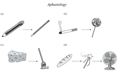
LAST se compose :
• D’une épreuve de dénomination d’images, choisies pour leur fréquence sémantique et
visuelle ainsi que pour leur niveau de difficulté phonémique;
• D’une épreuve de répétition (un mot et une phrase concrète) ;
• D’une série automatique (le comptage, peu soumis aux connaissances académiques) ;
• D’une épreuve de désignation d’images (quatre images parmi quatre pièges : sémantique,
visuel, phonétique proche, phonétique lointain) choisies elles aussi pour leur fréquence
visuelle et sémantique ;
• D’une épreuve d’exécution de trois ordres (simple, semi- complexe et complexe).
Au total, le patient obtient un score sur 15. LAST doit pouvoir être administré aussi bien
par un orthophoniste que par un non-spécialiste du langage (médecin, étudiant, infirmière, autre
rééducateur), avec la même fiabilité. Cette évaluation doit pouvoir se faire au lit du malade, dès
son arrivée, et participer au processus décisionnel thérapeutique (thrombolyse). Enfin, nous avons
créé deux versions de cette échelle, LAST-A et LAST-B, qui devront être strictement
équivalentes afin de pouvoir être administrées alternativement à différentes étapes de l’évolution
du patient pour permettre une évaluation quantitative de l’évolution.
Validation de l’outil :
Nous avons d’abord présenté l’échelle à 50 témoins, tous âges, sexes et niveau socio-culturels
confondus, afin de s’assurer qu’aucun item ne posait de difficulté ou ne présentait d’ambiguïté.
Puis nous avons testé :
• la validation interne de LAST et sa validité inter‐examinateur sur 300 patients consécutifs
en phase aigüe d’un AVC.
• sa validité externe et l’équivalence des versions A et B sur 150 patients présentant une
aphasie chronique (afin d’administrer également le « gold standard » BDAE pour
comparer les deux résultats, ce qui n’est pas envisageable sur des patients en phase aigüe
en raison de leur fatigabilité et de la longueur de passation de ces tests).
• le temps de passation moyen sur 50 patients consécutifs en phase aigüe d’un AVC.
Résultats et répercussions
:
La validation interne a montré qu’aucun item ne montrait d’effet plafond ou plancher, ni
de redondance. La validation externe, par rapport au BDAE, la batterie d’évaluation du langage
classiquement utilisée en phase de chronicité, nous a permis d’établir un cut-off à 14 : un score
inférieur à 15 doit alors justifier la passation d’autres évaluations plus poussées, tant sur le plan
arthrique que phasique, et permettre ainsi la prise en charge en rééducation précoce si cela
s’avère nécessaire. Les deux versions LAST-A et LAST-B sont strictement équivalentes. Le
temps de passation de LAST est en moyenne de deux minutes. La validité inter-juge montre que
les résultats sont fiables quelque soit l’examinateur.
LAST (Language Screening test) est le premier test screening publié et validé en français.
Il permet de dépister un trouble phasique en phase aigue d’un AVC, une amélioration de la
connaissance du tableau clinique du patient, une meilleure analyse du déficit, afin que la présence
d’une aphasie soit prise en compte dans le processus décisionnel de thrombolyse. Depuis sa
publication, LAST est de plus en plus présente dans les Unités de soins intensifs
neurovasculaires, et son utilisation s’étend à d’autres services (voir chapitre 3). Au-delà de son
intérêt pour la pratique clinique quotidienne, Les caractéristiques de LAST et la qualité de sa
validation en font potentiellement un outil de choix pour évaluer l’aphasie dans le cadre de
futures études interventionnelles à la phase aigue de l’accident vasculaire.
Audrey Chacon, Claire Join-Lambert, David Adams and Christian Denier
Constance Flamand-Roze, Bruno Falissard, Emmanuel Roze, Lisa Maintigneux, Jonathan Beziz,
Language Screening Test (LAST)
Print ISSN: 0039-2499. Online ISSN: 1524-4628
Copyright © 2011 American Heart Association, Inc. All rights reserved.
is published by the American Heart Association, 7272 Greenville Avenue, Dallas, TX 75231
Stroke
doi: 10.1161/STROKEAHA.110.609503
2011;42:1224-1229; originally published online April 12, 2011;
Stroke.
http://stroke.ahajournals.org/content/42/5/1224
World Wide Web at:
The online version of this article, along with updated information and services, is located on the
http://stroke.ahajournals.org/content/suppl/2011/04/12/STROKEAHA.110.609503.DC1.html
Data Supplement (unedited) at:
http://stroke.ahajournals.org//subscriptions/ Information about subscribing to Stroke is online at:
Subscriptions:
http://www.lww.com/reprints Information about reprints can be found online at:
Reprints:
Permissions and Rights Question and Answer document.
process is available in the
Request Permissions in the middle column of the Web page under Services. Further information about this Once the online version of the published article for which permission is being requested is located, click Stroke can be obtained via RightsLink, a service of the Copyright Clearance Center, not the Editorial Office. in Requests for permissions to reproduce figures, tables, or portions of articles originally published Permissions:
by guest on June 21, 2013
http://stroke.ahajournals.org/
Validation of a New Language Screening Tool for Patients
With Acute Stroke
The Language Screening Test (LAST)
Constance Flamand-Roze, ST; Bruno Falissard, MD, PhD; Emmanuel Roze, MD, PhD; Lisa Maintigneux, ST; Jonathan Beziz, ST; Audrey Chacon, ST; Claire Join-Lambert, MD;
David Adams, MD, PhD; Christian Denier, MD, PhD
Background and Purpose—Standard aphasia scales such as the Boston Diagnosis Aphasia Evaluation are inappropriate
for use in acute stroke. Likewise, global stroke scales do not reliably detect aphasia, and existing brief aphasia screening scales suitable for patients with stroke have several limitations. The objective of this study was to generate and validate a bedside language screening tool, the Language Screening Test, suitable for use in the emergency setting.
Methods—The Language Screening Test comprises 5 subtests and a total of 15 items. To avoid retest bias, we created 2
parallel versions of the scale. We report the equivalence of the 2 versions, their internal and external validity, and their interrater reliability. We validated the scale by administering it to 300 consecutive patients within 24 hours after admission to our stroke unit and to 104 stabilized patients with and without aphasia using the Boston Diagnosis Aphasia Evaluation as a reference.
Results—The 2 versions of the Language Screening Test were equivalent with an intraclass correlation coefficient of 0.96.
Internal validity was good; none of the items showed a floor or ceiling effect with no redundancy and good internal consistency (Cronbach ! 0.88). External validation against the Boston Diagnosis Aphasia Evaluation showed a sensitivity of 0.98 and a specificity of 1. Interrater agreement was near perfect (intraclass correlation coefficient, 0.998). The median time to complete the Language Screening Test was approximately 2 minutes. Importantly, the Language Screening Test does not need to be administered by a speech and language therapist.
Conclusions—This comprehensively validated language rating scale is simple and rapid, making it a useful tool for bedside
evaluation of patients with acute stroke in routine clinical practice. (Stroke. 2011;42:1224-1229.)
Key Words: aphasia ! diagnostic tool ! rating scale ! stroke ! validation study
P
oststroke aphasia is a major source of disability, poten-tially leading to impaired communication, reduced social activity, depression, and a lower probability of resuming work.1– 4Despite some controversy, early detection of aphasia after stroke may improve rehabilitation by taking advantage of the synergy between intensive speech therapy and early neural reorganization.5–7Tools capable of detecting aphasia and evaluating its severity during the acute phase of stroke might help to improve early rehabilitation and to predict outcome.8Standard aphasia rating scales such as the Western Aphasia Battery, the Boston Diagnostic Aphasia Evaluation (BDAE), and the Boston Naming Test are not appropriate for use during the acute phase of stroke.7,9 –11In particular, these scales take too long to complete and must be administered byspeech and language therapists.9 –11 Global stroke rating scales such as the National Institutes of Health Stroke Scale and the Scandinavian Stroke Scale include language items and have been developed for use in acute settings,12–17but they do not reliably detect aphasia.8 Several attempts have been made to develop and validate brief aphasia screening scales suitable for patients with acute stroke,5,18 –25 but all have inherent structural limitations, including7(1) inclusion of written language subtests, the results of which are influ-enced by hemiplegia and illiteracy5,19 –23,25; (2) use of com-plex visual material inappropriate for patients with stroke with neurovisual deficits19,20; (3) inclusion of subtests the results of which are markedly influenced by attention/exec-utive dysfunction19,20; (4) excessively lengthy
administra-Received November 23, 2010; final revision received February 11, 2011; accepted February 14, 2011.
From the Assistance Publique–Hoˆpitaux de Paris (C.F.-R., L.M., J.B., A.C., C.J.-L., D.A., C.D.), CHU Biceˆtre, Service de Neurologie, Le Kremlin Biceˆtre, France; INSERM U669 (B.F.), Maison de Solenn, Paris, France; Assistance Publique–Hoˆpitaux de Paris (E.R.), CHU Pitie´-Salpeˆtrie`re, Service de Neurologie, Paris, France; INSERM (E.R.), UMRS 952, and CNRS, UMR 7224, Universite´ Pierre et Marie Curie-Paris-6, Paris, France; INSERM (E.R.), UMRS 975, and CNRS 7225–CRICM, CHU Pitie´-Salpeˆtrie`re, Universite´ Pierre et Marie Curie-Paris-6, Paris, France; Universite´ Paris Sud 11 (C.J.-L., D.A.), Paris, France; and INSERM (D.A., C.D.), U788, Le Kremlin Biceˆtre, France.
The online-only Data Supplement is available at http://stroke.ahajournals.org/cgi/content/full/STROKEAHA.110.609503/DC1.
Correspondence to Constance Flamand-Roze, ST, Service de Neurologie, Centre Hospitalo-Universitaire de Biceˆtre, 78 rue du ge´ne´ral Leclerc, 94275 Le Kremlin Biceˆtre cedex, France. E-mail constance.flamand-roze@bct.aphp.fr
© 2011 American Heart Association, Inc.
Stroke is available at http://stroke.ahajournals.org DOI: 10.1161/STROKEAHA.110.609503
1224 by guest on June 21, 2013
http://stroke.ahajournals.org/
tion22; (5) difficulties with administration or scoring5,18,23,25; and (6) IQ dependency.21Some of these scales also have poor sensitivity for the detection of language disorders and a paucity of information on their validity and reliability.5,20
We therefore developed a brief language screening scale, named the Language Screening Test (LAST), for the assess-ment of patients with acute stroke. LAST incorporates the following features: (1) no written material; (2) no complex visual material; (3) no evaluation of verbal executive func-tion; and (4) suitability for bedside administration by persons who are not speech and language therapists. We report the validity, reliability, sensitivity, and specificity of LAST.
Patients and Methods Scale Construction
LAST was developed as a formalized quantitative scale for screening language functions, including comprehension and expression. The initial, qualitative design phase focused on item generation and construction. We chose to exclude verbal fluency subtests, the results of which are strongly influenced by changes in attention/executive function, and also written language subtests that are unsuitable for hemiplegic and illiterate patients. We generated several preliminary versions of the scale, which were evaluated internally and then refined because of the following weaknesses: (1) too lengthy to administer (too many items); (2) in the naming task, inadequate use of real daily objects such as watches and pens, instead of images, which are less ambiguous (for example, the pen of 1 examiner is different from the pen of another one); and (3) in the picture recognition task, inadequate use of color pictures, which may provide semantic clues. We selected the items by consensus and eliminated any ambiguities by administering the scale to 50 healthy volunteers (data not shown).
The final version of LAST consists of 5 subtests and a total of 15 items (Figure 1). The patient has 5 seconds to answer each question, and the answer is scored as either 1 (perfect answer) or 0 (imperfect answer, including arthric errors, and failure to answer). The maxi-mum score is therefore 15. There are 2 subscores, namely an expression index (naming, repetition, and automatic speech; maxi-mum score 8 points) and a receptive index (picture recognition and verbal instructions; maximum score 7 points).
The test is administered on a simple sheet held in portrait orientation. The front side corresponds to the expression index with 5 pictures to be named facing the patient and the instructions facing the examiner. The other side corresponds to the receptive index with 8 pictures (4 to be indicated with a finger and 4 trap pictures) facing the patient and the instructions facing the examiner (see Supplemen-tal Data; http://stroke.ahajournals.org).
Each subtest is composed as follows (Figure 1):
(1) “Naming” subtest: naming of 5 black-and-white pictures specially drawn for the test. The pictures were selected for their everyday familiarity (subjective verbal frequency) and for the image evoking value of the noun.26 Standard
syn-onyms and abbreviations are accepted (alligator for crocodile, TV for television, etc). The maximum score is 5 points. (2) “Repetition” subtest: repetition of 1 concrete 4-syllable noun
and 1 8-word sentence containing 11 syllables and 3 conso-nantal groups. One self-correction is accepted. The maximum score is 2 points, 1 for the isolated word and 1 for the sentence.
(3) “Automatic speech” subtest: the patient counts from 1 to 10. No mistakes or omissions are accepted. The score is 1 or 0. (4) “Picture recognition” subtest: recognition of 4 black-and-white pictures drawn specially for the test and selected for their high image-evoking value and sorted by their subjective verbal frequency. This subtest includes 2 phonologic traps (close and distant), 1 semantic and 1 visual. The maximum score is 4.
(5) “Verbal instructions” subtest: execution of 3 verbal orders— simple, semicomplex, and complex—involving the use of part of the body or simple objects in the room. The patient is asked to precisely execute the verbal order. The maximum score is 3. Having developed a version of the scale that we considered suitable for validation (LAST-a), we then generated a second, parallel version (LAST-b). Each item on the 2 scales was different (except for the automatic speech item, see subsequently) but strictly matched to obtain 2 equivalent versions of the scale. For example, the pictures (naming subtest and recognition subtest) used in the 2 versions were each matched for their visual and verbal frequency, and the words and sentences used for the repetition subtest were matched for their consonantal content. Several series can be used to assess automatic speech, but counting is the most universally acquired (days of the week and the alphabet, for example, are more influenced by sociocultural status). Counting to 10 was thus used in both versions (see Supplemental Data).
Patients and Instruments
To validate the scale, we included both “acute” and “chronic” patients. We first prospectively enrolled consecutive “acute” pa-tients, that is, admitted with suspected acute stroke to our stroke unit during a 7-month period. They were tested within 24 hours after their admission. During the same period, we enrolled stabilized patients (hospitalized or ambulatory) seen in our neurology department, but not in the stroke unit, who were able to complete the entire BDAE comprehensive language evaluation. These “chronic” patients were considered aphasic or nonaphasic on the basis of their BDAE results. The BDAE is a standard scale widely used for comprehensive evaluation of aphasia. Its 28 subtests evaluate oral comprehension, oral agility, repetition, naming, oral reading, reading comprehension, and writing and take between 1.5 and 2 hours to administer.10Both
“acute” and “chronic” patients were excluded if they had any of the following characteristics: (1) history of dementia or of severe psychiatric disorders; (2) deafness or blindness; (3) nonnative French language; and (4) altered consciousness. The study was approved by the ethics committee of Pitie´-Salpeˆtrie`re Hospital, Paris. Demo-graphic data were collected for all the patients and the National Institutes of Health Stroke Scale score was recorded for the “acute” patients. A schematic representation of the study design is shown in Figure 2.
Validation of LAST
LAST was validated on the basis of (1) the equivalence of the 2 versions of the instrument; (2) the internal validity of the 2 versions of the instrument (item analysis, reliability, and factor structure); (3) external validity (comparison with the BDAE scale); and (4) inter-rater reliability.
Figure 1. Design of the Language Screening Test (LAST). LAST
comprises 5 subtests and a total of 15 items. Each item is scored 1 (perfect answer) or 0 (imperfect answer, including arthric errors, and failure to answer) after an interval of 5 sec-onds. The maximum score is 15.
by guest on June 21, 2013
http://stroke.ahajournals.org/
To test the equivalence of the 2 versions of LAST, the “chronic” aphasic patients were asked to complete LAST-a followed by LAST-b with a 1-minute rest period.
To assess the internal validity of the scale, consecutive “acute” patients completed either LAST-a or LAST-b within 24 hours after admission. The 2 versions were used in alternation for each new patient (only 1 version per patient).
To assess external validity, aphasic and nonaphasic “chronic” patients were asked to complete the BDAE language evaluation followed by either LAST-a or LAST-b on the same day and administered by 2 different and blinded examiners.
Interrater reliability was assessed in the “acute” patients. Four examiners pairs were used, consisting of a speech and language therapist with another speech and language therapist, a student, a nurse, or a neurologist. All the examiners received a 5-minute explanation on how to administer the test. Blinded assessment was ensured as follows. Two examiners were present at the bedside. The first examiner administered LAST to the patient (result used for internal validity), reading aloud 1 by 1 the different subtest, at the same time as the second examiner, who could hear the first examiner but could not see the results he or she recorded, simultaneously scored the same version.
The median time for scale completion was calculated for 50 new consecutive “acute” patients.
Statistical Analysis
The concordance of the 2 versions of LAST was assessed by calculating the intraclass correlation coefficient (ICC) from the 2 total scores (equivalent to a quadratic weighted !).
Internal validity was assessed in 3 steps. First, we closely inspected the score distribution for each item to detect a floor or
ceiling effect, and the Pearson correlation matrix was used to detect item redundancy. Second, the number of underlying dimensions was determined by parallel analysis, which consists of representing a traditional screeplot with simulations.27,28 Third, we calculated
Cronbach " coefficient, a measure of reliability based on internal correlation of the items on the scale.
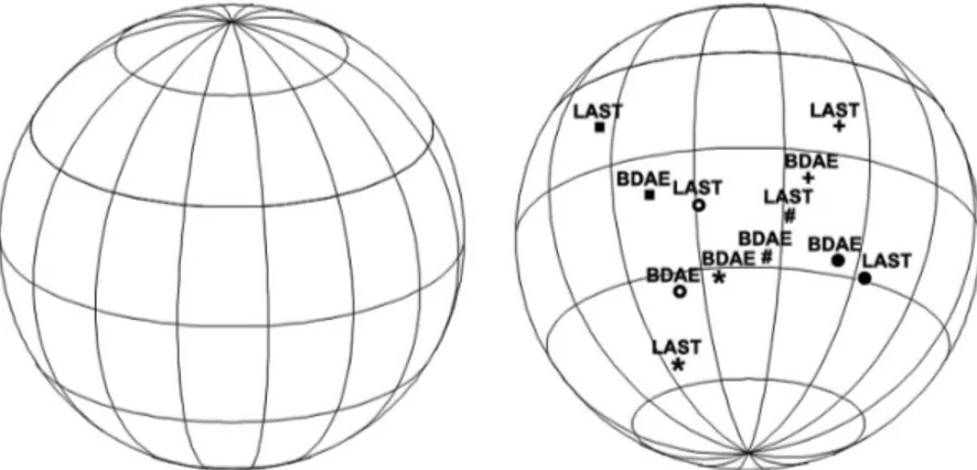
To evaluate external validity, sensitivity and specificity were assessed with respect to the BDAE. We represented the correlations between the LAST and BDAE subtests on a sphere (Figure 3)29and
with the receiver operating characteristic curve (Figure 4). The ICC was used to appreciate interrater reliability.
R 2.11.1 software and the “psy” library were used for all analyses.30
Results Sample Description
Three hundred forty consecutive unselected “acute” patients were admitted to our stroke unit for suspected acute stroke during a 7-month period. Thirty-six patients were excluded (nonnative French speakers [n!24], history of dementia or severe psychiatric disorders [n!6], deafness or blindness [n!3], altered consciousness [n!3]) and 4 could not be evaluated for logistic reasons. The remaining 300 “acute” patients were included in the internal validity and interrater reliability assessments (159 men and 141 women; mean age 62.6 years ["14.9]; mean National Institutes of Health Stroke Scale score 3.5 ["5.1]; Figure 2).
Figure 2. Schematic representation of
the Language Screening Test (LAST) val-idation process. Please note that the time taken to administer LAST was estimated using 50 new consecutive patients who are not represented on this figure.
Figure 3. Spherical representation of
the correlation matrix of the Language Screening Test (LAST) subtests and cor-responding Boston Diagnostic Aphasia Evaluation (BDAE) items. # Naming (LAST: 5 items/5 points; BDAE: 35 items/105 points); G word repetition (LAST: 1 item/1 point; BDAE: 20 items/10 points); f sentence repetition (LAST: 1 item/1 point; BDAE: 16 items/16 points); * automatic speech (LAST: 1 series/1 point; BDAE: 3 series/9 points); ● picture recognition (LAST: 4 items/4 points; BDAE: 36 items/72 points); # verbal instructions (LAST: 3 orders/3 points; BDAE: 5 orders/15 points).
1226 Stroke May 2011
by guest on June 21, 2013
http://stroke.ahajournals.org/