HAL Id: tel-00398008
https://tel.archives-ouvertes.fr/tel-00398008v2
Submitted on 13 Jul 2009
HAL is a multi-disciplinary open access
archive for the deposit and dissemination of sci-entific research documents, whether they are pub-lished or not. The documents may come from teaching and research institutions in France or abroad, or from public or private research centers.
L’archive ouverte pluridisciplinaire HAL, est destinée au dépôt et à la diffusion de documents scientifiques de niveau recherche, publiés ou non, émanant des établissements d’enseignement et de recherche français ou étrangers, des laboratoires publics ou privés.
Survivin and Aurora B kinase, two targets in the search
for anti - mitotic drugs; identification of a new class of
Aurora kinase inhibitors.
Thi My-Nhung Hoang
To cite this version:
Thi My-Nhung Hoang. Survivin and Aurora B kinase, two targets in the search for anti - mitotic drugs; identification of a new class of Aurora kinase inhibitors.. Life Sciences [q-bio]. Université Joseph-Fourier - Grenoble I, 2008. English. �tel-00398008v2�
Université Joseph Fourier-Grenoble 1
Chimie et Science du Vivant(Arrêtés ministériels du 5 juillet 1984 et du 30 mars 1992)
THÈSE
pour obtenir le titre de
Docteur de l’Université Joseph Fourier Discipline Biologie
Présentée et soutenue publiquement par
HOANG Thi My Nhung
Le 31 Janvier 2008
SURVIVIN AND AURORA B KINASE, TWO
TARGETS IN THE SEARCH FOR ANTI-MITOTIC
DRUGS; IDENTIFICATION OF A NEW CLASS
OF AURORA KINASE INHIBITORS.
COMPOSITION DU JURY
Véronique Baldin Rapportrice Claude Prigent Raporteur Dinh Duy Khang Examinateur Marylin Vantard Présidente Stéfan Dimitrov Examinateur
Tran Cong Yen Directeur de thèse (VNU) Annie Molla Directrice de thèse (UJF)
Thèse préparée au sein du Centre de Recherche INSERM Mécanismes d’assemblage et de régulation de l’appareil génétique
Acknowledgements
First of all, I would like to express my deepest gratitude to Dr. Annie Molla for her
supervision, patient guidance, and encouragement from the beginning of my research in
France as well as giving me excellent advice throughout this study. I also want to thank
her for assistances in preparing and writing this thesis.
I would like to warmly thank to my thesis co-supervisor, Dr. Tran Cong Yen, for his
invaluable support and enthusiasm to my graduate experience. I could never have
embarked and started all of this without his prior teachings in biology and thus opened
up unknown areas to me.
My special thanks to Dr. Stefan Dimitrov for giving me the opportunity to work on his
equipment and for his assistances, suggestions in my research.
I gratefully thank Dr. Véronique Baldin and Dr. Claude Prigent for reviewing my thesis
and for their constructive comments on this manuscript.
I would like to thank Dr. Marylin Vantard and Dr. Dinh Duy Khang for accepting to
be members of the committee.
My stay and my PhD in France is financial supported by a Scholarship from Sandwich
Programme, a project initiated and managed from Ministry of Education and Training
of Vietnam and partly from French. I also thank to ARC organization which supported
for the last year of my thesis.
It was a pleasure for me to work with all wonderful people in our lab here in IAB. I
would like to give many thanks to the fellow graduated students before me, Gaelh,
Marlene, Flore, and Fabienne Sirot, for sharing experiences and knowledge during the
time they were here. My thanks are given to another Fabienne for “science” discussions
and the pleasure working together in the lab. I would also acknowledgment Véro for
helping me in protein extraction and purification technique, thank for her technical
assistance. I am grateful to my colleagues: Andre, Thierry, Eric, Sophie, Fang, Jean,
Amina…
I would like to thank to the members of INSERM U823 for their support and their
comradeship. Thank Aude for her indispensable help dealing with administration and
bureaucratic matter during my stay in IAB.
I am very grateful to members of platform, Alexei and Jack, whose expertise,
understanding and patience in microscope are very helpful for my work.
I also want to thank Catherine Souchier for helps in using confocor microscope.
A great thank to my department, Faculty of Biology, Hanoi University of Science,
Hanoi National University (VNU) for supporting and creating good conditions for me
to do my PhD.
To my best French friend, Cedric Badowski, I gratefully acknowledge him for his
kindness and useful advices during the lifetime I spent in France. He provides me
unflinching encouragement and support in various ways. I have much benefited by
advice and guidance from him who always kindly grants me his time even for answering
some of my unintelligent question about microscope and molecular technique…. I am
much indebted to him!
My years as a PhD student would not have been as much fun without my friends:
Khoat, Nam, Dung, Hung, Vi Thuy, Thai, Chi, Lien…with whom I shared the
unforgettable moments. Thanks!
It is pleasure to express my gratitude wholeheartedly to Annie’s family for their
kindness hospitality during my stay in Grenoble. Thank Thierry for his assistance on
editing my thesis writing.
Finally, I take this opportunity to express my profound gratitude to my grand family:
my beloved parent, parent in law, my brothers, sisters for their inseparable support and
patience during my study in France.
Especially, I wish to express all my warmest thanks and deepest appreciations to my
dear husband and my little daughter for their love and sacrifices for me. Without my
two loves, I would not finish my thesis!!!
Abstract
The chromosomal passenger complex (CPC) plays a key role in mitosis : controlling both chromosome segregation, spindle tension, anaphase onset and cytokinesis. The complex is composed of four proteins: INCENP, Aurora B kinase, Survivin and Borealin. Taking into account that Survivin is phosphorylated by Aurora B and has a pivotal role in the complex, we have studied the phosphomimetic mutant SurvivinT117E. Survivin phosphorylation is required for anaphase onset and the phospho-mutant is poorly linked to centromere. Moreover it exhibits a dominant negative function in cytokinesis, preventing abscission.
In a search for Aurora kinase inhibitors we have identified a new class of Aurora B kinase inhibitors that prevents Histone H3 phosphorylation, impairs mitotic spindle checkpoint. Moreover these molecules prevent tumor cell proliferation. These inhibitors are interesting tools for understanding CPC function and represent a new lead for the development of anti-cancer drugs.
Survivin and Aurora B kinase, which are expressed exclusively in mitosis, are thus two druggable targets for new anti-mitotic therapies.
Key words: Mitosis, chromosomal passenger complex, Aurora kinase, Survivin, mitotic checkpoint.
Résumé
Le complexe passager joue un rôle clé en mitose: contrôlant à la fois la ségrégation des chromosomes, la tension du fuseau, l’entrée en anaphase et la cytodirèse. Le complexe est composé de quatre protéines: INCENP, la kinase Aurora B, Survivine et Boréaline. Sachant que la protéine Survivine est phosphorylée par Aurora B et qu’elle a un role pivot au sein du complexe, nous avons étudié un mutant mimant sa phosphorylation: Survivine T117E. La phosphorylation de Survivine est nécessaire à la transition Métaphase/ Anaphase. Le mutant Survivine T117E est faiblement lié aux centromères en métaphase et agit comme un dominant négatif de la cytodirèse, empêchant la séparation des deux cellules filles.
Lors de la recherche d’inhibiteurs des Aurora kinases, nous avons identifié une nouvelle classe de molécules qui inhibent la phosphorylation de l’histone H3 et le point de contrôle du fuseau. Ces molécules préviennent la prolifération des cellules tumorales. Ces composés sont des outils intéressants pour étudier la fonction du complexe passager et représentent un nouveau motif moléculaire pour le développement de drogues anti-cancéreuses.
Survivine et Aurora B kinase dont l’expression est restreinte à la mitose sont deux cibles pour de nouvelles thérapies anti-mitotiques.
Mots clé : Mitose, complexe passager, Aurora kinase, Survivine, point de contrôle du fuseau.
ABBREVIATIONS
APC/C : ATP: BIR: CDE/CHR: Cdks: CENP-A: CENP-E: CIN: CPC: CPP: DAPI: DMSO: DNA: DTT: EDTA: FACS: FRAP: GDP: GFP: GST: GTP: HP1: HRP: IAPs: INCENP: IPTG: kDa: MCAK: MCC: MPF : MKLP1: OD600: PBS: PFA: PP1: RNA: rpm: SAC: SDS: siRNA: TD-60: TEA: v/v: w/v:Anaphase promoting complex/Cyclosome Adenosine 5'-triphosphate
Baculovirus IAP Repeat
Cell cycle Dependent Element/ Cell cycle genes Homology Region; Cyclin-dependent kinases
Centromere protein A Centromere protein E Chromosomal INstability
Chromosomal Passenger Complex Chromosomal Passenger Protein 4',6-diamidino-2-phenylindole Dimethyl Sulfoxide
Deoxyribo Nucleic Acid Dithiothreitol
Ethylene Diamine Tetraacetic Acid Fluorescence Activated Cell Sorting
Fluorescent Recovery After Photobleaching Guanosine Di-Phosphate
Green Fluorescent Protein Glutathione S-Transferase Guanosine Tri-Phosphate Heterochromatin Protein 1 Horse Radish Peroxidase Inhibitors of apoptosis proteins INner CENtromere Protein
Isopropyl-ß-D-1-thiogalactopyranoside kilo Dalton
Mitotic Centromere Associated Kinesin Mitotic Checkpoint Complex
M-phase promoting factor Mitotic Kinesin-Like Protein 1 Optical Density at 600 nm Phosphate Buffer Saline ParaFormAldehyde Protein Phosphatase 1 Ribo Nucleic Acid round per minute
Spindle Assembly Checkpoint Sodium dodecyl sulfate silencing interference RNA Telophase Disc 60 kDa Triethanolamine volume/volume weight/volume
List of Figures and Tables
Introduction
Figure 1: Overview of cell cycle. In eukaryotic cells, the cell cycle is characterized by four distinct phases (G1, S, G2 and M phase)
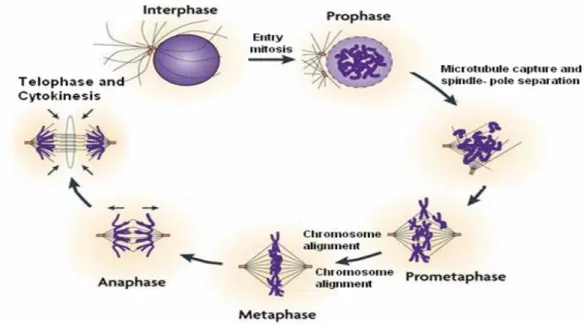
Figure 2 Steps in mitosis
Figure 3 Schematic diagram of Cytokinesis
Figure 4 The centrosome duplication cycle
Figure 5 Spindle assembly checkpoint signalling
Figure 6 Organization of the vertebrate kinetochore/centromere
Figure 7 Localization of passenger protein during mitosis
Figure 8 Three Aurora kinases in human
Figure 9 The model of kinetochore-microtubule attachments
Figure 10.1. A model of the mechanisms to correct syntelic chromosome mal-orientations during cell division
Figure 10.2. Aurora B contributes to the correction of merotelic attachments by promoting microtubule destabilization
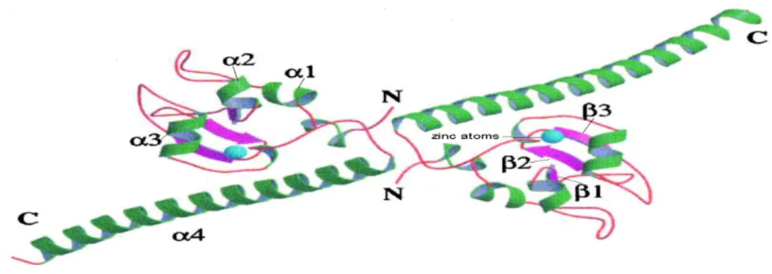
Figure 11 Survivin is a dimer
Figure 12 Survivin variants
Figure 13 Post-transcriptional modifications of Survivin
Figure 14 A model for Survivin functions in genetic fidelity and spindle formation
Figure 15 INCENP and interacting proteins
Figure 16 Interaction between passenger proteins in the complex
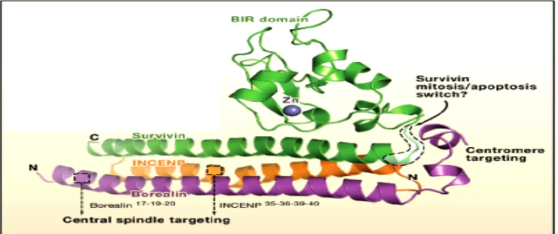
Figure 17 Survivin , INCENP and Borealin form a ternary complex
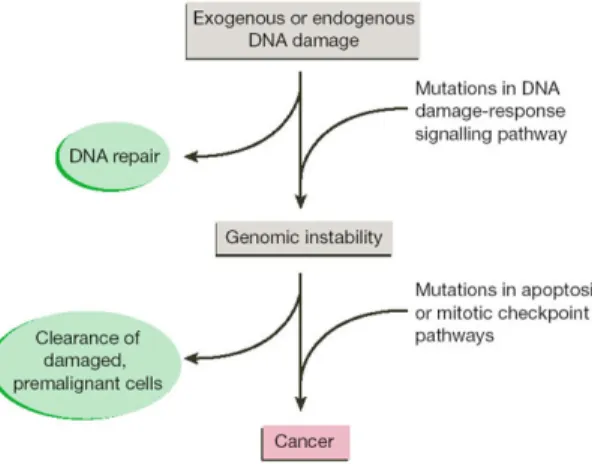
Figure 18 Genomic instability leads to cancer
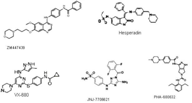
Figure 19 Aurora kinase inhibitors
Table 1 Mitotic spindle checkpoint components and complex
Table 2 Aurora B substrates in vertebrates and yeast
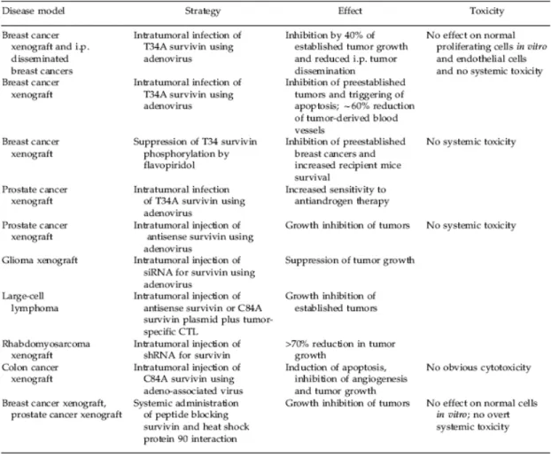
Table 3 Anti-Survivin strategies and interventions
Materials and Methods
Figure 20 Schema of the possible situations in a FRAP experiment
Table 4 Primary antibodies used in the experiments
Table 5 Secondary antibodies used in the experiments
Results
Figure 21 Phosphorylated Survivin has effect on the stable spindle assembly checkpoint
Figure 22 Phosphorylated Survivin impairs the localisation of MKLP1 at the spindle
midzone/midbody
Figure 23 Purification of both the kinase domain of Aurora A and Aurora B kinase
Figure 24 High throughput screening results
Figure 25 Hit from the Villeurbanne library
Figure 26 Flavone compounds
Figure 27 The best hit found in the Curie-CNRS library
Figure 28 The level of Cyclin B1 decreases in the presence of benzopyrido[e]indole
Figure 29 Surivin-GFP and Aurora-GFP share the same behavior in the presence of
Aurora kinase inhibitors
Figure 30 Localisation of Survivin and INCENP in the absence of Aurora B
Figure 31 Time-lapse experiments with HeLa cells expressing Survivin-GFP
Figure 32 Binary choice at the Metaphase/Anaphase transition
Table of content
INTRODUCTION ...5
CHAPTER I: CELL CYCLE...7
I.1. Phases in cell cycle. ...7
I.1.2. M phase ...9
I.1.3. Control of cell cycle...12
I.1.4. Mitotic slippage ...13
I.2. Two important structures in cell division: Mitotic spindle and Centromeres ...13
I.2.1. Mitotic spindle...13
I.2.2. Centromeres ...19
CHAPTER II: CHROMOSOMAL PASSENGER PROTEINS ...23
II.1. General localization of chromosomal passenger proteins ...23
II.2. Members of chromosomal passenger proteins ...23
II. 2. 1. Aurora B...24
II.2.1.1. Aurora B: a member of Aurora kinases...24
II.2.1.2. Aurora B regulation ...26
II.2.1.3. Aurora B substrates ...27
II.2.1.4. Aurora B function ...28
II.2.2. Survivin ...32
II.2.2.1. Survivin: a member of the IAP family ...32
II.2.2.2. Survivin isoforms...33
II.2.2.3. Survivin regulation...34
II.2.2.4. Role of Survivin in cell division...36
II.2.2.5. Role of Survivin in cell death...38
II.2.3. INCENP (Inner CENtromeric Protein)...39
II.2.4. Borealin/Dasra B: a novel chromosomal passenger protein ...40
II.2.5. TD-60 telophase disc 60 ...40
II.3. Chromosomal passenger complex CPC) ...41
II.3.1. Interaction between passenger proteins in the CPC ...42
II.3.1.1. The links between passenger proteins in the complex ...42
II.3.1.2. The interaction between passenger proteins is required for the right localization of the CPC...44
CHAPTER III: CHROMOSOMAL PASSENGER PROTEINS AND CANCER...47
III.1. Passenger proteins as possible targets for cancer therapy...47
III.1.1. Expression of passenger proteins in cancer...47
III.1.2. Passenger proteins and their role in aneuploidy promotion...48
III.2. Chromosomal passenger proteins and cancer therapy ...49
III.2.1. Aurora-kinase inhibitors...49
III.2.2. Survivin: a target for cancer therapy...50
CHAPTER I: BIOCHEMISTRY...55
I.1. Cells extracts...55
I.2. Electrophoresis...55
I.2.1. SDS-polyacrylamide gel electrophoresis...55
I.2.2. Transfer of protein from SDS-polyacrylamidee gel to solid supports. ...55
I.2.3. Immunoblotting...56
CHAPTER II: MOLECULAR BIOLOGY...57
II.1. Agarose electrophoresis ...57
II.1. Competent cells...57
II.2. Bacterial transformation ...57
II.3. DNA Miniprepations...57
CHAPTER III: EXPRESSION OF PROTEIN IN E. coli AND PURIFICATION...59
III.1. Protein over-expression...59
III.2. Protein extraction ...59
III.3. Protein purification...59
III.4. Storage of purified proteins ...60
III.5. Preparing sample for SDS-polyacrylamide gel Electrophoresis ...60
CHAPTER IV: PROTEIN KINASE ASSAY ...61
IV.1. Protein kinase assay ...61
IV.2. High throughput screening (HTS)...61
CHAPTER V: CELL CULTURE...63
V.1. Cell lines...63
V.2. Conservation of cells...63
V.3. Drugs used...63
V.4. Synchronisation ...64
V.4.1 Double Thymidine block (early S-phase block) ...64
V.4.2. Thymidine-Nocodazole block (mitotic block) ...64
V.5. FACS (Fluorescence Activated Cell Sorting)...64
V.7. Cell proliferation and viability ...64
V.7. Cell transfection...65
V.7.1 Plasmids...65
V.7.2 siRNA transfection ...66
CHAPTER VI: MICROSCOPIC TECHNIQUE ...67
VI.1. Immunofluorescence...67
VI.2. Time – lapse experiments ...68
CHAPTER VII: BUFFER ...69
VII.1. Biochemistry ...69
VII.2. Immunofluoresence ...69
VII.3. Protein extraction and purification ...70
RESULTS...71
CHAPTER I: ROLE OF SURVIVIN PHOSPHORYLATION BY AURORA B IN MITOSIS ...73
I.1. Role of Survivin T117E mutant in mitosis...75
I.2. Consequence of Survivin T117E over-expression in mitosis...84
I.2.1 SurvivinSRT117E-GFP mutant affects the spindle checkpoint ...84
I.2.2 Phosphorylated Survivin is required for the correct localization of MKLP1.84 CHAPTER II: IDENTIFICATION OF AURORA KINASE INHIBITORS ...87
II.1. Kinase purification...89
II.2. Setting up the protein-kinase assay ...89
II.3. High throughput screening...90
II.4 Characterization of the hits ...91
II.4.1 Flavone inhibits Aurora kinase in vitro ...91
II.4.2 Benzo[e]pyridoindoles a new class of Aurora kinase inhibitors ...93
II.5 Aurora kinase inhibitors, interesting tools for studying the CPC ...127
II.4.2. The effect of compound on the level of Cyclin B proteins ...127
II.4.3. Aurora kinase activity is required for accurate localization of CPC on centromere...128
DICUSSION & PERSPECTIVES ...131
Chapter I : Role of Survivin phosphorylation by Aurora B in mitosis ...133
Chapter II: Search for Aurora B kinase inhibitors...135
Perspectives ...139
INTRODUCTION
CHAPTER I: CELL CYCLE
CHAPTER II: CHROMOSOMAL PASSENGER PROTEINS
CHAPTER III: CHROMOSOMAL PASSENGER PROTEINS AND
CHAPTER I: CELL CYCLE
Cell is the basic unit of all living organisms (Rudolf Virchow) and the name “cell cycle” was given by Howard and Pecl (1953). Definitely, cell cycle or cell-division cycle is an ordered set of events, culminating in cell growth and division into two daughter cells. It is a very important process by which a single cell fertilized egg develops into a mature organism and by which hair, skin, blood cells and some organs are renewed.
I.1. Phases in cell cycle.
The goal of cell cycle is to produce two genetically identical cells from one precursor cell. This requires the replication of each chromosomal DNA, the separation of the two set of chromosomes into the daughter cells and the physical division of cell to produce two identical daughter cells. Details of cell cycle vary from organism to organism and may occur at different times in an organism’s life. We will concentrate the description mostly to superior eukaryotes.
In the cell cycle, there are different phases named: Go, G1, S, G2 and M phase (Figure 1). Go, G1, S and G2 phase are known as Interphase. The G1 phase stands for “GAP 1”. The S phase stands for “Synthesis” because in this stage DNA replication occurs. The G2 phase stands for “GAP 2”. The M phase itself is composed of two stages: mitosis (chromosomes separation) and cytokinesis (cytoplasmic division). We will first describe the different phases of the cycle and then, its regulation.
I.1.1. Interphase
During this period cells are engaged in metabolism and prepared for mitosis. Visually, this period is inactivity but in fact, on molecular level, it is quite an active time for the cells. In interphase, the cells replicate their nuclear DNA, produce protein and increase in size to ensure that after dividing, new created daughter cells are quite identical. Interphase is divided into three phases although these various stages are not morphologically distinguishable. Each phase of the interphase has a distinct set of specialized biochemical processes that prepare the cell for initiation of the cell division.
a. Go phase
The Go phase is a quiescent period in the cell cycle. In fact, when cells are unable to get through the G1 restriction point, they enter the Go phase. This usually occurs in response to a lack of growth factors or nutrients. During the Go phase, the cell cycle machinery is dismantled and cyclins and cyclin-dependent kinases disappear. Cells remain in the Go phase until there is a pressure for them to divide. For instance, epidermal
fibroblasts remain in the Go phase until stimulated by growth factors, which are generated in response to the wound healing process. Although many cells in the Go phase may die along with the organism, this does not mean that cells that enter the Go phase are destined to die. Go represents not simply the absence of signals for mitosis but also the active repression of the transcription of genes needed for mitosis. Cancer cells cannot enter Go and are destined to repeat the cell cycle indefinitely.
b. G1 phase
The G1 phase is the major period of cell growth. During this stage, cells produce RNA, synthesize proteins and increase in size. New organelles are also synthesized, so the cells need both structural proteins and enzymes. In this phase, there is an important activated cell cycle control mechanism (G1 checkpoint). At the restriction point, cells assess external and internal stimuli and decide whether to pause or to divide. A cell can pause in G1 and then, enter in Go phase. Committed cells go on S phase.
c. S phase
Following G1, cells enter S phase (short for Synthesis phase). During this phase, replication occurs. The complete DNA instructions of the cells have to be duplicated. So at the end of this stage, each chromosome has two identical DNA double helix molecules. The flawless operation of this cycle is important not only for the maintenance of cell viability but also, for preventing genetic instability that could potentially drive tumourigenesis (Blow and Dutta, 2005). DNA damage takes place often during this phase, and DNA repair is Figure 1. Overview of cell cycle. In eukaryotic cells, the cell cycle is characterized by four distinct phases (G1, S, G2 and M phase). In Go phase, cells enter a state of resting or quiescence.
The regulatory molecules (cyclins and cyclin depend kinases –Cdk) which control the cell cycle are also indicated. (Modified from http://www.med.unibs.it/~marchesi/dna.html)
initiated following the completion of replication. Incomplete DNA reparation may flag cell cycle checkpoint, which in turn, halts the progression in the cell cycle.
d. G2 phase
After successful completion of DNA synthesis, cells enter in the G2 phase. This is the final subphase of the cell cycle interphase. In this period, nucleus is still well defined and surrounded by the nuclear envelope; chromosomes are not clearly discerned in nucleus since they are still in the form of loosely packed chromatin fibres. During this phase, the cell will continue to grow and synthesize proteins. At the end of this gap there is another control checkpoint (G2 checkpoint) which determines whether the cells can initiate M phase or must extend the gap for further cell growth. G2 checkpoint helps to maintain genomic stability since it prevents cells from entering mitosis if DNA is damaged. It provides an opportunity for DNA repair and thus, stops the proliferation of damaged cells.
I.1.2. M phase
During M phase, separation of chromosomes plays a key role for producing two genetically identical daughter cells. In a typical somatic cell cycle, M phase comprises mitosis and cytokinesis. Mitosis is a complex and highly regulated process by which a cell separates its duplicated genome into two identical halves. The main purpose of mitosis is to segregate sister chromatids into two nascent cells, such that each daughter cell inherits one complete set of chromosomes (Nigg, 2001a). During cell division, an inaccurate separation of chromosomes can lead to an abnormal chromosome number and create genomic instability, a crucial step in the development of human cancer (Draviam et al., 2004). In cytokinesis cytoplasm, organelles and cell membrane are split equally into two daughter cells. This stage occurs in conjunction with mitosis.
a. Mitosis
- Prophase: prophase is a process in which chromatin condenses into a highly ordered structure called chromosome (Figure 2). Each chromosome which has been duplicated during an earlier S phase now contains two identical copies of itself, called sister
chromatids. The sister chromatids attach together in a specialized region of the chromosome
known as the centromere (see part I.3.2a). Chromosome condensation is accompanied by extensive phosphorylations of both histone and non-histone proteins. Phosphorylation of the core histone H3 at serine 10 is required for proper chromosome segregation in at least some organisms (Hans and Dimitrov, 2001; Nigg, 2001a).
- Prometaphase: this stage occurs immediately after prophase and sometime, is considered as a part of prophase. Early during this period, nuclear membrane breakdown,
microtubules radiate from centrosomes and invade the nuclear space. Then they quickly interact with the kinetochores on the chromosomes. This is called an opened mitosis and occurs in most multicellular organisms. Some protists, such as algae, undergo a variation called closed mitosis where the microtubules are able to penetrate an intact nuclear envelope (Ribeiro et al., 2002). During prometaphase, kinetochores are initially captured by a single microtubule that extends from a spindle pole and are then transported pole ward along the microtubule (Tanaka et al., 2005) (Figure 5). The kinetochore will provide the pulling force which is necessary to separate two chromatids. Throughout mitosis, microtubule-kinetochore interactions are highly dynamic (Nigg, 2001b).
- Metaphase: This phase is characterized by the highly condensed chromosomes alignment in the middle of the cell. In fact, early events in metaphase start with the later events of prometaphase when kinetochores attach microtubules. The centromeres of chromosomes convene themselves on the metaphase plate (or equatorial plate) - an imaginary line that is equidistant from the two centrosome poles (Figure 2). Unattached chromosomes apparently generate a signal that delays progression to anaphase until all sister chromatin are attached to the spindle apparatus. Such a signal is called the mitotic spindle checkpoint (see part I.2.1c). This process ensures the equal segregation of sister chromatids to the two daughter cells (Yu, 2006). In human cells, there is also a DNA decatenation-sensitive mitotic checkpoint which functions independently of DNA damage or spindle assembly checkpoint response (Skoufias et al., 2004).
Figure 2. Steps in mitosis. Mitosis is a continual and dynamic process. The sequence events in
mitosis are divided into five subphases: prophase, prometaphase, metaphase, anaphase and telophase. (Jackson et al., 2007).
- Anaphase: Anaphase onset starts only when the checkpoint is turned off. Within anaphase, two distinct processes occur. During early anaphase, the chromatids simultaneously separate and sister chromatids are pulled apart towards their respective centrosomes at the poles. In the late anaphase, the microtubules elongate and slide relatively to each other to drive the spindle poles further apart.
- Telophase: This is the last stage of mitosis. At telophase, chromosomes are re-condensed into chromatin and progressively nuclear membrane is reconstituted. At telophase, the non-kinetochore microtubules continue to lengthen, elongating the cell even more. Both sets of chromosomes, now surrounded by the new nucleus membrane, unfold back into chromatin. Each daughter nucleus has a complete copy of the genome of its parent cell.
b. Cytokinesis.
Cytokinesis is the final event in the cell cycle and whereby the cytoplasm of a single cell is divided to spawn two daughter cells. It usually occurs meanwhile the nuclear envelope is reforming, although they are distinct processes. The goal of cytokinesis is to physically separate a mother cell into two identical daughter cells.
In animal cells, a cleavage furrow develops where the metaphase plate used to be, pinching off the separated nuclei. This furrow contains actin, myosin, and other proteins that are organized into a contractile ring called the actomyosin ring (Figure 3). The ring then, ingresses generating a membrane barrier between the cytoplasmic content of each daughter. The ingressing furrow constricts components of the spindle midzone into a focused structure called the midbody (Guertin et al., 2002). Many signalling proteins are found in the midbody during cytokinesis, including GTPases, phosphatases, kinases and proteases-proteins as well as components of the DNA-damage or spindle-assembly checkpoints (Zeitlin and Sullivan, 2001). In fact, the final event in cytokinesis is the furrow “sealing” that generates two completely separate cells. Recently, it was shown that Rab 35, a GTPase
Figure 3. Schematic diagram of Cytokinesis.
Major features and important organels are indicated. (Modified from
http://www.sparknotes.com/biology/cellreproduction/ mitosis/section3.rhtml)
protein, is required for the final abscission by controlling Septin 2 and PiP2 subcellular distribution during cell division (Kouranti et al., 2006).
I.1.3. Control of cell cycle
Controls during the cell cycle are necessary to bring about an orderly progression through both S-phase and mitosis and to couple these two processes with cell growth (Nurse, 1997; Castro et al., 2005). How cell division (and thus, tissue growth) is controlled is very complex.
There are two main key classes of regulatory molecules that determine the cell cycle progression: the cyclins and cyclin-dependent kinases (Cdks) (Norbury and Nurse, 1991) (Figure 1). The most prominent mitotic kinase is the Cdk1-the founding member of cell cycle regulation. These molecules are major control switches within the cell cycle, causing the cell to move from G1 to S (G1 cyclin-Cdk complexes) such as Cyclin D/Cdk4; Cyclin D/Cdk 6; Cyclin E/Cdk2; or from G2 to M (Mitotic Cyclin-Cdk complexes) such as Cyclin B/Cdk1 (also knows as Cdc2). This complex which was first called M-phase promoting factor (MPF) is the key regulator on one hand of the G2-M transition of the cell cycleand on another hand of the APC/C activation (Brito and Rieder, 2006).
There are also some non-cyclin proteins involved in the regulation of the cell cycle. For example, p53 is a protein whose function is to block the cell cycle when DNA is damaged. If the damages are severe this protein can lead cells to apoptosis (cell death). To note mutations of p53 are commonly reported in transformed cells (May and May, 1999).
The nuclear protein p21WAF1 (p21), p27KIP1 (p27), and p57KIP2 are universal Cdk inhibitors (Sherr, 1995; Sherr, 1996; Weinberg and Denning, 2002). By inhibiting Cdks, they interrupt the transition from G1 to S phase in the cell cycle. Expression of p21 is induced by wild-type p53 in response to DNA damage. This mechanism is necessary for the maintenance of genomic fidelity. Perturbation of p53 functions leads to the loss of the checkpoint control mediated by p21, thus causing DNA instability. Therefore, expression of p21 protein is expected to be low when normal functions of the p53 are lost. However, p21 protein accumulation may also be induced through p53 independent pathways (Amatya et
al., 2001). p27 protein is another Cdk inhibitor, which inhibits cyclin complexes by a
posttranslational degradation through ubiquitin-dependent proteasomal proteolysis. Thoroughly, cells are blocked at the entry into S phase (Schiffer et al., 1999).
Anaphase promoting complex (APC) promotes degradation of structural proteins associated with the chromosomal kinetochores. APC also targets the mitotic cyclins for degradation, ensuring telophase and cytokinesis progression. Recently, studies brought to
light additional mitotic kinases to the regulation of M phase progression, including members of the Polo, the Aurora and the NIMA (never in mitosis A) families (Nigg, 2001b).
I.1.4. Mitotic slippage
We have described previously that cells pausing in G1 can escape in Go. Arrested mitotic cells may also benefit of such a binary choice. In the presence of an active spindle assembly checkpoint (SAC), cells escape from mitotic arrest and enter the next G1 as multinucleated. This is called mitotic slippage (Blagosklonny, 2007; Brito and Rieder, 2006; Rieder and Maiato, 2004).
As we said above, the sequential activation and inactivation of cyclin-dependent protein kinases ensure the proper timing and order of cell cycle events. Activated Cyclin B/Cdk1 complex regulates cells entry into mitosis. The destruction of cyclin B is essential for proper exit from mitosis (Raff et al., 2002). When the spindle assembly checkpoint is not satisfied (for instance, drug-mitotic arrest such as Nocodazole, Taxol treatment) cells will delay exit from mitosis by preventing the APC complex. However, in the absence of transcription, cells may die in prolonged mitotic arrest, the SAC does not arrest cells permanently and therefore cells may eventually escape from mitotic arrest without dividing. They enter in G1 as multinucleated cells. These cells either do not reproduce anymore or die (Blagosklonny, 2007; Brito and Rieder, 2006; Rieder and Maiato, 2004).
I.2. Two important structures in cell division: Mitotic spindle and Centromeres I.2.1. Mitotic spindle
The function of mitotic spindle is to segregate chromosomes during cell division. This structure contributes to the mitotic eukaryotic cytoskeleton. It consists of a bundle of microtubules joined at the ends but spread out in the middle. It is mostly ellipsoid in shape. At the pointed ends, known as spindle poles, microtubules are nucleated by the centrosomes. Forces that are generated by the attachment between microtubules to the kinetochores will pull chromosomes into alignment along the spindle midzone in order to build up the metaphase plate. Since the center of the spindle specifies the plate along which the cell will divide during cytokinesis, this ensures that each daughter cell will receive one of each chromatid.
a. Centrosomes.
The centrosome is the main microtubule-organizing centre (MTOC) of the cell and also regulates cell-cycle progression. It was discovered, in 1888, by Theodor Boveri and was described as the special organ of cell division.
Centrosomes are composed of two orthogonally arranged centrioles surrounded by an amorphous mass of pericentriolar material (PCM). Centrioles are very important in the cell division process. They organize the PCM which in turn, plays a role in the mitotic spindle assembly. Centrosomes are associated with the nuclear membrane during interphase. The centrosomes nucleate and organize microtubules, playing a role in mitosis. Their position is thought to dictate the organization of the cell microtubule network (Manneville and Etienne-Manneville, 2006).
During cell division the centrioles are duplicated, so a pair will be available for each daughter cell (Figure 4). Each new paired set of centrioles is composed of one original centriole. The centrosome itself replicates once and only once per cell cycle (Mazia, 1987). Two centrosomes form spindle poles and direct the formation of bipolar mitotic spindle. The presence of more than two centrosomes (chromosome amplification) severely disturbs mitosis process and cytokinesis via formation of more than two spindle poles. It will lead to increase the chromosome segregation errors (chromosome instability - CIN) (Deans et al., 2003; Fukasawa, 2005). Loss of certain tumour suppressor proteins leads to centrosome amplification, which in turn destabilizes chromosomes (Fukasawa, 2005). Moreover, loss of BRCA1 ubiquitination also results in centrosomes amplification (Sankaran and Parvin, 2006).
Figure 4. The centrosome duplication cycle.
Specific activation of Cdk2/cyclin E triggers initiation of centrosome duplication in late G1. Centrosome duplication begins with the physical separation of the paired centrioles, which is followed by formation of procentrioles near the proximal ends of each pre-existing centriole. During S and G2 phases, procentrioles elongate, and two centrosomes progressively recruit PCM. In late G2, the daughter centriole of the parental pair acquires appendages (shown as red wedges), and two identical centrosomes are generated. During mitosis, two duplicated centrosomes form spindle poles, and direct the formation of bipolar mitotic spindles. Upon cytokinesis, each daughter cell receives one centrosome. (Modified from Fukasawa, 2005).
The centrosome is a platform required for a multitude of cellular functions, including organization of signalling pathways, cellular responses to stress and cell cycle transition (Doxsey, 2005). Many regulators of the cell cycle associate with the centrosome, so cells that have lost their centrosomes arrest in the next cell cycle (Rieder et al., 2001). Recent researches suggest that centrosomes may have their own genome, as previously described for mitochondria and chloroplasts. There may be an interconnection between the functional network of DNA damage response and the dynamic structural organization of eukaryotic cells through centrosomes (Loffler et al., 2006).
b. Microtubules
Microtubules are cytoskeletal structures assembled from mostly α/β-tubulin heterodimers that play an essential role in many cellular processes, such as cell motility, organelle transport, maintenance of cell polarity, and cell division (Nogales, 2000). The tubulin dimers polymerize end to end in protofilaments. Another important feature of microtubule structure is polarity with α-tubulin at one end of the polymer (the minus end) and β-tubulin at the other (the plus end). The plus end explores the cellular space, switching rapidly between phases of growth and shrinkage, a behaviour called dynamic instability. (Moritz et al., 2001; Lansbergen and Akhmanova, 2006). Centrosome-nucleated microtubules probe the cytoplasm with their plus ends to search and capture chromosomes in prometaphase. γ-Tubulin, a protein related to α/β-tubulin, does not assemble into microtubules. In most cells, it is found at centrosomes. γ-Tubulin has been found in two main protein complexes: the γ-tubulin ring complex (γTuRC) and the γ-tubulin small complex (γTuSC) (Moritz et al., 2001).
Microtubules are intrinsically dynamic, in that they alternate abruptly and stochastically between two stages of growth and shortening, a phenomenon termed “dynamic instability” (Karsenti et al., 2006; Mitchison and Kirschner, 1984). These stages can generate force, in addition to motor proteins that move along the microtubule. The major microtubule motor proteins are kinesin, which generally moves towards the (+) end of the microtubule, and dynein, which generally moves towards the (-) end. Three microtubule motors – CENP-E, dynein and MCAK and several microtubule-binding proteins have been also localized at metazoan kinetochores (Garrett and Kapoor, 2003). An accurate control of microtubule dynamics is required for kinetochore tension generation and chromosome alignment during mitosis (Zhou et al., 2002). Yang Ge et al. have developed the single-fluorophore imaging in order to examine microtubule organization in the vertebrate meiotic spindle and they proposed that the mechanical integrity and force transmission across the spindle must be regulated by dynamic crosslinking of loosely connected microtubules (Yang
paclitaxel or docetaxel) used in the treatment of cancer, blocks dynamic instability by stabilizing GDP-bound tubulin to the microtubules. Thus, there is no more depolymerization and the microtubule does not shrink back. Nocodazole, Colchicine and Vinblastine have the opposite effect, blocking the polymerization of tubulin into microtubules. Noscapin does not significantly promote or inhibit microtubule polymerization but alters the steady state dynamics of microtubule assembly (Zhou et al., 2002).
c. Mitotic spindle checkpoint.
The spindle checkpointis a survey mechanism that delays anaphase onset untilall chromosomes are correctly attached in a bipolar fashionto the mitotic spindle (May and Hardwick, 2006). It is an evolution conserved mechanism, which ensures that cells with misaligned chromosomes do not exit mitosis and therefore do not divide. The spindle assembly checkpoint prevents thus aneuploidy (Bharadwaj et al., 2004). It is an active signal produced by improperly attached kinetochores. As chromosome attachment to the spindle microtubules is a stochastic process, not all chromosomes achieve alignment at the spindle equator at the same time (Yu, 2006). Even a single unaligned chromosome can prevent the onset of anaphase (Rieder et al., 1995). When sister kinetochores are properly attached to opposite spindle poles, forces in the mitotic spindle generate tension at the kinetochores. The interdependence between tension and microtubules attachment makes it difficult to determine whether these signals are separable. But some recent evidences show that defects in tension act as the primary checkpoint signal (Pinsky and Biggins, 2005).
The spindle checkpoint inhibits the anaphase-promoting complex/cyclosome (APC/C) leading to the delay of anaphase onset. APC/C is a multiprotein E3 ubiquitin ligase that ubiquitynates a range of cell-cycle regulators, targetingthem for degradation by the 26S proteasome (Castro et al., 2005; Yamano et al., 2004). Once all kinetochores have bipolar attachments, the checkpointis switched off and APC/C is activated (May and Hardwick, 2006). Then, anaphase onset starts. The molecular basic of the dynamic signalling carried out by spindle checkpoint proteins is listed in Table 1. Many proteins are involved in either activation or silencing of checkpoint. Some play a role in detecting the signal which is generated as a result of the lack of microtubule occupancy and kinetochore tension (Chan and Yen, 2003); some regulate APC/C activities; and finally others are involved in silencing the checkpoint. Recent works in yeast, frogs and mammals have outlined the signalling of spindle assembly checkpoint (Figure 5). For example Mad2 has been proposed as a specific marker of unattached kinetochores, whereas Aurora B (in complex with the other passenger proteins) is required for sensing the spindle tension. The activation of spindle checkpoint starts by the recruitment of spindle checkpoint proteins. The kinases Mps1 and Bub1 act in concert with BubR1; and the CENP-E kinesin triggers the rapid recruitment of the Mad1–
Mad2 complex to the kinetochore (Tan et al., 2005). The mitotic arrest deficiency 2 (Mad2) inhibits APC/C through binding to its mitotic-specific activator - Cdc20. Binding of Mad2 to Cdc20 involves a large conformational change of Mad2 and requires the Mad1–Mad2 interaction in vivo (Yu, 2006). BubR1 functions synergistically with Mad2 in inhibiting Cdc20-APC activity (Tan et al., 2005; Zhou et al., 2002). MCC complex, which contains the BubR1–Mad2–Bub3–Cdc20 proteins, strongly inhibits APC/C activity, causing a delay in anaphase.
As soon as all chromosomes are properly attached to the kinetochore microtubules and aligned at the metaphase plate, the spindle checkpoint is turned off. Mad1/Mad2 and BubR1 are transported away from the kinetochore along microtubules by dynein, preventing further inhibitory signalling (May and Hardwick, 2006). p31commet (p31C) binds to ‘closed’ conformation of Mad2 involved in the checkpoint switch off. The binding of microtubules to CENP-E downregulates BubR1 kinase activity and results in the silencing of the checkpoint (Mao et al., 2003). Activated Cdc20-APC catalyzes the ubiquitination of securins, leading to their degradation through proteosome-mediated proteolysis. Degradation of securins in turn causes the release of separases. The free separases destroys cohesin (the molecular glue holding sister chromatids together), thus allowing chromatids to be pulled to oppositepoles and finally anaphase starts.
Trigger anaphase onset
Checkpoint
components Comment
Core checkpoint proteins
Mad1 Binds to Mad2 and recruits it to kinetochore; also localises Mad2 to the nuclear periphery (NP) in interphase; required for checkpoint activation in response to tension; binds to Bub1 and Bub3 upon checkpoint activation in budding yeast. Mad2
Binds to Bub1 and Cdc20; exist in two conformations (‘closed’ C-Mad2 on binding Mad1 and Cdc20, or ‘open’ O-Mad2 when unbound); interacts with Cdc20 and Bub1/Mad3 to form MCC which inhibits the APC; excess Mad2 inhibits the APC/C in many experimental systems.
BubR1/Mad3 Binds to Bub3; interacts with Mad2 and Cdc20 to form MCC; C-terminal kinase domain of BubR1is activated by CENP-E. Bub1
Protein kinase; binds to Bub2; requirement for recruiting the other checkpoint proteins differs depending on systems; reported substrates include Bub3, Mad 2 and adenomatous polyposis coil; kinase activity is not required for the checkpoint arrest.
Bub3 Binds to Bub1 and MCC components; required for the Bub1 and BubR1 localization to the kinetochore. Mps1/Mph1 Protein kinase; phosphorylates Mad1 in vitro; excess activates the checkpoint; required for the recruitment of Mad1; Mad2 and CENP-E to the kinetochore.
Other protein required for checkpoint function
CENP-E Kinesin family member; binds to BubR1; stimulates BubR1 activity; required for capture and stabilization of microtubules at the kinetochore; only found in higher eukaryotes.
RZZ complex Complex of Rod, ZW10 and Zwilch, in higher eukaryotes only recruits dynein and Mad1/Mad2 to the kinetochore. Cdc20 APC/C activator; by binding to Mad2 and BubR1, it forms the MCC, which inhibits APC/C activation. Aurora B Protein kinase; chromosomal passenger protein binds INCENP and Survivin; required for checkpoint in response to the lack of tension but not attachment. P31comet Binds specifically to the ‘closed’ conformation of Mad2; excess disrupts
checkpoint signalling; involved in checkpoint switch off.
Dynein/Dynactin Minus-end-directed motor that transports Mad2 and BubR1 away from kinetochore; though to be required for switching off the checkpoint; dynactin complex recruits ‘cargo’ to dynein.
Downstream of the checkpoint
APC/C E3 ubiquitin ligase; target mitotic regulators for destruction by the proteasome; downstream effector of the checkpoint. Securin Binds and inhibits Separase
Separase Protease; cleaves the cohesin subunit Scc1and breaks the cohesion ring
Cohesin Protein complex of Scc1, Smc1and Smc3 that form a ring around sister chromatids, holding them together; cleavage of Scc1 is required for sister separation
The complexes MCC: the mitotic checkpoint complex (MCC);
Mad1-Mad2; Mad2-Cdc20; Bub3-Bub1; Bub3-BubR1; Mad2-Cdc20-BubR1-Bub3; BubR1-CENP-E; BubR1-Cdc20 Rod-ZW10-Zwilch;
AuroraB-INCENP-Survivin-Borealin (chromosomal passenger complex) Table 1: Mitotic spindle checkpoint components and complex (May and Hardwick, 2006).
I.2.2. Centromeres a. Centromere
The short definition of centromere is the point where the two chromatids are linked and where the microtubules attach. It plays as a key role in sister chromatid adhesion, in the kinetochore formation, in the pairing of homologous chromosomes and is also involved in the control of gene expression. Centromeres are highly complex chromosomal substructures involved in essential aspects of chromosome transmission during cell division.
Centromere consists of large arrays of repetitive DNA (e.g. satellite DNA) where the sequence within individual repeat elements is similar but not identical. The complex array of DNA repeats found in human centromeres has suggested a subunit model for the assembly of mammalian centromeres (Zinkowski et al., 1991). In fission yeast, centromeric repeats are transcribed into small interference RNA precursors (pre-siRNA) which are processed by Dicer to direct the formation of heterochromatine (Djupedal et al., 2005; Hall
et al., 2003). RNAi recruitment around centromeric chromatin maintains this specialized
structure (Hall et al., 2003; Volpe et al., 2002). The centromeric DNA is normally in a heterochromatin state that is probably essential for its function. Centromeric function depends upon a specialized centromeric organization where distinct domains of CENP A and dimethyl K4 histone H3 are positioned on the near surface of the chromosomes (Greaves et al., 2007). But histone variants H2AZ and non histone proteins SMC3 seem also to participate to this peculiar centromere organization that may vary slightly from chromosome to chromosome (Greaves et al., 2007; Jansen et al., 2007). In all active centromeres, the normal histone H3 is replaced in part with CENP-A, a centromere-specific variant (Sullivan et al., 2001). The presence of CENP-A is believed to be important for the assembly of the kinetochore on the centromere and may play a role in the epigenetic inheritance of the centromere site. CENP-A participates to the early organization of centromeric chromatin structure during interphase (Regnier et al., 2005). Assembly of new CENP-A nucleosomes is restricted to a demonstrating coupling progression through mitosis and maturation of the next generation of centromere (Jansen et al., 2007).
Within the cell nucleus, centromeres undergo changes in their intranuclear localization during the cell cycle. They have been found to associate or colocalize with subnuclear structures, such as the nucleolus or nuclear bodies. Dynamic interactions between centromeres and other nuclear substructures may be important in controlling gene expression as well as centromere function. The structure-function relationships of centromere components and their dynamic interplay within the nucleus will provide insight
into centromere function and may help to understand mechanisms underlying genome instability (Figure 6).
b. Kinetochores
Kinetochores are large protein complexes which assemble on the centromere and link the chromosome to microtubule mitotic spindle during mitosis and meiosis. The kinetochore contains two regions: the inner kinetochore, which is tightly associated with the centromeric DNA and provides the foundation for building the kinetochore–microtubule interface and the outer kinetochore which interacts with microtubules. In budding yeast, more than 60 kinetochore components have been identified so far by genetic and biochemical approaches and these proteins organize into at least 14 different subcomplexes (McAinsh et al., 2003). In animal cells, there are presently more than 80 proteins that show either exclusive or partial localization at kinetochores during mitosis (Gassmann et al., 2007) (Figure 6). The kinetochore is a dynamic structure whose composition is cell cycle-dependent. Most kinetochore components assemble in late G2 and prophase whereas others appear after nuclear envelope breakdown leading to competent kinetochores which are able Figure 6. Organization of the vertebrate kinetochore/centromere showing the locations of some of its component proteins and the relationship between them (updated from Pluta et al.,
1995,Rieder and Salmon, 1998), (modified from Chan and Yen, 2003). Red arrow indicates the enzymatic relationship between the trimethyl-lysine 9 modification of histone H3 and the suv39h1 methyltransferase. There are some complexes among these proteins. All protein complexes with their subunits are cartooned as a single shape. The yellow shapes indicate kinetochore MT-binding proteins. The green shapes indicate plus-end MT-binding proteins.
to engage spindle microtubules (Chan and Yen, 2003). These transient mitotic components leave the kinetochore at different times during mitosis. In contrast, constitutive components, such as the inner kinetochore component CENP-A, are boundto chromatin throughout the cell cycle. They ‘mark’ the future kinetochore location on the decondensed interphasic chromatin (Maiato et al., 2004). Many of these proteins are now known to be directly involved either in microtubule attachment or in the quality control of the attachments (Chan and Yen, 2003).
Since the kinetochore is a complex proteinaceous structure that mediates and monitors the attachment of spindle microtubules to chromosomes, it is essential for chromosome alignment and segregation of sister chromatids. Kinetochores are initially captured in prometaphase by a single microtubule. After kinetochores being captured by microtubules, they are transported along the microtubules toward the spindle pole. This process is regulated by ATP-driven motor proteins of the Kinesin and Dynein families. Each sister kinetochore must eventually attach to K-fibers from opposite poles. This is known as sister kinetochore bi-orientation or amphitelic attachment (Gassmann et al., 2007). This process is very important for the correct segregation of genetic information into daughter cells (Tanaka et al., 2005).
CHAPTER II: CHROMOSOMAL PASSENGER PROTEINS
II.1. General localization of chromosomal passenger proteins
Chromosomal passenger proteins (CPP) were first described by Cooke et al. (1987). To date, five proteins were described: including INCENP (INner CENtromere Protein) (Cooke et al., 1987), Aurora B kinase (Bischoff et al., 1998), Survivin (Ambrosini et al., 1997), Borealin (Gassmann et al., 2004) and the fifth protein: Telophase disc 60 TD-60) (Andreassen et al., 1991; Martineau-Thuillier et al., 1998).
Chromosomal passenger proteins are mostly absent in interphase. They are present, in the nuclear, in late G2 and their expression peaks in G2/M. Chromosomal passenger proteins are characterized by a peculiar localization during mitosis (Figure 7). In prophase, passenger proteins associate along the length of the condensing chromosomes and gradually concentrate at centromeres. At prometaphase and metaphase, they accumulate in the inner centromere. At anaphase onset, they leave the chromosomes and are transferred to the central spindle in association with microtubules. Finally, passenger proteins are concentrated in the midbody during cytokinesis (Cooke et al., 1987; Vagnarelli and Earnshaw, 2004).
II.2. Members of chromosomal passenger proteins
Although the chromosomal passenger proteins play together a key role in many processes during mitosis and assemble in complex, they each have their own characteristics. Here we will introduce mostly Aurora B kinase and Survivin, then briefly INCENP, Borealin and TD-60.
Prophase Metaphase Anaphase Telophase
Figure 7: Localization of passenger protein during mitosis. Aurora B (green); kinetochore (pink);
II. 2. 1. Aurora B
II.2.1.1. Aurora B: a member of Aurora kinases
Aurora proteins are a family of serine/threonine kinases. This family is conserved form yeast to human. In yeast, there is only one Aurora kinase gene. In Drosophila melanogaster and Caenorhabditis elegans, two kinases are present whereas in mammals, this family comprises three members: Aurora A, B and C (Meraldi et al., 2004). Aurora kinases play a key role in many processes of cell division and cytokinesis, such as centrosome cycle, spindle assembly, chromosome condensation, microtubule–kinetochore attachment and the spindle checkpoint control, .…
The three mammalian Aurora kinases share similar structure like the catalytic domain flanked by a very short C-terminal tail. But they differ in the length and sequence of the N-terminal domain (Figure 8). Aurora A and B exhibit 70% identity in the carboxyl-terminal catalytic domain (Carmena and Earnshaw, 2003). Aurora kinases present differences in their expression patterns, subcellular localization, and timing of activities. However, all three Aurora kinases are overexpressed in many types of cancer. Aurora kinases are thus potential drug targets for cancer therapy (Carmena and Earnshaw, 2003; Giet et al., 2005).
II.2.1.1.1. Aurora A
The original Aurora allele was identified in a screen for Drosophila melanogaster mutant, and is now classified as Aurora A (also termed AIK, STK6, or STK15) (Carmena and Earnshaw, 2003; Udayakumar et al., 2006). Aurora A is expressed at a low level during Figure 8: Three Aurora kinases in human. They share similar structures, as the catalytic domain,
the activation-loop domain and the D-box (Destruction box). Aurora A encompases both the A-box and D-box-activating domains (DAD). These Aurora kinases differ in the length and sequence of their N-terminal domains (Carmena and Earnshaw, 2003). The numbers on the right indicate their respective size in amino acids.
G1 and S phases, and peaks during G2/M phase (Katayama et al., 2003). It localizes on the duplicated centrosomes from the end of S phase to the beginning of G1. From metaphase to anaphase, Aurora A is located on the microtubules close to the spindle poles and then concentrates in the midbody during cytokinesis. The expression level of Aurora A falls down during mitotic exit and entry into G1 phase of the next cell cycle (Katayama et al., 2003). Aurora A is called the polar kinase due to its presence either on or close to centrosomes (Sugimoto et al., 2002).
Aurora-A kinase is necessary for centrosome separation and maturation as well as for spindle assembly (Brown et al., 2004; Prigent and Giet, 2003). Aurora-A is first activated, in late G2 phase, on the centrosome and Bora (Aurora Borealis), an Aurora A partner, may be responsible for such activation at mitosis onset (Hutterer et al., 2006). By using Clostridium difficile toxin B, Ando et al. have shown that Rho GTPases are also involved in centrosomal activation of Aurora-A during G2/M transition (Ando et al., 2007). Moreover, Aurora A is also activated by TPX2 on the mitotic spindle (Anderson et al., 2007; Bayliss et al., 2003; Giet et al., 2005), by protein phosphatase inhibitor-2 (I-2) (Satinover et al., 2004) and by Ajuba in G2 phase (Hirota et al., 2003; Prigent and Giet, 2003). Aurora A is degradated in late mitosis by Cdh1/Fizzy, a related form of the anaphase-promoting complex (APC) (Carmena and Earnshaw, 2003; Giet et al., 2005) and this process depends on both intact A- and D-boxes (Nguyen et al., 2005).
Mutations of either cysteines 8, 33 and 49 or of phenylalanine 31 in the N terminus of Aurora-A prevent its ubiquitination (Briassouli et al., 2006). Aurora A is in complex with PP2A (a protein phosphatase) at the cell poles during mitosis. PP2A dephosphorylates the serine 51 of Aurora A, a residue located in the A box, and thoroughly controls the degradation of Aurora A kinase (Horn et al., 2007).
The human Aurora A gene lies within the locus 20q13 that is amplified in many forms of cancer. In addition, Aurora A is overexpressed in several solid tumours (Barr and Gergely, 2007; Meraldi et al., 2004).
II.2.1.1.2. Aurora B
Aurora B has been identified in Drosophila, in yeast and mammalian cells. It is known as Ipl-1 (Chan and Botstein, 1993), Aurora 1 (Bischoff et al., 1998), Aik2 (Kimura
et al., 1998), Ark2 (Shindo et al., 1998), and Aim-1 (Terada et al., 1998). The human Aurora B gene is located on 17p13.1, very close to p53, in a region often susceptible to
allelic loss in tumours. Like Aurora A, Aurora B expression peaks at G2/M phase and maximum kinase activity is reached at transition during metaphase to anaphase (Katayama
domain kinase of Aurora B shares higher identity with Aurora C catalytic domain than with Aurora A domain. A small C-terminal sequence of Aurora B is responsible for its localization and function, but its N- terminal sequence seems not to be required. (Scrittori et
al., 2005). The role and regulation of Aurora B will be described in details hereafter.
II.2.1.1.3. Aurora C
The unique member of Aurora kinases that has only been described in mammals is Aurora C. To date, little is known about this protein. It was found overexpressed in testis and certain tumour cell lines such as HepG2, HuH7, MDA-MB-453 and HeLa cells (Giet et
al., 2005; Meraldi et al., 2004). The Aurora C gene is located on human chromosome 19
(locus 19q13) (Bernard et al., 1998). Aurora-B and Aurora-C exhibit a high degree of similarity (92% similarity and 85% identity) within their catalytic domain. Surprisingly, Aurora C was found to co-immunoprecipitate with Aurora B and the behaviour of Aurora-C,like Aurora-B, is typical of chromosomal passenger proteins (Chen et al., 2005; Li et al., 2004). Moreover, Li et al. have also indicated that Aurora-C interacts with INCENP and both cooperate for phosphorylating histone H3 (Chen et al., 2005; Sasai et al., 2004). Aurora C can also be associated with Aurora B and Survivin in vivo (Lens et al., 2006; Yan
et al., 2005) and may even rescue Aurora-B-depleted cells (Giet et al., 2005; Sasai et al.,
2004). Conversely, overexpression of an Aurora C kinase-deficient mutant disrupts the Aurora B-INCENP complex and induces polyploidy (Chen et al., 2005). Recently, Aurora C was known to be essential for male fertility in men (Dieterich et al., 2007), and mice (Kimmins et al., 2007).
II.2.1.2. Aurora B regulation
As in many other kinases, its phosphorylation state directly regulates its kinase activity. Aurora B as well as A is activated by phosphorylation (Walter et al., 2000; Murnion et al., 2001). In Xenopus leavis, it has been described that Aurora B is phosphorylated at multiple sites throughout the cell cycle. Aurora B seems to be involved in a kinase cascade since it was reported to be regulated by both the Raf kinase inhibitory protein (Eves et al., 2006), the checkpoint kinase Chk1 (Zachos et al., 2007) and the C. elegans Tousled like kinase (Han et al., 2005). Aurora B is also autophosphorylated at Thr 232. This phosphorylation at Thr 232 is an essential regulatory mechanism for Aurora B activation (Yasui et al., 2004). Protein phosphatase 1 (PP1), presents in kinetochores, is known to antagonize Aurora B activity (Hsu et al., 2000; Sugiyama et al., 2002; Trinkle-Mulcahy et al., 2003).
The regulation of Aurora B activity depends on its interactions with INCENP and Survivin (these interactions are described in more detail part III.2.1a). Chan and colleagues
(Kang et al., 2001) published that the in vitro activity of the budding yeast Aurora B (Ipl1p) is stimulated 10- to 20-fold by the direct binding of the INCENP homolog (Sli15p). INCENP is a specific substrate of Aurora B kinase and in turn, acts as the targeting subunit that localizes Aurora B to kinetochores (Adams et al., 2000). The binding of INCENP to Aurora B increases Aurora B kinase activity towards other substrates (Kang et al., 2001). Aurora B activation is triggered by autophosphorylation after its association with INCENP (Bishop and Schumacher, 2002). All that explain why INCENP is called a “substrate activator” of Aurora B. Bolton showed that in Xenopus leavis, the addition of recombinant Survivin protein to Aurora B immunoprecipitations stimulates 10-fold the mitotic kinase activity (Bolton et al., 2002). Survivin binding could increase Aurora B kinase activity. All these data means that Aurora B kinase activity is stimulated by its association with INCENP and Survivin and is fully active within the whole passenger protein complex. (Andrews et
al., 2003; Giet et al., 2005; Katayama et al., 2003).
Recent studies of Monaco et al. indicated that Aurora B physically and specially associates with PARP-1 (poly(ADP-ribose) polymerase 1. Moreover, Poly(ADP-ribosyl)ation of Aurora B by PARP-1 inhibits its kinase activity in response to DNA damage (Monaco et al., 2005).
Aurora B is degradated by the ubiquitin-proteasome pathway via APC/C. Nguyen et
al. have indicated that this degradation depends on intact KEN and A-boxes (Nguyen et al.,
2005). The conserved D-box at the COOH terminus is involved in the specific recognition ofAurora B by APC/C-Cdh1. Kinetics of the degradation of Aurora B bythe APC/C-Cdh1 pathway suggest that this degradation may be coupled to the initiation, progression, or completion of cytokinesis (Stewart and Fang, 2005).
II.2.1.3. Aurora B substrates
Aurora B has an over-expanding number of mitotic substrates in vitro. These substrates are very diverse, consisting of passenger proteins, chromosome-associated proteins, and proteins involved in cytokinesis as well as microtubule associated proteins (Table2).
Aurora B substrates Phases in mitosis Passenger proteins
Survivin (Wheatley et al., 2004)
INCENP (Bishop and Schumacher, 2002)
Borealin (Gassmann et al., 2004; Hayama et al., 2007)
Chromosome associated proteins
Shugoshin (Sgo 1) (Resnick et al., 2006) Histone H3 (Hsu et al., 2000)
Histone H2A (Brittle et al., 2007)
Ndc80 complexe (Cheeseman et al., 2002) Ndc10p (Biggins et al., 1999)
Boi1 and Boi2 (Norden et al., 2006) REC-8 (Buonomo et al., 2000)
CENP-A (Zeitlin et al., 2001b)
Topoisomerase II alpha (Morrison et al., 2002) BubR1 (Lampson and Kapoor, 2005)
Tousled kinase TLK-1 (Han et al., 2005)
Cytoskeleton associated proteins Dam1p(Cheeseman et al., 2002)
MCAK (Andrews et al., 2004)
MgcRac1GAP (Minoshima et al., 2003) Stathmin(Gadea and Ruderman, 2006) MKLP1 (Andrews, 2005)
MRLC (Andrews, 2005) GFAP (Andrews, 2005) Septin 1 (Qi et al., 2005)
Desmin (Goto et al., 2003; Kawajiri et al., 2003) Vimentin (Kawajiri et al., 2003)
Myosin II regulatory light chain (Murata-Hori et al., 2000)
metaphase metaphase/anaphase non determined prophase G2/metaphase pro/metaphase metaphase metaphase metaphase metaphase/anaphase metaphase metaphase metaphase G2/metaphase metaphase metaphase metaphase metaphase anaphase cytokinesis cytokinesis cytokinesis cytokinesis cytokinesis cytokinesis
II.2.1.4. Aurora B function
II.2.1.4.1. Aurora B is involvde in chromosome condensation and cohesion
Phosphorylation of histone H3 on serine 10(Ser-10) is one of the main epigenetic modification of chromatin in mitosis. There is a precise spatial and temporal correlation between this process and the initial stages of chromatin condensation (Hendzel et al., 1997; Nowak and Corces, 2004). In mammaliancell lines, a coincidence between chromosome condensation andphosphorylation of histone H3 on another residue, serine 28(Ser-28), has also been documented (Goto et al., 2002). Aurora B has been demonstrated to be responsible for both histone H3 phosphorylation on Ser-10 and Ser-28 during mitosis (Carmena and Earnshaw, 2003; Goto et al., 2002). Actually, Aurora B-mediated histone H3 phosphorylation is required for the recruitment of condensin and the other chromosomal
Table 2: Aurora B substrates in vertebrates and yeast.