Publisher’s version / Version de l'éditeur:
Molecular Microbiology, 79, 4, pp. 940-953, 2010-12-30
READ THESE TERMS AND CONDITIONS CAREFULLY BEFORE USING THIS WEBSITE. https://nrc-publications.canada.ca/eng/copyright
Vous avez des questions? Nous pouvons vous aider. Pour communiquer directement avec un auteur, consultez la première page de la revue dans laquelle son article a été publié afin de trouver ses coordonnées. Si vous n’arrivez pas à les repérer, communiquez avec nous à PublicationsArchive-ArchivesPublications@nrc-cnrc.gc.ca.
Questions? Contact the NRC Publications Archive team at
PublicationsArchive-ArchivesPublications@nrc-cnrc.gc.ca. If you wish to email the authors directly, please see the first page of the publication for their contact information.
NRC Publications Archive
Archives des publications du CNRC
This publication could be one of several versions: author’s original, accepted manuscript or the publisher’s version. / La version de cette publication peut être l’une des suivantes : la version prépublication de l’auteur, la version acceptée du manuscrit ou la version de l’éditeur.
For the publisher’s version, please access the DOI link below./ Pour consulter la version de l’éditeur, utilisez le lien DOI ci-dessous.
https://doi.org/10.1111/j.1365-2958.2010.07504.x
Access and use of this website and the material on it are subject to the Terms and Conditions set forth at
The zinc cluster transcription factor Ahr1p directs Mcm1p regulation of
Candida albicans adhesion
Askew, Christopher; Sellam, Adnane; Epp, Elias; Mallick, Jaideep; Hogues,
Hervé; Mullick, Alaka; Nantel, Andre; Whiteway, Malcolm
https://publications-cnrc.canada.ca/fra/droits
L’accès à ce site Web et l’utilisation de son contenu sont assujettis aux conditions présentées dans le site LISEZ CES CONDITIONS ATTENTIVEMENT AVANT D’UTILISER CE SITE WEB.
NRC Publications Record / Notice d'Archives des publications de CNRC:
https://nrc-publications.canada.ca/eng/view/object/?id=8fb0a4ad-e3e1-4fe8-9f54-5bb4391f1709
https://publications-cnrc.canada.ca/fra/voir/objet/?id=8fb0a4ad-e3e1-4fe8-9f54-5bb4391f1709
The zinc cluster transcription factor Ahr1p directs Mcm1p
regulation of
Candida albicans adhesion
mmi_7504 940..953Christopher Askew,1,2Adnane Sellam,1,3Elias Epp,1,2
Jaideep Mallick,1Hervé Hogues,1Alaka Mullick,1,4
André Nantel1,3and Malcolm Whiteway1,2*
1Biotechnology Research Institute, National Research
Council of Canada, Montréal, Québec, Canada H4P 2R2.
2Department of Biology, McGill University, Montréal,
Québec, Canada H3A 1B1.
3Department of Anatomy and Cell Biology, McGill
University, Montréal, Québec, Canada H3A 1B1.
4Départment de Microbiologie et Immunologie,
l’Université de Montréal, Montréal, Québec, Canada H3T 1J4.
Summary
Biofilm development by Candida albicans requires cell adhesion for the initial establishment of the biofilm and the continued stability after hyphal devel-opment occurs; however, the regulation of the process has not been fully established. Using chro-matin immunoprecipitation coupled to microarray analysis (ChIP-chip) we have characterized a regulon containing the Mcm1p factor that is required for the initial surface adhesion during biofilm formation. In the yeast Saccharomyces cerevisiae several Mcm1p regulons have been characterized in which regulatory specificity is achieved through cofactors binding a sequence adjacent to the Mcm1p binding site. This new Mcm1p regulon in C. albicans also requires a cofactor, which we identify as the transcription factor Ahr1p. However, in contrast to the other yeast regu-lons, Ahr1p alone binds the target promoters, which include several key adhesion genes, and recruits Mcm1p to these sites. Through transcription profiling and qPCR analysis, we demonstrate that this Ahr1p– Mcm1p complex directly activates these adhesion genes. When the regulatory circuit was disrupted by deleting AHR1, the strain displayed reduced adher-ence to a polystyrene surface. We also demonstrate a role for the regulon in hyphal growth and in virulence.
Our work thus establishes a new mechanism of
Mcm1p-directed regulation distinct from those
observed for other Mcm1p co-regulators.
Introduction
Candida albicans is a commensal fungus inhabiting the
skin, gastrointestinal and genitourinary tracts of warm-blooded animals such as humans. However, C. albicans is also an opportunistic pathogen that can colonize and invade host tissues causing potentially lethal systemic infections. In fact, Candida species are the most com-monly isolated agent in human fungal infections with C.
albicans accounting for more than half of these cases
(Wisplinghoff et al., 2004; Leroy et al., 2009).
Part of the success of C. albicans as a pathogen is attributed to its development of biofilm structures that protect the fungus from host defence mechanisms and decrease its sensitivity to most antifungal drugs (Hawser and Douglas, 1995; Chandra et al., 2001; Ramage et al., 2002), although the precise mechanisms of this resis-tance are not well understood (Vediyappan et al., 2010). An initial step in biofilm formation is adherence of yeast cells to a surface. After a basal layer is established, the biofilm matures by developing hyphal filaments and pro-ducing an extracellular matrix. The result is a stable, dense mass of yeast, pseudohyphal and hyphal cells embedded within this matrix of carbohydrates and proteins. C. albicans is able to adhere to a variety of surfaces both in vitro and in vivo; these include implanted medical devices such as catheters and heart valves. Biofilm development on these devices is frequently linked with nosocomial infections (Douglas, 2003; Kojic and Darouiche, 2004) and the resistance of biofilms towards antifungals significantly complicates treatment. Often catheters must be removed to allow for effective antifun-gal therapy (Mermel et al., 2001) and thus the under-standing of biofilm regulation is of significant medical interest.
Adhesion is critical for the development of the initial basal layer as well as the stability of the mature biofilm. The Agglutinin-Like Sequence (ALS) gene family of cell surface glycoproteins and the cell surface hyphal protein
HWP1 have a primary role in adhesion in C. albicans.
Deletion of ALS1, ALS2, ALS3 or HWP1 has been shown
Accepted 2 December, 2010. *For correspondence. E-mail malcolm. whiteway@cnrc-nrc.gc.ca; Tel. (+1) 514 496 6146; Fax (+1) 514 496 5143.
(2011) 79(4), 940–953 䊏
to disrupt biofilm formation (Zhao et al., 2005; 2006; Nobile et al., 2006a; Sellam et al., 2009a). With the exception of ALS2, these genes are known to be under the positive regulation of the transcription factor Bcr1p (Nobile and Mitchell, 2005). A bcr1 deletion strain has a severe adhesion defect resulting in the formation of a rudimentary biofilm (Nobile and Mitchell, 2005; Nobile
et al., 2006b). Outside of Bcr1p, little is known about the
regulation of adhesion genes in C. albicans, although it is probable that any additional adhesion regulators will be
Candida-specific transcription factors. C. albicans and the
model yeast Saccharomyces cerevisiae diverged from a common ancestor approximately 235–841 million years ago (Heckman et al., 2001; Douzery et al., 2004), and unlike C. albicans, S. cerevisiae does not naturally form biofilms or have orthologues of the ALS gene family,
HWP1 or BCR1.
In general, transcription factors are bipartite in nature, possessing distinct DNA binding and activation domains. The requirement of a strong protein–DNA interaction constrains the flexibility of the DNA binding domain to evolve, resulting in several well-conserved domains that form the basis of transcription factor families. One of these families, the zinc cluster proteins, is characterized by a signature motif (CX2CX6CX5–12CX2CX6–8C) contain-ing six conserved cysteine residues that bind two zinc atoms (for review see MacPherson et al., 2006). This family of regulators is found exclusively in fungi. In S.
cerevisiae, zinc cluster transcription factors are involved
in regulating a broad range of cellular processes includ-ing primary and secondary metabolism, mitosis and meiosis, chromatin remodelling, stress response and multidrug resistance (MacPherson et al., 2006). Interest-ingly, while S. cerevisiae possesses 54 such factors, C.
albicans contains 77 (Braun et al., 2005). Thus, there
are many zinc cluster proteins in C. albicans with no obvious orthologues in S. cerevisiae. These factors rep-resent regulators that are potentially involved in
Candida-specific processes such as virulence, the
white–opaque transition, hyphal growth and biofilm for-mation.
This study characterizes one of these Candida-specific zinc cluster factors encoded by ORF19.7381. Provisionally named ZCF37 in the Candida Genome Database (CGD, http://www.candidagenome.org/), our findings demonstrate that Zcf37p is the DNA binding factor that recruits Mcm1p to target promoters that lack canonical Mcm1 binding sites, establishing a new mechanism of Mcm1p-directed regulation. This Zcf37p– Mcm1p complex plays an important role in adhesion by directly activating adhesion genes, and is also involved in hyphal growth and in virulence. As a result, we have named ORF19.7381 as AHR1 for Adhesion and Hyphal Regulator.
Results
Identification of a Candida-specific zinc cluster transcription factor
Candida albicans possesses many uncharacterized
puta-tive zinc cluster transcription factors. Our group recently characterized CaNdt80p, a transcription factor involved in regulating ergosterol biosynthesis, hyphal growth, cell separation and virulence (Sellam et al., 2009b; 2010). Interestingly, a putative zinc cluster transcription factor,
AHR1 (ORF19.7381), was significantly downregulated in
the ndt80 expression profile under hyphal-inducing con-ditions (Sellam et al., 2010), suggesting that Ahr1p might contribute to the inability of the ndt80 deletion strain to form hyphae. As well, it was reported that an ahr1 deletion strain exhibits morphological defects when cultured on Spider media at 30°C or on either YPD or Lee’s media at 37°C (Homann et al., 2009).
Sequence analysis of C. albicans Ahr1p revealed puta-tive orthologues only in the Candida-clade of the Saccha-romycotina subphylum (data not shown), a pattern similar to that previously observed for CaNdt80p (Sellam et al., 2010). Searches directed outside of the Candida-clade showed homology strictly in the well-conserved zinc cluster DNA binding domain. Our lab and others have used the technique of chromatin immunoprecipitation coupled to microarray analysis (ChIP-chip) to characterize
C. albicans transcription factors and provide insights into
the evolution of its transcriptional networks (Borneman
et al., 2007; Hogues et al., 2008; Tuch et al., 2008; Askew et al., 2009; Nobile et al., 2009; Lavoie et al., 2010).
Therefore, we decided to perform ChIP-chip with Ahr1p as this factor represents a potential Candida-specific mor-phological regulator.
Location profiling reveals that Ahr1p binds the promoters of genes involved in biofilm formation
We performed two biological replicates of the ChIP-chip experiment under standard growth conditions (log phase in YPD media at 30°C) and hybridized half of each sample to our full-genome microarray that contains single-intergenic probes. The enriched targets of the two repli-cates were highly similar (P = 4.82 ¥ 10-121) so one replicate was chosen to hybridize to a tiling array for detailed analysis. There were 275 sites with a smoothed-peak intensity greater than twofold. Assigning these peaks to particular genes (Table S1, see Experimental
procedures for criteria) and taking into account multiple
peaks in the same gene’s promoter resulted in 182 gene targets. Gene ontology (GO) analysis of these targets revealed enrichment for biofilm formation, filamentous growth, adhesion, regulation of RNA metabolism and amino acid transport (Fig. 1A). While approximately 60%
of C. albicans’ genes have an orthologue in S. cerevisiae (based on the latest CGD orthologue mapping, March 29, 2010, http://www.candidagenome.org), only 41% of Ahr1p targets (74 out of 182 genes) have a S. cerevisiae ortho-logue, suggesting that the factor is important for regulat-ing Candida-specific processes.
Ahr1p recognizes a characteristic zinc cluster factor motif
The binding sites of Ahr1p were analysed for the presence of a motif. Zinc cluster factors commonly bind as dimers to a pair of CGG triplets organized as direct, indirect or inverted repeats that are separated by a sequence of variable length (MacPherson et al., 2006). De novo motif analysis by MEME of the top Ahr1p peak intensity targets (at least fivefold) showed a strong enrichment for one motif (E-value = 1.0 ¥ 10-73) (Fig. 1B). This sequence shows characteristics of a typical zinc cluster factor motif with CGG/GCC triplets separated by eight bases.
Ahr1p recruits Mcm1p to Ahr1p target sites
Intriguingly, the motif enriched in the Ahr1p ChIP-chip targets is identical to a motif previously reported for Mcm1p in C. albicans (Tuch et al., 2008). Mcm1p is a highly conserved fungal regulator with a MADS-box domain that functions as both a DNA binding and a dimer-ization domain. Mcm1p has been well studied in S.
cer-evisiae where it has been shown to be involved in
modulating distinct regulons controlling the cell cycle,
mating and arginine metabolism (for review see Messen-guy and Dubois, 2003). Mcm1p functions as a combina-torial regulator; it requires different cofactors to achieve regulatory specificity, with the cofactor binding to a sequence adjacent to the Mcm1p binding site. ChIP-chip of Mcm1p in C. albicans showed that a subset of the targets (110 of the 761 total targets) had a non-canonical motif distinct from the standard MADS-box type motif [CC(A/T)6GG] found at the other targets (Tuch et al., 2008). This binding pattern was unique to C. albicans as ChIP-chip of Mcm1p in S. cerevisiae and the dairy yeast
Kluyveromyces lactis showed enrichment only for the
canonical Mcm1p motif (Tuch et al., 2008). The C.
albicans-specific non-canonical Mcm1p motif was the
same one we discovered among the Ahr1p targets, and as seen with the Ahr1p sites, this subset of Mcm1p targets was enriched for genes involved in adhesion and biofilm formation (Tuch et al., 2008).
As the Mcm1p non-canonical motif has characteristics of a zinc cluster factor motif and it is unlikely Mcm1p recognizes two distinct motifs, we hypothesized that Ahr1p is responsible for binding to these non-canonical motif targets and directing Mcm1p to regulate gene expression. To test this hypothesis we performed ChIP-chip of Mcm1p in both a wild-type strain [Mcm1p(WT)] and a strain deleted for AHR1 [Mcm1p(ahr1)]. We had previ-ously performed ChIP-chip with Mcm1p to demonstrate the validity of our TAP-tag approach (Lavoie et al., 2008) compared with the Mcm1p antibody method employed by Tuch et al. (2008). However, we did not hybridize Mcm1p to a tiling array in that previous work so we repeated the
Fig. 1. Ahr1p binds many promoter targets involved in biofilm formation and recognizes a typical zinc cluster motif.
A. ChIP-chip of Ahr1p was performed and the 182 gene targets with a peak intensity greater than twofold were analysed for GO
enrichment. GO categories with P< 0.0005 are displayed with the P-value indicated. Only the general categories of adhesion and filamentous growth are shown although more specific categories related to these processes were enriched but were omitted because of redundancy.
B. Consensus motif representing the binding site of Ahr1p based on the top peak intensity targets (> 5-fold enrichment). The sequence surrounding each peak point (covering five probes, 300 bp) was sent to MEME
(http://meme.sdsc.edu/meme4_5_0/intro.html) and the most significant motif is reported.
B
Biofilm formation Filamentous growth Adhesion
Amino acid transport
GO category bound by Ahr1p (%)
1.49E-06 6.01E-06 2.53E-04 4.87E-04
A
Regulation of RNA metabolism 3.89E-04 0 10 20 30 䊏ChIP-chip experiment of Mcm1p in the wild-type strain. As a control, before hybridizing to the tiling array we hybrid-ized half the sample to the single-intergenic-probe full-genome array. The enriched targets were highly similar to our previously published list (P = 6.88 ¥ 10-182) (Lavoie
et al., 2008). Two biological replicates of Mcm1p(ahr1)
were performed and showed highly similar results with the single-intergenic-probe array (P< 10-300). One replicate was chosen for hybridization to the tiling array.
Comparison of the Mcm1p(WT) and Ahr1p ChIP-chip targets with the tiling array revealed a strong overlap between the two experiments. All of the top 50 Ahr1p peak intensity sites were bound by Mcm1p(WT) (Table 1). Of the 182 gene targets of Ahr1p, 179 were bound by Mcm1p(WT) with a peak intensity greater than 1.5 with 146 having an enrichment of more than 2. Addi-tionally, Mcm1p(WT) bound the promoters of 352 genes that were not Ahr1p targets (see Table S2 for complete gene targets).
Including the Mcm1p(ahr1) ChIP-chip results (see Table S3 for complete gene targets) in the analysis showed a clear interaction between Mcm1p and Ahr1p. Of the top 50 Ahr1p peak intensity targets, 48 were affected by the loss of AHR1 with 42 having peak intensities less than twofold in Mcm1p(ahr1) (Table 1). Comparing Mcm1p(WT) vs. Mcm1p(ahr1), GO analysis revealed that the P-value of enrichment was reduced for the Ahr1p-related processes including biofilm formation (1.07 ¥ 10-06 vs. 2.79 ¥ 10-04), filamentous growth (9.49 ¥ 10-07 vs. 1.29 ¥ 10-04) and adhesion (1.53 ¥ 10-06vs. 2.54 ¥ 10-03) but increased for Ahr1p-independent processes such as arginine metabolism (2.12 ¥ 10-04vs. 4.47 ¥ 10-07).
We performed motif analysis with the highest peak intensity targets (at least fivefold). For Mcm1p(WT), MEME analysis showed enrichment of two motifs corre-sponding to the Mcm1p canonical motif (E-value = 3.7 ¥ 10-110) and non-canonical motif (E-value = 2.5 ¥ 10-17) (Fig. 2A) as previously reported (Tuch et al., 2008). For Mcm1p(ahr1), MEME analysis revealed only the canonical motif (E-value = 5.8 ¥ 10-167) with a higher enrichment in the data set compared with Mcm1p(WT) (Fig. 2A). Therefore, based on GO and motif analysis, Ahr1p is required for Mcm1p to recognize the non-canonical motif. Thus, from here on, the Mcm1p motif refers to the canonical motif for Mcm1p, and the Ahr1p motif refers to the non-canonical motif that represents Mcm1p binding directed by Ahr1p.
The Mcm1p targets can be divided into four groups. The first group (type 1) is the largest and represents Mcm1p targets that were unaffected by the loss of AHR1 (Fig. 2Bi). Mcm1p binds these targets directly as the binding sites generally contained Mcm1p motifs and were not significantly bound by Ahr1p. The second group (type 2) consists of targets that were bound by Mcm1p(WT), but
Table 1. Mcm1p ChIP-chip fold enrichments of the top Ahr1p binding sites. Gene namea Ahr1p fold enrichment Mcm1p(WT) fold enrichment Mcm1p(ahr1) fold enrichmentb CTA4 20.67 12.67 – RME1c 15.73 7.95 – orf19.699 14.96 9.47 – CAM1 13.03 7.37 – SPO11/RCR1 12.53 11.17 – orf19.7152 11.19 6.99 – EFG1c 10.55 10.73 – COG2/RFG1c 10.48 11.33 – CDC8/SVL3 10.41 5.92 – AHR1/orf19.7380c 10.36 7.95 – N/A 10.09 7.24 – CTN2 9.91 3.76 – N/A 9.90 6.47 – MDR1d 9.52 12.88 6.99 N/A 9.46 7.10 – AHR1 9.15 10.55 2.04 GIT2 9.14 5.94 – ALS1 9.07 7.06 – orf19.6805c 9.00 6.69 – ENA21 8.87 7.87 – TEC1c 8.67 9.03 – GCN4 8.48 3.88 – orf19.2332/orf19.2333 7.98 5.62 – N/A 7.87 7.12 – orf19.7306 7.61 9.35 2.24 EMC9/orf19.1906 7.60 5.25 – CSR1 7.45 6.85 – orf19.251d 7.04 8.06 5.66 orf19.6996 7.03 6.48 – orf19.4459 6.98 6.67 – FCR1 6.95 5.46 – NTA1/orf19.851 6.86 5.50 – GAP4 6.75 4.30 – APN2/orf19.1835 6.73 4.94 – orf19.4531d 6.70 8.90 8.45 orf19.450c 6.66 6.95 – SFK1c 6.40 6.52 – PHM7/orf19.2169 6.37 10.38 – AAF1c 6.35 5.44 – PHO2c 6.16 4.73 – N/A 6.16 4.08 – orf19.2724 6.15 3.60 3.58 N/A 6.12 5.19 – RPC31 6.08 4.93 – CRZ2c 6.00 5.36 – PYR2 5.91 2.84 – SFK1c 5.78 5.70 3.02 FAA4c 5.73 5.92 3.64 SOD5/orf19.2059 5.59 3.18 – GPX2 5.40 4.23 –
a. Peaks at the 3′ ends of adjacent genes could not be associated
with a gene and are called N/A.
b. Fold enrichments < 2 are indicated with ‘–’.
c. Binding site is > 2 kb upstream of the gene’s start codon. d. Mcm1p canonical motif at binding site among Mcm1p(ahr1)
Log 2 (fold enrichment) -1 0 1 2 3 -1 0 1 2 3 4 -0.5 0 0.5 1 1.5 2 -1 0 1 2 3 4 Log 2 (fold enrichment) Log 2 (fold enrichment) MDR1 MDR1 ASE1 ASE1 ALS1 ALS1 HWP1HWP1 -1300 -500 -2200 -1000 -2000 -1000 -1500 -1000 -500 Mcm1p motif Ahr1p motif -1 0 1 2 3 ARG1 ARG1 -1000 -500
A
B
Mcm1p(WT) Mcm1p(ahr1) Canonical (Mcm1p) motif Canonical (Mcm1p) motifNon-canonical (Ahr1p) motif
i ii iii Ahr1p Mcm1p(WT) Mcm1p(ahr1) -0.5 0 0.5 1 1.5 2 2.5 3 3.5 CSH1 CSH1 -500 -1000 WCE Myc IP HA IP Myc HA GFP IP
C
Mcm1p Ahr1p 䊏lost all binding in Mcm1p(ahr1) (Fig. 2Bii). These targets were strongly bound by Ahr1p and generally possessed the Ahr1p motif. Note in Fig. 2Bii how the Ahr1p and Mcm1p(WT) peaks overlap indicating a common binding site. Mcm1p binds this group of targets, which includes several adhesion genes and hyphal regulators involved in biofilm formation (ALS1, ALS4, HWP1, EFG1 and TEC1), indirectly through Ahr1p. The third group (type 3) is the smallest and is characterized by a reduction but not com-plete loss of Mcm1p(ahr1) binding compared with Mcm1p(WT) (Fig. 2Biii). These targets were bound by Ahr1p but analysis of these promoter regions often revealed the presence of both the Mcm1p and Ahr1p motifs. As Mcm1p is able to bind these targets both directly and indirectly, loss of AHR1 only disrupts the indirect interaction. In the case of MDR1, the Mcm1p(ahr1) peak is narrower than the Mcm1p(WT) peak and has shifted towards the Mcm1p motif demonstrating loss of the indirect interaction through Ahr1p while main-taining a direct interaction through the Mcm1p motif (Fig. 2Biii). The final group (type 4) represents targets with a higher peak intensity in Mcm1p(ahr1) compared with Mcm1p(WT) and is discussed later.
To confirm an interaction between Ahr1p and Mcm1p at the protein level, we performed immunoprecipitations using an Ahr1p HA-tagged and Mcm1p Myc-tagged strain. In an anti-HA antibody immunoprecipitation we were able to detect both Ahr1p and Mcm1p (Fig. 2C). As well, we were able to detect both proteins in an anti-Myc antibody immunoprecipitation. Therefore, Ahr1p and Mcm1p interact with each other, further supporting our conclusions that Ahr1p recruits Mcm1p to Ahr1p target promoters. Although it is impossible to accurately quan-titate proteins during immunoprecipitations, essentially no Ahr1p was found in the extracts after co-immunoprecipitation with tagged Mcm1p, while consid-erable Mcm1p was detected remaining after the Ahr1p immunoprecipitation (data not shown). This result sug-gests that the majority of Ahr1p is complexed with Mcm1p while only a small fraction of Mcm1p is associ-ated with Ahr1p, which correlates with the observation that Mcm1p forms several other protein complexes (Tuch et al., 2008).
The Ahr1p–Mcm1p complex directly activates the expression of adhesion genes
To gain insight into the role of the Ahr1p–Mcm1p complex, we focused on the effect of deleting AHR1. This approach was used as Mcm1p is recruited to virtually all of Ahr1p’s targets but Mcm1p is also involved in other Ahr1p-independent regulons. Therefore, deleting AHR1 selec-tively disrupts the Ahr1p–Mcm1p complex without interfering with the other Mcm1p regulatory circuits. Fur-thermore, MCM1 is an essential gene (Rottmann et al., 2003) so deletion analysis is not possible.
We first performed an expression profile with an ahr1 deletion strain (log phase in YPD media at 30°C) to comple-ment the ChIP-chip profile and determine which genes the Ahr1p–Mcm1p complex directly controls. Comparing expression levels between the ahr1 deletion strain and the wild type and using a 1.5-fold cut-off, 92 genes were expressed at lower levels in the mutant while 93 genes were expressed at higher levels (Tables S4 and S5). The most significantly downregulated genes showed a strong correlation with the Ahr1p ChIP-chip results and this sug-gests that Ahr1p functions mainly as a transcriptional acti-vator. Of the top 15 genes with reduced expression in the
ahr1 strain, 13 were bound while only 3 of the top 15
upregulated genes were bound. Strikingly, four of the top nine downregulated genes are involved in adhesion (ALS1, ALS2, ALS4 and CSH1).
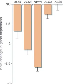
The adhesion gene HWP1 was not differentially expressed even though Ahr1p bound its promoter. However, HWP1 is expressed at higher levels in biofilm cells compared with free-living yeast cells (Garcia-Sanchez et al., 2004). Therefore, we determined the expression levels of a set of adhesion genes in the ahr1 strain compared with the wild type during the initial adhe-sion step in biofilm formation on polystyrene (Fig. 3). Under these conditions HWP1 was downregulated in the ahr1 strain, demonstrating that the Ahr1p–Mcm1p complex acti-vates the gene during adhesion to polystyrene. We also measured BCR1 expression to determine whether any changes in adhesion gene expression could be indirectly attributed to this factor but we observed no change (0.98 ⫾ 0.16).
Fig. 2. Ahr1p directs Mcm1p binding to the non-canonical motif.
A. Consensus motifs representing the binding sites of Mcm1p in a wild-type strain and an ahr1 deletion strain based on ChIP-chip. The sequence surrounding (covering five probes, 300 bp) the top peak intensity targets (> 5-fold enrichment) was sent to MEME
(http://meme.sdsc.edu/meme4_5_0/intro.html) and the most significant high-complexity motifs are reported.
B. ChIP-chip binding curves representing Ahr1p, Mcm1p(WT) and Mcm1p(ahr1) binding for type 1 (i), type 2 (ii) and type 3 (iii) Mcm1p targets. Data points for the curves were plotted at each probe position of 60 bp intervals. Values were determined by taking the mean fold enrichment of each probe and the surrounding four probes.
C. Immunoprecipitations with an Ahr1p HA-tagged and Mcm1p Myc-tagged strain were performed with cells grown in YPD media at 30°C. The top labels refer to the antibody used in the immunoprecipitation and the labels on the right refer to the primary antibody used to probe the membrane. The GFP antibody was used as a negative control and WCE represents the whole cell extract.
Disrupting the Ahr1p–Mcm1p complex affects adhesion ability and hyphal growth
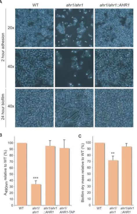
During analysis of the adhesion gene expression levels, we observed that after the 2 h incubation on polystyrene there was a clear reduction in the number of ahr1 adhered cells compared with the wild type (Fig. 4A). This adhesion defect was also observed when silicone was used as the substrate (data not shown). For the polystyrene sample, a semi-quantitative XTT assay was performed to measure the metabolic activity of the cells and thus approximate the number of cells. The wells containing the ahr1 cultures had about one-third of the metabolic activity of the wells containing the wild-type cultures (Fig. 4B). The ahr1/
AHR1-TAP strain was included to demonstrate that the
TAP-tag used for the ChIP-chip experiment did not inter-fere with the function of Ahr1p. The ahr1 strain was still able to develop a biofilm with filamentation, although the density was reduced because of the initial adhesion defect (Fig. 4A). This result was reflected by the reduction in the biofilm dry mass (Fig. 4C).
Although we observed hyphal formation in the ahr1 strain during biofilm development, we wanted to further investigate the role of the Ahr1p–Mcm1p complex in hyphal growth as our initial hypothesis was that Ahr1p was a morphological regulator and the ChIP-chip of Ahr1p
was enriched for targets involved in filamentous growth. On solid media at 37°C with YPD, YPD and serum, or M199 pH 8, the ahr1 colonies were unable to form wrinkles or filaments compared with the wild type (Fig. 5). In liquid media, the ahr1 strain was still able to filament when induced with serum but the average hyphal length of the ahr1 cells was significantly shorter (P< 0.0001,
t-test) than either the wild-type or complemented strains
(Table S6). Therefore, the Ahr1p–Mcm1p complex is involved in the regulation of adhesion and hyphal growth.
An ahr1 strain has attenuated virulence in a systemic infection mouse model
As the Ahr1p–Mcm1p complex was involved in regulating adhesion and showed defects in the yeast–hyphal switch, we tested whether deleting AHR1 affects the virulence in a systemic infection C57BL/6J mouse model. However, because the ahr1 mutant (CAS12) was constructed in a uracil auxotrophic background (BWP17), which can influ-ence the interpretation of virulinflu-ence results (Lay et al., 1998), we created a new ahr1 deletion strain (CaEE534) in the SN95 background. The SN95 strain does not have any influence on virulence as shown by different labs using various mouse models (Noble and Johnson, 2005; Epp
et al., 2010a). The SN95 ahr1 deletion strain (CaEE534)
showed filamentation and biofilm phenotypes similar to strain CAS12 (data not shown). In the C57BL/6J mouse model, the ahr1 strain (CaEE534) displayed significant attenuated virulence compared with both the wild-type (SN95, P = 0.0152, log–rank test) and complemented strains (CaEE573, P = 0.0032) (Fig. 6A). We also observed that the ahr1-injected mice accumulated a sig-nificantly higher fungal burden at the time when the mice became moribund (Fig. 6B), suggesting that the mice can tolerate higher levels of the attenuated pathogen. Together, our data suggest that the Ahr1p–Mcm1p regulon is impor-tant for the full pathogenicity of C. albicans.
Discussion
The sequencing of multiple Candida species has allowed for a detailed comparative genomic analysis between the genomes of the Saccharomycotina subphylum (Butler
et al., 2009). A major goal of such an investigation is to
identify the genomic basis for the phenotypic differences in pathogenicity. As the zinc finger transcription factor family is more abundant in Candida species compared with
Sac-charomyces species (Butler et al., 2009), these
transcrip-tion factors are ideal candidates for regulators of Candida-specific processes important for virulence including the yeast–hyphal transition and biofilm formation. By investi-gating the Candida-specific zinc cluster transcription factor Ahr1p in C. albicans, we were able to identify a new Mcm1p -3
-2.5 -2 -1.5
NC ALS1 ALS4 HWP1 ALS3
Fold change in gene expression
ALS9
Fig. 3. The Ahr1p–Mcm1p complex activates ALS1, ALS4 and
HWP1 under adhesion-promoting growth conditions. RNA was
extracted after 2 h of adhesion to a polystyrene surface and mRNA expression of selected adhesion genes was determined by qPCR for the ahr1 strain (CAS12) compared with the wild-type strain (CAS19). ‘NC’ indicates no change in gene expression. ALS3 and
ALS9 were included as comparison references as neither Ahr1p
nor Mcm1p bound their promoters and their expression was unchanged in the ahr1 transcription profile.
co-regulator involved in virulence and virulence-related processes. Using ChIP-chip and co-immunoprecipitation, we established that Ahr1p binds its target promoters, including several adhesion genes, through a zinc cluster factor motif, and that it interacts with and recruits Mcm1p to these sites. Previously observed Mcm1p regulons required both Mcm1p and the cofactor to bind adjacently to the DNA. Therefore, our study not only established an interac-tion between Mcm1p and Ahr1p but also a new mechanism of Mcm1p-directed regulation.
This new mode of Mcm1p regulation appears to be a recent development; the Ahr1p motif is only found in C.
albicans and the closely related C. dubliniensis as shown
by promoter analysis of the Mcm1p non-canonical motif target orthologues across 32 fungal species (Tuch et al., 2008), although the complex may have evolved earlier and just recently moved to this set of targets. If the regulon is a recent development, it is possible that the mode of Ahr1p–Mcm1p regulation is still in a transitory state. Originally, Ahr1p and Mcm1p might have each
Fig. 4. Disrupting the Ahr1p–Mcm1p complex affects adherence to polystyrene and reduces biofilm density. Strains used were WT (CAS19), ahr1/ahr1 (CAS12),
ahr1/ahr1::AHR1 (CAS13) and ahr1/AHR1-TAP (CAS15).
A. Overnight cultures were resuspended in RPMI media at OD600= 1 and 100 ml was added to each well in a 96-well plate. After 2 h of rocking incubation at 37°C, the non-adherent cells were removed, the wells were washed with PBS, and pictures were taken at 20¥ (top panel) and 40¥ (middle panel) magnification. Fresh RPMI media was added, the plate was incubated with rocking for 24 h at 37°C to allow for biofilm
development, and pictures were then taken at 40¥ magnification (bottom panel).
B. An XTT assay was performed after the 2 h adhesion step. Absorbencies were reported relative to the WT strain. The symbol ‘***’ indicates a significant difference (P< 0.0001,
t-test) compared with both wild-type and
complemented strains.
C. Biofilm dry masses were determined after 24 h of development. Masses were reported relative to the WT strain. The symbol ‘**’ indicates a significant difference (P< 0.01,
t-test) compared with both wild-type and
bound DNA separately, similar to the situation observed with the other known yeast Mcm1p regulons (Messenguy and Dubois, 2003). Over time, Ahr1p and Mcm1p might then have evolved to interact at a protein level with DNA providing a scaffold for this to occur. Eventually, the two proteins developed a strong enough interaction that Mcm1p no longer needed to directly bind DNA. Without selective pressure acting on the Mcm1p motif at the Ahr1p–Mcm1p targets, it was lost for most of the targets, resulting in the type 2 targets that are completely depen-dent on Ahr1p DNA binding, while it was retained at a few sites, the current type 3 targets containing both Ahr1p and Mcm1p motifs. Alternatively, the type 2 targets might rep-resent the ancestral mode of regulation developed by an initial protein–protein interaction between Mcm1p and Ahr1p. Over time, Mcm1p binding sites developed for a few targets allowing Mcm1p to directly interact with the DNA. In this scenario, the regulon is evolving towards type 3 targets and the standard mode of Mcm1p regulation.
Mcm1p type 2 targets demonstrate the potential of ChIP-chip to detect indirect protein–DNA interactions and falsely associate a motif with a transcription factor. Recent
studies have developed methods that attempt to address this problem by distinguishing direct and indirect transcrip-tion factor binding events (Gordan et al., 2009; Zhu et al., 2009). It was found that 16% of the ChIP-chip data sets from S. cerevisiae correlated with indirect binding of the factor (Gordan et al., 2009). The ability to determine whether a transcription factor is binding directly or indi-rectly is useful in predicting protein complexes, just as we showed in this study.
We further investigated the functional role of this newly formed Mcm1p complex in C. albicans. Our results established that the Ahr1p–Mcm1p complex is a direct activator of several key adhesion genes and that disrupting this complex by deleting AHR1 resulted in reduced adherence of C. albicans cells to polystyrene
Fig. 5. The Ahr1p–Mcm1p complex controls the yeast–hyphal switch. Cells were serially diluted and a representative dilution is shown. Strains used were WT (CAS19), ahr1/ahr1 (CAS12) and
ahr1/ahr1::AHR1 (CAS13). Pictures were taken after 4 days for
M199 pH 8 and after 2 days for the other conditions. Although the
ahr1/ahr1::AHR1 strain did not revert the phenotype for YPD and
M199 pH 8 at 37°C, three independently constructed heterozygous strains (CAS25) showed the same phenotype as the complemented strain (data not shown), indicating that AHR1 shows
haplo-insufficiency under some hyphal growth conditions.
0 20 40 60 100 80 Sur vi ving mice (%) Days post-injection 0 2 4 6 8 10 12 14 A WT ahr1/ahr1::AHR1 ahr1/ahr1 B lo g CFU / kidne y 5 6 7 8
***
**
ns WT ahr1/ahr1::AHR1 ahr1/ahr1Fig. 6. An ahr1 strain displays attenuated virulence in a C57BL/6J mouse model.
A. Survival curves of mice infected with WT (SN95), ahr1/ahr1 (CaEE534) and ahr1/ahr1::AHR1 (CaEE573) strains. Mice were monitored daily according to approved protocols.
B. Comparison of the kidney fungal load of mice infected with WT (SN95), ahr1/ahr1 (CaEE534) and ahr1/ahr1::AHR1 (CaEE573) strains. Kidney fungal burdens were assessed at the time of sacrifice when mice were determined to be moribund. Mutant-infected mice survived longer and accumulated a statistically significantly higher fungal burden compared with both WT and revertant-infected mice as indicated (***P< 0.001; **P< 0.01; ns P > 0.05).
and silicone surfaces. The ahr1 strain was able to develop a biofilm, but with a reduced biomass. As well, the ahr1 strain had defects in the yeast–hyphal switch under several different growth conditions. Although we analysed the ahr1 strain in order to selectively disrupt the Ahr1p–Mcm1p complex, it should be noted that over-expression of MCM1 has been shown to result in increased adhesion as well as defects in the yeast– hyphal transition (Rottmann et al., 2003).
Our results, combined with previous studies on Bcr1p, suggest that the Ahr1p–Mcm1p complex and Bcr1p regulate adhesion genes separately. Ahr1p or Mcm1p did not bind the promoter of Bcr1p and BCR1 expres-sion was not affected by deletion of AHR1. As well,
AHR1 expression was not affected by deletion of BCR1
(Nobile and Mitchell, 2005). Furthermore, the Ahr1p– Mcm1p complex and Bcr1p have some different adhe-sion targets as Ahr1p–Mcm1p solely regulate ALS4 while Bcr1p solely regulates ALS3 (Nobile and Mitchell, 2005; Nobile et al., 2006b).
Disrupting the Ahr1p–Mcm1p interaction caused Mcm1p to bind new sites in which the peak intensity was greater in the ahr1 mutant strain compared with the wild-type strain (wild-type 4 targets). Analysis of this group of targets revealed that these sites are almost exclusively found near protein translation genes including most of the ribosomal protein encoding genes. Interestingly, many of the peaks were at the 3′ end of these genes so our peak analysis algorithm would either not associate the peak with a gene or assign the peak to a different gene. The peak intensity of the type 4 targets was generally just above the cut-off in the ahr1 strain (2- to 2.5-fold) and often there was a weak signal in the wild-type strain. Generally, no Mcm1p motif was present. Therefore, it appears that Mcm1p very weakly associates with the translational machinery genes and disrupting the interac-tion with Ahr1p enhances this associainterac-tion.
Our study primarily focused on the role of the Ahr1p– Mcm1p complex in regulating adhesion; however, our results suggest additional roles for the regulon. As pre-viously mentioned, there is a link between biofilm devel-opment and drug resistance. Mcm1p has been shown to regulate MDR1, which encodes a key drug transporter that is upregulated in many fluconazole-resistant strains, although it was concluded that elements upstream of the Mcm1p canonical motif were also required for MDR1 activation (Riggle and Kumamoto, 2006). Our results demonstrated that Mcm1p binds to the MDR1 promoter at its canonical motif independently of Ahr1p and is also recruited to a non-canonical motif by Ahr1p. This Ahr1p motif is upstream of the Mcm1p motif and is located in a region identified as containing a cis-acting sequence of
MDR1 (Harry et al., 2005; Hiller et al., 2006). As well,
Cdr1p, another key drug transporter upregulated in
fluconazole-resistant strains, is also a type 3 Mcm1p target. Although the ahr1 strain shows no difference in sensitivity to fluconazole compared with the wild type (Homann et al., 2009), the role of the Ahr1p–Mcm1p complex in regulating drug transporters is an area of interest.
Links have also been established between biofilm for-mation and mating in C. albicans. Pheromone induces mating in opaque cells but promotes substrate adhesion and enhanced biofilm development in white cells (Daniels
et al., 2006). Both white and opaque cell responses to
pheromone involve the same receptors and MAP-kinase pathway but activate a different transcription factor (Yi
et al., 2008). While Cph1p is the target of the opaque cell
pheromone response pathway (Yi et al., 2008), Tec1p is the downstream target of the white cell pheromone response pathway (Sahni et al., 2010). Tuch et al. (2008) identified that the subset of non-canonical Mcm1p targets includes several regulators of the white–opaque switch and strongly overlaps with the binding targets of Wor1p. As Ahr1p recruits Mcm1p to the promoter regions of TEC1 and the white–opaque regulators WOR1, WOR2 and
EFG1, the complex’s role in this aspect of cell function is
another area of interest.
Our study provides insight into a Mcm1p regulon con-trolled by a Candida-specific zinc cluster factor involved in the regulation of important pathogenic characteristics such as adhesion and the yeast–hyphal transition. The reduced virulence of the ahr1 strain in a systemic infec-tion mouse model further highlights the potential of zinc cluster proteins as antifungal drug targets. Zinc cluster proteins have a common structural domain, are fungal specific and regulate diverse cellular processes; there-fore, the targeted disruption of the zinc cluster domain would certainly result in severe consequences to the cell. Continuing to characterize the zinc cluster family members unique to C. albicans will allow for more insight into the regulation of Candida-specific transcrip-tional networks that are critical for the virulence of this fungus.
Experimental procedures
C. albicans strain construction and media
The C. albicans strains used in this study are listed in Table S7. Unless otherwise stated, cells were grown at 30°C in media containing 1% yeast extract, 2% peptone, 2% dex-trose (YPD) and supplemented with uridine (50 mg ml-1).
Gene disruption of AHR1 was done as described previ-ously for NDT80 (Sellam et al., 2009b) in a BWP17 back-ground using URA3 and HIS1 PCR deletion cassettes, resulting in ahr1 deletion strain CAS12. For an auxotrophic control, the plasmid CIp20 (Dennison et al., 2005) was digested with StuI and transformed into BWP17, resulting in strain CAS19. The ahr1/ahr1::AHR1 complemented strain
(CAS13) was created using the SAT1-flipper cassette as previously done for TYE7 and GAL4 (Askew et al., 2009). The AHR1 gene along with 500 bp upstream and down-stream homology regions of AHR1 were cloned into plasmid pSFS2A (Reuss et al., 2004), resulting in plasmid pCA4. We selected clones that replaced the HIS1 deletion cassette and then restored histidine prototrophy by transformation with NruI-digested pGEM-HIS1 (Wilson et al., 1999). As some filamentation phenotypes were not reverted, independent
AHR1/ahr1 heterozygous deletion strains (CAS25) were
created with the HIS1 PCR deletion cassette and then trans-formed with StuI-digested CIp10 (Murad et al., 2000). For ChIP-chip experiments, Ahr1p and Mcm1p were tagged chro-mosomally with a TAP-URA3 PCR product (Lavoie et al., 2008) in either a BWP17 or ahr1 background (strains CAS11, CAS23 and CAS24). For the ahr1 strain (CAS12) back-ground, the URA3 marker from the deletion cassette was recycled using 5-fluoroorotic acid. To ensure that the TAP-tag did not interfere with the function of Ahr1p, the HIS1 PCR deletion cassette was used to delete the untagged allele of strain CAS11, resulting in strain CAS15. For immunoprecipi-tations, Ahr1p was tagged with a HA-HIS1 PCR product and Mcm1p was tagged with a MYC-URA3 PCR product (Lavoie
et al., 2008) in a SN76 background, resulting in strain CAS18.
Correct integration for all tags was confirmed by PCR and Western blots were used to verify protein expression. For the mouse study, a new ahr1 deletion strain (CaEE534) and complemented strain (CaEE573) were constructed in the SN95 background to eliminate any URA3 positional effects (Lay et al., 1998). The nourseothricin marker was used as previously described (Epp et al., 2010b). Briefly, 300 bp of upstream and downstream homology regions of AHR1 were cloned into plasmid pSFS2A (Reuss et al., 2004), resulting in plasmid pEE95, to sequentially delete both alleles of AHR1. Plasmid pCA4 was used to construct the revertant strain (CaEE573) as was done to create CAS13 in the BWP17 background.
ChIP-chip
ChIP-chip experiments were performed as previously described (Askew et al., 2009) with two biological replicates for each condition. Half of the labelled samples were hybrid-ized to single spot full-genome (ORF and intergenic) micro-arrays containing 11 817 70-mer oligonucleotide probes (Lavoie et al., 2008). Targets with a fold enrichment > 1.5 were compared between the two replicates to ensure a high degree of overlap. The other half of one replicate was then chosen for hybridization to a custom designed whole-genome tiling array (Askew et al., 2009). Normalization and peak detection were performed as previously described (Askew
et al., 2009). Peaks located within 2 kb upstream and 120 bp
downstream (to account for location uncertainty of the tiling array) of a start codon were assigned to the gene and reported in Tables S1–S3 with ORFs annotated as ‘dubious’ or ‘spurious’ removed from the analysis. In the case of diver-gent promoters both genes were included provided the peak was within 2 kb of each gene’s start codon. Genes with multiple peaks in the promoter were counted only once for GO analysis, which was performed as previously described (Lavoie et al., 2010).
Immunoprecipitations and Western blotting
An Ahr1p HA-tagged and Mcm1p Myc-tagged strain (CAS18) was grown to OD600= 1.0 in YPD media at 30°C. Cells were harvested by centrifugation, washed with IP150 buffer [50 mM Tris-HCl (pH 7.4), 150 mM NaCl, 2 mM MgCl2, 0.1% NP40], and lysed by vortexing with glass beads in IP150 buffer supplemented with a protease inhibitor cocktail tablet (Roche) and 1 mM phenylmethylsulfonyl fluoride (PMSF). The protein extract was clarified by centrifugation and incu-bated at 4°C with beads conjugated with anti-Myc mouse monoclonal antibody (9E10), GFP rabbit polyclonal anti-body (Santa Cruz) or anti-HA rabbit polyclonal antianti-body (Roche). Following incubation, beads were washed three times with IP150 buffer, boiled with SDS gel loading buffer and resolved in 4–20% gradient SDS polyacrylamide gels. The separated polypeptides were transferred onto a nitrocel-lulose membrane and analysed by Western blotting using anti-HA (3F10) or anti-Myc (9E10) monoclonal antibodies.
RNA extraction, transcription profiles and qPCR analysis
RNA extraction and transcription profiles were performed and analysed as previously described (Askew et al., 2009). Four biological replicates were done (YPD at 30°C) comparing the wild-type (BWP17) and ahr1 deletion (CAS12) strains and gene lists were created using a fold change in expression > 1.5 or < 0.67 and a t-test P-value < 0.05.
Cells for qPCR analysis were prepared as described for the XTT adhesion assays below except 2 ml of OD600= 0.5 cell suspension was added to each well in a six-well poly-styrene plate (Nunclon). After a 2 h incubation at 37°C both adherent and non-adherent cells were collected with a cell scraper and the RNA was extracted. qPCR analysis was performed as previously described (Askew et al., 2009) except with the Mx3005P QPCR System (Agilent). Samples were done in triplicate and four biological replicates were performed.
Adhesion and biofilm analysis
XTT assays were carried out as previously described (Ramage et al., 2001; Kelly et al., 2004). Briefly, overnight YPD cultures were washed twice with PBS and resuspended in RPMI 1640 (Gibco) supplemented with L-glutamine to OD600= 1. Ninety-six-well polystyrene plates (Costar) were used and 100 ml of cells was added to each well. The plates were placed in a rocking incubator at 37°C for 2 h. The media and any non-adherent cells were removed and the wells were washed three times with PBS. After washing, 100 ml of a freshly prepared XTT-menadione solution (0.5 g l-1 XTT in PBS and 1 mM menadione in acetone) was added to sample and control wells. The plate was incubated in the dark for 2 h at 37°C and the colorimetric change resulting from XTT reduction was measured at 490 nm. Five biological replicates done in triplicate were performed.
Determination of biofilm dry mass was determined based on previous studies (Palanisamy et al., 2010) with 2 ml of OD600= 0.5 cell suspension added to each well in a six-well polystyrene plate (Nunclon) for the 2 h adhesion step. After 䊏
washing with PBS, 2 ml of fresh media was added and the plate was incubated with rocking for an additional 24 h. Bio-films were collected and dried at 37°C for 48 h. Three bio-logical replicates done in triplicate were performed.
Virulence study
The mouse study was carried out as previously described (Mullick et al., 2004). Briefly, 8- to 12-week-old C57BL/6J mice (Jackson Laboratories, Bar Harbor, ME, USA) were inoculated via the tail vein with 200 ml of a suspension con-taining 3 ¥ 105 C. albicans in PBS. Ten mice, five female and five male, were used for each experimental group. Mice were closely monitored according to approved protocols and those mice showing extreme lethargy were considered moribund and were euthanized. Kaplan–Meier survival curves were created and compared with the log–rank test and the Kruskal–Wallis test was used to assess significance between fungal burdens (GraphPad Prism 5). The number of fungal cells per kidney was determined by removing kidneys aseptically and homogenizing in PBS before plating on YPD plates containing chloramphenicol (34 mg ml-1). All experimental procedures involving mice were approved by the Biotechnology Research Institute Animal Care Commit-tee, which operated under the guidelines of the Canadian Council of Animal Care.
Accession codes
Microarray data for ChIP-chip and transcription profiling experiments have been submitted to the NCBI Gene Expres-sion Omnibus (GEO) under the Accession Number GSE25174 (http://www.ncbi.nlm.nih.gov/geo).
Acknowledgements
Thanks to members of the Whiteway Lab, BRI Microarray Lab, and BRI Animal Facility, especially Mario Mercier, Patri-cia Liscourt, Jessy Tremblay and Khairul Islam for technical assistance. This work was supported by grants from the Canadian Institute for Health Research (CIHR) to A.M., A.N. and M.W. (CTP-79843, MOP-84341 and MOP-42516). C.A. was supported by a NSERC PGS Extension Scholarship. This is National Research Council manuscript 53136.
References
Askew, C., Sellam, A., Epp, E., Hogues, H., Mullick, A., Nantel, A., and Whiteway, M. (2009) Transcriptional regu-lation of carbohydrate metabolism in the human pathogen
Candida albicans. PLoS Pathog 5: e1000612.
Borneman, A.R., Gianoulis, T.A., Zhang, Z.D., Yu, H., Rozo-wsky, J., Seringhaus, M.R., et al. (2007) Divergence of transcription factor binding sites across related yeast species. Science 317: 815–819.
Braun, B.R., van Het Hoog, M., d’Enfert, C., Martchenko M., Dungan, J., Kuo, A., et al. (2005) A human-curated anno-tation of the Candida albicans genome. PLoS Genet 1: 36–57.
Butler, G., Rasmussen, M.D., Lin, M.F., Santos, M.A., Sak-thikumar, S., Munro, C.A., et al. (2009) Evolution of
patho-genicity and sexual reproduction in eight Candida genomes. Nature 459: 657–662.
Chandra, J., Mukherjee, P.K., Leidich, S.D., Faddoul, F.F., Hoyer, L.L., Douglas, L.J., and Ghannoum, M.A. (2001) Antifungal resistance of candidal biofilms formed on denture acrylic in vitro. J Dent Res 80: 903–908.
Daniels, K.J., Srikantha, T., Lockhart, S.R., Pujol, C., and Soll, D.R. (2006) Opaque cells signal white cells to form biofilms in Candida albicans. EMBO J 25: 2240–2252. Dennison, P.M., Ramsdale, M., Manson, C.L., and Brown,
A.J. (2005) Gene disruption in Candida albicans using a synthetic, codon-optimised Cre-loxP system. Fungal Genet
Biol 42: 737–748.
Douglas, L.J. (2003) Candida biofilms and their role in infection. Trends Microbiol 11: 30–36.
Douzery, E.J., Snell, E.A., Bapteste, E., Delsuc, F., and Phil-ippe, H. (2004) The timing of eukaryotic evolution: does a relaxed molecular clock reconcile proteins and fossils?
Proc Natl Acad Sci USA 101: 15386–15391.
Epp, E., Vanier, G., Harcus, D., Lee, A.Y., Jansen, G., Hallett, M., et al. (2010a) Reverse genetics in Candida albicans predicts ARF cycling is essential for drug resistance and virulence. PLoS Pathog 6: e1000753.
Epp, E., Walther, A., Lepine, G., Leon, Z., Mullick, A., Raymond, M., et al. (2010b) Forward genetics in Candida
albicans that reveals the Arp2/3 complex is required for
hyphal formation, but not endocytosis. Mol Microbiol 75: 1182–1198.
Garcia-Sanchez, S., Aubert, S., Iraqui, I., Janbon, G., Ghigo, J.M., and d’Enfert, C. (2004) Candida albicans biofilms: a developmental state associated with specific and stable gene expression patterns. Eukaryot Cell 3: 536–545.
Gordan, R., Hartemink, A.J., and Bulyk, M.L. (2009) Distin-guishing direct versus indirect transcription factor-DNA interactions. Genome Res 19: 2090–2100.
Harry, J.B., Oliver, B.G., Song, J.L., Silver, P.M., Little, J.T., Choiniere, J., and White, T.C. (2005) Drug-induced regu-lation of the MDR1 promoter in Candida albicans.
Antimi-crob Agents Chemother 49: 2785–2792.
Hawser, S.P., and Douglas, L.J. (1995) Resistance of
Candida albicans biofilms to antifungal agents in vitro. Anti-microb Agents Chemother 39: 2128–2131.
Heckman, D.S., Geiser, D.M., Eidell, B.R., Stauffer, R.L., Kardos, N.L., and Hedges, S.B. (2001) Molecular evidence for the early colonization of land by fungi and plants.
Science 293: 1129–1133.
Hiller, D., Stahl, S., and Morschhauser, J. (2006) Multiple cis-acting sequences mediate upregulation of the MDR1 efflux pump in a fluconazole-resistant clinical Candida
albi-cans isolate. Antimicrob Agents Chemother 50: 2300–
2308.
Hogues, H., Lavoie, H., Sellam, A., Mangos, M., Roemer, T., Purisima, E., et al. (2008) Transcription factor substitution during the evolution of fungal ribosome regulation. Mol Cell
29: 552–562.
Homann, O.R., Dea, J., Noble, S.M., and Johnson, A.D. (2009) A phenotypic profile of the Candida albicans regu-latory network. PLoS Genet 5: e1000783.
Kelly, M.T., MacCallum, D.M., Clancy, S.D., Odds, F.C., Brown, A.J., and Butler, G. (2004) The Candida albicans
CaACE2 gene affects morphogenesis, adherence and virulence. Mol Microbiol 53: 969–983.
Kojic, E.M., and Darouiche, R.O. (2004) Candida infections of medical devices. Clin Microbiol Rev 17: 255–267. Lavoie, H., Sellam, A., Askew, C., Nantel, A., and Whiteway,
M. (2008) A toolbox for epitope-tagging and genome-wide location analysis in Candida albicans. BMC Genomics 9: 578.
Lavoie, H., Hogues, H., Mallick, J., Sellam, A., Nantel, A., and Whiteway, M. (2010) Evolutionary tinkering with conserved components of a transcriptional regulatory network. PLoS
Biol 8: e1000329.
Lay, J., Henry, L.K., Clifford, J., Koltin, Y., Bulawa, C.E., and Becker, J.M. (1998) Altered expression of selectable marker URA3 in gene-disrupted Candida albicans strains complicates interpretation of virulence studies. Infect
Immun 66: 5301–5306.
Leroy, O., Gangneux, J.P., Montravers, P., Mira, J.P., Gouin, F., Sollet, J.P., et al. (2009) Epidemiology, management, and risk factors for death of invasive Candida infections in critical care: a multicenter, prospective, observational study in France (2005–2006). Crit Care Med 37: 1612–1618. MacPherson, S., Larochelle, M., and Turcotte, B. (2006) A
fungal family of transcriptional regulators: the zinc cluster proteins. Microbiol Mol Biol Rev 70: 583–604.
Mermel, L.A., Farr, B.M., Sherertz, R.J., Raad, I.I., O’Grady, N., Harris, J.S., and Craven, D.E. (2001) Guidelines for the management of intravascular catheter-related infections.
Clin Infect Dis 32: 1249–1272.
Messenguy, F., and Dubois, E. (2003) Role of MADS box proteins and their cofactors in combinatorial control of gene expression and cell development. Gene 316: 1–21. Mullick, A., Elias, M., Picard, S., Bourget, L., Jovcevski, O.,
Gauthier, S., et al. (2004) Dysregulated inflammatory response to Candida albicans in a C5-deficient mouse strain. Infect Immun 72: 5868–5876.
Murad, A.M., Lee, P.R., Broadbent, I.D., Barelle, C.J., and Brown, A.J. (2000) CIp10, an efficient and convenient integrating vector for Candida albicans. Yeast 16: 325– 327.
Nobile, C.J., and Mitchell, A.P. (2005) Regulation of cell-surface genes and biofilm formation by the C. albicans transcription factor Bcr1p. Curr Biol 15: 1150–1155. Nobile, C.J., Nett, J.E., Andes, D.R., and Mitchell, A.P.
(2006a) Function of Candida albicans adhesin Hwp1 in biofilm formation. Eukaryot Cell 5: 1604–1610.
Nobile, C.J., Andes, D.R., Nett, J.E., Smith, F.J., Yue, F., Phan, Q.T., et al. (2006b) Critical role of Bcr1-dependent adhesins in C. albicans biofilm formation in vitro and in vivo. PLoS Pathog 2: e63.
Nobile, C.J., Nett, J.E., Hernday, A.D., Homann, O.R., Dene-ault, J.S., Nantel, A., et al. (2009) Biofilm matrix regulation by Candida albicans Zap1. PLoS Biol 7: e1000133. Noble, S.M., and Johnson, A.D. (2005) Strains and strategies
for large-scale gene deletion studies of the diploid human fungal pathogen Candida albicans. Eukaryot Cell 4: 298– 309.
Palanisamy, S.K., Ramirez, M.A., Lorenz, M., and Lee, S.A. (2010) Candida albicans PEP12 is required for biofilm integrity and in vivo virulence. Eukaryot Cell 9: 266– 277.
Ramage, G., Vande Walle, K., Wickes, B.L., and Lopez-Ribot, J.L. (2001) Standardized method for in vitro antifun-gal susceptibility testing of Candida albicans biofilms.
Antimicrob Agents Chemother 45: 2475–2479.
Ramage, G., VandeWalle, K., Bachmann, S.P., Wickes, B.L., and Lopez-Ribot, J.L. (2002) In vitro pharmacodynamic properties of three antifungal agents against preformed
Candida albicans biofilms determined by time-kill studies. Antimicrob Agents Chemother 46: 3634–3636.
Reuss, O., Vik, A., Kolter, R., and Morschhauser, J. (2004) The SAT1 flipper, an optimized tool for gene disruption in
Candida albicans. Gene 341: 119–127.
Riggle, P.J., and Kumamoto, C.A. (2006) Transcriptional regulation of MDR1, encoding a drug efflux determinant, in fluconazole-resistant Candida albicans strains through an Mcm1p binding site. Eukaryot Cell 5: 1957–1968. Rottmann, M., Dieter, S., Brunner, H., and Rupp, S. (2003) A
screen in Saccharomyces cerevisiae identified CaMCM1, an essential gene in Candida albicans crucial for morphogenesis. Mol Microbiol 47: 943–959.
Sahni, N., Yi, S., Daniels, K.J., Huang, G., Srikantha, T., and Soll, D.R. (2010) Tec1 mediates the pheromone response of the white phenotype of Candida albicans: insights into the evolution of new signal transduction pathways. PLoS
Biol 8: e1000363.
Sellam, A., Al-Niemi, T., McInnerney, K., Brumfield, S., Nantel, A., and Suci, P.A. (2009a) A Candida albicans early stage biofilm detachment event in rich medium. BMC
Microbiol 9: 25.
Sellam, A., Tebbji, F., and Nantel, A. (2009b) Role of Ndt80p in sterol metabolism regulation and azole resistance in
Candida albicans. Eukaryot Cell 8: 1174–1183.
Sellam, A., Askew, C., Epp, E., Tebbji, F., Mullick, A., White-way, M., and Nantel, A. (2010) Role of the transcription factor CaNdt80p in cell separation, hyphal growth and virulence in Candida albicans. Eukaryot Cell 9: 634– 644.
Tuch, B.B., Galgoczy, D.J., Hernday, A.D., Li, H., and Johnson, A.D. (2008) The evolution of combinatorial gene regulation in fungi. PLoS Biol 6: e38.
Vediyappan, G., Rossignol, T., and d’Enfert, C. (2010) Inter-action of Candida albicans biofilms with antifungals: tran-scriptional response and binding of antifungals to beta-glucans. Antimicrob Agents Chemother 54: 2096–2111. Wilson, R.B., Davis, D., and Mitchell, A.P. (1999) Rapid
hypothesis testing with Candida albicans through gene disruption with short homology regions. J Bacteriol 181: 1868–1874.
Wisplinghoff, H., Bischoff, T., Tallent, S.M., Seifert, H., Wenzel, R.P., and Edmond, M.B. (2004) Nosocomial bloodstream infections in US hospitals: analysis of 24 179 cases from a prospective nationwide surveillance study.
Clin Infect Dis 39: 309–317.
Yi, S., Sahni, N., Daniels, K.J., Pujol, C., Srikantha, T., and Soll, D.R. (2008) The same receptor, G protein, and mitogen-activated protein kinase pathway activate different downstream regulators in the alternative white and opaque pheromone responses of Candida albicans. Mol Biol Cell
19: 957–970.
Zhao, X., Oh, S.H., Yeater, K.M., and Hoyer, L.L. (2005) Analysis of the Candida albicans Als2p and Als4p adhesins 䊏
suggests the potential for compensatory function within the Als family. Microbiology 151: 1619–1630.
Zhao, X., Daniels, K.J., Oh, S.H., Green, C.B., Yeater, K.M., Soll, D.R., and Hoyer, L.L. (2006) Candida albicans Als3p is required for wild-type biofilm formation on silicone elas-tomer surfaces. Microbiology 152: 2287–2299.
Zhu, C., Byers, K.J., McCord, R.P., Shi, Z., Berger, M.F., Newburger, D.E., et al. (2009) High-resolution DNA-binding specificity analysis of yeast transcription factors.
Genome Res 19: 556–566.
Supporting Information
Additional supporting information may be found in the online version of this article.
Please note: Wiley-Blackwell are not responsible for the content or functionality of any supporting materials supplied by the authors. Any queries (other than missing material) should be directed to the corresponding author for the article.