HAL Id: hal-02985330
https://hal.archives-ouvertes.fr/hal-02985330
Submitted on 2 Nov 2020HAL is a multi-disciplinary open access archive for the deposit and dissemination of sci-entific research documents, whether they are pub-lished or not. The documents may come from teaching and research institutions in France or abroad, or from public or private research centers.
L’archive ouverte pluridisciplinaire HAL, est destinée au dépôt et à la diffusion de documents scientifiques de niveau recherche, publiés ou non, émanant des établissements d’enseignement et de recherche français ou étrangers, des laboratoires publics ou privés.
Loop 1 of APOBEC3C regulates its antiviral activity
against HIV-1
Ananda Ayyappan Jaguva Vasudevan, Kannan Balakrishnan, Christoph G.
W. Gertzen, Fanni Borvetö, Zeli Zhang, Anucha Sangwiman, Ulrike Held,
Caroline Küstermann, Sharmistha Banerjee, Gerald Schumann, et al.
To cite this version:
Ananda Ayyappan Jaguva Vasudevan, Kannan Balakrishnan, Christoph G. W. Gertzen, Fanni Borvetö, Zeli Zhang, et al.. Loop 1 of APOBEC3C regulates its antiviral activity against HIV-1. Journal of Molecular Biology, Elsevier, In press, �10.1016/j.jmb.2020.10.014�. �hal-02985330�
Loop 1 of APOBEC3C regulates its antiviral
activity against HIV-1
Ananda Ayyappan Jaguva Vasudevan1,*,#,§, Kannan Balakrishnan1,2,#, Christoph G. W. Gertzen3,4,5, Fanni Borvető6, Zeli Zhang1,§§, Anucha Sangwiman1, Ulrike Held7, Caroline Küstermann7, Sharmistha Banerjee2, Gerald G. Schumann7,Dieter Häussinger1, Ignacio G. Bravo6, Holger Gohlke3,4, Carsten Münk1,*
1Clinic for Gastroenterology, Hepatology, and Infectiology, Medical Faculty, Heinrich Heine
University Düsseldorf, Düsseldorf, Germany. 2Department of Biochemistry, School of Life
Sciences, University of Hyderabad, Gachibowli, Hyderabad, India. 3Institute for
Pharmaceutical and Medicinal Chemistry, Heinrich Heine University Düsseldorf, Düsseldorf, Germany. 4John von Neumann Institute for Computing (NIC), Jülich Supercomputing Centre &
Institute of Biological Information Processing (IBI-7: Structural Biochemistry), Forschungszentrum Jülich GmbH, Jülich, Germany. 5Center for Structural Studies (CSS),
Heinrich Heine University Düsseldorf, Düsseldorf, Germany. 6Centre National de la Recherche
Scientifique, Laboratory MIVEGEC (CNRS, IRD, Uni Montpellier), Montpellier, France. 7Division
of Medical Biotechnology, Paul-Ehrlich-Institute, Langen, Germany.
# A.A.J.V and K.B contributed equally to this article.
Present address:
§Structural Cell Biology Group, Genome Integrity and Structural Biology Laboratory, National
Institute of Environmental Health Sciences (NIEHS), NIH, Research Triangle Park, NC 27709, USA.
§§La Jolla Institute for Immunology, La Jolla, CA 92037, USA.
*Address correspondence to Ananda Ayyappan Jaguva Vasudevan, Structural Cell Biology
Group, Genome Integrity and Structural Biology Laboratory, National Institute of Environmental Health Sciences (NIEHS), NIH, Research Triangle Park, NC 27709, USA. anand.jaguvavasudevan@nih.gov. Carsten Münk, Clinic for Gastroenterology, Hepatology, and Infectiology, Medical Faculty, Heinrich Heine University Düsseldorf, Building 23.12.U1.82, Moorenstr. 5, 40225 Düsseldorf, Germany, Tel: +49 (0) 211-81-10887, carsten.muenk@med.uni-duesseldorf.de.
Short title: Evolution and role of loop 1 of A3C
ABSTRACT
APOBEC3 deaminases (A3s) provide mammals with an anti-retroviral barrier by catalyzing dC-to-dU deamination on viral ssDNA. Within primates, A3s have undergone a complex evolution
via gene duplications, fusions, arms race and selection. Human APOBEC3C (hA3C) efficiently
restricts the replication of viral infectivity factor (vif)-deficient Simian immunodeficiency virus (SIVΔvif), but for unknown reasons, it inhibits HIV-1Δvif only weakly. In catarrhines (Old World monkeys and apes), the A3C loop 1 displays the conserved amino acid pair WE, while the corresponding consensus sequence in A3F and A3D is the largely divergent pair RK, which is also the inferred ancestral sequence for the last common ancestor of A3C and of the C-terminal domains of A3D and A3F in primates. Here, we report that modifying the WE residues in hA3C loop 1 to RK leads to stronger interactions with substrate ssDNA, facilitating catalytic function, which results in a drastic increase in both deamination activity and in the ability to restrict HIV-1 and LINE-1 replication. Conversely, the modification hA3F_WE resulted only in a marginal decrease in HIV-1Δvif inhibition. We propose that the two series of ancestral gene duplications that generated A3C, A3D-CTD and A3F-CTD allowed neo/subfunctionalization: A3F-CTD maintained the ancestral RK residues in loop 1, while diversifying selection resulted in the RK WE modification in Old World anthropoids’ A3C, possibly allowing for novel substrate specificity and function.
HIGHLIGHTS
• Loop 1 residues in primate APOBEC3C and closely related A3s are under positive selection
• A3C in monkeys and apes replaced the highly active residues RK to WE during A3C gene evolution
• Replacing WE residues in loop 1 of A3C with RK turns A3C into a superior restriction factor.
• Loop 1 residues RK caused enhancement of A3C catalytic activity by improved DNA interaction.
• The cytosine deamination hotspots and cell-wide distribution of A3C were not affected by loop 1 residues RK.
Keywords: APOBEC3C_A3F_cytidine deaminase, Sooty Mangabey monkey, human
INTRODUCTION
The APOBEC3 (A3) family of single-stranded (ss) DNA cytidine deaminases builds an intrinsic immune defense against retroviruses, retrotransposons, and other viral pathogens [1-4]. There are seven human A3 proteins that possess either one (A3A, A3C, and A3H) or two (A3B, A3D, A3F, and A3G) zinc (Z)-coordinating DNA cytosine deaminase motifs, Z motifs can be classified into three groups (Z1, Z2, Z3), but share the consensus signature HXE[X23-28]PC[X 2-4]C (where X indicates a non-conserved position) [5-9]. A3C is the only single-domain A3Z2
protein in humans. During primate evolution, the ancestor of the A3C gene duplicated several times and formed double-domain A3Z2-A3Z2 genes, which are A3D and A3F [6]. Initially, A3G was characterized as the factor capable of restricting infection of HIV-1 lacking Vif (viral infectivity factor) protein in non-permissive T cell lines and its biochemical properties and biological functions have been extensively studied [3, 10-13].
The encapsidation of A3s into the viral particles is crucial for virus inhibition [14-19]. During reverse transcription, viral core-associated A3 enzymes can deaminate cytidines (dC) on the retroviral ssDNA into uridines (dU). These base modifications in the minus-strand DNA cause coding changes and premature stop codons in the plus-strand viral genome (dG dA hypermutation), which impair or suppress viral infectivity [2, 11, 20-23]. In addition to the mutagenic activity of the virus-incorporated A3s, deaminase-independent mechanisms of restriction have been identified such as impeding reverse transcription or inhibiting DNA integration [24-29]. To counteract A3 mediated inhibition, lentiviruses evolved the Vif protein, which physically interacts with A3s, targeting them for polyubiquitination and proteasomal
degradation [30-32]. These A3-Vif interactions are often species-specific and an important factor reducing virus cross-species transmission [33-38].
In addition to A3G, A3D, A3F, and A3H were shown to restrict HIV-1 lacking vif (HIV-1Δvif) [2, 37, 39-42]. Recently, mutation signatures resulting from the catalytic activity of nuclearly localized A3s (especially A3A, A3B, and likely A3H) were reported in several cancer types [43-50]. The knowledge about A3C is rather sparse, it is distributed in both cytoplasm and nucleus [51] and does not seem to be a causative agent of chromosomal DNA mutations. In addition, human A3C is known to act as a potent inhibitor of Simian immunodeficiency virus from African green monkey (SIVagm) and from rhesus macaque (SIVmac), limits the infectivity of herpes simplex virus, certain human papillomaviruses, murine leukemia virus, Bet-deficient foamy virus, and hepatitis B virus, and represses the replication of LINE-1 (L1) endogenous retrotransposons [51-61]. In contrast, the restrictive role of A3C on HIV-1 is marginal and there are several contradictory findings regarding its viral packaging and cytidine deamination activity [42, 52, 62-64]. Notably, A3C is ubiquitously expressed in lymphoid cells [5, 52, 65, 66], mRNA expression levels of A3C are higher in HIV-infected CD4+ T lymphocytes [42, 52], and
significantly elevated in elite controllers compared to ART-suppressed individuals [67]. A3C was found to moderately deaminate HIV-1 DNA if expressed in target cells of the virus with the effect of increasing viral diversity rather than causing restriction [65].
The crystal structure of A3C and its HIV-1 Vif-binding interface have been solved [68]. The study revealed several key residues in the hydrophobic V-shaped groove formed by the α2 and α3 helices of A3C that facilitate Vif binding resulting in proteasome-mediated degradation of A3C [68]. We have extended this finding and identified additional Vif interaction sites in the
α4 helix of A3C [69]. Apart from a previous study that predicted putative DNA substrate binding pockets [57], biochemical and structural aspects of A3C enzymatic activity and their relevance for antiviral activity remain hitherto not well investigated [3, 4].
Recently, we have shown that increasing the catalytic activity of A3C by an S61P substitution in loop 3 is not sufficient to restrict HIV-1Δvif [70]. It is unknown why A3C can potently restrict SIVΔvif while HIV-1Δvif is largely resistant, despite the fact that wild-type (WT) human A3C possesses reasonable catalytic activity and is encapsidated efficiently into retroviral particles [70]. Here we set out to further explore the determinants of A3C’s restrictive capacity of HIV-1. We generated a synthetic open reading frame derived from sooty mangabey monkey genome
(smm, Cercocebus atys (torquatus) lunulatus) coding for an A3C-like protein (hereafter called
smmA3C-like protein) capable of restricting HIV-1 to similar or higher extents than human A3G. This A3C-like protein was reported to be resistant to HIV-1 Vif-mediated depletion [69]. Using this smmA3C-like protein as a tool, we dissected a novel structure-function relationship of hA3C and discovered the importance of loop 1 for A3C to achieve strong inhibition of HIV-1.
RESULTS
Identification of an A3Z2 protein with enhanced antiviral activity
To determine whether A3C from non-human primates can potently restrict HIV-1Δvif propagation, we produced HIV-1Δvif luciferase reporter virus particles with A3C (an A3Z2 protein) from human, rhesus macaque, chimpanzee (cpz), African green monkey (agm), and
with human A3G (an A3Z2-A3Z1 double domain protein), or with a synthetic smmA3C-like protein and tested the infectivity of the respective viral particles. Viral particles were pseudotyped with the glycoprotein of Vesicular stomatitis virus (VSV-G) and normalized by reverse transcriptase (RT) activity before infection. The firefly luciferase enzyme activity of infected cells was quantified two days post infection. Figure 1A shows the level of relative infectivity of HIV-1Δvif in the presence of the tested A3 proteins. Human, rhesus, chimpanzee, and African green monkey A3C proteins reduced the relative infectivity of HIV-1Δvif similarly by approximately 60 to 70%. Conversely, smmA3C-like protein inhibited HIV-1Δvif replication by more than one order of magnitude (Fig. 1A). Human A3G served as a positive control for major anti-HIV-1 activity. Viral vector-producing cells showed that expression levels of smmA3C-like protein and agmA3C were lower than those of A3Cs from human, rhesus, and cpz (Fig. 1B). Efficiency of viral incorporation of the smmA3C-like protein was similar to that of hA3G, but much lower compared to hA3C (Suppl. Fig. S1A).
The smmA3C-like construct was originally described to express A3C of sooty mangabey monkey [69]. However, using alignments of primate A3Z2 and related A3 proteins, we found that the open reading frame consists of exons from both smmA3C and smmA3F genes. We fused these exons during the PCR amplification step, which occurred because of the high sequence similarity and poor annotation of the smm genome (see discussion section). In the smmA3C-like construct, first (coding for amino acids 1MNPQIR6) and last “exon” (amino acids 153FKYC to EILE190) were derived from smmA3C (i.e, coding regions of exon 1 and exon 4 of the
smmA3C gene) while second (amino acids 7NPMK to FRNQ58) and third “exon” (amino acids 59VDPE to GYED152) in smmA3C-like were of smmA3F origin (smmA3F C-terminal domain, CTD,
smmA3C-like to the WT proteins, we cloned the genuine smmA3C and smmA3F-CTD. Immunoblot analysis of cell lysates confirmed that cellular expression of smmA3C-like and smmA3C (WT) were comparable, but the smmA3F-CTD construct failed to yield detectable levels of protein in transfected cells (Fig. 1C). In contrast to our expectations, only the smmA3C-like protein and not smmA3C showed enhanced cytidine deaminase activity (Fig. 1D). Not surprisingly, like hA3C[70], smmA3C-like protein formed intracellular RNAse resistant oligomers or high molecular mass (HMM) complexes and did not self-associate in the cytosol (data not shown).
Because restriction of HIV-1Δvif by smmA3C-like protein was similar to or slightly stronger than restriction by hA3G (Fig. 1A), we analyzed the DNA-editing capacity of these A3s during infection by “3D-PCR” [70, 71]. DNA sequences in which cytosines are deaminated by A3 activity contain fewer GC base pairs than non-edited DNA, resulting in a lower melting temperature than the original, non-edited DNA. Therefore, successful PCR amplification at lower denaturation temperatures (Td) (83.5 - 87.6°C) by 3D-PCR indicates the presence of
A3-edited sequences. 3D-PCR amplification of viral genomic cDNA with samples of cells infected with HIV-1Δvif viruses encapsidating hA3C, rhA3C, cpzA3C, or agmA3C yielded amplicons at Td
≥86.3°C, whereas the activity of smmA3C-like protein allowed to produce amplicons at Td
<84.2°C. In control reactions using virions produced in the presence of hA3G, PCR amplification of viral DNA was detectable at lower Td (85.2°C and weakly at 84.2°C) (Fig. 1E).
Importantly, using the vector control sample (no A3), PCR amplicons could be amplified only at higher Td (87.6°C). To study the effect of smmA3C-like protein in HIV-1Δvif, PCR products
generated on smmA3C-like protein-edited samples formed at 84.2°C were cloned and independent clones were sequenced. The novel smmA3C-like protein caused hypermutation
in HIV-1Δvif with a rate of 17.16% and predominantly favored the expected GA dinucleotide context (Suppl. Fig. S2A). Thus, smmA3C-like protein caused a higher G A mutation rate in HIV-1Δvif than our previously described enhanced activity mutant A3C.S61P (see Figs. 2A and 2B for sequence and structure), A3G and A3F [70]. In addition, we applied qualitative in vitro cytidine deamination assays using A3 proteins isolated from HIV-1Δvif and SIVagmΔvif viral particles [72, 73]. This PCR-based assay depends on the sequence change caused by A3s converting a dC dU in an 80-nucleotide (nt) ssDNA substrate harboring the A3C-specific TTCA motif. Catalytic deamination of dC dU by A3C is then followed by a PCR that replaces dU by dT generating an MseI restriction site. The efficiency of MseI digestion was monitored by using a similar 80-nt substrate-containing dU instead of dC in the recognition site. As expected, encapsidation of hA3C and hA3C.S61P into the HIV-1Δvif particles, did not yield a substantial product resulting from ssDNA cytidine deamination [70], whereas smmA3C-like protein generated high amounts of deamination products (Fig. 1F). Using smmA3C-like protein, the deamination products were observed even after transfection of 10-fold smaller amounts of expression plasmid during virus production. In contrast, A3C and A3C.S61P proteins isolated from SIVagmΔvif particles produced the expected deamination products, whereas smmA3C-like protein exhibited the strongest catalytic activity, regardless of whether encapsidated in SIVagmΔvif or HIV-1Δvif particles (Fig. 1F). Taken together, we conclude that smmA3C-like protein inhibits HIV-1 by cytidine deamination causing hypermutation of the viral DNA.
Identification of the regulatory domain of smmA3C-like protein that mediates HIV-1
Amino acid sequence identity and similarity between hA3C and smmA3C-like protein reach 77.9% and 90%, respectively (Fig. 2A). To facilitate the identification of distinct determinants of smmA3C-like protein that confer HIV-1 inhibition, ten different hA3C/smmA3C-like chimeras were constructed [69] (Fig. 2C). Next, viral particles containing different chimeric proteins were produced and their infectivity was tested. As shown in Fig. 2D, chimeras C2, C4, and C8 strongly reduced the infectivity of HIV-1Δvif. Especially, chimera C2 (hA3C harboring a swap of 36 residues of the smmA3C-like protein at the N-terminal end) inhibited HIV-1Δvif replication by about two orders of magnitude. In comparison, chimeras C6 and C9 reduced viral infectivity by only 72% relative to vector control (Fig. 2D).
Next, we determined the intracellular expression and virion incorporation efficiency of the chimeras by immunoblot analysis. Chimeras C2, C3, C5, C7, and C9, which contain residues 37 to 76 of hA3C (Fig. 2C), were more highly expressed than C1, C4, C6, and C10 (Suppl. Fig. S2B). Specifically, chimera C2 displayed higher protein levels than hA3C while C10 protein was below the detection threshold. Chimeras, C2, C4, C6, C7, and C9 were found to be encapsidated in HIV-1Δvif (Suppl. Fig. S2B, viral lysate). In particular, C3 and C5 were less efficiently packaged into viral particles although they were present at higher intracellular expression levels. Conversely, C6 produced less protein but its viral incorporation was higher than that of C3 or C5. In addition, we analyzed the in vitro cytidine deaminase activity of these chimeras as described above (Suppl. Fig. S2C). Here we used lysates of transfected HEK293T cells to readily evaluate the catalytic activity of the chimeric A3Cs. Only chimers C2 and C4 showed the level of deamination similar to those produced by smmA3C-like protein (Suppl. Fig. S2C). Taken together, chimeras C2 and C4 have the strongest HIV-1Δvif-restricting effect
among all tested chimeras and display corresponding in vitro deamination activity. Due to its superior antiviral activity, we mainly focused on chimera C2 in our following experiments.
Synergistic effects of residues in the RKYG motif of chimera C2 and smmA3C-like protein
control their potent antiviral activity
To identify the specific residues in chimera C2 that are essential for its anti-HIV-1 activity, we targeted two N-terminal motifs of C2, namely 13DPHIFYFH20 (shortly “DHIH”) and 25RKAYG29
(named “RKYG”) as presented in the sequence alignments of Fig. 2A, and generated variants of C2 by swapping one, two, or four amino acids with the analogous residues of hA3C as presented in Fig. 2E. First, we cloned the C2 variants C2.DH-YG (YGTQ motif of helix α1) and C2.RKYG-WEND (WEND motif of loop 1, Figs. 2A and 2B) and tested their anti-HIV-1 and deamination activity. This pilot experiment revealed that loop 1 motif RKYG but not α1 helix motif DHIH in C2 is essential for its activity (Fig. 2F and Suppl. Fig. S3A). Hence, we constructed the mutants C2.R25W, C2.K26E, C2.Y28N, and C2.G29D (Fig. 2E) and tested them for catalytic and antiviral activity. The results of the deamination assay further demonstrated that the DH motif in C2 is not relevant for its potent catalytic activity, as the C2.DH-YG acted similar to C2 (Suppl. Fig. S3A), but mutation of the RKYG motif in the RKYG-WEND variant resulted in a loss of deamination activity (Suppl. Fig. S3A). Interestingly, none of the single amino acid changes in RKYG (R25W, K26E, Y28N, and G29D) resulted in the loss-of-function of C2, albeit the catalytic activities of R25W and K26E were partially reduced (Suppl. Fig. S3A). Consistent with the data obtained from the in vitro assay, the chimeric C2.RKYG-WEND variant failed to restrict the infectivity of HIV-1Δvif (Fig. 2F). Immunoblot analysis of cell and viral lysates confirmed
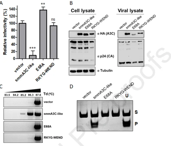
that cellular expression and viral encapsidation of these variants were comparable (Suppl. Fig. S3B). Finally, to test the in vivo DNA-editing capacity, we performed 3D-PCR analysis using C2, C2.DH-YG, and C2.RKYG-WEND variants. As presented in the 3D-PCR experiment of Suppl. Fig. S3C, only HIV-1Δvif particles produced in the presence of A3C chimera C2 and its mutant C2.DH-YG generated amplicons that were detected at low-denaturation temperature, and C2.RKYG-WEND behaved similar to the vector control. Likewise, replacing RKYG with WEND in the smmA3C-like protein (Fig. 2E) inhibited its antiviral activity (Figs. 3A and 3B), DNA-editing capacity of HIV-1 genomes (Fig. 3C), and catalytic activity in vitro (Fig. 3D) as did the active site mutant E68A.
The WE-RK mutation in loop 1 of hA3C determines its strong deaminase-dependent antiviral
function
Mutational changes of the RKYG motif to WEND residues in loop 1 of C2 and smmA3C-like protein resulted in complete loss of enzymatic functions and anti-HIV-1 activities (Figs. 2F, 3A, 3C, 3D, and Suppl. Figs. S3A and S3C). To identify the residues in hA3C that are critically required for the deaminase-dependent antiviral activity against HIV-1Δvif, we mutated the loop 1 of hA3C with 25WE26 to 25RK26 and 28ND29 to 28YG29 residues and compared their antiviral
capacity (see A3C alignment and ribbon diagram Figs. 2A and 2B). As controls, we included additional mutants such as a catalytically inactive non-Zn2+-coordinating C97 mutant,
A3C.C97S [57], and the variants A3C.S61P [70] and A3C.S188I [74] exhibiting enhanced deaminase activity. Compared to WT hA3C, WE-RK greatly enhanced inhibition of HIV-1Δvif and the ND-YG variant behaved like WT A3C, while S61P and S188I demonstrated only
marginally increased HIV-1Δvif restriction (Fig. 4A). Importantly, active site mutant A3C.C97S did not inhibit HIV-1Δvif (Fig. 4A). Enhancement of the antiviral activity of hA3C.WE-RK compared to WT hA3C neither appear to result from higher protein expression in the virus producer cells nor from differences in encapsidation, as demonstrated in a titration experiment that directly compared these features for both proteins (Suppl. Fig. S4A).
Next, we asked if the antiviral activity of A3C.WE-RK is deamination-dependent. To achieve this, we introduced the C97S mutation in A3C.WE-RK. Additionally, we compared the ancillary effect of mutants such as S61P [70] and S188I [74] by introducing these mutations in the WE-RK variant of A3C. As expected, the inhibitory activities of A3C.WE-WE-RK, A3C.WE-WE-RK.S61P, and A3C.WE-RK.S61P.S188I against HIV-1Δvif were abolished by the active site ablating mutation C97S, indicating the importance of the enzymatic activity of A3C (Fig. 4B). In comparison, introducing either the single mutation S61P or the double mutation S61P.S188I did not considerably change the activity of A3C.WE-RK (Fig. 4B). Immunoblot analysis of cell and viral lysates demonstrated that hA3C and all mutants (except A3C.WE-RK.S61P.S188I.C97S mutant) were expressed at comparable levels (Fig. 4C). However, viral incorporation of A3C.C97S, A3C.WE-RK.C97S, A3C.WE-RK.S61P.C97S, and WE-RK.S61P.S188I.C97S was slightly decreased relative to that of WT and mutant proteins that do not contain the C97S mutation (Fig. 4C). Moreover, we confirmed the effects of all mutants on HIV-1Δvif propagation by 3D-PCR (Fig. 4D) and deamination assays in vitro (Fig. 4E). In both assays, we found that the C97S mutation destroyed the function of all A3C variants. Thus, we conclude that the loop 1-mediated enhanced activity of hA3C.WE-RK is dependent on catalytic deamination.
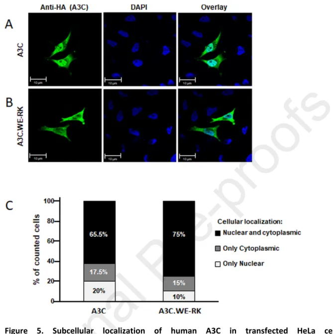
To address if the cellular localization of A3C is affected by the WE-RK mutations, we used confocal microscopy. HeLa cells were transfected with the HA-tagged hA3C or hA3C.WE-RK and the proteins were visualized by applying an anti-HA antibody. Both proteins, hA3C and hA3C.WE-RK were localized in cytoplasm and nucleus (Figs. 5A and 5B). This distribution was found in 65.5% and 75% of cells expressing hA3C or that of hA3C.WE-RK, respectively. Only 20% or 10% of the cells expressing hA3C or hA3C.WE-RK displayed these proteins solely in the nucleus, respectively (Fig. 5C). Together, we infer that hA3C and hA3C.WE-RK had a similar distribution in HeLa cells.
The RK-WE mutation in loop 1 moderately reduces the antiviral activity of hA3F
hA3C and hA3F-CTD display 77% sequence similarity, reflecting a common evolutionary origin [6]. Interestingly, the antiviral activity of hA3F is mediated by its CTD [75, 76]. Various loops within the A3F-CTD were recently investigated for their role in substrate binding and enzyme function [77] but it was not possible to unravel the antiviral activity of a protein consisting only of the A3F-CTD, mainly due to earlier reported difficulties in expressing this domain alone in human cells [70, 78]. The residues 25RK26 in loop 1 of smmA3C-like protein are derived from
exon 5 of the smmA3F gene, located in the CTD of A3F (Suppl. Fig. S1B) and are conserved in primate A3F proteins (see section evolution, below). To test the impact of RK residues in CTD loop 1 of the hA3F, we compared the antiviral activity of hA3F with A3F.RK-WE against HIV-1Δvif. hA3F and hA3F.RK-WE yielded similar amounts of protein and were equally efficiently encapsidated in HIV-1 particles (Fig. 6A). However, the HIV-1Δvif inhibiting effect of A3F.RK-WE was about 2-fold lower than WT A3F (Fig. 6B). Consequently, A3F.RK-A3F.RK-WE showed
decreased mutation efficiency compared with WT A3F (Figs. 6C and 6D), which is consistent with data presented in a recent report [77]. Thus, we conclude that loop 1 with its residues RK in CTD of A3F is important for the enzymatic function of hA3F.
Inhibition of human LINE-1 retrotransposition by A3C variants
Since A3C and A3F restrict endogenous human LINE-1 (L1) retrotransposition activity by 40-75% and 66-85%, respectively [51, 61, 79, 80], we set out to elucidate how the WE and the RK residues in loop 1 of both hA3C and hA3F affect the L1 inhibiting activity. To this end, we quantified the L1-inhibiting effect of human WT A3A, A3C, and A3F proteins and their mutants hA3C.WE-RK, hA3C.WE-RK.S61P, and hA3F.RK-WE by a dual-luciferase retrotransposition reporter assay [81]. In this cell culture-based assay, the firefly luciferase gene is used as the reporter for L1 retrotransposition and the Renilla luciferase gene is encoded on the same plasmid for transfection normalization (Fig. 7A). Consistent with previous reports, overexpression of hA3A, hA3C, and hA3F inhibited L1 reporter retrotransposition by approximately 94%, 68%, and 56%, respectively (Fig. 7B). The mutant hA3C.WE-RK restricted L1 more strongly (from 56% to ~96%), but the introduction of the additional S61P mutation in hA3C.WE-RK.S61P did not further increase the ability of the enzyme to restrict L1 mobilization (Fig. 7B). Notably, hA3F and the mutant hA3F.RK-WE exhibited a comparable level of L1 restriction, indicating that regions other than loop 1 of A3F-CTD and, probably, the NTD (N-terminal domain) of hA3F are involved in L1 restriction (Fig. 7B). Immunoblot analysis of cell lysates of co-transfected HeLa-HA cells demonstrated comparable expression of the L1 reporter and HA-tagged A3- and A3 mutant proteins (Suppl. Fig. S4B). Furthermore, compared
to the inhibition of L1 retrotransposition by hA3C and chimpanzee A3C (~60%), hA3C.S61P inhibited L1 reporter retrotransposition by 75% (Suppl. Fig. S4C and S4D). These findings indicate that the WE-RK mutation in hA3C enhances its L1-inhibiting activity. Based on the observed antiviral activity and the L1-restricting effect of hA3C.WE-RK on L1, we hypothesize that the introduction of these positively charged residues in hA3C significantly fosters its interaction with nucleic acids, which was recently reported to mediate its L1 inhibiting activity [61].
The positively charged residues R25 and K26 in A3C form salt-bridges with the backbone of
the ssDNA
To understand how the positively charged residues in loop 1 of A3C.WE-RK mediate the enhanced cytidine deamination activity, a structural model of hA3C variant hA3C-RKYG binding to ssDNA, based on the ssDNA-bound crystal structure of A3A was generated that shows a cytidine residue in the active center of hA3C.RKYG (Suppl. Fig. S5A). However, the ssDNA fragment (which was co-crystallized with hA3A) in this conformation is too short to interact with amino acids 25, 26, 28, and 29, which differ between hA3C WT and the hA3C.RKYG variant. Hence, this static binding mode model cannot explain why hA3C.RKYG has a higher cytidine deaminase activity than hA3C WT. To probe the impact of structural dynamics on residue-ssDNA interactions in order to explain the differences in A3C.WE-RK properties, this model was later subjected to molecular dynamics (MD) simulations.
To assess the binding to a longer ssDNA fragment, we generated a second complex model of ssDNA bound to the NTD of rhesus macaque A3G (rhA3G) [82], similar to the ssDNA-bound
A3F-CTD model built previously [83], and aligned the crystal structure of hA3C WT and the model of hA3C.RKYG to this complex (Figs. 8A, 8B, and 8C. Note that the A3G structure was used only for placing the DNA but not for modeling the protein part). This new model revealed that the positively charged residues R25 and K26 in hA3C.RKYG form salt-bridges with the backbone of the ssDNA (Fig. 8C) in contrast to hA3C WT (Fig. 8B). Thus, these two residues can form stronger interactions with ssDNA in hA3C.RKYG than their counterparts in hA3C, which may explain the enhanced cytidine deaminase activity of hA3C.WE-RK compared to hA3C (Fig. 4E). However, as the binding of ssDNA to NTDs, such as in the structure of rhA3G, differs from that in CTDs, we did not subject the former model to MD simulations.
We next performed five replicas of MD simulations of 2 µs length each for hA3C, hA3C.RKYG, and hA3C.S61P.S188I to assess the structural impact of the substitutions on the protein. For this, we used a hA3C crystal structure as starting structures and variants thereof generated by substituting respective residues. In all MD simulations, the cytidine remains bound to the Zn2+
ion in the active site. The root mean square fluctuations (RMSF), which describe atomic mobilities during the MD simulations, show distinct differences between the variants in the putative DNA-binding regions of the proteins: the RMSF of hA3C.RKYG and hA3C.S61P.S188I are up to 2 Å larger compared to hA3C WT in the regions carrying the substitutions (residues 21-32 for hA3C.RKYG and residues 55-67 for hA3C.S61P.S188I) (Suppl. Fig. S5B). This effect is specifically related to the respective substitutions, as no change in RMSF occurs for a variant in any region where it is identical to A3C WT. The increased movement of ssDNA-binding residues might improve the sliding of hA3C.RKYG and hA3C.S61P.S188I along the ssDNA, owing to more transient interactions with the ssDNA backbone. Conversely, the RMSF of loop
7 is up to 1 Å lower in both the hA3C.RKYG and hA3C.S61P.S188I variants compared to the hA3C WT (Suppl. Fig. S5B).
These results encouraged us to investigate possible interaction patterns between DNA and each of the three A3C variants that could be a result of the shift in loop 1 dynamics. For this purpose, we used the initial DNA-bound model of hA3C.RKYG with cytidine in the active center, modeled from the experimental A3A structure as described above, to generate DNA-bound complexes for hA3C WT and hA3C.S61P.S188I. While our MD simulations showed similar changes in the conformational dynamics of the loops as before (Suppl. Fig. S5B), we detected an interesting change in interactions between loop 1 residue R30 and the DNA. R30, which is present in all three variants and points away from the DNA in the A3C crystal structure, interacts more frequently with the DNA in both hA3C.S61P.S188I (16.4 ± 2.6% of the simulation time applying stringent criteria for H-bond formation (mean ± SEM for 10 trajectories)) and hA3C.RKYG (44.7 ± 2.7%) than in hA3C WT (0.1 ± 0.0%). In hA3C.RKYG, K26 similarly forms H-bonds with the DNA over 10.3 ± 2.8% of the MD trajectories, but, expectedly, E26 in hA3C WT and hA3C.S61P.S188I forms almost no H-bonds.
In addition, to rule out the possibility that the loop 7 residues might be influencing the loop 1 residues from binding DNA, we have analyzed the interaction between them. The average distance between the two loops in the absence of DNA is very similar for hA3C (12.1 ± 1.75 Å; SD, n=5000), hA3C.RKYG (12.7 ± 1.78 Å; SD, n=5000), and hA3C.S61P.S188I (12.12 ± 1.78 Å; SD, n=5000). Given the average distance of 12 Å it is not surprising that with the exception of N23 and A121, which are the only residues in spatial proximity and thus commonly interact, residues in loop 1 form H-bonds to those in loop 7 in less than 1% of the simulation time for
all variants. The average distance of any atom in residue 25 to residues in loop 7 is larger than 4.4 Å, suggesting that sustained interactions are unlikely.
Next, we used more lenient distance criteria suitable to evaluate the formation of interactions and evaluated, whether only the N-terminus (W25 in hA3C and hA3C.S61P.S188I and R25 in hA3C.RKYG) or only the C-terminus (R30 in all three variants) of loop 1, or both residues at the same time, interact with the DNA. In hA3C, only W25 interacts with the DNA in ~20% of the conformations (Suppl. Fig. S6A). In hA3C.S61P.S188I, interactions between W25 or R30 occur in ~20% of the conformations, thus showing an increase of a factor of 5 for R30 (Suppl. Fig. S6B). In hA3C.RKYG, both R25 and R30 simultaneously interact with DNA in ~29% of all investigated conformations besides the interactions of R30 with DNA alone in ~42% of the conformations (Suppl. Fig. S6C). Hence, these results suggest that W25 and R30 act additively in hA3C.S61P.S188I, whereas they act cooperatively in hA3C.RKYG. This correlates with the differences in activities, with hA3C.RKYG showing the highest activity against HIV-1Δvif.
WE-RK mutation in the loop 1 of A3C enhances the interaction with ssDNA
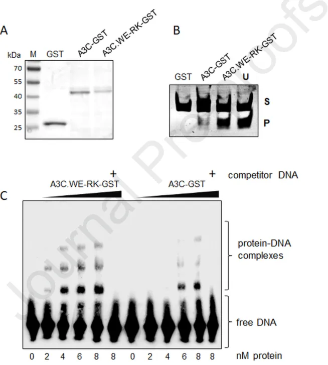
To validate our structural modeling analysis and to address if the interaction of hA3C and hA3C.WE-RK with the substrate ssDNA was differentially affected, we performed electrophoretic mobility shift assays (EMSA) using hA3C-GST (A3C fused to glutathione S-transferase, GST) and hA3C.WE-RK-GST purified from HEK293T cells (Fig. 9A). We first confirmed that the purified GST fusion proteins are catalytically active (Fig. 9B). As expected hA3C.WE-RK-GST displayed a stronger enzymatic activity than the WT equivalent and no activity with GST was detected (Fig. 9B). For EMSA, as a probe, we used a biotin-labeled ssDNA
oligonucleotide that harbors a TTCA motif in its central region [70, 84]. Because hA3C-GST is known to form a stable DNA-protein complex when the protein concentration reaches ≥ 20 nM [70], we decreased the amount of A3C and its mutant protein to specifically test their inherent DNA binding capacity. In a titration experiment with concentrations ranging from 2 to 8 nM in steps of 2 nM of hA3C-GST and hA3C.WE-RK-GST purified protein, we detected a clear trend in the formation of DNA–protein complexes for hA3C-GST and hA3C.WE-RK-GST (Fig. 9C). Intriguingly, DNA-protein complexes of hA3C.WE-RK-GST started appearing at the lowest protein concentration used (2 nM), while hA3C-GST-DNA complexes were detected at protein concentrations ≥ 6 nM. To confirm the specificity of the DNA–protein complexes, we competed for the reaction with unlabeled DNA carrying the same nucleotide sequence as the used probe in 200-fold excess relative to that probe. The addition of the competitor DNA to the sample containing the maximum (8 nM) amount of A3C protein, efficiently disrupted the protein-DNA complex formation (Fig. 9C and Suppl. Fig. S7). Together, data from structural modeling and EMSA experiments allowed us to conclude that the two amino acid-change in loop 1 of A3C boosts the ssDNA binding capacity of A3C. Importantly, the GST moiety did not affect the binding (Suppl. Fig. S7 and [70]).
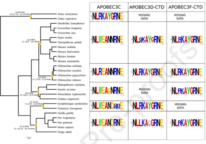
Evolution of A3Z2 loop 1 regions in primates
Because of the strong evolutionary relationship between A3C, the CTD of A3F, and related A3Z2 proteins [6], we performed a phylogenetic reconstruction for the A3Z2 domains in primates, using the A3Z2 sequences in the northern tree shrew as outgroup. Our analyses were performed at the A3Z2 domain level, separating the two Z2 domains of the
double-domain A3D and A3F proteins, thus generating five evolutionary units: the A3D-NTD, A3F-NTD, A3C, A3D-CTD and A3F-CTD (Suppl. Fig. S8). Remarkably, the results show that the A3Z2 domains underwent independent duplication in the two sister taxa, tree shrews and primates, as the three A3Z2 tree shrew sequences constitute a clear outgroup to all primate A3Z2 sequences. We identified a sharp clustering of the A3D-NTD and A3F-NTD on the one hand and of A3C, A3D-CTD, and A3F-CTD on the other hand. As to New World monkeys (Platyrrhini), we could only confidently retrieve A3C sequences from the white-faced sapajou Cebus
capucinus and from the Ma's night monkey Aotus nancymaae. These sequences from A3C New
World monkeys were basal to all Catarrhini (Old World monkeys and apes) A3C, A3D-CTD and A3F-CTD sequences, suggesting that the two gene duplications leading to the extant organization of A3C, A3D, and A3F occurred after the Platyrrhini/Catarrhini split 43.2 Mya (41.0 - 45.7 Mya) and before the Cercopithecoidea/Hominoidea (Old World monkeys/apes) split 29.44 Mya (27.95 - 31.35 Mya). The results show a tangled distribution within the A3D-NTD and A3F-A3D-NTD clade, and within the A3D-CTD and A3F-CTD clade. These confusing relationships are more obvious when comparing the phylogenetic reconstruction of the Z2 domains without imposing any topological constraint (Suppl. Fig. S8) with a tree in which monophyly of each of the large six clades identified was enforced (Suppl. Fig. S8). The tanglegram linking both, highlights those sequences whose phylogenetic position does not match the expected cluster, after the current annotation. Conversely, Catarrhini A3C sequences form a well-supported monophyletic taxon, and this A3C gene tree essentially adheres to the corresponding species tree (Fig. 10). Focusing exclusively on the nodes that we could identify with confidence, we performed ancestral phylogenetic inference of the most likely amino acid sequence for the A3 loop 1 (Suppl. Fig. S9) and, in parallel, performed a
consensus analysis of the extant sequences (Fig. 10 and Suppl. Fig. S9). Our results recover the well-conserved aromatic stacking stretch F[FY]FXF characteristic of all A3s. In the A3C, A3D-CTD, and A3F-CTD clade, we identified a small motif displaying striking divergent evolution flanked by conserved small hydrophobic amino acids. The most likely ancestral form is the amino acid motif LRKA, which is also the form present in extant New World monkeys A3C and the most common in extant A3F-CTD, while in the extant A3D-CTD the Arg residue is less conserved L[RLQ][KT]A (Fig. 10). Strikingly, in the ancestor of Catarrhini A3C at around 29.4 Mya (27.6-31.3 Mya), this motif had already evolved to LWEA (Suppl. Fig. S9), and this is the common extant form in Old World monkeys and apes (Fig. 10). Only subsequently, and exclusively in the Chlorocebus lineage (African green monkeys), this change was partly reverted to LREA by a TGG>CGG transition. This reversion should have occurred after the divergence within Cercopithecinae, around 13.7 Mya (10.7 - 16.6 Mya) and before the speciation within Chlorocebus at 3.42 Ma (2.05-4.15 Mya) (Fig. 10).
DISCUSSION
Compared to the many studies conducted over the past decade on the HIV-1 restriction factors A3G and A3F, investigations on A3C are very limited. A small number of studies have addressed the catalytic activity and substrate binding capacity of A3C [61, 70, 74, 85]. While the previously characterized hA3C mutants S61P and S188I boost the catalytic activity of the enzyme to a certain degree, none of these mutations is powerful enough to reduce the HIV-1Δvif infectivity to the level accomplished by A3G and they do not directly partake in catalytic activity [70, 74, 85]. Because our repeated attempts to express A3F-CTD in human cells were
not successful ([70] and Fig. 1C), we assayed A3C proteins from different Old World primate species. Due to the high level of nucleotide sequence identity between the A3C (A3Z2) paralogs (see discussion below) in the sooty mangabey monkey genome, we generated by missannotation a smmA3C-like protein with superior anti-HIV-1 and enzymatic activity. We have identified the key role of two positively-charged residues in loop 1 of this smmA3C-like protein (and of the hA3F-CTD), namely R25 and K26 in the RKYG motif. Replacing RKYG of smmA3C-like by the WEND form of this motif in hA3C abolished its anti-HIV-1 and catalytic activity. Importantly, the converse strategy of introducing the substitution WE-RK in the loop 1 of hA3C generated the potent, deaminase-dependent anti-HIV-1 enzyme hA3C.WE-RK. Consistent with these observations, our EMSA data demonstrate that residues in the loop 1 of A3C regulate protein-DNA interaction. Thus, we postulate that this more intense DNA-protein interaction is causative for the enhanced deamination activity and enhanced anti-HIV and anti-L1 activity. Similarly, Solomon and coworkers discussed that loop 1 residues of hA3G-CTD strongly interact with substrate ssDNA and that this interaction distinguishes catalytic binding from non-catalytic binding [86]. However, the loop 1 of A3 proteins likely has multiple functions, as loop 1 of A3A was found to be important for substrate specificity but not for substrate binding affinity [87], and loop 1 of A3H, especially its residue R26, plays a triple role for RNA binding, DNA substrate recognition, and catalytic activity likely by positioning the DNA substrate in the active site for effective catalysis [88]. In accordance, our study indicates that
25RK26 substitution in loop 1 of A3C provides the microenvironment that drives the flexibility
in substrate binding and enzymatic activity.
The binding model developed here rationalizes how hA3C.RKYG can interact with the negatively charged backbone of ssDNA via the positively charged loop 1 side chains of R25 and
K26 (Fig. 8C). Like our modeling strategy, Fang et al. [83] used their binding mode model of A3F-CTD with ssDNA to identify residues in the A3G-CTD important for ssDNA binding. Furthermore, the increased mobility of DNA binding regions carrying the substitutions in hA3C.RKYG and hA3C.S61P.S188I, respectively, compared to hA3C (Suppl. Fig. S5B) suggests that hA3C.RKYG and hA3C.S61P.S188I can better slide along the ssDNA than hA3C. The higher mobility of the residues may allow them to adapt more quickly to the passing ssDNA, which, together with likely stronger interactions with the ssDNA backbone, may explain the increased deaminase activity. This idea is corroborated by the MD simulations, in which the complexes including DNA loop 1 residues show more frequent interactions with the DNA in the case of hA3C.RKYG than in any of the other two variants, suggesting a stronger binding of the DNA; by contrast, in hA3C.S61P.S188I in 39.2% of the time either W25 or R30 interact with the DNA such that the DNA could be passed on from one residue to the other, assisting in the sliding-down mechanism while possibly also increasing binding affinity. In addition, loop 7 exhibits a decreased mobility in both hA3C.RKYG and hA3C.S61P.S188I compared to hA3C (Suppl. Fig. S5B). Decreased mobility of loop 7 has been shown to predict higher deaminase activity, DNA binding, and substrate specificity of A3G and A3F, and has been reported to be also relevant for antiviral activity of A3B and A3D [76, 89-91]. These structural findings can explain the differences in deaminase activity among the three variants.
Unexpectedly, our experiments also demonstrated that LINE-1 restriction by A3C, which was reported earlier to be deaminase-independent [61], is enhanced after expression of the A3C.WE-RK variant. These data suggest that the reported RNA-dependent physical interaction between L1 ORF1p and A3C dimers might be mediated by A3C loop 1, is partly dependent on the two amino acids W25 and E26 and is enhanced by the R25 and K26 substitutions. However,
L1 inhibition by A3F was not significantly altered by the A3F.RK-WE mutations, clearly indicating that other regions (and NTD) in A3F are likely to be relevant for L1 restriction. Because selection likely had to balance between anti-viral/anti-L1 activity and genotoxicity of A3 proteins, we wanted to characterize loop 1 residues during the evolution of the closely related A3Z2 proteins A3C, A3D CTD and A3F CTD in primates, all of them descendant of an ancestral Z2 domain that had undergone two duplication rounds [6]. In the most recent common ancestor of these enzymes that existed before the split Old World and New World primates (Catarrhini-Platyrrhini) around 43 Mya, we infer the ancestral form of the sequence of this motif in loop 1 to be LRKAYG. In New World monkeys, the A3C genes were not duplicated and are basal to the three sister clades of Catarrhini A3C, A3D-CTD, and A3F-CTD. In extant A3C sequences in New World monkeys, the loop 1 motif has notably remained unchanged and reads LRKAYG. In Catarrhini, on the contrary, the ancestral A3C sequence underwent two rapid rounds of duplication that occurred after the split with the ancestor of Platyrrhini, and before the split between the ancestors of Old World monkeys (Cercopithecoidea) and apes (Hominoidea), some 29 Mya [6]. A3F has since then been involved in an Red Queen arms race with retroviral genes [92]. In extant A3F-CTD sequences, the consensus form of the loop 1 remains LRKAYG, albeit with a certain variability of the R residue, which is exchanged with other positively charged amino acids. In extant A3D-CTD enzymes, this motif has undergone erosion, is more variable and reads L[RLQ][KT]A[YC]G. Interestingly, loop 1 in A3C experienced rapid and swift selective pressure to exchange the positively charged RK amino acids by the largely divergent chemistry of WE, yielding LWEAYG. This selective sweep occurred very rapidly, as this is the fixed form in all Catarrhini. Notoriously, and exclusively in the Chlorocebus lineage (African Green monkeys), this amino
acid substitution was partly reverted to LREAYG, which is the conserved sequence in the four Chlorocebus A3C entries available (Fig. 10).
Overall, our results suggest that the two duplication events that generated the extant A3C, A3D-CTD, and A3F-CTD sequences in Catarrhines released the selective pressure on two of the daughter enzymes allowing them to explore the sequence space and to evolve via sub/neofunctionalization, as proposed for Ohno’s in-paralogs [93]. Thus, the A3F-CTD form of the loop 1 diverged little from the ancestral chemistry and possibly maintained the ancestral function, while the release in conservation pressure on A3D-CTD allowed the enzyme loop 1 to accumulate mutations and diverge from the ancestral state. In turn, A3C was rapidly engaged into a distinct evolutionary pathway, which is unique due to the highly divergent chemistry of loop 1 but also because A3C is the only A3Z2 monodomain enzyme of the A3 family. It must also be noted that among the descendants of the ancestral A3C in Catarrhines, only extant A3C forms a well-supported monophyletic clade (Suppl. Figs. S8 and S9). Instead, in several instances and for different species, sequences annotated in the databases as A3D-CTD clustered together with sequences annotated as A3F-A3D-CTD, and vice versa, and the same is true for the corresponding N-terminal domains (see tanglegram Suppl. Fig. S8), overall resulting in a lack of support for common ancestry for the individual moieties of A3C and A3F, and preventing us from inferring the ancestral forms of the loop 1 in A3D-CTD and A3F-CTD. This lack of monophyly could simply reflect the lack of power of phylogenetic reconstruction or the potential for database misannotations when applied to genes undergoing complex evolution, including a full panel of duplications, deletions, adaptive radiation, differential selection among paralogs and Red Queen dynamics [3, 6, 92, 94, 95]. In this respect, the field is wanting for a systematisation of protocols and procedures for identifying selection
signatures in genes with complex evolutionary histories [96]. This lack of resolution could also reflect a biological basis of read-through of unmatured mRNAs resulting in differentially edited or in naturally chimeric mRNAs [9, 97, 98], which can hamper phylogenetic inference. Finally, the genetic architecture of the A3 locus, with the different gene copies located in tandem may favour non-homologous recombination between recently diverged, closely related sequences, and may also facilitate gene conversion between non-homologous alleles, overall leading to genetic information flow between gene copies and decoupling the true evolutionary history from our gene name and annotation-based phylogenetic reconstructions. The combined result of these novelty-generating mechanisms could be an enhanced inter-species or even inter-individual diversity in the A3 locus at either the genetic or the transcriptomic levels [98, 99]. The functional impact of such gene and mRNA diversity deserves further investigation, especially in the context of personalised medicine.
In conclusion, we postulate that the loop 1 region of A3s might have a conserved role in anchoring its ssDNA substrate for efficient catalysis and that weak deamination and anti-HIV-1 activity of hA3C might have been the result of losing DNA interactions in loop anti-HIV-1 during its evolution. It is thus possible that genes encoding A3C proteins with loop 1 residues with a higher ssDNA affinity were too genotoxic to benefit their hosts by superior viral and anti-L1 activity. Tao et al. [100] noted that the level of A3C preferentially increased upon treatment with artesunate (Art) and suggested that upregulated A3C is involved in the Art-induced DNA damage response[100]. Conceptually, we cannot rule out the possibility that the residues characterized here in loop 1 of hA3C might have an impact on recognition of unknown substrates or targets.
MATERIALS AND METHODS
Cell culture. HEK293T cells (ATCC CRL-3216) were maintained in Dulbecco’s high-glucose
modified Eagle’s medium (DMEM) (Biochrom, Berlin, Germany), supplemented with 10% fetal bovine serum (FBS), 2 mM L-glutamine, 50 units/ml penicillin, and 50 µg/ml streptomycin at 37°C in a humidified atmosphere of 5% CO
2. Similarly, HeLa-HA cells [101] were cultured in
DMEM with 10% FCS (Biowest, Nuaillé, France), 2mM L-glutamine and 20 U/ml penicillin/streptomycin (Gibco, Schwerte, Germany).
Plasmids. The HIV-1 packaging plasmid pMDLg/pRRE encodes gag-pol, and the pRSV-Rev for
the HIV-1 rev [102]. The HIV-1 vector pSIN.PPT.CMV.Luc.IRES.GFP expresses the firefly luciferase and GFP [103]. HIV-1 based viral vectors were pseudotyped using the pMD.G plasmid that encodes the glycoprotein of VSV (VSV-G). SIVagm luciferase vector system was described before [33]. All A3 constructs described here were cloned in pcDNA3.1 (+) with a C-terminal hemagglutinin (HA) tag. The smmA3C-like expression plasmid was generated by exon assembly from the genomic DNA of a white-crowned mangabey (Cercocebus torquatus
lunulatus), and the cloning strategy for smmA3C-like and the chimeras of hA3C/smmA3C-like
plasmid construction was recently described [69]. The expression vector for A3G-HA was generously provided by Nathaniel R. Landau. Expression constructs hA3C, rhA3C, cpzA3C, agmA3C and A3C point mutant A3C.C97S were described before [57, 60, 70].
Various point mutants hA3C.WE-RK, hA3C.ND-YG, hA3C.WE-RK.C97S, hA3C.WE-RK.S61P, hA3C.WE-RK.S61P.C97S, hA3C.WE-RK.S61P.S188I, hA3C.WE-RK.S61P.S188I.C97S,
hA3F.RK-WE, smmA3C-like.E68A were generated by using site-directed mutagenesis. Similarly, single or multiple amino acid changes were made in expression vectors to produce chimera 2 mutants (C2.DH-YG, C2.RKYG-WEND, C2.R25W, C2.K26E, C2.Y28N, and C2.G29D) and smmA3C-like.RKYG-WEND. To clone C-terminal GST-tagged hA3C, hA3C.WE-RK, the ORFs were inserted between the restriction sites HindIII and XbaI in the mammalian expression construct pK-GST mammalian expression vector [104]. Individual exons of authentic smmA3C and smmA3F and smmA3F-like genes exons were amplified and cloned in pcDNA3.1. All the primer sequences are listed in Suppl. Table 1.
Virus production and isolation. HEK293T cells were transiently transfected using
Lipofectamine LTX and Plus reagent (Invitrogen, Karlsruhe, Germany) with an appropriate combination of HIV-1 viral vectors (600 ng pMDLg/pRRE, 600 ng pSIN.PPT.CMV.Luc.IRES.GFP, 250 ng pRSV-Rev, 150 ng pMD.G with 600 ng A3 plasmid or replaced by pcDNA3.1, unless otherwise mentioned) or SIVagm vectors (1400 ng pSIVTan-LucΔvif, 150 ng pMD.G with 600 ng A3 plasmid) in 6 well plate. 48 h post-transfection, virion containing supernatants were collected and for isolation of virions, concentrated by layering on 20% sucrose cushion and centrifuged for 4 h at 14,800 rpm. Viral particles were re-suspended in mild lysis buffer (50 mM Tris (pH 8), 1 mM PMSF, 10% glycerol, 0.8% NP-40, 150 mM NaCl and 1X complete protease inhibitor).
Luciferase-based infectivity assay. HIV-1 luciferase reporter viruses were used to transduce
was determined by RT assay using Cavidi HS kit Lenti RT (Cavidi Tech, Uppsala, Sweden). Normalized RT amount equivalent viral supernatants were transduced. 48 h later, luciferase activity was measured using SteadyliteHTS luciferase reagent substrate (Perkin Elmer, Rodgau, Germany) on a Berthold MicroLumat Plus luminometer (Berthold Detection Systems, Pforzheim, Germany). Transductions were done in triplicates and at least three independent experiments were performed.
Immunofluorescence microscopy. 1x10⁵ HeLa cells grown on polyethylene coverslips
(Thermo Fisher Scientific) were co-transfected with plasmids for hemagglutinin (HA) tagged hA3C (0.25 μg) WT or hA3C.WE-RK (0.25 μg) using FuGENE transfection reagent (Promega, Wisconsin, USA). At day 2 post transfection, cells were fixed with 4% paraformaldehyde in phosphate-buffered saline (PBS) for 10 mins, permeabilized 0.1% Triton X-100 for 10 min, incubated with blocking solution (10% FBS in PBS) for 1 h, and then cells were stained with mouse anti-HA antibody (Covance, Münster, Germany) 1:1,000 dilution in blocking solution for 1 h. Donkey anti-mouse Alexa Fluor 488 (Covance) was used as a secondary antibody, 1:300 dilution in blocking solution for 1 h. Finally, DAPI was used to stain nuclei for 2 mins. The images were captured by using a 63x objective on Zeiss LSM 510 Meta laser scanning confocal microscopy (Carl Zeiss, Cologne, Germany). For the quantification of cellular localization of A3Cs, 40 randomly chosen transfected cells with A3C or A3C.WE-RK were categorized and quantified.
Immunoblot analyses. Transfected HEK293T cells were washed with PBS and lysed in
radioimmunoprecipitation assay buffer (RIPA, 25 mM Tris (pH 8.0), 137 mM NaCl, 1% glycerol, 0.1% SDS, 0.5% sodium deoxycholate, 1% Nonidet P-40, 2 mM EDTA, and protease inhibitor cocktail set III [Calbiochem, Darmstadt, Germany]) 20 min on ice. Lysates were clarified by centrifugation (20 min, 14800 rpm, 4°C). Samples (cell/viral lysate) were boiled at 95⁰C for 5 min with Roti load reducing loading buffer (Carl Roth, Karlsruhe, Germany) and subjected to SDS-PAGE followed by transfer (Semi-Dry Transfer Cell, Biorad, Munich, Germany) to a PVDF membrane (Merck Millipore, Schwalbach, Germany). Membranes were blocked with skimmed milk solution and probed with appropriate primary antibody, mouse anti-hemagglutinin (anti-HA) antibody (1:7,500 dilution, MMS-101P, Covance); goat anti-GAPDH (C-terminus, 1:15,000 dilution, Everest Biotech, Oxfordshire, UK); mouse anti-α-tubulin antibody (1:4,000 dilution, clone B5-1-2; Sigma-Aldrich, Taufkirchen, Germany), mouse anti-capsid p24/p27 MAb AG3.0 [105] (1:250 dilution, NIH AIDS Reagents); rabbit anti S6 ribosomal protein (5G10; 1:103
dilution in 5% BSA, Cell Signaling Technology, Leiden, The Netherlands). Secondary Abs.: anti-mouse (NA931V), anti-rabbit (NA934V) horseradish peroxidase (1:104 dilution, GE Healthcare)
and anti-goat IgG-HRP (1:104 dilution, sc-2768, Santa Cruz Biotechnology, Heidelberg,
Germany). Signals were visualized using ECL chemiluminescent reagent (GE Healthcare). To characterize the effect of the expression of A3 proteins and their mutants on LINE-1 (L1) reporter expression, HeLa-HA cells were lysed 48 h post-transfection using triple lysis buffer (20 mM Tris/HCl, pH 7.5; 150 mM NaCl; 10 mM EDTA; 0.1% SDS; 1% Triton X-100; 1% deoxycholate; 1x complete protease inhibitor cocktail [Roche]), clarified and 20 μg total protein were used for SDS-PAGE followed by electroblotting. HA-tagged A3 proteins and L1 ORF1p were detected using an anti-HA antibody (MMS-101P; Covance) in a 1:5,000 dilution
and the polyclonal rabbit-anti-L1 ORF1p antibody #984 [106] in a 1:2,000 dilution, respectively, in 1xPBS-T containing 5% milk powder. ß-actin expression (AC-74, 1:30,000 dilution, Sigma-Aldrich Chemie GmbH) served as a loading control.
Differential DNA denaturation (3D) PCR. HEK293T cells were cultured in 6-well plates and
infected with DNAse I (Thermo Fisher Scientific) treated viruses for 12 h. Cells were harvested and washed in PBS, the total DNA was isolated using DNeasy DNA isolation kit (Qiagen, Hilden, Germany). A 714-bp fragment of the luciferase gene was amplified using the primers 5’-GATATGTGGATTTCGAGTCGTC-3’ and 5’-GTCATCGTCTTTCCGTGCTC-3’. For selective amplification of the hypermutated products, the PCR denaturation temperature was lowered stepwise from 87.6°C to 83.5°C (83.5°C, 84.2°C, 85.2°C, 86.3°C, 87.6°C) using a gradient thermocycler. The PCR parameters were as follows: (i) 95°C for 5 min; (ii) 40 cycles, with 1 cycle consisting of 83.5°C to 87.6°C for 30 s, 55°C for 30 s, 72°C for 1 min; (iii) 10 min at 72°C. PCRs were performed with Dream Taq DNA polymerase (Thermo Fisher Scientific). PCR products were stained with ethidium bromide. PCR product (smmA3C-like sample only) from the lowest denaturation temperature was cloned using CloneJET PCR Cloning Kit (Thermo Fisher Scientific) and sequenced. smmA3C-like protein-induced hypermutations of eleven independent clones were analysed with the Hypermut online tool (https://www.hiv.lanl.gov/content/sequence/HYPERMUT/hypermut.html) [107]. Mutated sequences (clones) carrying similar base changes were omitted and only the unique clones were presented for clarity.
In vitro DNA cytidine deamination assay. A3 proteins expressed in transfected HEK293T cells,
virion-incorporated A3s, or purified GST fusion proteins were used as input. Cell lysates were prepared with mild lysis buffer 48 h post plasmid transfection. Deamination reactions were performed as described [72, 108] in a 10 µL reaction volume containing 25 mM Tris pH 7.0, 2 µl of cell lysate and 100 fmol single-stranded DNA substrate (TTCA: 5’-GGATTGGTTGGTTATTTGTATAAGGAAGGTGGATTGAAGGTTCAAGAAGGTGATGGAAGTTATGTTT GGTAGATTGATGG). Samples were treated with 50 µg/ml RNAse A (Thermo Fisher Scientific). Reactions were incubated for 1 h at 37˚C and the reaction was terminated by boiling at 95˚C for 5 min. One fmol of the reaction mixture was used for PCR amplification Dream Taq polymerase (Thermo Fisher Scientific) 95˚C for 3 min, followed by 30 cycles of 61˚C for 30 s and 94˚C for 30 s using primers forward 5’-GGATTGGTTGGTTATTTGTATAAGGA and reverse 5'-CCATCAATCTACCAAACATAACTTCCA. PCR products were digested with MseI (NEB, Frankfurt/Main, Germany), and resolved on 15% PAGE, stained with ethidium bromide (7.5 μg/ml). As a positive control, substrate oligonucleotides with TTUA instead of TTCA were used to control the restriction enzyme digestion [70].
L1 retrotransposition reporter assay. Relative L1 retrotransposition activity was determined
by applying a rapid dual-luciferase reporter based assay described previously [81]. Briefly, 2x10⁵ HeLa-HA cells were seeded per well of a six-well plate and transfected using Fugene-HD
transfection reagent (Promega) according to the manufacturer’s protocol. Each well was cotransfected with 0.5 µg of the L1 retrotransposition reporter plasmid pYX017 or pYX015 [81] and 0.5 µg of pcDNA3.1 or WT or mutant A3 expression construct resuspended in 3 µl
Fugene-HD transfection reagent and 100 µl GlutaMAX-I-supplemented Opti-MEM I reduced-serum medium (Thermo Fisher Scientific). Three days after transfection cultivation, the medium was replaced by complete DMEM containing 2.5 µg/ml puromycin, to select for the presence of the L1 reporter plasmid harboring a puroR-expression cassette. Next day, the medium was replaced once more by puromycin containing DMEM medium and 48 hours later, transfected cells were lysed to quantify dual-luciferase luminescence. Dual-luciferase luminescence measurement: Luminescence was measured using the Dual-Luciferase Reporter Assay System (Promega) following the manufacturer’s instructions. For assays in 6-well plates, 200 µl Passive Lysis Buffer was used to lyse cells in each well; for all assays, 20 µl lysate was transferred to a solid white 96-well plate, mixed with 50 µl Luciferase Assay Reagent II and firefly luciferase (Fluc) activity was quantified using the microplate luminometer Infinite 200PRO (Tecan, Männedorf, Switzerland). Renilla luciferase (Rluc) activity was subsequently read after mixing 50 µl Stop & Glo Reagent into the cell lysate containing Luciferase Assay Reagent II. Data were normalized as described in the results section. The retrotransposition-defective L1RP/JM111 (located on pYX015) was used as the reference Fluc vector and the normalized luminescence ratio (NLR) resulting from cotransfection of pYX015 and pcDNA3.1(+) was set as 1.
Protein sequence alignment and visualization. Sequence alignment of hA3C and
smmA3C-like protein was done by using Clustal Omega (http://www.ebi.ac.uk/Tools/msa/clustalo/). The alignment file was then submitted to ESPript 3.0 [109] (espript.ibcp.fr) to calculate the similarity and identity of residues between both proteins and to represent the pairwise
sequence alignment. Cartoon model of the crystal structure of A3C (PDB 3VOW) was constructed using PyMOL (PyMOL Molecular Graphics System version 1.5.0.4; Schrödinger, Portland, OR).
Structural model building of protein-DNA complexes. The structural models of hA3C or
hA3C.RKYG binding to ssDNA were generated by first aligning the X-ray crystal structure of rhA3G-NTD (PDB ID 5K82 [82]) onto the X-ray crystal structure of hA3F-CTD (PDB ID 5W2M [83]), the latter of which was co-crystallized with ssDNA. Subsequently, the hA3C X-ray crystal structure (PDB ID 3VOW [68]) was aligned onto the NTD of rhA3G, which is structurally similar to hA3C. The ssDNA and the interface region of hA3C were subsequently relaxed in the presence of each other using Maestro [110]. The same program was used to mutate hA3C to obtain the hA3C.RKYG and hA3C.S61P.S188I variants, which were again relaxed in the presence of the ssDNA. Similarly, we obtained hA3C, hA3C.RKYG, and hA3C.S61P.S188I ssDNA binding models based on the ssDNA-binding X-ray crystal structure of hA3A (PDB ID 5SWW [111]), a much relevant model similar to 6BUX [112]. These three DNA complex structures were later used for MD simulations as they include a cytidine residue in the active center.
Molecular dynamics simulations. hA3C, hA3C.RKYG, and hA3C.S61P.S188I were subjected to
MD simulations. For this, the above-mentioned structures without the DNA were N- and C-terminally capped with ACE and NME, respectively. The three variants were protonated with PROPKA [113] according to pH 7.4, neutralized by adding counter ions, and solvated in an octahedral box of TIP3P water [114] with a minimal water shell of 12 Å around the solute. The
Amber package of molecular simulation software [115] and the ff14SB force field [116] was used to perform the MD simulations. For the Zn2+-ions the Li-Merz parameters for two-fold
positively charged metal ions [117] were used. To cope with long-range interactions, the “Particle Mesh Ewald” method [118] was used; the SHAKE algorithm [119] was applied to bonds involving hydrogen atoms. As hydrogen mass repartitioning [120] was utilized, the time step for all MD simulations was 4 fs with a direct-space, non-bonded cut-off of 8 Å.
In the beginning, 17500 steps of steepest descent and conjugate gradient minimization were performed; during 2500, 10000, and 5000 steps positional harmonic restraints with force constants of 25 kcal mol-1 Å-2, 5 kcal mol-1 Å-2, and zero, respectively, were applied to the solute
atoms. Thereafter, 50 ps of NVT (constant number of particles, volume, and temperature) MD simulations were conducted to heat up the system to 100 K, followed by 300 ps of NPT (constant number of particles, pressure, and temperature) MD simulations to adjust the density of the simulation box to a pressure of 1 atm and to heat the system to 300 K. During these steps, a harmonic potential with a force constant of 10 kcal mol-1 Å-2 was applied to the
solute atoms. As the final step in thermalization, 300 ps of NVT-MD simulations were performed while gradually reducing the restraint forces on the solute atoms to zero within the first 100 ps of this step. Afterwards, five independent production runs of NVT-MD simulations with 2 µs length each were performed. For this, the starting temperatures of the MD simulations at the beginning of the thermalization were varied by a fraction of a Kelvin. MD simulation of those three variants in complex with ssDNA were performed similarly, treating the DNA with the OL15 force field [121] and performing ten independent production runs of NVT-MD simulations with 2 µs length each. To evaluate the interactions between loop
1 (residues 25-30) of the three variants and the ssDNA present in the complexes, we employed two different measures using CPPTRAJ [122]. First, we used the h-bond command to detect hydrogen bonds between residues in loop 1 and the ssDNA. Second, we measured the minimal distance of the side chain atoms, not including Cβ of the respective residues, and the DNA for
each snapshot of the MD simulations and correlated both (Suppl. Fig. S6), considering a larger distance cut-off of 4 Å to detect interactions between the side chains and DNA. The minimal distance over time for residue 30 is can be seen in Suppl. Fig. S10A-C and the root mean square deviation (RMSD) over time is shown in Suppl. Fig. S10D-F. The latter figure indicates that the systems structurally stabilized after ~250 ns.
Expression and purification of recombinant GST-tagged hA3C and hA3C.WE-RK from
HEK293T cells. Recombinant C-terminal GST-tagged hA3C and hA3C.WE-RK were expressed in
HEK293T cells and purified by affinity chromatography using Glutathione Sepharose 4B beads (GE Healthcare) as described previously [70]. Cells were lysed 48 h later with mild lysis buffer [50 mM Tris (pH 8), 1 mM PMSF, 10% glycerol, 0.8% NP-40, 150 mM NaCl, and 1X complete protease inhibitor and incubated with GST beads. After 2 h incubation at 4°C in end-over-end rotation, GST beads were washed twice with wash buffer containing 50 mM Tris (pH 8.0), 5 mM 2-ME, 10% glycerol and 500 mM NaCl. The bound GST hA3C and hA3C.WE-RK proteins were eluted with wash buffer containing 20 mM reduced glutathione. The proteins were 90-95% pure as checked on 15% SDS-PAGE followed by Coomassie blue staining. Protein concentrations were estimated by Bradford's method.