aDepartment of Biochemistry and Molecular Biology, and bBiostatistics Unit, Direction de la Recherche Clinique, University Hospital of Clermont-Ferrand, Clermont-Ferrand, France; cDepartment of Pediatric Emergency Medicine, University Hospital, Nantes, France; dDepartment of Paediatric and Adolescent Surgery, Medical University of Graz, Graz, Austria; eDepartment of Pediatric Emergency Medicine, University Hospital, Geneva, Switzerland; and fGReD, Université Clermont Auvergne, Centre National de la Recherche Scientifique, Institut National de la Santé et de la Recherche Médicale, Clermont-Ferrand, France
Dr Oris wrote the initial manuscript and participated, under the supervision of Dr Bouvier, in every step of the search strategy, quality assessment, and data extraction; Dr Pereira contributed to the data analysis, data interpretation, and assessment of study quality as well as critically reviewed the manuscript; Ms Durif supported the search strategy and quality assessment and also critically reviewed the manuscript; Drs Simon-Pimmel, Castellani, and Manzano participated in the meta-analysis by agreeing to share their original data in addition to critically reviewing the manuscript; Dr Sapin critically reviewed the manuscript; Dr Bouvier conceptualized and designed the study as well as codrafted and finalized the manuscript; and all authors approved the final manuscript as submitted and agree to be accountable for all aspects of the work.
To cite: Oris C, Pereira B, Durif J, et al. The Biomarker S100B and Mild Traumatic Brain Injury: A Meta-analysis. Pediatrics. 2018;141(6):e20180037
CONTEXT: The usefulness of S100B has been noted as a biomarker in the management of mild traumatic brain injury (mTBI) in adults. However, S100B efficacy as a biomarker in children has previously been relatively unclear.
OBJECTIVE: A meta-analysis is conducted to assess the prognostic value of S100B in predicting intracerebral lesions in children after mTBI.
DATA SOURCES: Medline, Embase, the Cochrane Central Register of Controlled Trials (CENTRAL), Web of Science, Scopus, and Google Scholar.
STUDY SELECTION: Studies including children suffering mTBI who underwent S100B measurement and computed tomography (CT) scans were included.
DATA EXTRACTION: Of 1030 articles screened, 8 studies met the inclusion criteria. RESULTS: The overall pooled sensitivity and specificity were 100% (95% confidence interval [CI]: 98%–100%) and 34% (95% CI: 30%–38%), respectively. A second analysis was based on the collection of 373 individual data points from 4 studies. Sensitivity and specificity results, obtained from reference ranges in children with a sampling time <3 hours posttrauma, were 97% (95% CI: 84.2%–99.9%) and 37.5% (95% CI: 28.8%–46.8%), respectively. Only 1 child had a low S100B level and a positive CT scan result without clinically important traumatic brain injury.
LIMITATIONS: Only patients undergoing both a CT scan and S100B testing were selected for evaluation.
CONCLUSIONS: S100B serum analysis as a part of the clinical routine could significantly reduce the number of CT scans performed on children with mTBI. Sampling should take place within 3 hours of trauma. Cutoff levels should be based on pediatric reference ranges.
The Biomarker S100B
and Mild Traumatic Brain
Injury: A Meta-analysis
Charlotte Oris, PharmD, a Bruno Pereira, PhD, b Julie Durif, MS, a Jeanne Simon-Pimmel, MD, c Christoph Castellani, MD, d Sergio Manzano, MD, e Vincent Sapin, PharmD, PhD, a, f Damien Bouvier, MD, PhDa, f
The incidence of traumatic brain injury (TBI) in children is high (691 per 100 000 in emergency departments), and TBI constitutes a leading public health problem.1, 2 Mild traumatic brain injury (mTBI), ranking on the Glasgow Coma Scale (GCS) from 13 to 15, 3 is one of the most common causes of pediatric hospital admission.4, 5 Children with mTBI account for 5% to 8% of visits to French pediatric emergency departments (60–100 per 100 000 children).6
Cranial computed tomography (CT) is a standard diagnostic tool for TBI among adults. In children, however, authors of several recent large-scale epidemiologic studies have described a link between radiation exposure from CT scans and future risk of cancer.7–9 Iatrogenic radiation with a cumulative dose of 50 mGy may triple the risk of leukemia, and cumulative doses of 60 mGy could triple the risk of brain tumors.7 Moreover, in an Australian study of 11 million children, researchers found a 24% increase in cancer risk among the 680 000 children who underwent CT scanning.8
Alternatively, children who may have experienced mTBI may be admitted for inpatient observation with subsequent CT scans performed only on those exhibiting clinical deterioration. This approach reduces radiograph exposure but is typically approximately one-third more costly than using CT scans for initial diagnosis.10, 11 Most CT scans and inpatient observations could be avoided because 93% of children suffering from mTBI have no intracerebral lesions.12 In this context, clinical decision algorithms have been developed to help clinicians identify those children with a very low risk of developing intracerebral lesions.13 Clinical prediction rules were first validated by the Pediatric Emergency Care Applied Research Network (PECARN) algorithm in the prospective cohort
of Kuppermann et al.14 More recently, Scandinavian guidelines for the initial management of mTBI in children were published, but these guidelines still need to be validated before they are put into extensive clinical use.15 The PECARN strategy suggests an algorithm in which children with mTBI can be divided into 3 risk categories (very low, intermediate, and high) according to their risk of developing clinically important brain injuries. The PECARN algorithm has significantly decreased the use of CT scans (decrease of 90%13) for all 3 risk categories. Despite validated guidelines, more needs to be done to reduce unnecessary pediatric CT scanning.
Serum biomarkers may be used as a supplementary tool to identify patients at risk for intracerebral lesions, tagging them for further imaging. S100B protein, which is well established as a sensitive TBI biomarker, is one of the calcium-binding proteins found in glial cells. It is a small dimeric cytosolic protein (21 kDa) consisting of ββ or αβ chains. It is involved in a variety of intracellular and extracellular regulatory activities.16, 17 After the onset of cerebral lesions, S100B is immediately released from damaged glial cells into the circulatory system and is subsequently eliminated by the kidneys, exhibiting a short half-life of ∼30 to 100 minutes.18–20 The potential use of S100B serum levels in reducing unnecessary CT scans among adults presenting with mTBI has been well established via many observational studies21–24 and confirmed in 2 interventional studies.25, 26 The addition of S100B detection to the Scandinavian guidelines for the management of mTBI decreased the need for CT scans among adults by one-third, with an accompanying significant reduction in cost of care from ∼€39 to €71 per patient.26, 27 Undén and Romner28 described these findings in a meta-analysis of adults with mTBI
and voiced the need for more studies among children. Further efforts should be focused on standardizing S100B interpretation in this
pediatric population by using specific reference ranges.15 Although authors of some studies have reported data from children with mTBI, 29–33 no meta-analysis has been conducted to date in which researchers summarize the use of S100B as a biomarker among this age group.
Our aim in this study was, thus, to perform a systematic review and meta-analysis to evaluate the efficiency of using determination of S100B serum levels to detect intracranial lesions in a pediatric trauma population. Additionally, data from 4 studies were analyzed. In this study, we focus on the 10%13 of the pediatric population with mTBI requiring a CT scan on the basis of application of a clinical algorithm.14
METHODS
Study Design and Patients
In this systematic review and meta-analysis, we followed the Preferred Reporting Items for Systematic Reviews and Meta-Analyses.34 A protocol was designed and registered with PROSPERO (CRD42017057500). Prospective cohort studies in which researchers included children suffering from mTBI with S100B measurement, undergoing CT scan or inpatient stay, and included the possibility of extraction of pertinent data (sensitivity and specificity) were eligible for inclusion in the review. Studies in which researchers included adults or children with a GCS score of ≤12 were excluded.
Meta-analysis of Prospective Cohort Studies
Search Strategy
The strategy used for the literature search was developed and conducted with the affiliated university
a search on the following electronic databases: Medline, Embase, the Cochrane Central Register of Controlled Trials (CENTRAL), Web of Science, Scopus, and Google Scholar. Appropriate combinations of controlled vocabulary and key words were used with adjustments made to account for differences in indexing across databases (Supplemental Tables 3 and 4). The search was augmented by scanning the reference lists of the initially identified relevant studies. The literature search was performed from inception to March 15, 2017.
Study Selection
The search process and eligibility assessment were conducted
independently by 2 authors (C.O. and J.D.), and discrepancies between their assessments were resolved by a third author (D.B.).
Data Extraction
Information related to study design, sample size, recruitment, patient characteristics (age, sex ratio, GCS score), laboratory aspects of S100B (type of assay, concentrations, reference ranges, sampling information, time between mTBI and blood sampling), comparison of CT scan versus S100B blood values (true-positives, positives, false-negatives, true-false-negatives, sensitivity, specificity, area under the curve [AUC], and cutoffs), and eventual clinical evolution (CE) was extracted from the studies and collected in table form. Only English-language data were used for extraction. Assessment of Study Quality We evaluated the quality of each study using criteria presented in the Quality Assessment of Diagnostic Accuracy Studies 2 (QUADAS-2).35 Assessments of study quality were conducted independently by 2 authors (C.O. and J.D.), and discrepancies between their
assessments were resolved by a third author (B.P.).
Meta-analysis of Individual Participant Data
The researchers of the 8 studies included in the meta-analysis used different assays to measure S100B serum values, which is a potential source of variability in the interpretation of S100B levels. In addition, there was high variance between the studies with regard to sampling time and S100B cutoff values. On the basis of these findings, we requested original data from the authors of studies that used the same automated assay on a Cobas analyzer (Roche Diagnostics, Meylan, France). Four authors were contacted, and 3 of these responded to our request. The data requested included age, sex, GCS score, mechanism of TBI, serum S100B concentrations, time between trauma and blood draw, and results of CT scans.
Statistics
Meta-analysis of Prospective Cohort Studies
Statistical analyses were conducted by the Biostatistics Unit from University Hospital of Clermont-Ferrand by using Comprehensive Meta-Analysis software (version 2; Biostat
Corporation) and Stata software (version 13; Stata Corp, College Station, TX). Baseline characteristics were summarized for each study sample and reported as mean (SD) and number (percent) for continuous and categorical variables, respectively, or median (minimum, maximum, and interquartile range [IQR]) in cases in which variables were not normally distributed. The sensitivity, specificity, negative predictive value (NPV), positive likelihood ratio (LR+), negative likelihood ratio (LR−), and 95% confidence interval (CI) were estimated by using random effects models assuming between- and within-study variability (DerSimonian and Laird approach). For sensitivity, specificity, and NPV, the statistical heterogeneity between results was assessed by examining forest
plots, 95% CIs, and I2, which are the most common metrics used for measuring the magnitude of between-study heterogeneity and are easily interpretable. I2 values range between 0% and 100%, and values of <25% are typically considered low, 25% to 50% are considered modest, and >50% are considered high.36 The likelihood ratio scattergram was plotted to show the summary point of likelihood ratios obtained as functions of mean sensitivity and specificity. A sensitivity analysis was also conducted to assess the influence on the global sensitivity, specificity, and NPV of the inclusion and exclusion of studies based on a funnel plot and I2. Type I error was fixed at α = .05.
Meta-analysis of Individual Participant Data
Statistical analyses were performed by using Stata software (version 13; Stata Corp) and R software (https:// cran. r- project. org/ ). For descriptive analyses, data are presented as medians (minimum, maximum, and IQR) because they were not normally distributed. In all analyses, between- and within-study variability were taken into account. To address the nonindependence of data due to study effect, random effects models were preferred over the usual statistical tests for measuring the influence of the GCS score and the origin of mTBI on S100B serum levels and to compare S100B median values between patients with intracerebral lesions identified by CT scans and patients without such lesions. A linear mixed model was used for S100B as a quantitative outcome. The normality of residuals and random effects was determined by using the Shapiro-Wilk test, and the logarithmic transformation of S100B was proposed to achieve the normality of dependent end point. A generalized linear mixed model, more precisely a logistic regression, was applied for S100B as a dichotomous outcome (S100B+ categorized according to usual references).
Then, estimates of sensitivity and specificity were obtained by first estimating the sensitivity and specificity for each study. Sensitivity, specificity, LR+, and LR− were then estimated by averaging the individual-specific estimates across studies. The variance of the estimate was the sample variance divided by the number of studies. Generalized estimating equation models with logit links and working independence correlation structure were also used to estimate the sensitivity, taking into account the correlation among patients in the same study. The same considerations were applied for the study of the receiver operating characteristics (ROC) curve.37 ROC curves and 95% CIs were estimated and compared, as proposed by Liu et al.38 The best cutoff value was determined according to clinical relevance and several indexes recommended in the literature: Youden, Liu, log-likelihood. A P value <.05 was considered statistically significant.
RESULTS
Meta-analysis of Prospective Cohort Studies
Literature Search Strategy
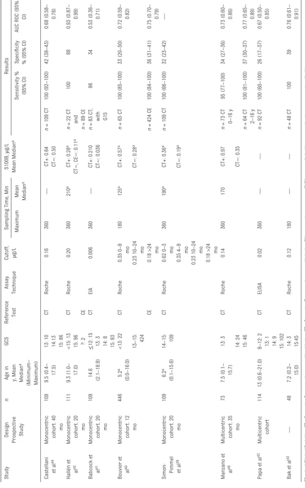
A total of 1623 studies were identified through the initial database search (Fig 1); 973 studies were excluded on the basis of a review of the title and abstract, leaving 57 studies for full review. Eight studies, 39–46 which included a total of 601 children, were selected for the meta-analysis. Characteristics of Included Studies In Table 1, we show the main characteristics of the eligible studies. The median number of patients was 109 (minimum: 48; maximum: 446; IQR: 100–112). In most of the studies, S100B serum values were measured via automated chemiluminescent immunoassay on a Cobas analyzer (Roche Diagnostics)39, 40, 43–46;
however, in 2 studies, researchers used manual assays.41, 42 Only 2 authors40, 43 recommended a maximum time of 3 hours instead of 6 hours between S100B blood draw and trauma.39, 41, 42, 44–46 Cutoff values for S100B differed between the studies (0.006 µg/L41– 0.20 µg/L45), and researchers interpreted S100B concentrations on the basis of reference ranges adapted for age in only 2 articles.39, 40 CT scan was the exclusive reference test in 6 studies, 39, 41–44, 46 whereas 2 authors40, 45 used both CT scan and CE. In most eligible studies, optimal sensitivity was shown, but in 1 study, researchers obtained a sensitivity of 86%.41 Specificity was relatively weak (26%–42%) with the exception of 1 cohort (88%).45 Areas under ROC curves ranged between 0.5341 and 0.93.45 The average prevalence of CT findings was 21.7%; extreme values were 8.3%39 and 35.4%.40
Quality Assessment and Risk of Bias in the Reviewed Studies
Seven studies39–42, 44, 46 fulfilled 5 or more of the QUADAS-2 tool criteria, with 1 study45 as an exception (Fig 2A). In 1 study, a risk of bias concerning patient selection was shown because there was no significant difference observed between the clinical condition and CT scan findings.46 In 1 other study, a high risk of bias was presented with regard to the flow, timing, and reference standard because a different reference standard (CT scan or CE) was used to evaluate the sensitivity and specificity of S100B.45 Overall Meta-analysis
The overall pooled sensitivity (Fig 2B) and NPV (Fig 2C) were 100% (with 95% CIs of 98%–100% and 99%–100%, respectively). I2 values were 0%. The overall pooled specificity was 41% (95%
FIGURE 1
TABLE 1
Characteristics of Studies Included in the Meta-analysis (
n = 8 studies) Study Design Prospective Study n
Age in y, Mean Median
a (Minimum – Maximum) GCS Reference Test Assay Technique Cutoff, µg/L
Sampling Time, Min
S100B, µg/L Mean Median a Results Maximum Mean Median a Sensitivity % (95% CI) Specificity % (95% CI) AUC ROC (95% CI) Castellani et a l 44 Monocentric cohor t. 40 mo 109 9.5 (0.4 – 17.5) 13: 10 CT Roche 0.16 360 — CT+: 0.64 n = 109 CT 100 (92 –100) 42 (38 –43) 0.68 (0.58 – 0.78) 14:13 CT − : 0.50 15: 86 Hall én et al45 Monocentric cohor t. 20 mo 111 9.3 (1.0 – 17.0) <15: 13 CT Roche 0.20 360 210 a CT+: 0.28 a n = 22 CT 100 88 0.93 (0.87 – 0.99) 15: 96 CT − , CE − : 0.11 a and ?: 2 CE n = 89 CE Babcock et al 41 Monocentric cohor t. 20 mo 109 14.6 (2.1 –18.9) ≤ 12: 15 CT EIA 0.006 360 — CT+: 0.210 n = 83 CT ; with G15 86 34 0.53 (0.36 – 0.71) 13: 3 CT − : 0.036 14: 8 15: 83 Bouvier et al 40 Monocentric cohor t. 12 mo 446 5.2 a (0.0 –16.0) <13: 22 CT Roche 0.35 0 –9 mo 180 125 a CT+: 0.57 a n = 65 CT 100 (85 –100) 33 (20 –50) 0.72 (0.59 – 0.82) 13 –15: 424 0.23 10 –24 mo CT − : 0.28 a CE 0.18 >24 mo n = 424 CE 100 (84 –100) 36 (31 –41) 0.75 (0.70 – 0.79) Simon- Pimmel et a l 39 Monocentric cohor t. 20 mo 109 6.2 a (0.1 –15.6) 14 –15: 109 CT Roche 0.62 0 –3 mo 360 190 a CT+: 0.36 a n = 109 CT 100 (66 –100) 32 (23 –42) — 0.35 4 –9 mo CT − : 0.19 a 0.23 10 –24 mo 0.18 >24 mo Manzano et al 46 Multicentric cohor t. 35 mo 73 7.5 (0.1 – 15.7) 13: 3 CT Roche 0.14 360 170 CT+: 0.97 n = 73 CT 0– 16 y 95 (77 –100) 34 (27 –36) 0.73 (0.60 – 0.86) 14: 24 CT − : 0.35 15: 46 n = 64 CT 2– 16 y 100 (81 –100) 37 (30 –37) 0.77 (0.65 – 0.89) Papa et a l 42 Multicentric cohor t 114 13 (0.6 –21.0) 9– 12: 2 CT ELISA 0.02 360 — — n = 92 CT 100 (60 –100) 26 (17 –37) 0.67 (0.50 – 0.85) 13: 1 14: 9 15: 102 Bak et a l 43 — 48 7.2 (0.2 – 15.0) 14: 3 CT Roche 0.12 180 — — n = 48 CT 100 39 0.76 (0.61 – 0.91) 15:45 CE −
, bad Clinical Evolution; CT
−
, Computer Tomography scan without lesion; CT+, Computer Tom
ography scan with lesion; EIA, enzyme immunoassay; ELISA, enzyme-linked immunosorbent assay;
—
, not applicable.
CI: 26%–57%) (Fig 2D), but results were significantly heterogeneous (I2 > 50%) as confirmed by the funnel plot, which revealed an important deviation in 1 study45
(Fig 2E). After removing this study, 45 the new overall pooled specificity (Fig 2F) was 34% (95% CI: 30%–38%) with an I2 value of 0%, the overall LR+ was 1.5 (95%
CI: 1.4–1.6), and the overall LR− was 0.04 (95% CI: 0.01–0.23), indicating that S100B is a marker of exclusion but not confirmation of intracranial lesions (Fig 3).
FIGURE 2
A, Risk of bias summary, QUADAS 2011. B, Forest plot showing the individual and pooled sensitivity of S100B for CT scans (n = 8 studies). C, Forest plot showing the individual and pooled NPV of S100B for CT scans (n = 8 studies). D, Forest plot showing individual and pooled specificity of S100B for CT scans (n = 8 studies). The sizes of markers are proportional to logarithms of the number of patients in the studies; horizontal lines represent 95% CIs. E, Funnel plot screening the distribution of 8 studies for specificity according to the average shown on the y-axis. F, Forest plot showing individual and pooled specificity (n = 7 studies). ☺ = low risk; ☹ = high risk; ? = unclear risk.
Meta-analysis of Individual Participant Data
Efficiency of S100B in Detecting Intracranial Lesions
A second analysis of sensitivity and specificity was based on data from 373 individuals (Table 2) included in 4 of the 8 studies.39, 40, 44, 46 Several ROC cutoffs were determined for a 100% sensitivity according to the sampling time (≤3 hours versus >3 hours posttrauma) and the age of the patient (≤2 years versus >2 years). Specificity was 17.5% (95% CI: 11.2%–25.5%) for a sampling time of ≤3 hours and 16.5% (95% CI: 12.5%–21.1%) in the overall population. Yet, among children ≤2 years of age, the specificity was largely superior for a sampling time of ≤3 hours (53.3% [95% CI: 34.3%–71.7%] vs 27.0% [95% CI: 16.6%–39.7%]). For a sensitivity of ∼100%, the better specificity (37.5% [95% CI: 28.8%–46.8%]) was not based on ROC cutoffs but rather on reference values. The sensitivity obtained from reference ranges in all 373 children was only 89.9% (95% CI: 80.2%–95.8%) against 97.0% (95% CI: 84.2%–99.9%) in children whose sampling time was <3 hours. The association between S100B serum levels and CT findings was tested on 373 patients. The median serum concentrations of S100B were 0.47 µg/L (minimum: 0.11; maximum: 5.54; IQR: 0.26–0.90) for patients with intracerebral lesions and 0.21 µg/L (minimum: 0.02;
maximum: 6.66; IQR: 0.13–0.39) for those without such lesions. The difference between these 2 groups was statistically significant (P < .001). Influence of the GCS Score on S100B Values
The influence of the GCS score on S100B values was tested on the data collected from 364 individuals included in 4 of the included studies.39, 40, 44, 46 On admission, GCS scores of 15, 14, and 13 were recorded for 262 patients (72.0%), 81 patients (22.2%), and 21 patients (5.8%), respectively. There was a significant positive association between S100B levels and GCS groups (GCS 13 vs 14:
P = .001; GCS 13 vs 15: P = .001) for
children with mTBI.
Influence of the Origin of mTBI on S100B Values
The influence of the origin of mTBI was tested on the data collected from 358 individuals (Fig 4) included in 2 studies.39, 40 Of these children, 39 road accidents, 259 domestic accidents, and 60 sport-related accidents were included. The median concentrations of S100B were 0.39 µg/L (minimum: 0.09; maximum: 5.65; IQR: 0.19–1.00), 0.29 µg/L (minimum: 0.05; maximum: 3.10; IQR: 0.18–0.44), and 0.18 µg/L (minimum: 0.02; maximum: 0.76; IQR: 0.12–0.31), for road accidents, domestic accidents, and sport-related accidents, respectively. The S100B+ percentages were 72%, 62%, and 50%, respectively. The difference
FIGURE 3
Likelihood ratio scattergram showing individual and pooled LR− and LR+ of S100B for CT scan (n = 7 studies). A, Exclusion and confirmation (with LR+ >10 and LR− <0.1). B, Confirmation only (with LR+ >10 and LR− >0.1). C, Exclusion only (with LR+ <10 and LR− <0.1). D, No confirmation and no exclusion (with LR+ <10 and LR− >0.1).
TABLE 2 Analysis of Sensitivity, Specificity, LR+, and LR− (n = 4 Studies)
n AUC 95% CI AUC PAUC Cutoff (Sensitivity =
100%), µg/L Optimal Sensitivity % (95% CI)
Specificity % (95% CI) LR+ (95% CI) LR− (95% CI) Total 373 0.74 0.68–0.80 <.001 0.108 100 (94.8–100) 16.5 (12.5–21.1) 1.2 (1.14–1.26) 0 ≤3 h 153 0.73 0.64–0.82 <.001 0.135 100 (89.4–100) 17.5 (11.2–25.5) 1.21 (1.12–1.32) 0 ≤2 y old 80 0.76 0.63–0.88 .001 0.202 100 (80.5–100) 27 (16.6–39.7) 1.37 (1.18–1.59) 0 >2 y old 293 0.74 0.67–0.80 <.001 0.108 100 (93.2–100) 19.5 (14.7–25.1) 1.24 (1.17–1.32) 0 ≤2 y old and ≤3 h 39 0.77 0.61–0.93 .02 0.311 100 (66.4–100) 53.3 (34.3–71.7) 2.14 (1.46–3.14) 0 >2 y old and ≤3 h 114 0.73 0.62–0.83 <.001 0.135 100 (85.8–100) 21.1 (13.2–31) 1.27 (1.14–1.41) 0 Reference valuesa 373 — — — — 89.9 (80.2–95.8) 49.7 (43.9–55.4) 1.79 (1.56–2.05) 0.2 (0.1–0.42) Reference valuesa ≤3 h 153 — — — — 97 (84.2–99.9) 37.5 (28.8–46.8) 1.55 (1.33–1.8) 0.08 (0.01–0.56) —, not applicable.
between the road accidents group and the domestic accidents group (P < .001) and the difference between the road accidents group and the sport-related accidents group (P < .001) were significant. Accordingly, S100B specificity could be higher after a sport-related trauma.
DISCUSSION
A meta-analysis was conducted to determine the efficacy of S100B as a biomarker in the management of mTBI in a pediatric population. In this study, we focus on the 10%13 of the pediatric population presenting at an emergency department with possible mTBI requiring a CT scan on the basis of application of a clinical algorithm.14 S100B protein has a strong NPV for intracranial lesions, and its use could reduce the number of CT scans among pediatric TBI patients. The strict integration of S100B into a clinical algorithm is essential. This is important because a patient with a low risk of intracerebral lesions (and therefore not eligible for S100B assay and CT scan) could have S100B positive value and so an unnecessary CT scan prescription.40
Our meta-analysis was based on 8 eligible studies, including 601 children with serum S100B testing and CT scans for mTBI. Among the 8 eligible studies, 39–46 the QUADAS-2 tool criteria revealed a high risk of bias for 1 of them.45 Excluding this study, 45 a low heterogeneity between studies (I2 = 0%) resulted. The overall pooled sensitivity and specificity obtained after the exclusion of that study were, respectively, 100% (95% CI: 98%–100%) and 34% (95% CI: 30%–38%). Consequently, S100B could reduce the use of CT scans in children with mTBI by 34%. The overall pooled LR− at 0.04 (95% CI: 0.01–0.23) confirms S100B protein as a marker of exclusion of intracranial lesions. This reduction could possibly
be improved by combining S100B with another biomarker, such as glial fibrillary acidic protein or ubiquitin c-terminal hydrolase-L1.47–49 Among the 8 articles, in 2 studies, 41, 42 researchers used manual enzyme immunoassays for S100B detection. However, automated assays present better analytical results with regard to precision, linearity, or accuracy, and appear to be a preferable option for S100B measurement.50, 51 Only Babcock et al41 presented a nonoptimal 100% sensitivity, and a manual assay was used in that study. Other studies52, 53 not included in our meta-analysis (because the authors also included moderate and severe TBI) revealed a nonoptimal sensitivity of ∼75% by using manual assays. These data may confirm the ability of automated assays to obtain better analytical results compared with manual assays. S100B interpretation differed across the 8 eligible studies. In only 2 studies, researchers interpreted S100B measurements using age-dependent reference ranges. In 2 studies, authors recommended a maximum interval of 3 hours between trauma and blood sampling rather than the 6 hours suggested by authors of the other studies. Despite a significant variability in regard to S100B interpretation, our meta-analysis revealed useful evidence of the role of S100B as a biomarker in the management of mTBI among children.
The authors of 4 studies39, 40, 44, 46 included in the meta-analysis who used the same automated assay (Roche Diagnostics method) shared their original data. Data from 373 individuals were examined and compiled. A sampling time of ≤3 hours did not improve the specificity in comparison with the overall population (17.5% vs 16.5%). However, among children ≤2 years of age, the specificity was largely superior with a sampling time of ≤3 hours (53.3% vs 27.0%). With this result, the importance of reference ranges for S100B serum levels during the first 2 years of life is supported. Indeed, for a sensitivity of ∼100%, the better specificity was not based on ROC cutoffs but on reference values. It should be mentioned that the sensitivity was 97.0% and not 100% because 1 child (>2 years old) had a petechial hemorrhage despite a negative S100B value, but the absence of a CT scan would not have impacted the management of this patient. Thus, the child’s age is essential in the interpretation of S100B concentration because protein values vary physiologically during the first 2 years of life. Reference ranges were recently determined over a large pediatric population54–59 and have recently been refined for children <4 months of age.60 Only 2 authors of studies included in this meta-analysis39, 40 took this fact into consideration, using S100B reference limits established in the study by Bouvier et al.55 We studied reference ranges based on age, but S100B concentrations are also affected by skin pigmentation and capillary sampling. S100B values for those with darker skin pigmentation are significantly higher because melanocytes synthesize a small amount of S100B protein under physiologic conditions.51 Capillary sampling, which is sometimes used when venous sampling is not possible, systematically increases S100B measurements.57 These 2 conditions could also decrease
FIGURE 4
Median concentrations of S100B by origin of TBI and proportion of S100B positive (white) and S100B negative (gray) (n = 2 studies). * P < .001
S100B specificity without impacting the 100% sensitivity. However, sensitivity could be affected by the sampling time (3 hours versus 6 hours) because of the short half-life of S100B (30–100 minutes). Thus, a delay of ≤3 hours is preferable to avoid the decline of S100B concentration below the cutoff level, despite the presence of intracranial lesions.21, 28 Our data were used to confirm the importance of this sampling time. Indeed, sensitivity was only 89.9% for a sampling time of <6 hours against 97.0% for a sampling time of ≤3 hours. In the same way, we only noted a correlation of S100B levels to GCS groups (13, 14, and 15) when the sampling time was <3 hours. Laribi et al61 validated S100B as an accurate screening tool for mTBI in adults, provided that the blood sampling was not delayed >3 hours posttrauma; delays beyond this time could result in false-negatives.
There are some limitations to our meta-analysis. As explained above, this study was focused on the 10%13 of the pediatric population with mTBI that require a CT scan on the basis of application of a clinical algorithm.14 Thus, only patients who had both a CT scan and who were evaluated for S100B concentration were selected for evaluation. This clinical selection bias explains the fact that the average prevalence of positive CT scan results (21.7%) in the included studies was higher than the values reported in the literature (0%–7%).12 The clinical conditions were often unclear with regard to CT scan realization. Manzano et al46 did not find any significant difference between clinical condition and CT scan findings, whereas Babcock et al41 determined a significant difference. With these results, a possible clinical selection bias concerning CT scan realization was confirmed, supporting the importance of using a PECARN clinical algorithm, as shown by Simon-Pimmel et al.39 Although the CT scan
remains the actual gold standard, CE plays an important role in the management of mTBI and is directly linked to PECARN clinical decision rules. These rules permit short hospitalization to evaluate CE for 23% of all mTBI.13 Evaluation of S100B serum levels could probably decrease the number of hospitalizations among these patients. In this context, only Bouvier et al40 evaluated the criteria of CE in a cohort of 424 patients, showing the potential of S100B to identify 100% of patients as bad CE with a potential decrease in unnecessary hospitalizations of 36%. This result needs to be further investigated in studies in which the potential economic impact of this biomarker is emphasized. For this purpose, the presence of persistent clinical signs (for example, presence at 48 hours and 3 weeks after the mTBI) will have to be monitored by using standardized medical interviews. The use of neurosurgery could be an additional criterion for study, but <1% of children with mTBI require neurosurgical intervention.13 Another limitation is the size of the CIs (from meta-analysis of prospective cohort studies and data on individual participants). Although our data, and interventional studies among adults, are promising for the use of S100B as an mTBI biomarker in the pediatric population, 25–27 a large multicenter study is missing for this population. In this context, a randomized, multicenter, open, prospective, interventional study (9 centers) in which researchers are using a stepped wedge cluster design with 2 arms (intervention group “S100B management” and control group “conventional management”) is in progress in France (clinical trial identifier NCT02819778). This stepped wedge cluster randomization (stratified by cluster size) was chosen to improve feasibility in emergency departments and to avoid the major risk of contamination bias in the control group.
CONCLUSIONS
A meta-analysis has been conducted to demonstrate the usefulness of S100B as a biomarker in the management of pediatric mTBI. S100B protein serum levels, in combination with the PECARN algorithm, could reduce the need for CT scans by one-third. In our additional analysis, based on 373 children, the importance of taking a blood sample ≤3 hours after trauma was underscored. Unlike for adults, reference ranges in children are fundamental to the interpretation of S100B levels. Finally, S100B represents a promising biomarker with 100% sensitivity. The limited specificity of S100B could be reevaluated for future research by using a combination of different brain biomarkers.
ACKNOWLEDGMENTS
We thank Mrs Nathalie Pinol from the affiliated university library for her assistance in refining the search strategy and Scribendi for the academic editing of the manuscript.
ABBREVIATIONS
AUC: area under the curve CE: clinical evolution CI: confidence interval CT: computed tomography GCS: Glasgow Coma Scale IQR: interquartile range LR−: negative likelihood ratio LR+: positive likelihood ratio mTBI: mild traumatic brain
injury
NPV: negative predictive value PECARN: Pediatric Emergency
Care Applied Research Network
QUADAS-2: Quality Assessment of Diagnostic Accuracy Studies 2 ROC: receiver operating
characteristic TBI: traumatic brain injury
REFERENCES
1. Thurman DJ. The epidemiology of traumatic brain injury in children and youths: a review of research since 1990. J Child Neurol. 2016;31(1):20–27 2. Trefan L, Houston R, Pearson G, et al.
Epidemiology of children with head injury: a national overview. Arch Dis
Child. 2016;101(6):527–532
3. Kristman VL, Borg J, Godbolt AK, et al. Methodological issues and research recommendations for prognosis after mild traumatic brain injury: results of the International Collaboration on Mild Traumatic Brain Injury Prognosis.
Arch Phys Med Rehabil. 2014;95(suppl
3):S265–S277
4. Cassidy JD, Carroll LJ, Peloso PM, et al; WHO Collaborating Centre Task Force on Mild Traumatic Brain Injury. Incidence, risk factors and prevention of mild traumatic brain injury: results of the WHO Collaborating Centre Task Force on Mild Traumatic Brain Injury.
J Rehabil Med. 2004;(suppl 43):28–60
5. Schutzman SA, Greenes DS. Pediatric minor head trauma. Ann Emerg Med. 2001;37(1):65–74
6. Jehlé E, Honnart D, Grasleguen C, et al; Comité de pilotage. Minor head injury (Glasgow Coma Score 13 to 15): triage, assessment, investigation and early management of minor head injury in infants, children and adults. Ann Fr
Med Urgence. 2012;2(3):199–214
7. Pearce MS, Salotti JA, Little MP, et al. Radiation exposure from CT scans in childhood and subsequent risk of leukaemia and brain tumours: a retrospective cohort study. Lancet. 2012;380(9840):499–505
8. Mathews JD, Forsythe AV, Brady Z, et al. Cancer risk in 680, 000 people exposed to computed tomography scans in childhood or adolescence: data linkage study of 11 million Australians. BMJ. 2013;346:f2360
9. Miglioretti DL, Johnson E, Williams A, et al. The use of computed tomography in pediatrics and the associated radiation exposure and estimated cancer risk.
JAMA Pediatr. 2013;167(8):700–707 10. Norlund A, Marké L-A, af Geijerstam J-L,
Oredsson S, Britton M; OCTOPUS Study. Immediate computed tomography or admission for observation after mild head injury: cost comparison in randomised controlled trial. BMJ. 2006;333(7566):469
11. Af Geijerstam JL, Britton M, Marké LA. Mild head injury: observation or computed tomography? Economic aspects by literature review and decision analysis. Emerg Med J. 2004;21(1):54–58
12. Homer CJ, Kleinman L. Technical report: minor head injury in children.
Pediatrics. 1999;104(6). Available at:
www. pediatrics. org/ cgi/ content/ full/ 104/ 6/ e78
13. Babl FE, Borland ML, Phillips N, et al; Paediatric Research in Emergency Departments International Collaborative. Accuracy of PECARN, CATCH, and CHALICE head injury decision rules in children: a prospective cohort study. Lancet. 2017;389(10087):2393–2402
14. Kuppermann N, Holmes JF, Dayan PS, et al; Pediatric Emergency Care Applied Research Network. Identification of children at very low risk of clinically-important brain injuries after head
trauma: a prospective cohort study [published correction appears in
Lancet. 2014;383(9914):308]. Lancet.
2009;374(9696):1160–1170 15. Astrand R, Rosenlund C, Undén J;
Scandinavian Neurotrauma Committee. Scandinavian guidelines for initial management of minor and moderate head trauma in children. BMC Med. 2016;14:33
16. Zimmer DB, Cornwall EH, Landar A, Song W. The S100 protein family: history, function, and expression.
Brain Res Bull. 1995;37(4):417–429
17. Donato R. S100: a multigenic family of calcium-modulated proteins of the EF-hand type with intracellular and extracellular functional roles. Int J
Biochem Cell Biol. 2001;33(7):637–668 18. Petzold A, Keir G, Lim D, Smith M,
Thompson EJ. Cerebrospinal fluid (CSF) and serum S100B: release and wash-out pattern. Brain Res Bull. 2003;61(3):281–285
19. Jönsson H, Johnsson P, Höglund P, Alling C, Blomquist S. Elimination of S100B and renal function after cardiac surgery. J Cardiothorac Vasc Anesth. 2000;14(6):698–701
20. Townend W, Dibble C, Abid K, Vail A, Sherwood R, Lecky F. Rapid elimination of protein S-100B from serum after minor head trauma. J Neurotrauma. 2006;23(2):149–155
21. Biberthaler P, Linsenmeier U, Pfeifer KJ, et al. Serum S-100B concentration provides additional information for the indication of computed tomography in patients after minor head injury: a prospective multicenter study.
Shock. 2006;25(5):446–453
DOI: https:// doi. org/ 10. 1542/ peds. 2018- 0037
Accepted for publication Mar 21, 2018
Address correspondence to Damien Bouvier, MD, PhD, Department of Biochemistry and Molecular Biology, Laboratoire de Biochimie Médicale, Centre de Biologie, CHU Gabriel Montpied, 58 Rue Montalembert, 63000 Clermont-Ferrand, France. E-mail: [email protected]
PEDIATRICS (ISSN Numbers: Print, 0031-4005; Online, 1098-4275). Copyright © 2018 by the American Academy of Pediatrics
FINANCIAL DISCLOSURE: The authors have indicated they have no financial relationships relevant to this article to disclose. FUNDING: No external funding.
22. Ingebrigtsen T, Romner B, Marup-Jensen S, et al. The clinical value of serum S-100 protein measurements in minor head injury: a Scandinavian multicentre study. Brain Inj. 2000;14(12):1047–1055
23. Müller K, Townend W, Biasca N, et al. S100B serum level predicts computed tomography findings after minor head injury. J Trauma. 2007;62(6):1452–1456 24. Bouvier D, Oddoze C, Ben Haim D, et al. Interest of S100B protein blood level determination for the management of patients with minor head trauma [in French]. Ann Biol Clin (Paris). 2009;67(4):425–431
25. Calcagnile O, Undén L, Undén J. Clinical validation of S100B use in management of mild head injury. BMC Emerg Med. 2012;12(1):13
26. Undén L, Calcagnile O, Undén J, Reinstrup P, Bazarian J. Validation of the Scandinavian guidelines for initial management of minimal, mild and moderate traumatic brain injury in adults. BMC Med. 2015;13:292 27. Calcagnile O, Anell A, Undén J. The
addition of S100B to guidelines for management of mild head injury is potentially cost saving. BMC Neurol. 2016;16(1):200
28. Undén J, Romner B. Can low serum levels of S100B predict normal CT findings after minor head injury in adults?: an evidence-based review and meta-analysis. J Head Trauma Rehabil. 2010;25(4):228–240
29. Filippidis AS, Papadopoulos DC, Kapsalaki EZ, Fountas KN. Role of the S100B serum biomarker in the treatment of children suffering from mild traumatic brain injury. Neurosurg
Focus. 2010;29(5):E2
30. Schiavi P, Laccarino C, Servadei F. The value of the calcium binding protein S100 in the management of patients with traumatic brain injury. Acta
Biomed. 2012;83(1):5–20
31. Mondello S, Schmid K, Berger RP, et al. The challenge of mild traumatic brain injury: role of biochemical markers in diagnosis of brain damage. Med Res
Rev. 2014;34(3):503–531
32. Papa L, Ramia MM, Kelly JM, Burks SS, Pawlowicz A, Berger RP. Systematic
review of clinical research on biomarkers for pediatric traumatic brain injury. J Neurotrauma. 2013;30(5):324–338
33. Heidari K, Vafaee A, Rastekenari AM, et al. S100B protein as a screening tool for computed tomography findings after mild traumatic brain injury: systematic review and meta-analysis.
Brain Inj. 2015;29(10):1–12
34. Moher D, Liberati A, Tetzlaff J, Altman DG; PRISMA Group. Preferred reporting items for systematic reviews and meta-analyses: the PRISMA statement.
BMJ. 2009;339:b2535
35. Whiting PF, Rutjes AWS, Westwood ME, et al; QUADAS-2 Group. QUADAS-2: a revised tool for the quality assessment of diagnostic accuracy studies. Ann
Intern Med. 2011;155(8):529–536 36. Higgins JPT, Thompson SG, Deeks JJ,
Altman DG. Measuring inconsistency in meta-analyses. BMJ. 2003;327(7414): 557–560
37. Liu H, Wu T. Estimating the area under a receiver operating characteristic (ROC) curve for repeated measures design. J Stat Softw. 2003;8(12):1–18 38. Liu H, Li G, Cumberland WG, Wu T.
Testing statistical significance of the area under a receiving operating characteristics curve for repeated measures design with bootstrapping.
J Data Sci. 2005;3(3):257–278 39. Simon-Pimmel J, Lorton F, Guiziou N,
et al. Serum S100β neuroprotein reduces use of cranial computed tomography in children after minor head trauma.
Shock. 2015;44(5):410–416 40. Bouvier D, Fournier M, Dauphin J-B,
et al. Serum S100B determination in the management of pediatric mild traumatic brain injury. Clin Chem. 2012;58(7):1116–1122
41. Babcock L, Byczkowski T, Mookerjee S, Bazarian JJ. Ability of S100B to predict severity and cranial CT results in children with TBI. Brain Inj. 2012;26(11):1372–1380
42. Papa L, Mittal MK, Ramirez J, et al. In children and youth with mild and moderate traumatic brain injury, glial fibrillary acidic protein out-performs S100β in detecting traumatic intracranial lesions on computed
tomography. J Neurotrauma. 2016; 33(1):58–64
43. Bak HU, Sung WY, Lee JY, et al. The usefulness of serum S-100 beta levels as a screening test for pediatric minor head trauma. J Korean Soc Emerg
Med. 2008;19(2):185–191
44. Castellani C, Bimbashi P, Ruttenstock E, Sacherer P, Stojakovic T, Weinberg A-M. Neuroprotein s-100B – a useful parameter in paediatric patients with mild traumatic brain injury? Acta
Paediatr. 2009;98(10):1607–1612
45. Hallén M, Karlsson M, Carlhed R, Hallgren T, Bergenheim M. S-100B in serum and urine after traumatic head injury in children. J Trauma. 2010;69(2):284–289
46. Manzano S, Holzinger IB, Kellenberger CJ, et al. Diagnostic performance of S100B protein serum measurement in detecting intracranial injury in children with mild head trauma. Emerg
Med J. 2016;33(1):42–46
47. Rhine T, Babcock L, Zhang N, Leach J, Wade SL. Are UCH-L1 and GFAP promising biomarkers for children with mild traumatic brain injury? Brain
Inj. 2016;30(10):1231–1238
48. Mondello S, Kobeissy F, Vestri A, Hayes RL, Kochanek PM, Berger RP. Serum concentrations of ubiquitin C-terminal hydrolase-L1 and glial fibrillary acidic protein after pediatric traumatic brain injury. Sci Rep. 2016;6:28203
49. Papa L, Silvestri S, Brophy GM, et al. GFAP out-performs S100β in detecting traumatic intracranial lesions on computed tomography in trauma patients with mild traumatic brain injury and those with extracranial lesions. J Neurotrauma. 2014;31(22):1815–1822
50. Smit LHM, Korse CM, Bonfrer JMG. Comparison of four different assays for determination of serum S-100B.
Int J Biol Markers. 2005;20(1):34–42 51. Bouvier D, Duret T, Rouzaire P, et al. Preanalytical, analytical, gestational and pediatric aspects of the S100B immuno-assays. Clin Chem Lab Med. 2016;54(5):833–842
52. Bechtel K, Frasure S, Marshall C, Dziura J, Simpson C. Relationship of serum S100B levels and intracranial injury in children with closed head
trauma. Pediatrics. 2009;124(4). Available at: www. pediatrics. org/ cgi/ content/ full/ 124/ 4/ e697
53. Berger RP, Adelson PD, Pierce MC, Dulani T, Cassidy LD, Kochanek PM. Serum neuron-specific enolase, S100B, and myelin basic protein concentrations after inflicted and noninflicted traumatic brain injury in children. J Neurosurg. 2005;103(suppl 1):61–68
54. Gazzolo D, Michetti F, Bruschettini M, et al. Pediatric concentrations of S100B protein in blood: age- and sex-related changes. Clin Chem. 2003; 49(6, pt 1):967–970
55. Bouvier D, Castellani C, Fournier M, et al. Reference ranges for serum
S100B protein during the first three years of life. Clin Biochem. 2011;44(10–11):927–929
56. Castellani C, Stojakovic T, Cichocki M, et al. Reference ranges for neuroprotein S-100B: from infants to adolescents. Clin Chem Lab Med. 2008;46(9):1296–1299
57. Astrand R, Romner B, Lanke J, Undén J. Reference values for venous and capillary S100B in children. Clin Chim
Acta. 2011;412(23–24):2190–2193 58. Spinella PC, Dominguez T, Drott
HR, et al. S-100β protein-serum levels in healthy children and its association with outcome in pediatric traumatic brain injury. Crit Care Med. 2003;31(3):939–945
59. Portela LV, Tort AB, Schaf DV, et al. The serum S100B concentration is age dependent. Clin Chem. 2002; 48(6, pt 1):950–952
60. Simon-Pimmel J, Lorton F, Masson D, Bouvier D, Hanf M, Gras-Le Guen C. Reference ranges for serum S100B neuroprotein specific to infants under four months of age. Clin Biochem. 2017;50(18):1056–1060
61. Laribi S, Kansao J, Borderie D, et al; Stic-S100 Study Groupa. S100B blood level measurement to exclude cerebral lesions after minor head injury: the multicenter STIC-S100 French study.
Clin Chem Lab Med. 2014;52(4):
DOI: 10.1542/peds.2018-0037 originally published online May 1, 2018;
2018;141;
Pediatrics
Castellani, Sergio Manzano, Vincent Sapin and Damien Bouvier
Charlotte Oris, Bruno Pereira, Julie Durif, Jeanne Simon-Pimmel, Christoph
The Biomarker S100B and Mild Traumatic Brain Injury: A Meta-analysis
Services
Updated Information &
http://pediatrics.aappublications.org/content/141/6/e20180037
including high resolution figures, can be found at: References
http://pediatrics.aappublications.org/content/141/6/e20180037#BIBL
This article cites 61 articles, 12 of which you can access for free at: Subspecialty Collections
_sub
http://www.aappublications.org/cgi/collection/traumatic_brain_injury
Traumatic Brain Injury
b
http://www.aappublications.org/cgi/collection/head_neck_injuries_su
Head and Neck Injuries following collection(s):
This article, along with others on similar topics, appears in the
Permissions & Licensing
http://www.aappublications.org/site/misc/Permissions.xhtml
in its entirety can be found online at:
Information about reproducing this article in parts (figures, tables) or
Reprints
http://www.aappublications.org/site/misc/reprints.xhtml
DOI: 10.1542/peds.2018-0037 originally published online May 1, 2018;
2018;141;
Pediatrics
Castellani, Sergio Manzano, Vincent Sapin and Damien Bouvier
Charlotte Oris, Bruno Pereira, Julie Durif, Jeanne Simon-Pimmel, Christoph
The Biomarker S100B and Mild Traumatic Brain Injury: A Meta-analysis
http://pediatrics.aappublications.org/content/141/6/e20180037
located on the World Wide Web at:
The online version of this article, along with updated information and services, is
http://pediatrics.aappublications.org/content/suppl/2018/04/27/peds.2018-0037.DCSupplemental
Data Supplement at:
by the American Academy of Pediatrics. All rights reserved. Print ISSN: 1073-0397.
the American Academy of Pediatrics, 345 Park Avenue, Itasca, Illinois, 60143. Copyright © 2018 has been published continuously since 1948. Pediatrics is owned, published, and trademarked by Pediatrics is the official journal of the American Academy of Pediatrics. A monthly publication, it