Characterization and Informed Design of Downregulating
Anti-Epidermal Growth Factor Receptor Antibodies
by
Jamie Berta Spangler B.S. Biomedical Engineering The Johns Hopkins University, 2006
MASSACHUSETTS INSTITUTE OF TECHNOLOGY
L BRARIES
ARCHIVES
SUBMITTED TO THE DEPARTMENT OF BIOLOGICAL ENGINEERING IN PARTIALFULFILLMENT OF THE REQUIREMENTS FOR THE DEGREE OF
DOCTOR OF PHILOSOPHY IN BIOLOGICAL ENGINEERING at the
MASSACHUSETTS INSTITUTE OF TECHNOLOGY
September 2011
©2011 Jamie Berta Spangler. All Rights Reserved
The author hereby grants to MIT permission to reproduce and to distribute publicly paper and electronic copies of this thesis document in whole or in part in any medium now known or
herafter created.
A
n
Jamie Spangler Department of Biological Engineering June 16, 2011
K. Dane Wittrup
C. P. Dubbs Professor of Chemical & Biological Engineering
Thesis Supervisor
Forest M. White Associate Professor of Biological Engineering Chairman, Committee for Graduate Students
Committee Members
Douglas A. Lauffenburger (Chairman)
Ford Professor of Biological Engineering, Chemical Engineering, & Biology
Forest M. White
Associate Professor of Biological Engineering
K. Dane Wittrup
Characterization and Informed Design of Downregulating Anti-Epidermal Growth Factor Receptor Antibodies
by
Jamie Berta Spangler
Submitted to the Department of Biological Engineering on June 16, 2011 in partial fulfillment of the requirements for the degree of
Doctor of Philosophy in Biological Engineering
ABSTRACT
Due to its common dysregulation in epithelial-based cancers and the extensive characterization of its role in tumor growth, epidermal growth factor receptor (EGFR) has long been an attractive target for monoclonal antibodies. Intense research has culminated in the approval of two
antibody-based drugs against EGFR for cancer treatment, with numerous others in clinical trials. However, therapeutic efficacy of these drugs has been disappointingly low due to autocrine signaling, receptor mutation, and transport limitations, necessitating novel antibody designs and mechanisms of action. Recently, it was reported that treatment with combinations of antibodies can induce receptor clustering, leading to synergistic receptor downregulation and anti-tumor activity. The aim of this thesis is to elucidate the details of this phenomenon and to exploit this mechanism to design more effective therapeutic antibodies targeting EGFR.
We first illuminate several key aspects of combination antibody-induced clustering. By
screening a panel of pairwise combinations, we show that the most potently downregulating pairs consist of two non-competitive antibodies that target EGFR extracellular domain 3. We further find the mechanism underlying downregulation to be consistent with recycling inhibition. Lastly, in contrast to the agonism associated with ligand-induced downregulation, we demonstrate that combination mAb-induced downregulation does not activate EGFR or its downstream effectors and it leads to synergistic reduction in migration and proliferation of cells that secrete autocrine ligand.
To enhance antibody binding and induced receptor clustering, we design multispecific antibody-based constructs that engage up to four distinct epitopes on EGFR. We engineer two classes of constructs: one consisting of a full EGFR-specific antibody fused to the variable domain of a
second anti-EGFR antibody and the other consisting of a full EGFR-specific antibody fused to one or more EGFR-targeted tenth type three domains of human fibronectin. Both classes of constructs induce robust receptor clustering and downregulation in the absence of signal
activation. In vitro downregulation correlates well with in vivo inhibition of tumor growth in several mouse xenograft tumor models and mutational analysis demonstrates that the efficacy of our fusions is attributable to both signaling effects and antibody-dependent cell-mediated
cytotoxicity. Our multi-epitopic strategy may be readily applied to other receptor systems to form the basis for a new category of antibody-based therapeutics.
Thesis Supervisor: K. Dane Wittrup
Jamie B. Spangler
ACADEMIC BACKGROUND
Massachusetts Institute of Technology, Cambridge, MA (September, 2006 -present)
e PhD Candidate in Department of Biological Engineering, Bioengineering Track
e Thesis Project: Characterization and informed design of downregulating anti-EGFR antibodies
The Johns Hopkins University, Baltimore, MD (September, 2002 - January, 2006)
e B.S. in Biomedical Engineering, Chemical Engineering Concentration, French Minor e Graduated in 2006 with General and Departmental Honors
RESEARCH EXPERIENCE
K. Dane Wittrup Laboratory, Massachusetts Institute of Technology, Cambridge, MA
Graduate Research Fellow (January, 2007 -present)
e Characterized the dynamics and structural dependence of epidermal growth factor receptor (EGFR) trafficking and signaling in response to treatment with combination antibody therapeutics that induce receptor clustering and down-regulation
* Elucidated the kinetic mechanism of steady-state surface EGFR downregulation using an abstracted model of receptor trafficking
* Designing novel multispecific antibodies targeting multiple epitopes of EGFR incorporating scFv and human tenth type III fibronectin domains to cluster and down-regulate receptor as a potential therapeutic strategy
e Quantifying receptor down-regulation and signaling as well as inhibition of cancer cell migration and proliferation following treatment with engineered and pre-existing anti-EGFR antibodies
* Validating results of kinetic and in vitro assays with spheroid models and mouse tumor xenografts * Developing fibronectin clones targeting other receptor tyrosine kinases implicated in anti-EGFR therapy
resistance for use in multispecific constructs
National Institute of Allergy and Infectious Disease, Lab of Molecular Microbiology, Bethesda, MD
Supervisor: Dr. Jonathan Silver, Head of Biophysical Virology Section Post-Baccalaureate Research Fellow (January - August, 2006)
* Probed biophysical mechanism of HIV-1 viral-mediated membrane fusion and entry into cells using molecular biology techniques in tandem with high-sensitivity imaging
e Studied antibody neutralization kinetics of vesicular stomatitis virus (VSV) using sensitive luciferase-based infectivity assay
- Constructed device for imaging bilayer lipid membranes and quantum dot-labeled retroviral particles
Kalina Hristova Laboratory, The Johns Hopkins University, Baltimore, MD
Undergraduate Research Assistant (January, 2003 - December, 2005)
* Investigated consequences of cysteine mutations and disulfide bond stability on fibroblast growth factor receptor 3 (FGFR3) dimerization and cell signaling
" Performed unprecedented measurements of dimerization free energy changes due to pathogenic mutations
in receptor protein via FRET experiments in detergents and liposomes
e Conducted solid-phase peptide synthesis and peptide analysis for FGFR3 characterization * Examined effects of achondroplasia mutation in rat DNA on protein expression
PUBLICATIONS
1. Spangler JB, Neil JR, Abramovitch S, Yarden Y, White FM, Lauffenburger DA, & Wittrup KD. Combination
antibody treatment down-regulates epidermal growth factor receptor by inhibiting endosomal recycling. Proc Natl Acad Sci US A. 2010. 107(30):13252-7.
2. You M, Spangler J, Li E, Han X, Ghosh P, & Hristova K. Effect of pathosgenic cysteine mutations on FGFR3 transmembrane domain dimerization in detergents and lipid bilayers. Biochemistry. 2007. 4(39):11039-46.
3. Iwamoto T, You M, Li E, Spangler J, Tomich JM, & Hristova K. Synthesis and initial characterization of
FGFR3 transmembrane domain: Consequences of sequence modifications. Biochim Biophys Acta. 2005.
ORAL PRESENTATIONS
MIT Koch Institute Retreat (October, 2010)
Spangler J & Wittrup KD. Informed design and therapeutic evaluation of epidermal growth factor receptor antibodies.
Biomedical Engineering Society Annual Meeting (October, 2010)
Spangler J & Wittrup KD. Characterization and informed design of down-regulating epidermal growth factor receptor antibodies.
MIT Koch Institute Seminar (April, 2010)
Spangler J & Wittrup KD. Design and evaluation of epidermal growth factor receptor antibodies that induce clustering and consequent down-regulation.
MIT Cell Decision Processes Seminar (March, 2010)
Spangler J & Wittrup KD. Characterization of down-regulating epidermal growth factor receptor antibodies.
POSTER PRESENTATIONS
International Conference on Biomolecular Engineering (January, 2011)
Spangler J, Epstein B, Ross B, & Wittrup KD. Design and preclinical evaluation of novel multispecific antibody-based constructs targeting EGFR.
Protein Society Annual Meeting (August, 2010)
Spangler J, Epstein B, Ross B, Murray M, & Wittrup KD. Characterization and informed design of down-regulating EGFR antibodies.
Cambridge Health Institute Protein Engineering Summit (May, 2010)
Spangler J, Neil J, & Wittrup KD. Characterization of downregulating EGFR antibodies. IBC Antibody Engineering Conference (December, 2009)
Spangler J, Neil J, & Wittrup KD. Synergistic down-regulation of EGFR induced by combination and multispecific antibody treatment.
IBC Drug Discovery Conference (August, 2009)
Spangler J, Hackel B, & Wittrup KD. Synergistic down-regulation and antagonism of EGFR induced by combination antibody treatment.
IBC Antibody Engineering Conference (December, 2008)
Spangler J, Manzari M, & Wittrup KD. Characterization of EGFR down-regulation induced by combination antibody treatment.
Biophysical Society Annual Meeting (February, 2006)
Spangler J, You M, Li E, & Hristova K. SDS-PAGE analysis of disulfide bonding in pathogenic FGFR3 cysteine mutations.
Howard Hughes Poster Presentation (August, 2005)
Spangler J, You M, Li E, & Hristova K. Measurements of the comparative dimerization of pathogenic FGFR3 transmembrane domain mutants using SDS-PAGE analysis.
PATENTS
Wittrup KD, Epstein B, Ross B, & Spangler J. Bispecific antibodies directed against tyrosine kinase receptors. 2010. Patent Application # 61/375765 (Provisional).
Wittrup KD, Hackel B, & Spangler J. Engineered proteins including mutant fibronectin domains. 2010.
TEACHING AND SUPERVISORY EXPERIENCE
Teaching Assistant, Massachusetts Institute of Technology
Biomolecular Kinetics and Cellular Dynamics (September - December, 2008)
An in-depth study of kinetic and equilibrium mathematical models of biomolecular interactions, as well as the application of these quantitative analyses to biological problems across a wide range of levels of organization, from individual molecular interactions to populations of cells
* Held tutorials, recitations, and office hours with a particular emphasis on MATLAB applications
e Assisted with the formulation, administration, and evaluation of homework assignments and examinations
Supervision of Undergraduate Student Projects
Elizabeth Rosalia (December, 2010 -present)
Evaluating cell proliferation in autocrine EGFR ligand-driven models of cancer.
Fangdi Sun (December, 2010 -present)
Therapeutic analysis of multispecific EGFR-targeted antibodies in tumor xenograft models. Mandana Manzari (September, 2010 - present)
Engineering antibody-fibronectin fusion constructs with increased valency, specificity, and therapeutic potency. Mariah Murray (June, 2010 -present)
Analysis of cellular growth and migration in response to multispecific antibody-fibronectin fusion treatment. Brian Ross (April, 2010 -present)
In vitro characterization and in vivo evaluation of a bispecific antibody targeting multiple epitopes of EGFR. Alexandra Doolittle (December, 2009 - March, 2010)
High-resolution imaging of antibody-induced EGFR clusters via deconvolution microscopy. Benjamin Epstein (September, 2009 - May, 2010)
Design and mechanistic characterization of a bispecific antibody targeting multiple epitopes of EGFR.
PROFESSIONAL SKILLS
* Programming experience in MATLAB and Java
e Expertise in protein synthesis, characterization, and analysis
e Extensive experience with molecular biology, cell biology, and yeast surface display e Proficiency in flow cytometry and bead-based immunoassays
* Familiarity with confocal and deconvolution microscopy * Experience with mouse xenograft models
HONORS AND AWARDS
* International Conference on Biomolecular Engineering Top Poster Award 2011 * Repligen Koch Institute for Integrative Cancer Research Fellowship 2010 * IBC Antibody Engineering Student Poster Scholarship 2008, 2009
e National Defense Science and Engineering Graduate Fellowship 2007
* MIT Graduate Presidential Fellowship 2006
e Howard Hughes Undergraduate Research Fellowship 2005 e Alpha Eta Mu Beta Biomedical Engineering Honor Society 2005
* Tau Beta Pi Engineering Honor Society 2004
* William R. Roberts Leadership Award 2004
* National Society of Collegiate Scholars Honor Society 2003
e National Merit Scholarship 2002
SOCIETY MEMBERSHIPS
* Biomedical Engineering Society 2010
ACKNOWLEDGMENTS
This thesis is the culmination of the efforts of many individuals who made invaluable contributions to both the vision and execution of this project.
I would first and foremost like to thank my advisor, Dane, for his incredible guidance, insight, and
inspiration from start to finish. His ability to balance excitement with patience, direction with independence, and encouragement with constructive feedback has been a driving force behind this project. Dane never ceases to amaze me by keeping scientific contact rolling at all hours of the day and with the seemingly inexhaustible reservoir of ideas and directions he provides. It has been a privilege to learn from the best in terms of both my scientific and professional development.
I would also like to thank my thesis committee members, Doug and Forest, for offering their unique
perspectives and experience throughout the course this project. It was an early suggestion from Doug and Forest to explore alternative model systems that originally got my project off the ground. This token of advice and others like it were instrumental in galvanizing and advancing the work described in this thesis.
One of the most cherished aspects of my graduate school experience has been the interactions I have shared with my colleagues in the Wittrup Lab. It has been an honor to work with such a brilliant, creative, and passionate group of scientists who are always willing to make time for a rich scientific discussion or to offer experimental or technical guidance. Drs. Ginger Chao, Shanshan Howland, Greg Thurber, Stephen Sazinsky, Mike Schmidt, Kelly Orcutt, and Margie Ackerman provided excellent technical assistance and expertise with protein production and characterization. In addition to
spearheading the Fn3 work and isolating the three EGFR-binding clones used in our Ab-Fn3 fusions, Ben Hackel offered exceptional scientific and technical guidance. John Rhoden and Xiaosai Yao offered extensive assistance with animal studies and Tiffany Chen contributed insight and experimental assistance with effector function analysis. Chris Pirie and Jordi Mata-Fink participated in valuable scientific
discussions and offered constructive advice. Seymour de Picciotto has provided helpful insight into the ErbB network and Nicole Yang, Cary Opel, Alice Tzeng, Byron Kwon, and Michael Santos have provided useful input for experimental and project directions.
Throughout this project, we were the beneficiaries of extremely helpful and dedicated collaborators. Sivan Abramovitch in Yosef Yarden's lab generously provided us with antibodies and Steven Wiley kindly provided us with autocrine ligand-expressing cell lines. Jason Neil in Forest White's lab performed a global phosphotyrosine mass spectrometry screen on one of our downregulating antibody cominations. Justin Pritchard of Doug Lauffenburger's lab and Mike Lee of Mike Yaffe's lab provided instruction and reagents for high-throughput signaling assays.
The incredibly talented undergraduates that I have had the opportunity to supervise over the past five years have contributed a great deal to the work presented in this thesis and, perhaps unwittingly, have played a formative role in the development of my mentorship skills. Mandana Manzari, Mariah Murray, and Elizabeth Rosalia assisted with Ab-Fn3 fusion development; Benjamin Epstein, Brian Ross, and Fangdi Sun contributed to the BS28 work; and Alexandra Doolittle assisted with live cell imaging.
Finally, I would like to thank my family and friends for the unwavering support and encouragement they have provided me throughout my graduate school career. In particular, I would like to thank my parents, Bonnie and David, for instilling in me from a young age the value of hard work and dedication. I thank my brothers, Ben and Joey, for being constant sources of support and inspiration. Last, but certainly not least, I would like to thank my fianc6 Brett for being there for me in every way that I have needed him and for always believing in me and encouraging me to pursue my dreams.
TABLE OF CONTENTS
1. Background
Epidermal Growth Factor Receptor (EGFR) Structure and Signaling...10
E G FR Trafficking...14
Linking EGFR to Cancer...18
Anti-EGFR Therapeutics...19
Antibody-Induced Clustering...22
Thesis Contributions...23
R eferences... 25
2. Characterization of Receptor Clustering and Downregulation Induced by Combination Antibody Treatment Introduction ... 35
R esults... 36
D iscussion... 52
Materials and Methods...54
R eferences... 60
3. Design of Multispecific Antibodies Targeting Both Wild Type and Mutant EGFR Introduction ... 65
R esults... 70
D iscussion... 78
Materials and Methods...82
R eferences... 84
4. Design of Downregulating Multispecific Antibody-Fibronectin Fusions Introduction ... 89
R esults... 92
D iscussion ... 110
M aterials and M ethods...113
R eferences...118
5. Therapeutic Evaluation of Engineered Multispecific Antibodies and Elucidation of the Mechanistic Basis for Tumor Inhibition Introduction ... 123
R esu lts...124
D iscussion ... 139
Materials and Methods...141
R eferences...147
6. Conclusions and Future Perspectives C onclusions...152
Future Perspectives...153
R eferences...156
Appendices A. Basic Trafficking Model...158
B. Comprehensive Phosphotyrosine Mass Spectrometry Dataset...162
LIST OF ABBREVIATIONS
EGFR Epidermal growth factor receptor
EGF Epidermal growth factor
RTK Receptor tyrosine kinase
TGFa Transforming growth factor Alpha MAPK Mitogen-activated protein kinase PI3K Phosphoinositide 3-kinase
mAb Monoclonal antibody
Ab Antibody
SD Standard deviation
PE Phycoerythrin
PBS Phosphate buffered saline
BSA Bovine serum albumin
PBSA Phosphate buffered saline containing 0.1% BSA
Ka Equilibrium dissociation constant
FBS Fetal bovine serum
CHO Chinese hamster ovary
HMEC Human Mammary Epithelial Cells
DMEM Dulbecco's modified Eagle medium
EDTA Ethylenediaminetetraacetic acid
IgG Immunoglobulin G
HEK Human embryonic kidney
SDS-PAGE Sodium dodecyl sulfate polyacrylamide gel electrophoresis
Fn3 Tenth type 3 domain of human fibronectin Ab-Fn3 Fusion Antibody-Fibronectin domain fusion PMSF Phenylmethanesulfonylfluoride
CDC Complement-dependent cytotoxicity
ADCC Antibody-dependent cell-mediated cytotoxicity SEM Standard error of the mean
1. Background
Epidermal Growth Factor Receptor Structure and Signaling
The epidermal growth factor receptor (EGFR, also known as ErbB 1), is one of four members of the ErbB family of transmembrane proteins, which is responsible for mediating the effects of growth factors such as epidermal growth factor (EGF), transforming growth factor-a (TGF-a), amphiregulin, and neuregulins (1-3). The four ErbB receptors (EGFR, ErbB2, ErbB3, and ErbB4) are prototypical receptor tyrosine kinases that homo- and heterodimerize in the presence of ligand to form ten functional signaling complexes. Receptor interaction results in trans-phosphorylation of tyrosine residues in the constituent receptors' intracellular domains, recruiting phosphotyrosine-binding adaptor proteins which in turn activate downstream pathways. Each ErbB receptor has distinct ligand-binding and dimerization propensities; For instance ErbB2 does not bind to any known ligand and ErbB3 has an inactive kinase domain, making both of these receptors dependent upon heterodimerization for signaling. Notably, ErbB2 is the favored heterodimeric partner of the other ErbB family receptors and its preferred interaction partner is ErbB3, resulting in a potently signaling heterodimer (4).
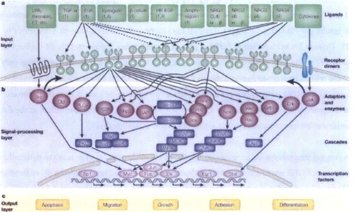
The interplay between ErbB family members creates a horizontal and vertical signaling network that leads to activation of a variety of pathways, including the mitogen-activated protein kinase (MAPK) pathway, the protein kinase C (PKC) pathway, and the phosphoinositide 3-kinase (P13K) pathway. Activation of these pathways ultimately converges on transcription factors, whose modulation leads to an array of cellular responses including growth, adhesion, migration, differentiation, and apoptosis. The ErbB family signaling network as it is currently understood is
summarized in Figure 1.1 (2). This diagram encapsulates the complexity of the network, yet eloquently abstracts ErbB family signaling into an input layer of ligand binding, a signal
processing layer of adaptor protein recruitment and transcription factor regulation, and an output layer of cell behaviors.
a
Figure 1.1. Schematic of the ErbB signaling pathway including stimulating ligands, ErbB family receptors, effector proteins, and downstream transcription factors regulated by the indicated signaling cascades, from (2), reproduced with permission. Note that the diagram is abstracted into input signal, signal processing network, and output response layers.
Among the members of the ErbB family, EGFR is by far the best characterized. EGFR is a 170-kDa protein that is 1186 amino acids in length. Over 20% of the receptor mass is contributed by N-linked glycosylation, which is requisite for both localization and function (5). Like the other ErbB family receptors, EGFR consists of an extracellular region (domains I-IV), a
transmembrane (TM) domain, a juxtamembrane (6) domain, a tyrosine kinase domain (TK), and a carboxyl-terminal region (CT), as shown in Figure 1.2. ErbB family members share 37%-49% sequence identity and the three-dimensional folds of ErbB receptors are similar, except for possible divergence in the C-terminal domains (7).
151 312 481 621 644 687 955 1186
D2 D4 TM CT
Figure 1.2: Schematic of the four extracellular domains, the transmembrane domain, and the intracellular domains (tyrosine kinase and c-terminal domain) of EGFR. Numbers represent amino acid positions along the receptor.
The EGFR extracellular region is composed of four domains. Domains 1 and 3 are often called
LI and L2, respectively, since their fold resembles that of the leucine-rich domain of receptors in
the insulin growth factor family (8). Ligands bind between these two leucine-rich domains
(9-11). Domain 3 was identified as the predominant ligand binding domain by epitope studies that
showed EGF and TGF-a have sub-micromolar affinities for an EGFR fragment containing only L2 and small adjacent sections of domains 2 and 4 (12, 13). Domains 2 and 4 are often referred to as CR1 and CR2, respectively, as their sequences are cysteine-rich and consist of numerous
short segments linked by disulfide bonds. Domain 2 plays a key role in dimerization, as the CR1 domains of partner receptors directly interact in EGFR homodimers (11).
The crystal structures of both the monomeric (9) and homodimeric (11) forms of the extracellular domains of EGFR were recently solved, although domain 4 was either disordered (9) or missing
(11) from the crystallized protein. The dimeric structure was solved conjugated to EGF and
TGF-a. EGF ligand binds to the receptor in two different states: a high-affinity state (10-50 pM) and a low-affmity state (1-2 nM), possibly differing in dimeric conformations or activity (14).
EGF ligand interacts with domains 1 and 3 of the tethered receptor monomer, distal from the
dimerization interface. It is believed that binding of a growth factor induces a 1300 rigid body rotation that transitions EGFR into the dimeric conformation, as shown in Figure 1.3 (15).
2 130' 2 IV IV'qi Donwir' IV A mxkdaes3- 7 0604to I N
Tethered Monomer Extended Dimer
Figure 1.3: Crystal structures of the tethered monomeric (9) and extended dimeric (11) forms of the EGFR extracellular domains. Domain 4 has been added to the dimer for context using its coordinates from the monomer. Domain 1 is shown in red, domains 2 and 4 are colored green, and domain 3 is shown in blue and red. EGF ligand is shown in teal. The domain 2 dimerization contacts between interacting receptors are marked with asterisks. Figure reproduced with permission from reference (15).
EGFR dimers exist in 2:2 receptor:ligand complexes, although dimerization is entirely receptor-mediated. The dimerization interface is localized to domain 2, as shown in Figure 1.3. Residues 242-259 of each EGFR molecule contact and interact with domain 2 residues of the partner receptor (15). Additional domain 2 contacts occur between residues 193-195, 204-205, and
279-280 of partner receptors (11). Domain 4 may play a role in the dimerization interface as it has
been reported that peptides that mimic modules 6 and 7 of this extracellular domain can disrupt EGFR homo- and heterodimerization (16). Also, domain 4 mutations have been shown to obstruct ligand binding and tyrosine phosphorylation, but the role of domain 4 in dimerization has yet to be fully elucidated due to the inability to crystallize this region (17). The orientation and interacting residues in the EGFR dimer are depicted in Figure 1.3.
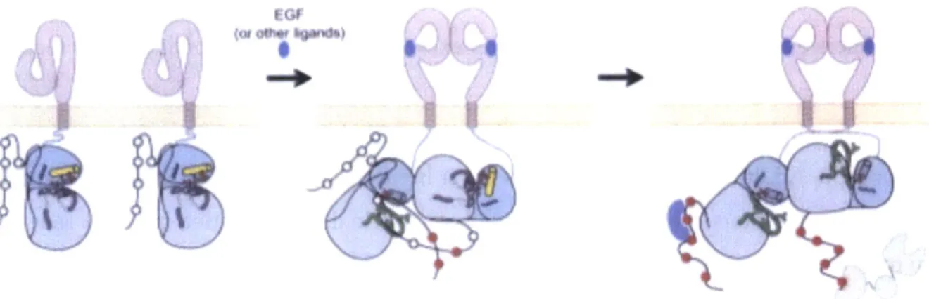
Although the structure of the extracellular domain and its rearrangement upon ligand-mediated dimerization are now well understood, the mechanism of kinase activation was, until recently, poorly defined. Conflicting structural data showed the active kinase domain crystallized in both symmetric and asymmetric formats (18, 19). Further mutational and biochemical analysis by Zhang and colleagues demonstrated that dimerization of the extracellular domain relieves the autoinhibition of the intracellular kinase domain through formation of an asymmetric kinase dimer in which the C-terminal lobe of one kinase domain interacts with the N-terminal lobe of its partner receptor in an analogous manner to the interaction between cyclin and cyclin-dependent kinases. This interaction exposes the active site on one receptor, allowing for kinase activity. The two receptors can dynamically switch positions to enable trans-phosphorylation of both receptors, as shown in Figure 1.4 (19).
i ( '
Figure 1.4. Schematic of EGFR kinase domain activation following EGF activation. Ligand-induced symmetric extracellular dimerization (pink) effects the rearrangement of intracellular tyrosine kinase domains to relieve autoinhibition and enable activation in a structurally similar manner to the cyclin/cyclin-dependent kinase interaction. The active kinase domain then trans-phosphorylates its partner receptor. The two receptors may switch positions to allow for activation of both molecules. Figure reproduced with permission from (19).
EGFR signaling is critical for epithelial cell development and its knockout in mice results in death either in mid-gestation, at birth, or postnatal day 20, depending on the genetics of the particular mouse strain (20-23). In contrast, mice show much less sensitivity to the absence of EGFR ligands, suggesting extensive redundancy in their function. Knockout of EGF and amphiregulin have no palpable effects on mouse phenotype (24) and knockout of TGF-a results only in slight abnormalities in the eye and in hair follicles (25). Even simultaneous knockout of
EGF, amphiregulin, and TGF-a results in relatively mild defects including slight growth
retardation and irregularities in the small intestine (26).
EGFR Trafficking
The structural and signaling properties of EGFR are intimately linked to its trafficking kinetics. In the absence of ligand, the vast majority of EGFR is localized to the cell surface (80-90%), but it is constantly shuttled in and out of the cell with an internalization halftime of 30 minutes and a rapid rate of recycling (27). Since the metabolic half-life of EGFR is 10-14 hours in fibroblasts and epithelial cells and 20-48 hours in transformed cells, receptors cycle through the endocytic pathway dozens of times with little risk of degradation (Figure 1.5) (1, 28, 29).
Once a ligand binds to and activates EGFR, the rate of internalization increases 5-10 fold and the degradation fraction is augmented at the expense of recycling (27, 30). This phenomenon,
known as receptor downregulation, is thought to be a negative feedback mechanism to attenuate growth factor signaling.
Oe~deton I u M Wd bne*e
... .
Figure 1.5. Schematic of EGFR trafficking with known time constants labeled. EGFR is constitutively shuttled in and out of the cell with an internalization halftime of 30' and a high probability of rapid recycling. Upon ligand stimulation, internalization is accelerated five- to tenfold and recycled fraction is dramatically reduced, shunting receptors to late endosomes and, ultimately, degradation in the lysosome. This figure is reproduced with permission from (1).
interact with the receptor. Cbl family
Acceleration of endocytosis rate is a direct consequence of recruitment to the clathrin-mediated endocytosis pathway compared to basal internalization routes. Clathrin-mediated
endocytosis is by far the fastest means of receptor internalization and is the predominant endocytic route for activated receptor under physiological conditions (31). However, additional modes of internalization involving lipid rafts, caveolae, membrane ruffling, and pinocytosis have also been described (32-36). Unlike clathrin-mediated internalization, though, these alternative
mechanisms do not accelerate EGFR endocytosis compared to constitutive kinetics. Also,
accumulating evidence shows that these pathways are active only in the presence of extremely high concentrations of EGF, well above those observed physiologically (37-39).
Immediately following activation of EGFR, Cbl proteins bind to the EGFR phosphotyrosine domain through complexation with the adapter protein Grb2 or a presently uncharacterized protein (referred to as "X" in Figure 1.6), both of which proteins, which have E3 ubiquitin ligase activity, monoubiquitinate EGFR with the assistance of E2 ubiquitin conjugating enzymes (31). These proteins are in turn phosphorylated by Src, increasing their interaction with the Cbl-interacting protein CIN85. CIN85 is constitutively associated with endophilins, which regulate
clathrin-mediated endocytosis, suggesting a role for this protein in internalization. However, the role of
CIN85 in trafficking of membrane-bound EGFR remains undefined as its presence has not yet
been detected in coated pits.
Ubiquitinated EGFR is recognized by ubiquitin binding domains (UBDs) of the EGFR pathway substrates 15 (EPS 15) and 15R (Eps 15R) as well as epsin, which collaborate to recruit clathrin to the cell surface and facilitate the formation of a complex between the tetramer AP-2 and the receptor to be internalized. AP-2 drives clathrin assembly and thus the formation of the 0.2-tm vesicles known as clathrin-coated pits. Receptors enter these pits and are pinched off from the membrane with the aid of dynamin (40). The internalized clathrin-coated vesicle is subsequently uncoated and fuses with an early endosome.
It should be noted that there is evidence of a ubiquitination-independent route of clathrin-mediated endocytosis. Mutation of the EGFR ubiquitination sites did not impact internalization rate, implying that Cbl-mediated ubiquitination is not strictly required for rapid endocytosis (39, 41, 42). Also, siRNA knockdown of the UBD-containing epsin, Eps 15, and Eps 15R failed to inhibit clathrin-dependent internalization of EGFR (32, 43). Ubiquitin- and
Grb2/Cbl-independent mechanisms of EGFR recruitment to clathrin-coated pits are currently being investigated.
In contrast with ubiquitination, kinase domain activity is requisite for clathrin-mediated endocytosis. Mutation of the kinase domain's catalytic lysine and competitive inhibition of kinase activation result in significant reductions in endocytosis rate in the presence of ligand (on the order of basal endocytosis) and inefficient recruitment to clathrin-coated pits (44-48). Thus, accelerated endocytosis through clathrin-coated pits requires intact kinase activity.
From the early endosome, a receptor may be sorted for recycling back to the surface (known as quick recycling). Unbound receptors are typically shunted to this pathway whereas active EGF-bound receptors are rarely recycled and remain in the endosome as it matures into the late
endosome, also known as a multi-vesicular body (MVB). Within the MVB, activated
adaptor molecule, and HRS, hepatocyte-growth-factor-regulated tyrosine-kinase substrate) through its UBDs. The receptor is then encapsulated into the intraluminal vesicles of MVBs, which subsequently fuse with lysosomes, where protein degradation is carried out by hydrolases
(49). Small amounts of recycling (known as late recycling) also occur from MVBs. The EGFR internalization pathway and approximate timescales for relevant events are summarized in Figure
1.5 and a more detailed depiction of the endocytic pathway is provided in Figure 1.6.
Ubiquitin
C e a W UBID
Chi x Phosphorylated tyroine
' AP-2 binding site
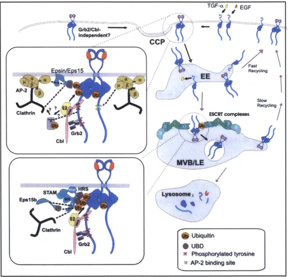
Figure 1.6. Model of EGFR endocytosis and intracellular trafficking following ligand stimulation. Briefly, EGF or TGF-a binding to EGFR (blue) induces dimerization and phosphorylation. The Grb2-Cbl adaptor protein complex is then recruited to phosphotyrosine sites in the C-terminal domain of the receptor. The RING domain of Cbl in turn recruits E2 enzymes to ubiquitinate the receptor. Ubiquitinated receptor is recognized by ubiquitin binding domains (UBDs) of epsin, Eps15, and Epsl5R, which are associated with AP-2 and clathrin heavy chain, the principle constituent of clathrin-coated pits (CCPs). Note that there may also be Grb2-Cbl complex-independent modes of
CCP recruitment. CCPs containing EGFR fuse with early endosomes (EEs) and receptor may either be rapidly
recycled or remain encapsulated in endosomes as they mature into multivesicular bodies (MVBs). Ubiquitinated EGFR that remains in MVBs interacts with the UBDs of the ESCRT-O complex (consisting of HRS and STAM) as well as Eps15b, resulting in its localization to internal MVB vesicles. MVB then fuses with primary lysosomes, where vesicle-bound EGFR-EGF complexes are degraded. Figure reproduced with permission from (31).
Grb2/CbI.--Independent?
CCP
mn
Internalization has historically been viewed exclusively as a mechanism for quenching receptor signaling, but it is now known that most if not all EGFR in endosomes is capable of signal transduction (50, 51). However, the signaling pathways that are activated and the duration of activation differ for internalized receptors compared with their surface-bound counterparts (52). While PLC-y and P13K pathway activation are reported to be restricted to surface-bound
receptors, the ras-dependent MAPK can be activated from both surface-bound and intracellular receptors (53, 54). Endosomal signaling has also been linked to tumor development. For instance, nuclear localization of EGFR has been observed following endocytosis and this localization is enhanced in cancer cells, but the role that nuclear EGFR plays in signal transduction or cancer development has yet to be established (55).
Aside from receptor internalization, an additional mechanism for signal attenuation is dephosphorylation of internalized EGFR at the endoplasmic reticulum by protein tyrosine phosphatase-1B (PTP1B) (56). However, the relationship between receptor deactivation and sorting is unclear at present (57).
EGFR trafficking and internalization are also affected by heterodimerization. All other ErbB family members exhibit slower internalization and less efficient degradation sorting than EGFR
(58-61), resulting in modulation of EGFR trafficking kinetics depending on dimer distribution.
In particular, ErbB2 is the preferred heterodimerization partner of EGFR and the formation of a complex between these two ErbB family members results in enhanced affinity for EGF, but reduced internalization and degradation (20, 27). Consequently, EGFR downregulation is decreased and EGFR signaling persists for longer time periods in the presence of excess ErbB2.
Linking EGFR to Cancer
For the past three decades, EGFR has been under intensive study due to its common
overexpression in epithelial-based tumors. EGFR expression is found at increased levels in many head, neck, breast, bladder, prostate, kidney, non-small-cell lung cancers, and gliomas
expression has been linked to poor clinical outcome, making EGFR a particularly attractive target for anti-cancer therapeutics (62, 63).
In addition to aberrant expression, mutations of the EGFR gene that dysregulate receptor
signaling are often detected in cancer cells. Heterozygous somatic mutations including deletions, insertions, and point mutations have been observed in the EGFR kinase domain, particularly in lung cancer patients (64-66). These mutations strengthen receptor interactions with ATP, amplifying autophosphorylation and boosting cell survival (67, 68).
Numerous non-kinase domain EGFR mutants have also been observed, particularly in tumors with EGFR gene amplification. Mutants with deletions, sequence duplications, and defective kinase regulatory signals have been reported. As many as 20% of glioblastomas express EGFR variants (69, 70), the most common of which is EGFRvIII (71), a splice mutant that deletes residues 6-273 of the receptor (most of extracellular domains 1 and 2). Due to the absence of domain 1-mediated tetheringin EGFRvIII, it is locked in the extended dimeric conformation. The TK domain is thus constitutively active, independent of ligand binding (57). Other non-kinase rearrangements within the ErbB 1 gene such as large deletions, point mutants, and insertions are also common, particularly in gliomas (72).
Finally, ErbB receptors have been linked to cancer in the context of autocrine signaling. Expression of an ErbB receptor in conjunction with an excess of its ligand can lead to unregulated cell growth. Autocrine production of TGF-a and EGF in cancer patients is correlated with increased mortality (73, 74). Also, autocrine signaling by neuregulin has been
shown to induce uncontrolled proliferation of human vestibular schwannoma cells (75). Thus, anti-cancer therapeutics must often compete with high concentrations of endogenous ligands in
vivo.
Anti-EGFR Therapeutics
Extensive overexpression and dysregulation of EGFR in a wide variety of cancer forms make this receptor an attractive therapeutic target. The two main compounds that have been used for
targeting EGFR are small-molecule tyrosine kinase inhibitors (TKIs) and anti-EGFR monoclonal antibodies (mAbs).
TKIs block activation of the EGFR TK domain by binding to the intracellular Mg-ATP binding site, obstructing ATP binding and subsequent autophosphorylation. The best-characterized TKIs are gefitinib and erlotinib, both of which are synthetic anilinoquinazolines that inhibit EGFR TK activity with nanomolar IC50 values. Complementing its primary action of blocking ATP
binding, gefitinib has also been shown to indirectly reduce angiogenesis (76). Treatment with gefitinib or erlotinib inhibits tumor cell proliferation, probably through induction of cell cycle arrest or apoptosis, and both drugs were recently approved by the FDA in the treatment of non-small-cell lung cancer (77, 78). There are several other TKIs in clinical trials, including
canertinib, lapatinib, AG-1478, and HKI-272 (57).
mAbs are Y-shaped homodimeric proteins that consist of two identical heavy and two identical light chains. The structure of a standard immunoglobulin G (IgG) is shown in Figure 1.7. The heavy and light chains contain an N-terminal variable fragments (VH and VL, respectively).
Each variable fragment has a beta barrel structure with conserved beta sheets (the framework regions) separated by three hypervariable loops known as the complementarity determining regions that confer specificity to a target antigen (79, 80). In addition to variable domains,
antibody heavy chains consist of three constant domains (CH1, CH2, and CH3) and the light chains include one constant domain (CL), all of which are uniform for a particular antibody isotype. The
Figure 1.7. Schematic of the assembly and structure of IgG heavy chain CH2 and CH3 constant antibody. The structure of the heavy (left) and light (midde) chains domains are collectively termed the
are presented. These chains assemble via four disulfide bridges to
form the symmetric 2:2 dimer shown at right. Note that the heavy Fc region, and interact with
chain consists of an N-terminal variable domain and three constant
domains whereas the light chain consists of an N-terminal variable complement or Fcy receptors found
domain and one constant domain. on several types of leukocytes,
Antibodies assemble into 2:2 heavy:light chain complexes, linked by four disulfide bonds: One between CHI and CL of each arm and two in the hinge region (N-terminal end of the CH2 domain). Crystallization of a full, intact human IgG revealed substantial asymmetry emanating from the extensive flexibility in antibody structure (82). Their ability to specifically target an antigen of interest and to foment an immune response have made mAbs attractive therapeutic molecules. In cancer treatment alone, there are ten clinically approved antibody-based drugs, with 165 others currently undergoing clinical trials (6).
mAbs specific for EGFR function both by recruiting cytotoxic lymphocytes or other white blood cells to enhance the immune response to cancer in a process known as antibody-dependent cell-mediated cytotoxicity (ADCC) (81) and by directly inhibiting EGFR signaling. The first antibody-based EGFR therapeutic to receive clinical approval was cetuximab (humanized form of murine mAb 225) (83). Originally isolated from immunized mice, mAb 225 was
reconstructed as a chimeric human Immunoglobulin GI (IgGl) for clinical use. With a 100-fold greater affinity for EGFR than the native EGF ligand, mAb 225 directly competes with ligand binding to domain III, blocking dimerization and autophosphorylation (84, 85). Its binding has been shown to enhance EGFR internalization and degradation in some cell lines (86, 87). mAb
225 treatment has been shown to induce Gi-phase cell-cycle arrest and enhanced apoptosis in a
range of human tumor cell lines (88, 89). Incidentally, mAb 225 also inhibits vascular endothelial growth factor (VEGF), further enhancing its anti-tumor activity (90).
Following successful phase III clinical trials, mAb 225 was recently approved to treat metastatic colorectal cancer (91) and squamous cell carcinoma of the head and neck (92, 93). Other monoclonal antibodies targeting the EGFR ligand-binding domain include the FDA-approved panitumumab and several compounds undergoing clinical trials, including matuzumab and hR-3 (94, 95).
A monoclonal antibody (mAb 806) that specifically targets EGFRvIII was recently discovered (96). mAb 806 binds to a cysteine loop at the end of EGFR extracellular domain II, a
conformational epitope that is exposed only when the receptor transitions into the open
targeting the ligand-binding domain, it is undergoing clinical testing for both monotherapy and combination therapy. A recent phase I clinical trial of mAb 806 demonstrated specific targeting of the mutant receptor and no significant toxicity (100).
Despite recent advances in the development of monoclonal antibody therapeutics against EGFR, large deficiencies remain in the efficacy of these drugs. For instance, mAb 225 showed an 11% response rate as a monotherapy and a 23% response rate in combination with chemotherapy in metastatic colorectal cancer and showed a response rate of 13% in squamous cell carcinoma of the head and neck (Erbitux@ prescribing information). The other clinically approved anti-EGFR
antibody, panitumumab, had only an 8% response rate in metastatic colorectal cancer (Vectibix@ prescribing information). It should also be noted that these antibodies are ineffective against
EGFRvIII and tumor cells that secrete high levels of autocrine EGFR ligands due to their reliance on ligand competition for efficacy. In addition, poor tumor penetration, autocrine signaling, acquired resistance, and receptor mutation hinder drug performance (101). It is therefore of interest to develop complementary therapeutic strategies to enhance mAb efficacy.
Antibody-Induced Clustering
It was recently established that particular combinations of non-competitive anti-EGFR mAbs synergistically reduce surface receptor levels both in vitro and in vivo. This downregulation of receptor is independent of tyrosine kinase activity and leads to enhanced tumor cell killing and prolonged survival in mouse xenograft models of cancer (102-105). Consistency between downregulation levels and combination efficacy in mouse models was also reported for ErbB2
(106). Friedman et al proposed that antibody synergism results from the formation of large
clusters of crosslinked receptors on the cell surface following combination mAb treatment (102). Biochemical evidence for the formation of higher-order clusters of EGFR was presented by Zhu
et al using the tyrosine kinase inhibitor decorin (107).
In particular, inclusion of the 806 monoclonal antibody in combination antibody treatments could be of significant utility in targeting EGFR. Due to its selectivity for mutant or overactive receptors and its neutralizing activity, it may be able to evade some of the shortcomings of mAbs that compete with ligand binding (96, 98). In a recent study, it was demonstrated that EGFR
undergoes synergistic downregulation in the presence of mAb 806 and mAb 528, an antibody with similar specificity and binding affinity to mAb 225 (103). This study points again to the possibility of receptor crosslinking and clustering, which can impact both trafficking and downstream signaling.
We hypothesized that receptor clustering could lead to downregulation through internalization enhancement or degradation fraction augmentation. We note that the endocytic machinery is
saturable (37), thus improving the efficiency of internalization via simultaneous entry of multiple clustered receptors could enhance the rate of receptor internalization compared to that of
untreated cells. Additionally, whereas unoccupied single receptors are rapidly recycled back to the membrane following internalization, oligomeric receptors are preferentially retained in
endosomes through maturation and lysosomal fusion (31, 108), which could potentially result in enhancement of EGFR degradation.
Receptor clustering could be advantageous in that it would complement existing therapeutic mechanisms by downregulation of EGFR and its signaling. Also, a clustering strategy would be independent of ligand binding, allowing for efficacy in constitutively active systems involving mutated receptor or dysregulated ligand, which resist treatment by currently approved
therapeutics. The characterization and, ultimately, the exploitation of antibody-mediated clustering holds promise as a novel strategy for therapeutic EGFR targeting in cancer.
Thesis Contributions
At the outset of this thesis work, the idea of antibody-induced clustering was just beginning to be explored. The objective of our project was to characterize the reproducibility and scope of this phenomenon and to isolate its mechanistic basis to gain insights that would inform design of EGFR-downregulating compounds for therapeutic applications. In the work described herein, we identify the requirements for antibody-induced downregulation, elucidate its kinetic mechanism, and evaluate its efficacy in a simulated tumor environment. We then apply our insights to the design of two novel classes of multispecific antibodies that show therapeutic promise in both in vitro and in vivo models of cancer.
In Chapter 2, we illuminate several crucial properties of combination antibody-induced
clustering. By screening a panel of pairwise antibody combinations, we show that co-treatment with non-competitive mAbs targeting distinct epitopes on EGFR elicits up to 80% receptor downregulation. Receptor downregulation induced by mAb combinations is found to be
inversely proportional to receptor density and requires bivalency of both constituent antibodies. We further find that mAb pairs consisting of two non-competitive antibodies that target EGFR domain 3 show the highest downregulation activity. The mechanism underlying downregulation is found to be consistent with recycling inhibition. Importantly, in contrast to the agonism associated with ligand-induced downregulation, we find that combination mAb-induced downregulation does not activate EGFR or downstream signaling pathways and that it leads to synergistic reduction in migration and proliferation of autocrine ligand-secreting cells.
After establishing the parameters for combination mAb-induced downregulation, we attempt to enhance antibody binding and induced receptor clustering through avidity effects. We employ two novel design approaches for our multispecific antibodies: (1) Fusion of the a full anti-EGFR antibody with the variable domain fragment of a second EGFR-targeted antibody (described in Chapter 3) and (2) Fusion of a full EGFR-specific antibody fused to one or more EGFR-binding variants of the tenth type three domain of human fibronectin (detailed in Chapter 4). We
demonstrate that both classes of constructs induce robust receptor clustering and reproducible downregulation on a range of cancerous cell lines. We also establish that these multispecific fusions downregulate both through blockade of receptor recycling and via acceleration of endocytosis and that downregulation is independent of receptor surface density. To motivate therapeutic application, we show that multispecific compounds do not agonize EGFR signaling and that they inhibit motility and growth of cells that aberrantly express autocrine ligand.
We present evidence in Chapter 5 that both of our multispecific strategies effectively curtail growth in tumor xenograft models that resist treatment with standard of care antibodies and that
in vivo inhibition of tumor growth channels in vitro receptor downregulation. In particular, we
show dramatic control of tumors that contain genetic mutations in effector proteins downstream of EGFR (raf and ras). Through mutational analysis, we demonstrate that ligand inhibition,
In summary, we have rigorously characterized and defined requirements for antibody-induced downregulation and applied this information to the design of novel classes of EGFR-targeted antibody-based therapeutics. The modularity of our constructs and the generality of our system suggests extensibility of the multi-epitopic strategy to other ErbB family members and, more generally, to receptor proteins of therapeutic interest.
References
1. Sorkin A & Von Zastrow M (2002) Signal transduction and endocytosis: close encounters of many kinds. Nat Rev Mol Cell Biol 3(8):600-614.
2. Yarden Y & Sliwkowski MX (2001) Untangling the ErbB signalling network. Nat Rev
Mol Cell Biol 2(2):127-137.
3. Hynes NE & Lane HA (2005) ERBB receptors and cancer: the complexity of targeted inhibitors. Nat Rev Cancer 5(5):341-354.
4. Tzahar E, et al. (1996) A hierarchical network of interreceptor interactions determines signal transduction by Neu differentiation factor/neuregulin and epidermal growth factor.
Mol Cell Biol 16(10):5276-5287.
5. Slieker LJ, Martensen TM, & Lane MD (1986) Synthesis of epidermal growth factor receptor in human A431 cells. Glycosylation-dependent acquisition of ligand binding activity occurs post-translationally in the endoplasmic reticulum. JBiol Chem
261(32):15233-15241.
6. Reichert J (2011) Antibodies for cancer: Past, present, and future. Seventh Annual
Protein Engineering Summit.
7. Jorissen RN, et al. (2003) Epidermal growth factor receptor: mechanisms of activation and signalling. Exp Cell Res 284(l):31-53.
8. Garrett TP, et al. (1998) Crystal structure of the first three domains of the type-I insulin-like growth factor receptor. Nature 394(6691):395-399.
9. Ogiso H, et al. (2002) Crystal structure of the complex of human epidermal growth factor and receptor extracellular domains. Cell 110(6):775-787.
10. Garrett TP, et al. (2002) Crystal structure of a truncated epidermal growth factor receptor extracellular domain bound to transforming growth factor alpha. Cell 110(6):763-773.
11. Ferguson KM, et al. (2003) EGF activates its receptor by removing interactions that autoinhibit ectodomain dimerization. Mol Cell 11(2):507-517.
12. Kohda D, et al. (1993) A 40-kDa epidermal growth factor/transforming growth factor alpha-binding domain produced by limited proteolysis of the extracellular domain of the epidermal growth factor receptor. JBiol Chem 268(3):1976-1981.
13. Lemmon MA, et al. (1997) Two EGF molecules contribute additively to stabilization of the EGFR dimer. EMBO J 16(2):281-294.
14. Lin CR, et al. (1986) Protein kinase C phosphorylation at Thr 654 of the unoccupied EGF receptor and EGF binding regulate functional receptor loss by independent mechanisms.
Cell 44(6):839-848.
15. Burgess AW, et al. (2003) An open-and-shut case? Recent insights into the activation of EGF/ErbB receptors. Mol Cell 12(3):541-552.
16. Berezov A, et al. (2002) Disabling receptor ensembles with rationally designed interface peptidomimetics. JBiol Chem 277(31):28330-28339.
17. Saxon ML & Lee DC (1999) Mutagenesis reveals a role for epidermal growth factor receptor extracellular subdomain IV in ligand binding. JBiol Chem
274(40):28356-28362.
18. Stamos J, Sliwkowski MX, & Eigenbrot C (2002) Structure of the epidermal growth factor receptor kinase domain alone and in complex with a 4-anilinoquinazoline inhibitor.
JBiol Chem 277(48):46265-46272.
19. Zhang X, Gureasko J, Shen K, Cole PA, & Kuriyan J (2006) An allosteric mechanism for activation of the kinase domain of epidermal growth factor receptor. Cell 125(6):1137-1149.
20. Citri A & Yarden Y (2006) EGF-ERBB signalling: towards the systems level. Nat Rev
Mol Cell Biol 7(7):505-516.
21. Miettinen PJ, et al. (1995) Epithelial immaturity and multiorgan failure in mice lacking epidermal growth factor receptor. Nature 376(6538):337-341.
22. Threadgill DW, et al. (1995) Targeted disruption of mouse EGF receptor: effect of genetic background on mutant phenotype. Science 269(5221):230-234.
23. Sibilia M & Wagner EF (1995) Strain-dependent epithelial defects in mice lacking the
EGF receptor. Science 269(5221):234-238.
24. Luetteke NC, et al. (1999) Targeted inactivation of the EGF and amphiregulin genes reveals distinct roles for EGF receptor ligands in mouse mammary gland development.
Development 126(12):2739-2750.
25. Mann GB, et al. (1993) Mice with a null mutation of the TGF alpha gene have abnormal skin architecture, wavy hair, and curly whiskers and often develop corneal inflammation.
Cell 73(2):249-261.
26. Troyer KL, et al. (2001) Growth retardation, duodenal lesions, and aberrant ileum architecture in triple null mice lacking EGF, amphiregulin, and TGF-alpha.
Gastroenterology 121(1):68-78.
27. Wiley HS (2003) Trafficking of the ErbB receptors and its influence on signaling. Exp
Cell Res 284(l):78-88.
28. Stoscheck CM & Carpenter G (1984) Characterization of the metabolic turnover of epidermal growth factor receptor protein in A-431 cells. J Cell Physiol 120(3):296-302.
29. Burke PM & Wiley HS (1999) Human mammary epithelial cells rapidly exchange empty EGFR between surface and intracellular pools. J Cell Physiol 180(3):448-460.
30. Worthylake R, Opresko LK, & Wiley HS (1999) ErbB-2 amplification inhibits down-regulation and induces constitutive activation of both ErbB-2 and epidermal growth factor receptors. JBiol Chem 274(13):8865-8874.
31. Sorkin A & Goh LK (2009) Endocytosis and intracellular trafficking of ErbBs. Exp Cell
Res 315(4):683-696.
32. Sigismund S, et al. (2005) Clathrin-independent endocytosis of ubiquitinated cargos.
Proc Natl Acad Sci U S A 102(8):2760-2765.
33. Chinkers M, McKanna JA, & Cohen S (1979) Rapid induction of morphological changes in human carcinoma cells A-431 by epidermal growth factors. J Cell Biol 83(1):260-265. 34. Haigler HT, McKanna JA, & Cohen S (1979) Direct visualization of the binding and
internalization of a ferritin conjugate of epidermal growth factor in human carcinoma cells A-43 1. J Cell Biol 81(2):382-395.
35. Yamazaki T, et al. (2002) Role of Grb2 in EGF-stimulated EGFR internalization. J Cell
36. Orth JD, Krueger EW, Weller SG, & McNiven MA (2006) A novel endocytic mechanism of epidermal growth factor receptor sequestration and internalization. Cancer Res
66(7):3603-3610.
37. Wiley HS (1988) Anomalous binding of epidermal growth factor to A431 cells is due to the effect of high receptor densities and a saturable endocytic system. J Cell Biol
107(2):801-810.
38. Lund KA, Opresko LK, Starbuck C, Walsh BJ, & Wiley HS (1990) Quantitative analysis of the endocytic system involved in hormone-induced receptor internalization. JBiol
Chem 265(26):15713-15723.
39. Jiang X & Sorkin A (2003) Epidermal growth factor receptor internalization through clathrin-coated pits requires Cbl RING finger and proline-rich domains but not receptor polyubiquitylation. Traffic 4(8):529-543.
40. Waterman H & Yarden Y (2001) Molecular mechanisms underlying endocytosis and sorting of ErbB receptor tyrosine kinases. FEBS Lett 490(3):142-152.
41. Huang F, Kirkpatrick D, Jiang X, Gygi S, & Sorkin A (2006) Differential regulation of
EGF receptor internalization and degradation by multiubiquitination within the kinase
domain. Mol Cell 21(6):737-748.
42. Huang F, Goh LK, & Sorkin A (2007) EGF receptor ubiquitination is not necessary for its internalization. Proc Natl A cad Sci USA 104(43):16904-16909.
43. Huang F, Khvorova A, Marshall W, & Sorkin A (2004) Analysis of clathrin-mediated endocytosis of epidermal growth factor receptor by RNA interference. JBiol Chem
279(16):16657-16661.
44. Chen WS, et al. (1989) Functional independence of the epidermal growth factor receptor from a domain required for ligand-induced internalization and calcium regulation. Cell
59(1):33-43.
45. Wiley HS, et al. (1991) The role of tyrosine kinase activity in endocytosis,
compartmentation, and down-regulation of the epidermal growth factor receptor. JBiol
Chem 266(17):11083-11094.
46. Sorkina T, Huang F, Beguinot L, & Sorkin A (2002) Effect of tyrosine kinase inhibitors on clathrin-coated pit recruitment and internalization of epidermal growth factor receptor.
47. Sorkin A, Waters C, Overholser KA, & Carpenter G (1991) Multiple
autophosphorylation site mutations of the epidermal growth factor receptor. Analysis of kinase activity and endocytosis. JBiol Chem 266(13):8355-8362.
48. Lamaze C & Schmid SL (1995) Recruitment of epidermal growth factor receptors into coated pits requires their activated tyrosine kinase. J Cell Biol 129(1):47-54.
49. Bache KG, Raiborg C, Mehlum A, & Stenmark H (2003) STAM and Hrs are subunits of a multivalent ubiquitin-binding complex on early endosomes. JBiol Chem
278(14):12513-12521.
50. Wang Y, Pennock S, Chen X, & Wang Z (2002) Endosomal signaling of epidermal growth factor receptor stimulates signal transduction pathways leading to cell survival.
Mol Cell Biol 22(20):7279-7290.
51. Pennock S & Wang Z (2003) Stimulation of cell proliferation by endosomal epidermal growth factor receptor as revealed through two distinct phases of signaling. Mol Cell Biol
23(16):5803-5815.
52. Schmidt MH, Furnari FB, Cavenee WK, & Bogler 0 (2003) Epidermal growth factor receptor signaling intensity determines intracellular protein interactions, ubiquitination, and internalization. Proc Natl Acad Sci USA 100(11):6505-65 10.
53. Haugh JM, Huang AC, Wiley HS, Wells A, & Lauffenburger DA (1999) Internalized epidermal growth factor receptors participate in the activation of p21 (ras) in fibroblasts. J
Biol Chem 274(48):34350-34360.
54. Haugh JM & Meyer T (2002) Active EGF receptors have limited access to Ptdlns(4,5)P(2) in endosomes: implications for phospholipase C and PI 3-kinase signaling. J Cell Sci 115(Pt 2):303-3 10.
55. Lo HW, et al. (2005) Novel prognostic value of nuclear epidermal growth factor receptor in breast cancer. Cancer Res 65(l):338-348.
56. Haj FG, Verveer PJ, Squire A, Neel BG, & Bastiaens PI (2002) Imaging sites of receptor dephosphorylation by PTP 1 B on the surface of the endoplasmic reticulum. Science
295(5560):1708-1711.
57. Sebastian S, et al. (2006) The complexity of targeting EGFR signalling in cancer: from expression to turnover. Biochim Biophys Acta 1766(1):120-139.
58. Waterman H, Sabanai I, Geiger B, & Yarden Y (1998) Alternative intracellular routing of ErbB receptors may determine signaling potency. JBiol Chem 273(22):13819-13827.
59. Baulida J, Kraus MH, Alimandi M, Di Fiore PP, & Carpenter G (1996) All ErbB
receptors other than the epidermal growth factor receptor are endocytosis impaired. JBiol
Chem 271(9):5251-5257.
60. Baulida J & Carpenter G (1997) Heregulin degradation in the absence of rapid receptor-mediated internalization. Exp Cell Res 232(1):167-172.
61. Waterman H, Alroy I, Strano S, Seger R, & Yarden Y (1999) The C-terminus of the kinase-defective neuregulin receptor ErbB-3 confers mitogenic superiority and dictates endocytic routing. EMBO J 18(12):3348-3358.
62. Nicholson RI, Gee JM, & Harper ME (2001) EGFR and cancer prognosis. Eur J Cancer
37 Suppl 4:S9-15.
63. Earp HS, 3rd, Calvo BF, & Sartor CI (2003) The EGF receptor family--multiple roles in proliferation, differentiation, and neoplasia with an emphasis on HER4. Trans Am Clin
Climatol Assoc 114:315-333; discussion 333-314.
64. Lynch TJ, et al. (2004) Activating mutations in the epidermal growth factor receptor underlying responsiveness of non-small-cell lung cancer to gefitinib. N Engl J Med
350(21):2129-2139.
65. Paez JG, et al. (2004) EGFR mutations in lung cancer: correlation with clinical response to gefitinib therapy. Science 304(5676):1497-1500.
66. Pao W, et al. (2004) EGF receptor gene mutations are common in lung cancers from "never smokers" and are associated with sensitivity of tumors to gefitinib and erlotinib.
Proc Natl A cad Sci US A 101(36):13306-13311.
67. Tracy S, et al. (2004) Gefitinib induces apoptosis in the EGFRL858R non-small-cell lung cancer cell line H3255. Cancer Res 64(20):7241-7244.
68. Sordella R, Bell DW, Haber DA, & Settleman J (2004) Gefitinib-sensitizing EGFR mutations in lung cancer activate anti-apoptotic pathways. Science 305(5687):1163-1167.
69. Ekstrand AJ, et al. (1991) Genes for epidermal growth factor receptor, transforming growth factor alpha, and epidermal growth factor and their expression in human gliomas in vivo. Cancer Res 51(8):2164-2172.