ORIGINAL ARTICLE
Use of diffusion-weighted imaging (DWI) in PET/MRI for head
and neck cancer evaluation
Marcelo A. Queiroz&Martin Hüllner&Felix Kuhn&
Gerhardt Huber&Christian Meerwein&Spyros Kollias&
Gustav von Schulthess& Patrick Veit-Haibach
Received: 5 March 2014 / Accepted: 16 July 2014 / Published online: 5 August 2014 # Springer-Verlag Berlin Heidelberg 2014
Abstract
Objective The purpose of this study was to analyze whether diffusion-weighted imaging (DWI) adds significant informa-tion to positron emission tomography/magnetic resonance imaging (PET/MRI) on lesion detection and characterization in head and neck cancers.
Methods Seventy patients with different head and neck can-cers were enrolled in this prospective study. All patients underwent sequential contrast-enhanced (ce) PET/computed tomography (CT) and cePET/MRI using a tri-modality PET/ CT-MR setup either for staging or re-staging. First, the DWI alone was evaluated, followed by the PET/MRI with conven-tional sequences, and in a third step, the PET/MRI with DWI was evaluated. McNemar’s test was used to evaluate differ-ences in the accuracy of PET/MRI with and without DWI compared to the standard of reference.
Results One hundred eighty-eight (188) lesions were found, and of those, 118 (62.8 %) were malignant and 70 (37.2 %) were benign. PET/MRI without DWI had a higher accuracy in
detecting malignant lesions than DWI alone (86.8 % vs. 60.6 %, p<0.001). PET/MRI combined with DWI detected 120 concurrent lesions (89 malignant and 31 benign), PET/ MRI alone identified 48 additional lesions (20 malignant and 28 benign), and DWI alone detected 20 different lesions (nine malignant and 11 benign). However, lesions detected on DWI did not change overall staging. SUV maximum and mean were significantly higher in malignant lesions than in benign lesions. DWI parameters between malignant and benign le-sions were not statistically different.
Conclusion The use of DWI as part of PET/MRI to evaluate head and neck cancers does not provide remarkable informa-tion. Thus, the use of DWI might not be needed in clinical PET/MRI protocols for the staging or restaging of head and neck cancers.
Keywords PET/MRI . DWI . Head and neck cancer . SUV . ADC . b-value
Introduction
Head and neck cancers (HNC) are among the most prevalent cancers, accounting for more than 550,000 cases annually worldwide [1]. The primary risk factors associated with HNC include tobacco use, alcohol consumption, human pap-illomavirus (HPV) infection (for oropharyngeal cancer), and Epstein-Barr virus (EBV) infection (for nasopharyngeal can-cer) [2]. Standard treatments for head and neck cancers in-clude radiation therapy and surgery, and for certain types of head and neck cancer, chemotherapy. Survival is partly poor and has partly improved over the past three decades [3], possibly related to a better tumor staging system and a strict follow-up protocol, including the imaging approach.
M. A. Queiroz (*)
:
M. Hüllner:
F. Kuhn:
G. von Schulthess:
P. Veit-HaibachDepartment of Medical Radiology, Nuclear Medicine, University Hospital Zurich, Zurich, Switzerland
e-mail: [email protected] F. Kuhn
:
P. Veit-HaibachDepartment of Medical Radiology, Diagnostic and Interventional Radiology, University Hospital Zurich, Zurich, Switzerland M. Hüllner
:
S. KolliasDepartment of Medical Radiology, Neuroradiology, University Hospital Zurich, Zurich, Switzerland
G. Huber
:
C. MeerweinDepartment of Otorhinolaryngology, University Hospital Zurich, Zurich, Switzerland
18
F-fluorodeoxy-D-glucose (FDG) positron emission tomography/magnetic resonance imaging (PET/MRI) appears to be an excellent tool for imaging evaluation of head and neck cancers due to the high soft-tissue contrast provided by the MR component and the possibly better characterization of HNC. The definition of MR protocols for PET/MRI is chal-lenging due to the vast amount of differently weighted se-quences, but acquisition time should ideally be no longer than a standard PET/CT with contrast media. Furthermore, because of the availability of the PET component, the information provided by the MR component should be complementary or confirmatory to the PET information, but not redundant.
Diffusion-weighted imaging (DWI) was initially established as a functional MR technique that helps in detecting acute stroke, and has been researched for a wide variety of extracra-nial applications as well [4,5]. It analyses the structure of a biologic tissue at a microscopic level based on the motion of water molecules. The differences in water mobility are quanti-fied by an apparent diffusion coefficient (ADC). The ADC reflects the signal loss on DWI that occurs with increasing b-values and is inversely correlated with tissue cellularity [6–8]. FDG is a glucose analogue that accumulates in tumor cells after its transformation to FDG-6-phosphate by hexokinase, which cannot be oxidized further through glycolysis [9]. The semi-quantitative assessment of the glucose metabolism in these cells is expressed by the standardized uptake value (SUV). The SUV is defined as the FDG uptake in a tumor over a certain time interval, considering tracer decay, the administered dose of the PET tracer, and the patient’s body weight [10].
In HNC, DWI has been widely used for tissue characteri-zation for primary tumors and lymph node (LN) metastases, prediction and monitoring of treatment response, and differ-entiation of recurrent tumors from post-therapeutic changes [11]. However, even if DWI and FDG do show different molecular phenomena, in the context of PET/MRI with the FDG-PET component available, the additional DWI compo-nent might actually represent redundant information to FDG-PET. Furthermore, one study has shown that DWI does not improve lesion detection in a PET/MRI protocol for whole-body cancer staging in a mixed oncologic population [12].
Thus, the aim of this study was to analyze whether the addition of a DWI sequence to PET/MRI adds significant additional information concerning lesion detection and char-acterization in patients with head and neck cancer.
Materials and methods Patient population
A total of 157 adult patients who underwent PET/CT-MR between February 2012 and March 2013, either for staging
or re-staging of various head and neck cancers, were enrolled in this prospective study. The inclusion criterion was the presence of at least one suspicious lesion on full-diagnostic PET/MRI, regardless of the type of treatment for recurrent lesions. The exclusion criteria of the present study included: unwillingness to undergo an additional MR exam, claustro-phobia, MR-incompatible medical devices (e.g., cardiac pace-makers, neurostimulators, cochlear implants, and insulin pumps), or possible metallic fragments in the body. The institutional ethics committee approved this study and signed informed consent was obtained from all subjects prior to the examination.
PET/CT and MR imaging
Sequential PET/CT, ceCT, and ceMR were performed on a tri-modality PET/CT-MRI setup (full ring, time-of-flight Discov-ery PET/CT 690, 3T DiscovDiscov-ery MR 750 w, both GE Healthcare, Waukesha, WI, USA). The dedicated MR- and CT-compatible shuttle transfer mechanism connecting the MR system and the PET/CT system allowed for PET/CT scanning that was free of radiofrequency (RF) coil-induced artifacts, and also made it possible to ascertain the placement of dedi-cated RF coils for MR imaging without repositioning of the patient [13,14].
In accordance with the European Association of Nuclear Medicine (EANM) procedure guidelines for PET imaging, patients fasted for at least 4 h prior to injection of a standard dose of 4.5 MBq per kg body weight [15]. After an uptake time of 30 min, the patients were positioned on the shuttle table in the MR suite and the MR acquisition covering the region between the orbital roof and the cranial end of the sternum was initiated. The images were acquired using a dedicated RF coil (32-Channel HD Head-Neck-Spine, GE Healthcare). The applied MR pulse sequences (PS) included an axial, T1-weighted (T1w), three-dimensional (3D), spoiled-gradient echo pulse sequence (LAVA), an axial, two-point, Dixon-based, T2-weighted (T2w) gradient echo se-quence (IDEAL), an axial, two-point, Dixon-based, ceT1w-gradient echo sequence (IDEAL), coronal and sagittal, two-point, Dixon-based, ceT1w-gradient echo sequences (LAVA flex), and an axial DWI. All images were acquired with a slice thickness of 4 mm within a total duration of the MR of 20– 25 min (additional scanning parameters are listed in Table1). The intravenously (IV) injected amount of contrast medium (Omniscan, GE Healthcare) was 0.2 mL/kg body weight at a flow rate of 1.5 ml/s. After completion of the MR scan, the coils were removed and the patient was transferred to the PET/ CT scanner, still being positioned on the shuttle board. This ensures an identical position of the patient during the acquisi-tion of both the PET/CT and the MR exam.
Non-enhanced low-dose CT and PET emission data were acquired from the mid-thigh to the vertex of the skull.
PET data was acquired in 3D time of flight (TOF) mode with a scan duration of 2 min per bed position, an overlap of bed positions of 23 %, and an axial field of view (FOV) of 153 mm. The emission data was corrected for attenuation using the low-dose CT (CTAC), and was then iteratively reconstructed (matrix size 256×256 pixels, Fourier rebinning (VIP mode), VUE Point FX (3D) with three iterations, 18 subsets).
Image processing
The acquired PET, CT, and ceMR images were transmitted to a dedicated review workstation (Advantage Workstation, Ver-sion 4.5, GE Healthcare), which enables the review of the PET, CT, and ceMR images side by side or in fused/overlay mode (PET/CT; cePET/MRI). Due to the calibrated tri-modality system, no software-based image registration was necessary. A previously conducted study validated the image registration accuracy with less than 4 mm of lateral misalign-ment between CT, PET, and MR data sets, similar to the intrinsic error assessed with phantom measurements [16].
Image analysis
All images were analysed in consensus by a board-certified nuclear medicine physician/radiologist and a radiologist with substantial experience in PET/CT image reading. The pres-ence of at least one suspicious lesion on full-diagnostic PET/ MRI was mandatory for further evaluation.
DWI-only, PET/MRI-only, and PET/MRI with DWI im-ages were analyzed concerning the detection and characteri-zation of lesions. First, only the DW sequence was evaluated,
with thresholds applied as mentioned below. Then, the PET/ MRI with axial T2w fat-suppressed, axial LAVA, and multiplanar ceT1 were analyzed. Last, PET/MRI with DWI was evaluated.
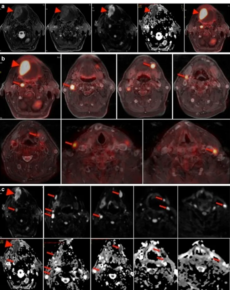
On DWI, a malignant lesion was defined if the mean ADC (ADCmean) value was lower than and 1.2×10⁻³mm2/s, based on reports available in the literature [17–22]. Additionally, several other thresholds were applied, ranging from 1.0× 10⁻³ mm2/s to 1.5 × 10⁻³ mm2/s, in order to identify the threshold with best accuracy. So, the lesions were quantitative-ly defined as: 1 (higher than 1.4×10⁻³mm2/s); 2 (between 1.2 and 1.4×10⁻³mm2/s); 3 (between 1.0 and 1.2×10⁻³mm2/s); and 4 (lower than 1.0×10⁻³mm2/s).
For PET/MRI, a malignant lesion was defined based on both functional and morphological criteria. The functional criterion used for the PET compound was a maximum SUV (SUVmax) of at least two-fold higher than the surrounding background activity (as published before). The morphological criteria for malignancy on MRI included: (1) a mass-like lesion with irregular borders and contrast enhancement; (2) enlarged lymph nodes (LN) greater than 1.0 cm in the short axis (and 1.5 cm for angular lymph nodes), cystic, with a necrotic centre, round-shaped, in a cluster formation, with an irregular boundary of the LN capsule and/or extra capsular LN spread. If there were discordant findings between PET and MRI, the combination of the most relevant findings (morpho-logical and functional) was taken into account (e.g., an en-larged and irregular LN was considered malignant even if there was no FDG uptake) [23].
The lesions were additionally classified both on PET/CT and MRI using a likelihood evaluation ranging from 1 to 4 (1, negative (meaning no suspicious lesion detected); 2, probably Table 1 MR acquisition parameters
Parameter T1w LAVA T2w IDEAL ceT1w LAVA flex DWI EPI-STIR
Repetition time/echo time (ms) 8.1/2.1 5188/80 6.2/1.7 5500/66.1
Echo train length NA 23 NA NA
Flip angle (°) 15 90 15 90
Inversion time (ms) NA NA NA 250
Parallel imaging acceleration factor 2 2 2 2
Receiver bandwidth (kHz) 83.33 83.33 166.67 250 Field of view (cm) 24 24 24 24 Matrix 320×256 320×256 220×220 320×256 b-value (s/mm) NA NA NA 0 and 800 NEX NA NA NA 1 Number of directions NA NA NA 3 Acquisition time 00:57 03:38 03:50 01:39
T1w LAVA T1-weighted, spoiled-gradient echo pulse sequence; T2w IDEAL Two-point, Dixon-based, 3D T2-weighted gradient echo sequence; ceT1w IDEAL Two-point, Dixon-based, 3D enhanced, T1-weighted gradient echo sequence; ceT1w LAVA flex Two-point, Dixon-based, 3D contrast-enhanced, T1-weighted gradient echo sequence; DWI Diffusion-weighted imaging sequence; EPI-STIR Echo planar imaging–short-time inversion recovery; NEX Number of excitations; NA Not applicable
benign; 3, likely (lesion likely to be malignant); and 4, very likely (lesion with very high suspicion of malignancy).
The standard of reference consisted of the histopathology (n=65) of the detected lesions, clinical evaluation (n=59), and imaging follow-up including all other imaging modali-ties (n=64).
ADC values were measured for each pixel with b-factors of 0 and 800 s/mm2 using the standard software on the worksta-tion (GE Healthcare, Waukesha, WI, USA). The ADC values were evaluated within a manually drawn, oval region of interest (ROI) placed carefully within the center of the lesions, avoiding apparent cystic changes or necrosis. The b-values were also calculated using the same software after placing the ROI on the DW image (b=800 s/mm2).
Anatomical localization, SUVmax, mean SUV (SUVmean), ADCmean,and mean and maximum b-values (b-valuemeanand b-valuemax, respectively) were also assessed.
Statistical analysis
All statistical tests were performed using SPSS Statistics Version 21 (IBM, Armonk, NY, USA). P-values<0.05 were considered statistically significant. Wilcoxon's signed-rank test was used for the comparison of the likelihood evaluation in PET/MRI and DWI. Spearman’s correlation analysis was performed to evaluate the correlation between SUVmax, SUVmean, b-valuemax, b-valuemean , and ADCmean. The Mann–Whitney Test was applied to analyze the difference of SUVmax, SUVmean, b-valuemax, b-valuemean, and ADCmeanin malignant and non-malignant lesions. McNemar's test was used to evaluate differences in the accuracy of PET/MRI and DWI compared to the standard of reference.
Results
One hundred eighty-eight (188) lesions were identified in 70 patients (53 men, 17 women; mean age 63.8 years, range 26– 86 years).
The remaining 87 patients showed no FDG-positive or suspicious lesions on MRI in PET/MRI of the head and neck area, and were therefore excluded.
Of the 188 lesions, 118 were malignant (37 tumors, 74 lymph node metastases, and seven soft-tissue metastases). Of the malignant lesions, 41 lesions were confirmed by histopa-thology, 44 by imaging, and 33 by clinical follow-up.
Additionally, 70 benign lesions were detected (56 inflammatory/reactive lymph nodes, 10 unspecific findings, and four Whartin’s tumors) and confirmed by clinical and imaging follow-up.
Of the overall 70 patients with lesions, 16 underwent imag-ing for primary stagimag-ing and 54 for follow-up/re-stagimag-ing. Mean follow-up time after PET/MRI was 196 days (range, 43 [this
patient died]–394, median 180 days). Forty-one patients were alive without disease at the end of the follow-up phase, 23 were alive with disease, and six were dead as a result of disease.
The majority of the primary tumor histology (tumor histol-ogy at initial staging) was squamous-cell carcinoma (83.1 %). Overall primary tumor staging was as follows: one patient was T0 (1.6 %), 17 were T1 (26.6 %), 19 were T2 (29.7 %), nine were T3 (14.1 %), and 18 were T4 (28.1 %). The initial N-staging was N0 in 25 patients (39.1 %), N1 in eight (12.5 %), N2 in 29 (45.3 %), and N3 in two (3.1 %). The overall patient and tumor characteristics are summarized in Table2.
PET/MRI without DWI had a higher accuracy in detecting malignant lesions than DWI alone (86.8 % vs. 60.6 %, p<0.001). PET/MRI read jointly with DWI had a slightly lower accuracy, although without statistical significance (86.8 % vs. 84.0 %, p>0.05; see Table 3). The PET/MRI likelihood evaluation was significantly different from the DWI evaluation (p=0.001; Table4). PET/MRI was superior to DWI for all different ADCmeanthreshold settings, mainly reflected by higher sensitivity (see Table5).
DWI missed 48 lesions detected by PET/MRI. Thirty-one lesions were not detected by DWI due to technical reasons, such as MR artifacts (due to metal, movement) and lesion location at the edge of the field of view (FOV). The remaining 17 lesions had no restricted diffusion and were therefore missed by DWI. Of those 48 lesions missed by DWI, 20 were malignant (nine tumors [seven recurrent and two primary], nine recurrent lymph nodes, and two metastasis). Fourteen lesions of those 20 (six tumors, six recurrent lymph nodes, and two metastasis) were missed based on technical issues (artifacts). The remain-ing six malignant lesions (three tumors and three recurrent lymph nodes) did not show restricted diffusion (Fig.1).
DWI added 20 lesions to the PET/MRI findings, and of those, 11 were inflammatory/reactive lymph nodes and nine were malignant lymph nodes. However, none of these nine malignant lymph nodes changed the overall staging since other lymph nodes defining the N-stage were already detected in PET/MRI without DWI (Fig.2).
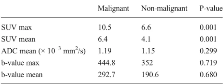
Concerning the quantitative values of PET and DWI, there was a significant difference (p= 0.001) in SUVmax and SUVmean between malignant and non-malignant lesions (10.5 vs 6.6 and 6.4 vs 4.1, respectively). ADCmeanas well as b-valuemeanand b-valuemaxshowed no statistical significant difference between those lesions (Table6).
PET and DWI values were significantly different between subjects referred for staging and re-staging (Table7).
Discussion
In this study, it has been shown that PET/MRI is superior to DWI alone and that PET/MRI with DWI does not achieve a higher accuracy than PET/MRI without DWI in patients with
head and neck tumors. However, DWI is able to add different pathological lesions, although not changing the final staging. Therefore, DWI is not essential in clinical PET/MRI protocols for the staging or re-staging of head and neck cancers.
General aspects
PET/MRI currently is emerging as a potential diagnostic tool that combines the functional PET information on tumor me-tabolism with the excellent anatomical correlation provided by different PS in MRI [13,24,25]. Furthermore, MRI can potentially offer additional physiological sequences that may provide information on tumor microstructure, e.g., with DWI sequences [10,26,27].
DWI has been promoted as a useful imaging tool to detect and characterize malignant lesions and predict tumor response in oncology [28]. In head and neck cancer single-modality MRI, DWI is often performed for tumor detection and char-acterization, to monitor treatment response, and for the differ-entiation of recurrence from post-radiation changes [11,29,
30]. However, the quality of the DWI sequence can be severe-ly distorted by susceptibility artifacts, particularsevere-ly in the head and neck area due to dental implants, and has a relatively low specificity [29–31].
Diagnostic accuracy
In our patient population, PET/MRI had a significantly higher diagnostic accuracy and negative predictive value than DWI alone, independent of different thresholds selected for the ADC. Our study showed a DWI sensitivity varying from 44.6 to 82.2 %, depending on the ADC threshold used. Table 2 Patient and tumor characteristics
No. of patients 70
Histological type, no (%)
SCC 59 (83.1)
MECb, Adenocarcinoma 2 (2.8) AdCCb, Follicular B-cell lymphoma, odontogenic
keratocyst
1 (1.4) RMSb, Spindle-cell-like carcinoma, melanoma,
papillary carcinoma and osteosarcoma Primary sitea, no (%) Oral cavity 17 (23.9) Mesopharynx 16 (22.5) Epipharynx 8 (11.3) Hypopharynx 8 (11.3) Larynx 7 (9.9) Maxilla 5 (7.0) Skin 3 (4.2) Parotid space 2 (2.8)
Thyroid, mandible, nasal cavity, CUPband lymphoma involvement
1 (1.4) Treatment, no.
Primary staging patients (treatment after imaging) 16
- Surgery (with flap) 9 (4)
- RT 9
- CT 8
- ND 7
Re-staging patients (treatment before imaging) 54 - Surgery (with flap) 35 (16)
- RT 43
- CT 37
- ND 31
Lesions detection 188
PET/MRI and DWI 120
Only PET/MRI 48 Only DWI 20 Malignant 118 Tumor 37 Lymph node 74 Metastasis 7 Benign 70 Inflammatory/reactive 56 Unspecific 10 Whartin tumors 4
a1 simultaneous SCC (Floor of the mouth and hypopharynx) b
SCC Squamous-cell carcinoma; MEC Mucoepidermoid carcinoma; AdCC Adenoid cystic carcinoma; RMS Rhabdomyosarcoma; CUP Cancer unknown primary
Table 3 Sensitivity, specificity, positive predictive value (PPV), negative predictive value (NPV), and accuracy of PET/MRI with DWI, PET/MRI without DWI, and DWI alone (using ADCmeanthreshold for malignant lesions of 1,2)
PET/MRI without DWI PET/MRI with DWI DWI Sensitivity 90.4 % 99.1 % 54.8 % Specificity 80.8 % 60.3 % 69.9 % PPV 88.1 % 79.7 % 74.1 % NPV 84.3 % 97.8 % 49.5 % Accuracy 86.7 %* 84.0 % 60.6 %* * statistically significant
Table 4 Likelihood evaluation of PET/MRI and DWI. Numbers repre-sent the evaluation of the lesion found in all patients (number of patients)
PET/MRI DWI
1 (No) 39 77
2 (Probably) 30 26
3 (Very likely) 24 28
However, the specificity decreases at the same time (from 65.9 % to 31.7 %). The current literature is controversial regarding ADC thresholds in oncological imaging. Razek
and co-workers have shown that ADC values for residual or recurrent head and neck tumors were significantly lower than that for post-treatment changes [32]. Other publications have demonstrated that ADC was significantly higher in metastatic lymph nodes than in benign lymphadenopathy [33]. However, the ADC thresholds usually differ when analyzing HNC be-fore treatment and after therapy [11]. Since our population includes patients referred for primary staging and for follow-up/recurrence imaging, identifying one ideal threshold value that could fit both at the same time is practically impossible. This is probably reflected in the high number of false-negative findings observed by DWI (22/142). This leads to the limited value of integrating DWI into clinical PET/MRI protocols. On the other hand, PET/MRI is able to clearly detect pathologies both during the initial staging and the follow-up using the same technique.
The superiority of PET/MRI as compared to DWI is also reflected by the different likelihood evaluations. It is already known that multimodality imaging provides information that is superior to morphological or functional methods alone for the detection of malignant lesions [10]. The introduction of PET/CT has demonstrated improved diagnostic performance over PET alone by reducing the number of false-positive findings in patients with initial staging and follow-up of head and neck malignancy [34,35]. Studying a population of oro-and hypopharyngeal SCC patients, Chan oro-and colleagues have shown that PET/CT affords higher diagnostic capability than whole-body MRI in detecting residual/recurrent tumors or associated second primary tumors [36]. However, when inte-grating DWI into the PET/MRI protocol in our patient popu-lation, no improvement could be found.
Malignant and non-malignant lesions
Another point regarding the ability of PET/MRI to detect and characterize malignancies was the significant difference of SUVmax between malignant and benign lesions. This finding is well-known in the PET and PET/CT literature. Ghanooni and colleagues, for example, found that SUVmax values were significantly higher in malignant than in benign, post-treatment lesions in head and neck cancer patients [37]. Table 5 Sensitivity, specificity, and accuracy of DWI with different ADC thresholds
DWI Threshold 1.0×10⁻³mm2/s Threshold 1.2×10⁻³mm2/s Threshold 1.3×10⁻³mm2/s Threshold 1.4×10⁻³mm2/s Threshold 1.5×10⁻³mm2/s Sensitivity 35.7 % 54.8 % 62.6 % 71.3 % 73.0 % Specificity 75.3 % 69.9 % 60.3 % 50.7 % 49.3 % Accuracy 51.1 % 60.6 % 61.7 % 63.3 % 63.8 %
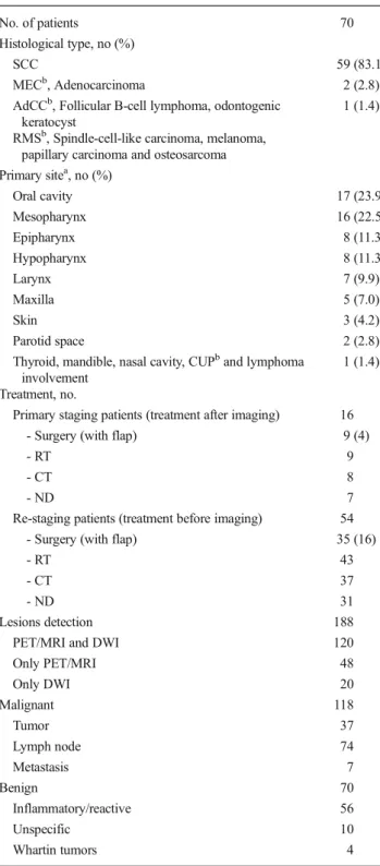
Fig. 1 Thirty-five-year-old male with recurrence of a mesopharynx carcinoma in the right rosenmuller fossa. PET/MRI accurately detected it, but DWI showed no restriction (ADCmean=1.73×10⁻³mm2/s)
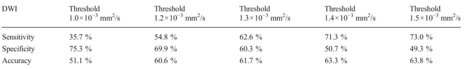
Fig. 2 a–c Sixty-nine-year-old man with squamous cell carcinoma of the base of the tongue. a Axial T2w, Axial ceT1w, DWI, ADC, and PET/ MRI showing the primary tumor in the base of the tongue. b PET/MRI in
different levels showing seven metastatic lymph nodes. c DWI (top) and ADC map (bottom) in different levels showing nine metastatic lymph nodes. In both PET/MRI and DWI, N-staging remains N2c
Conversely, the quantitative DWI parameters such as b-values and ADCs of malignant vs. benign lesions didn’t show any statistical difference between malignant and non-malignant lesions. This finding is partially in contradiction to the current literature, which states that DWI is able to differentiate benign from malignant lesions in primary staging of head and neck cancers using ADC with different threshold values [6,11], and is also able to differentiate changes related to treatment from loco-regional recurrence [32, 38]. One potential explanation for these conflicting results is that our b-values are partly lower than previously reported in the literature (800 vs. 1,000–2,000). It is known that the ADC value decreases when the b-value increases beyond 1,000 s/ mm2 [30]. The decrease in the observed ADC with an in-creasing b-value is explained by the decay of biexponential signal intensity [30]. Thus, this requires further investigation as to whether higher b-values should be used in head and neck cancer protocols. The problem with higher b-values is that MR imaging time increases and integration into a clinically acceptable PET/MRI protocol is thus even more difficult.
PET values and DWI parameters
Our study did not show a correlation between SUVmaxand ADC or b-values. Recent studies have demonstrated for head and neck cancers that SUVmax is inversely correlated to ADC ratio between different b-values [39] or ADC ratio between minimum and mean [40]. However, Nakajo and co-authors
have shown a negative and significant correlation between SUVmax and ADC mean, studying 26 patients with HNSCC before treatment [41]. Our results are probably different due to the fact that our population is heterogenous (as it is in any clinical routine), including different histologic types of HNC and not only primary tumors.
When PET values and DWI values for different popula-tions are compared, it has been found that they yield similar results. A malignant lesion at primary staging, for example, shows a high SUVmax and b-value, and low ADC. Thus, the information provided by DWI is superfluous, since it does not provide any information that wasn't already available from the PET/MRI.
Evaluating 30 patients with different malignancies, Borra and co-workers recently have shown the prognostic potential of b-values compared to PET uptake. The b-values represent the average signal intensity from the native DW images for each ROI, and are therefore easy to use. It was found that a high b-value was more indicative of malignant FDG uptake than the traditionally used signal loss on ADC maps [42]. However, we could not demonstrate this in our patient popu-lation with HNC. Thus, b-values might be useful in certain oncological settings, but possibly not in HNC.
Limitations
One limitation of this study was a mixed-population analysis, which included primary staging and follow-up/recurrence exams, as well as different histologic types. This might have limited the ability of the ADC threshold to differentiate benign from malignant lesions. However, it does not render the con-clusion invalid, since our purpose was to evaluate the utility of DWI in a PET/MRI protocol for a routine, clinical HNC population, not specifically for primary staging or follow-up. Furthermore, different ADC thresholds were applied without significant differences concerning the overall accuracy com-pared to significant differences in SUV. Another limitation was the lack of pathological confirmation of all lesions, how-ever, this is not always possible in a clinical setting. The reading process may also be considered a limitation, since two independent readers could possibly enhance the results. The DWI-only evaluation might not be highly relevant in a routine clinical setting, since DWI should be read in conjunc-tion with a complete diagnostic MRI protocol. However, to account for similar DWI studies that are available in the current literature, it has been included.
Conclusion
The use of DWI as part of PET/MRI procedures to evaluate head and neck cancers did not provide diagnostically relevant Table 6 Quantitative values of PET and in malignant and non-malignant
lesions
Malignant Non-malignant P-value
SUV max 10.5 6.6 0.001
SUV mean 6.4 4.1 0.001
ADC mean (× 10−3mm2/s) 1.19 1.15 0.299
b-value max 444.8 352 0.719
b-value mean 292.7 190.6 0.680
Table 7 Quantitative values of PET and DWI in primary staging and follow-up/recurrence patients
Primary Follow-up P-value
SUVmax 11.0 7.9 0.003
SUVmean 6.7 4.8 0.005
b-value max 503.5 323.9 0.016
b-value mean 338.6 178.9 0.001
information in our study. Thus, the use of DWI might not be needed in clinical PET/MRI protocols for the staging or restaging of head and neck cancers.
Disclosures This research project was supported by an institutional research grant from GE Healthcare. Patrick Veit-Haibach received IIS Grants from Bayer Healthcare and Siemens Medical Solutions, and speaker fees from GE Healthcare. Gustav von Schulthess is a grant recipient of GE Healthcare funding and receives speaker fees from GE Healthcare. The other authors declare no other conflicts of interest.
References
1. Jemal A, Bray F, Center MM, Ferlay J, Ward E, Forman D. Global cancer statistics. CA Cancer J Clin. 2011;61:69–90.
2. Epidemiology and risk factors for head and neck cancer [Internet]. [cited 2014 Feb 14]. Available from: http://www.uptodate.com/ contents/epidemiology-and-risk-factors-for-head-and-neck-cancer#H1.
3. Hustinx R, Lucignani G. PET/CT in head and neck cancer: an update. Eur J Nucl Med Mol Imaging. 2010;645–51.
4. Thoeny H. Diffusion-weighted MRI, in head and neck radiology: applications in oncology. Cancer Imaging. 2010;10:209–14. 5. Thoeny HC, De Keyzer F. Extracranial applications of
diffusion-weighted magnetic resonance imaging. Eur Radiol. 2007;17:1385– 93.
6. Srinivasan A, Mohan S, Mukherji SK. Biologic imaging of head and neck cancer: the present and the future. AJNR Am J Neuroradiol. 2012;33:586–94.
7. Friedrich KM, Matzek W, Gentzsch S, Sulzbacher I, Czerny C, Herneth AM. Diffusion-weighted magnetic resonance imaging of head and neck squamous cell carcinomas. Eur J Radiol. 2008;68: 493–8.
8. Herneth AM, Guccione S, Bednarski M. Apparent diffusion coeffi-cient: a quantitative parameter for in vivo tumor characterization. Eur J Radiol. 2003;45:208–13.
9. Higgins KA, Hoang JK, Roach MC, Chino J, Yoo DS, Turkington TG, et al. Analysis of pretreatment FDG-PET SUV parameters in head-and-neck cancer: tumor SUVmean has superior prognostic value. Int J Radiat Oncol Biol Phys. 2012;82:548–53.
10. Sadick M, Schoenberg SO, Hoermann K, Sadick H. Current onco-logic concepts and emerging techniques for imaging of head and neck squamous cell cancer. GMS Curr Top Otorhinolaryngol Head Neck Surg. 2012;11:1–24.
11. Thoeny HC, De Keyzer F, King AD. Diffusion-weighted MR imag-ing in the head and neck. Radiology. 2012;263:19–32.
12. Buchbender C, Hartung-Knemeyer V, Beiderwellen K, Heusch P, Kühl H, Lauenstein TC, et al. Diffusion-weighted imaging as part of hybrid PET/MRI protocols for whole-body cancer staging: does it benefit lesion detection? Eur J Radiol. 2013;82:877–82.
13. Veit-Haibach P, Kuhn FP, Wiesinger F, Delso G, von Schulthess G. PET-MR imaging using a tri-modality PET/CT-MR system with a dedicated shuttle in clinical routine. MAGMA. 2013;26:25–35. 14. Kuhn FP, Crook DW, Mader CE, Appenzeller P, von Schulthess GK,
Schmid DT. Discrimination and anatomical mapping of PET-positive lesions: comparison of CT attenuation-corrected PET images with coregistered MR and CT images in the abdomen. Eur J Nucl Med Mol Imaging. 2012;44–51.
15. Boellaard R, O’Doherty MJ, Weber WA, Mottaghy FM, Lonsdale MN, Stroobants SG, et al. FDG PET and PET/CT: EANM procedure
guidelines for tumour PET imaging: version 1.0. Eur J Nucl Med Mol Imaging. 2010;37:181–200.
16. Kuhn FP, Wiesinger F, Wollenweber SD, Samarin A, Von Schulthess G SD. Sequential integrated PET/CT-MR system: comparison of image registration accuracy of PET/CT versus PET/MR. Melbourne, Australia: International Society for Magnetic Resonance in Medicine (ISMRM); 2012.
17. Lee M-C, Tsai H-Y, Chuang K-S, Liu C-K, Chen M-K. Prediction of nodal metastasis in head and neck cancer using a 3T MRI ADC map. Am J Neuroradiol. 2013;34:864–9.
18. Zhang Y, Chen J, Shen J, Zhong J, Ye R, Liang B. Apparent diffusion coefficient values of necrotic and solid portion of lymph nodes: differential diagnostic value in cervical lymphadenopathy. Clin Radiol. 2013;68:224–31.
19. Abdel Razek AAK, Nada N. Role of diffusion-weighted MRI in differentiation of masticator space malignancy from infection. Dentomaxillofac Radiol. 2013;42:20120183.
20. Perrone A, Guerrisi P, Izzo L, D’Angeli I, Sassi S, Mele L Lo, et al. Diffusion-weighted MRI in cervical lymph nodes: differentiation between benign and malignant lesions. Eur J Radiol. 2011;77:281–6. 21. Holzapfel K, Duetsch S, Fauser C, Eiber M, Rummeny EJ, Gaa J. Value of diffusion-weighted MR imaging in the differentiation be-tween benign and malignant cervical lymph nodes. Eur J Radiol. 2009;72:381–7.
22. Vandecaveye V, De Keyzer F. Head and neck squamous cell carci-noma: value of diffusion-weighted MR imaging for nodal staging. Radiology. 2009;251:134–46.
23. Queiroz MA, Hüllner M, Kuhn F, Huber G, Meerwein C, Kollias S, et al. PET/MRI and PET/CT in follow-up of head and neck cancer patients. Eur J Nucl Med Mol Imaging. 2014. doi: 10.1007/s00259-014-2707-9.
24. Platzek I, Beuthien-Baumann B, Schneider M, Gudziol V, Langner J, Schramm G, et al. PET/MRI in head and neck cancer: initial experi-ence. Eur J Nucl Med Mol Imaging. 2013;40:6–11.
25. Appenzeller P, Mader C, Huellner MW, Schmidt D, Schmid D, Boss A, et al. PET/CT versus body coil PET/MRI: how low can you go? Insights Imaging. 2013;4:481–90.
26. Buchbender C, Heusner TA, Lauenstein TC, Bockisch A, Antoch G. Oncologic PET/MRI, Part 1: tumors of the brain, head and neck, chest, abdomen, and pelvis. J Nucl Med. 2012;53:928–38. 27. Chawla S, Kim S, Dougherty L, Wang S, Loevner LA, Quon H, et al.
Pretreatment diffusion-weighted and dynamic contrast-enhanced MRI for prediction of local treatment response in squamous cell carcinomas of the head and neck. Am J Roentgenol. 2013;200:35– 43.
28. Heijmen L, Verstappen MCHM, Ter Voert EEGW, Punt CJ, Oyen WJG, de Geus-Oei L-F, et al. Tumour response prediction by diffusion-weighted MR imaging: ready for clinical use? Crit Rev Oncol Hematol Elsevier Ireland Ltd. 2012;83:194–207.
29. Vandecaveye V, De Keyzer F, Dirix P, Lambrecht M, Nuyts S, Hermans R. Applications of diffusion-weighted magnetic resonance imaging in head and neck squamous cell carcinoma. Neuroradiology. 2010;52:773–84.
30. Hwang I, Choi S, Kim Y. Differentiation of recurrent tumor and posttreatment changes in head and neck squamous cell carcinoma: application of high b-value diffusion-weighted imaging. Am J Neuroradiol. 2013;1–7.
31. Sepahdari AR, Politi LS, Aakalu VK, Kim HJ, Abdel Razek a a K. Diffusion-weighted imaging of orbital masses: multi-institutional data support a 2-ADC threshold model to categorize lesions as benign, malignant, or indeterminate. Am J Neuroradiol. 2013;1–6. 32. Abdel Razek AAK, Kandeel AY, Soliman N, El-shenshawy HM,
Kamel Y, Nada N, et al. Role of diffusion-weighted echo-planar MR imaging in differentiation of residual or recurrent head and neck tumors and posttreatment changes. Am J Neuroradiol. 2007;28: 1146–52.
33. Sumi M, Sakihama N, Sumi T, Morikawa M, Uetani M, Kabasawa H, et al. Discrimination of metastatic cervical lymph nodes with diffusion-weighted MR imaging in patients with head and neck cancer. Am J Neuroradiol. 2003;24:1627–34.
34. Ishikita T, Oriuchi N, Higuchi T, Miyashita G, Arisaka Y, Paudyal B, et al. Additional value of integrated PET/CT over PET alone in the initial staging and follow up of head and neck malignancy. Ann Nucl Med. 2010;24:77–82.
35. Goerres GW, Schuknecht B, Schmid DT, Stoeckli SJ, Hany TF. Positron emission tomography/computed tomography for staging and restaging of head and neck cancer: comparison with positron emission tomography read together with contrast-enhanced comput-ed tomography. Clin Imaging. 32:431–7.
36. Chan S-C, Wang H-M, Yen T-C, Lin C-Y, Chin S-C, Liao C-T, et al. 18
F-FDG PET/CT and 3.0-T whole-body MRI for the detection of distant metastases and second primary tumours in patients with untreated oropharyngeal/hypopharyngeal carcinoma: a comparative study. Eur J Nucl Med Mol Imaging. 2011;38: 1607–19.
37. Ghanooni R, Delpierre I, Magremanne M, Vervaet C, Dumarey N, Remmelink M, et al.18F-FDG PET/CT and MRI in the follow-up of
head and neck squamous cell carcinoma. Contrast Media Mol Imaging. 2011;6:260–6.
38. Srinivasan A, Dvorak R, Perni K, Rohrer S, Mukherji SK. Differentiation of benign and malignant pathology in the head and neck using 3T apparent diffusion coefficient values: early experience. Am J Neuroradiol. 2008;29:40–4.
39. Choi SH, Paeng JC, Sohn C-H, Pagsisihan JR, Kim Y-J, Kim KG, et al. Correlation of 18F-FDG uptake with apparent diffusion coeffi-cient ratio measured on standard and high b value diffusion MRI in head and neck cancer. J Nucl Med. 2011;52:1056–62.
40. Ho K-C, Lin G, Wang J-J, Lai C-H, Chang C-J, Yen T-C. Correlation of apparent diffusion coefficients measured by 3T diffusion-weighted MRI and SUV from FDG PET/CT in primary cervical cancer. Eur J Nucl Med Mol Imaging. 2009;36:200–8.
41. Nakajo M, Kajiya Y. FDG PET/CT and diffusion-weighted imaging of head and neck squamous cell carcinoma. Clin Nucl Med. 2012;37: 475–80.
42. Borra R, Catalano O, Catana C, McDermott S, Blake M, Sahani D, et al. Comparison of SUVand whole body diffusion imaging findings in oncological imaging with hybrid PET-MRI. Soc Nucl Med Annu Meet Abstr. 2013;54:1408.