www.microbialcell.com
Conventional and emerging roles of the energy sensor
Snf1/AMPK in Saccharomyces cerevisiae
Paola Coccetti
1,2,*, Raffaele Nicastro
1,3and Farida Tripodi
1,21 Department of Biotechnology and Biosciences, University of Milano-Bicocca, Milan, Italy. 2 SYSBIO, Centre of Systems Biology, Milan, Italy.
3 Present address: Department of Biology, University of Fribourg, Fribourg, Switzerland.
* Corresponding Author:
Paola Coccetti, Department of Biotechnology and Biosciences, University of Milano-Bicocca, Milan, Italy; E-mail: [email protected]
INTRODUCTION
Cell growth and proliferation require a high amount of
en-ergy for biosynthetic pathways. Cells take enen-ergy from
nu-trient intake and both unicellular and multicellular
eukary-otes have evolved systems that allow dynamic sensing of
en-ergy sources, mainly sugars. The class of Snf1/AMPK
(Su-crose non-fermenting/AMP-activated protein kinase) plays
a key role as a guardian of cellular energy [1]. They are highly
conserved serine/threonine kinases and their primary role
is the integration of signals regarding nutrient availability
and environmental stress, ensuring the adaptation to those
conditions and cell survival [2].
Here we discuss the mechanisms of action of Snf1, a
member of the Snf1/AMPK family in Saccharomyces
cere-visiae and its conventional roles in the regulation of
metab-olism, stress response and aging. In addition, we also focus
on recent advances showing emerging functions of Snf1 on
the modulation of key processes such as endocytosis and
cellular trafficking as well as cell cycle, proliferation and
me-tabolism.
SNF1 COMPLEX COMPOSITION
Protein kinase Snf1 in yeast is a heterotrimeric complex
made by the catalytic α subunit Snf1, a regulatory β subunit
(alternatively Gal83, Sip1 and Sip2) and the γ subunit Snf4
[3].
The catalytic α subunit (encoded by the SNF1 gene) was
identified in a screening of mutants unable to grow in
pres-ence of sucrose as carbon source [4]. The Snf1 subunit is
constitutively expressed and constituted by a catalytic
N-terminal domain and a C-N-terminal regulatory region. The
regulatory region presents a short autoinhibitory sequence
(AIS) (380 - 415 aa) and a domain which mediates the
inter-actions with the β subunits of the complex. The
autoinhibi-tory domain interacts with both the regulaautoinhibi-tory subunit Snf4
and the kinase domain of Snf1. The interaction with Snf4
re-lieves the inhibition of the AIS allowing the phosphorylation
of Thr210 residue of Snf1 that determines its activation
[5, 6].
In S. cerevisiae, three β subunits (Gal83, Sip1 and Sip2)
are present. They share partially redundant functions,
since only the triple mutant sip1Δsip2Δgal83Δ strain shows
ABSTRACT All proliferating cells need to match metabolism, growth and cell
cycle progression with nutrient availability to guarantee cell viability in spite of
a changing environment. In yeast, a signaling pathway centered on the effector
kinase Snf1 is required to adapt to nutrient limitation and to utilize alternative
carbon sources, such as sucrose and ethanol. Snf1 shares evolutionary
con-served functions with the AMP-activated Kinase (AMPK) in higher eukaryotes
which, activated by energy depletion, stimulates catabolic processes and, at
the same time, inhibits anabolism. Although the yeast Snf1 is best known for
its role in responding to a number of stress factors, in addition to glucose
limi-tation, new unconventional roles of Snf1 have recently emerged, even in
glu-cose repressing and unstressed conditions. Here, we review and integrate
avail-able data on conventional and non-conventional functions of Snf1 to better
un-derstand the complexity of cellular physiology which controls energy
homeo-stasis.
doi: 10.15698/mic2018.11.655 Received originally: 29.06.2018; in revised form: 23.08.2018, Accepted 23.08.2018, Published 29.09.2018.Keywords: budding yeast, metabolism, stress response, aging, transcription, signaling, cell cycle, endocytosis, DNA damage, glucose repression.
Abbreviations:
growth defects when glycerol or ethanol are added as
car-bon sources [7, 8]. The β subunits contain a conserved
C-terminal sequence in which two domains are present: the
KIS domain (Kinase Interacting Sequence) that mediates the
interaction with the α-subunit Snf1 [9] and the ASC domain
(Association with SNF1 kinase Complex) that allows the
interaction with Snf4 [10]. Differently, the N-terminal
se-quence is specific for each β subunit and confers a different
subcellular localization pattern to each protein. All three
proteins are mainly cytoplasmic in presence of high glucose
concentrations. Upon glucose depletion, Sip1 relocalizes to
the vacuolar membrane, Gal83 relocalizes to the nucleus,
while Sip2 remains cytoplasmic [11]. Thus, the role of the β
subunits is to interact with Snf1 and to modulate its
subcel-lular localization [11, 12]. The particular localization of the
kinase complexes with different β subunits confers
special-ized functions. For example, Sip1 alone is not able to sustain
growth on ethanol or glycerol and determines a very low
ki-nase activity of the complex [13], Sip2 function seems to be
involved in the mechanism of cellular aging [14], while Gal83
plays its main role in the Snf1-dependent transcriptional
regulation, since in low glucose it determines the nuclear
lo-calization of the Snf1 complex thank to its NLS (Nuclear
Localization Signal). On the contrary, NES (Nuclear Export
Signal), present on the sequence of Gal83, allows the exit
from the nucleus of the complex when high glucose
concen-trations are available [15]. In addition, Gal83 mediates the
interaction of Snf1 with some substrates, such as the
tran-scription activator Sip4 [16] and the trantran-scriptional
appa-ratus [17]. It has also been shown that deletion of the
glyco-gen binding domain (GBD) of Gal83 leads to a constitutive
activation of Snf1, which results able to modulate the
ex-pression of some Snf1-regulated genes also in high glucose
concentrations [18]. The GBD domain of Gal83 also interacts
with the Reg1/Glc7 phosphatase complex, responsible for
Snf1 inactivation [18]. Taken together those data suggest
that Gal83 plays a dual role regulating nuclear localization
of Snf1 in low glucose and guaranteeing its inactivation in
high glucose.
Similarly to SNF1, the gene encoding the γ subunit, SNF4,
was identified by isolation of a sucrose non fermenting
mu-tant [4]. Snf4 is a constitutively expressed protein that binds
both the α and β subunits of the Snf1 complex [10, 19]. The
role of Snf4 is to relieve the inhibition of Snf1 interacting
with its AIS domain, stabilizing the Snf1 complex in the
ac-tive conformation [20]. In fact, SNF4 deletion causes a
decreased kinase activity of Snf1, whereas deletion of the
AIS domain of Snf1 fully complement the phenotype of a
snf4Δ strain [19, 20]. Remarkably, the activating
phosphor-ylation of Thr210 residue of Snf1 is still detectable in a snf4Δ
strain [21] and in high glucose Snf4 seems to be required for
the proper inactivation of Snf1 mediated by the
phospha-tase complex Reg1/Glc7 [18]. Thus, these findings indicate
that Snf4 plays a complex role in the regulation of Snf1.
REGULATION OF SNF1 ACTIVITY
Snf1 complex is activated through phosphorylation of the
Thr210 of the α subunit by one of the three constitutively
active upstream kinases Sak1, Tos3 and Elm1 [22, 23] (Fig.
1). This phosphorylation is essential for Snf1 activity, since
the sak1Δtos3Δelm1Δ strain shows the same phenotype of
a snf1Δ strain, such as growth defects in presence of limiting
glucose or alternative carbon sources like glycerol or
ethanol [22].
Although Snf1 phosphorylation is a key step for its
activa-tion, a non-phosphorylatable Snf1 mutant (Snf1-T210A)
re-tains a low catalytic activity, originating intermediate
phenotypes [24, 25]. Also, the mutation of the lysine which
constitutes the ATP binding site in the kinase domain
(Snf1-K84R), which for many aspects mimics the loss of Snf1
protein, still confers a slight catalytic activity [26-29].
On the other side, in response to high glucose
concentra-tions Snf1 is inactivated through dephosphorylation of
Thr210 by the Gcl7 protein phosphatase (also known as
FIGURE 1: Schematization of the activa-tion of Snf1 and its main convenactiva-tional functions. Snf1 complex is composed by
the α subunit Snf1, the ƴ subunit Snf4 and one of three alternative β subunits Gal83, Sip1 or Sip2. Snf1 is phosphorylated on T210 by the upstream kinases Sak1, Tos3 and Elm1, while it is de-phosphorylated by the phosphatase complex Glc7/Reg1.
When active, Snf1 phosphorylates
transcription factors which regulate the ex-pression of genes involved in glucose transport, stress response and glucose re-pression. In addition, Snf1 directly phos-phorylates some metabolic enzymes. See text for details.
PP1), which is targeted to Snf1 by the adaptor subunit Reg1
[30, 31]. Reg1 interacts both with Glc7 and Snf1 when
glu-cose is largely available in the culture medium and loss of
Reg1 leads to the constitutive activation of Snf1 [32-34]. It
has been reported that in high glucose concentration, Hxk2
(Hexokinase 2) regulates the activity of PP1 and
conse-quently the activation of Snf1 kinase [31].
Active Snf1 phosphorylates serine and threonine residues
contained in the consensus pattern Φ-x-R-x-x-S/T-x-x-x-Φ,
where Φ is a hydrophobic residue [35].
Differently from its mammalian homolog AMPK, yeast
protein kinase Snf1 is not allosterically activated by AMP
[36]. However, it was demonstrated that ADP molecules are
able to bind the γ subunit Snf4, preventing Snf1
dephosphorylation mediated by Glc7 [37, 38].
The structure of the kinase domain of Snf1 showed that
it is a dimer, which represents an inactive form of the kinase,
since Thr210 is inaccessible for phosphorylation by the
acti-vating kinases [39]. Although these results suggest the
exist-ence of another layer of regulation of Snf1 activity, further
investigation is required to better elucidate its physiological
relevance.
Interestingly, some recent evidence indicates additional
mechanisms that regulate Snf1 activity: (i) phosphorylation
of Ser214, inside the activation loop, downregulates Snf1
function [40]; (ii) SUMOylation of the catalytic α subunit
Snf1 inhibits its activity, possibly by attenuating its levels in
the cell and/or favoring the inactive conformation of the
ki-nase [41]; (iii) the SAGA acetyl transferase complex
deubiq-uitylates Snf1 affecting the stability of the complex and its
kinase activity [42]; (iv) the ubiquitin-associated motif (UBA)
of the α subunit Snf1 indirectly regulates SNF1 gene
expres-sion and Snf1 interaction with the γ subunit Snf4 [43].
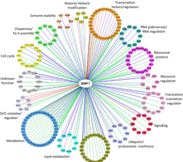
NETWORK OF SNF1 PHYSICAL INTERACTORS
A total of 216 proteins physically interacting with Snf1 are
annotated in SGD (Saccharomyces Genome Database,
http://www.yeastgenome.org
), 92 of which are also Snf1
substrates (identified by high throughput or low throughput
assays). In Figure 2 we clustered them on the base of their
function. Apart from the most known interactors involved in
Snf1 complex regulation, transcription, histone
modifica-tion, signaling and metabolism, there are many proteins
which regulate translation, ribosome function, intracellular
transport/trafficking and cell cycle. In addition, some of
them are also proteins of the ubiquitin/proteasome
machin-ery, chaperones and Fe/S cluster proteins (Fig. 2).
Remarka-bly, only a few of them have been extensively investigated
for the physiological relevance of Snf1-dependent
phos-phorylations, suggesting that many functions of Snf1 are still
to be discovered.
CONVENTIONAL ROLES OF SNF1
Snf1 and the regulation of transcription
The most studied function of Snf1 is the regulation of
tran-scription, involving more than 400 genes [46]. Snf1 acts both
on transcription factors and on chromatin remodeling
[47-50], as also highlighted by the number of its interactors
be-longing to these two classes of proteins (Fig. 2).
Mig1 is the most important glucose-regulated
transcrip-tional repressor [51]. Mig1 is phosphorylated by Snf1 on
four sites when glucose is scarce, causing the activation of a
NES (Nuclear Export Signal) sequence that causes its
trans-location from the nucleus to the cytoplasm through the
ex-portin Msn5 [48, 52, 53]. Important in the regulation of Mig1
is hexokinase Hxk2, which interacts with the transcriptional
repressor directly in the nucleus to avoid its
phosphoryla-tion by Snf1, thus providing a link between glucose
metabo-lism and transcription of glucose-repressed genes [54]. Mig1
represses about 90 genes, including those coding for
en-zymes required for the metabolism of sucrose (SUC2),
malt-ose (the MAL regulon) and galactmalt-ose (GAL4) [55].
Further-more, Mig1 controls the expression of high-affinity glucose
transporters, required when glucose is scarce (HXT2, HXT4)
[56], represses TPS1, essential for the metabolism of
treha-lose [57] and genes coding for enzymes of the TCA cycle
[58].
Besides Mig1, Snf1 regulates the activity of other
tran-scription factors. Cat8 and Sip4, which bind Carbon Source
Responsive Elements (CSRE), regulate the expression of
glu-coneogenic genes [59] and are activated by Snf1
phosphor-ylation [49, 60]. Cat8 activates the expression of
glucose-re-pressed genes alongside transcription factor Adr1, which is
itself a target of Snf1 [61, 62]. Moreover, in a fine
mecha-nism of positive feedback, the CAT8 gene is activated by
Snf1 through inhibition of Mig1 [47]. In addition, Gcn4, the
transcription factor responsible for the expression of genes
involved in amino acid biosynthesis, is also regulated by Snf1
when in complex with the β subunit Gal83 or Sip1, but not
Sip2 [63].
Snf1 has been reported to phosphorylate Ser10 of
his-tone H3 and to promote the acetylation on Lys14 of hishis-tone
H3 by Gcn5, a component of the SAGA complex [50].
Snf1-mediated regulation of histone H3 is involved in the
expres-sion of ADY2 gene. In fact, Snf1 stimulates the binding of
Gcn5 and the acetylation of histone H3 at ADY2 promoter,
promoting the transcription of this gene [64].
Snf1 and the regulation of metabolism
Besides its role in regulating the transcription of several
genes involved in metabolism, Snf1 directly regulates,
through phosphorylation, important metabolic enzymes. In
fact, together with the class of transcription factors and
reg-ulators, proteins linked to metabolism are the most
abundant among Snf1 interactors (Fig. 2). Probably the most
impactful function exerted by Snf1 as a direct regulator of
metabolism is the regulation of the acetyl-CoA carboxylase
Acc1 [65]. In yeast, loss of Snf1 causes a dramatic
accumu-lation of fatty acids and the carbon overflow into the
fatty acid biosynthetic pathway has been shown to cause
in-ositol auxotrophy mediated by the impairment of INO1
expression [65, 66]. Moreover, the excessive alloca-
tion of carbon into fatty acids causes a depletion of the
intracellular acetyl-CoA pool, and thus a global reduction of
acetylation of histones, of Swi4, the DNA-binding protein of
the transcription factor SBF [67], and of the β-subunit Sip2
[68].
Snf1 was also shown to phosphorylate Pfk27, the second
isoform of 6-phosphofructo-2-kinase [69]. Upon glucose
re-moval, Snf1 phosphorylates Pfk27 in its N-terminal domain,
leading to the SCF
Grr1-dependent degradation of Pfk27 [69].
In particular, Snf1-dependent phosphorylation is required
to promote Pfk27 association with the F-box protein Grr1
[69], thus leading to Pfk27 turnover and consequently to a
reduction of fructose-2,6-bisphosphate. The importance of
Pfk27 turnover is highlighted by the fact that expression of
a non-phosphorylatable and non-degradable Pfk27 protein
inhibits growth on glycerol [69].
Moreover, Snf1 phosphorylates Gpd2, the
glycerol-3-phosphate dehydrogenase required for anaerobic growth,
thus inhibiting glycerol synthesis during the diauxic shift. In
fact, it was reported that Snf1 phosphorylates Gpd2 on
Ser72 priming Gpd2 for subsequent phosphorylation on
Ser75, probably by Yck1 [70].
FIGURE 2: Network of Snf1 physical interactors. The network reports the known physical associations obtained from SGD (Saccharomyces Genome
Database, http://www.yeastgenome.org). Interactors are clustered according to their function and colored differently. When the interactor is also a substrate of Snf1 according to the Yeast Kinase Interaction Database (KID, http://www.moseslab.csb.utoronto.ca/KID/; [44]), the edge is colored in red if phosphorylation was analyzed by low throughput assays (LTP in vitro kinase assays; in vitro phosphorylation site mapping; in vivo phosphorylation site mapping; phosphorylation reduced or absent in kinase mutant) or in blue if phosphorylation was assayed only by high throughput analysis (protein chip data for in vitro phosphorylated substrate; HTP in vitro phosphorylation). Data visualization and analysis was performed with Cytoscape [45].
Snf1 and PKA crosstalk
In yeast, the main pathway activated by glucose is the PKA
pathway, involved in metabolism, growth and proliferation
[71-73]. Targets of PKA include glycolytic and
gluconeoge-netic enzymes, proteins involved in the metabolism of
storage carbohydrates, transcription factors regulating
stress response, ribosomal biogenesis, and carbohydrate
metabolism. Active PKA directly stimulates glycolysis, cell
growth and cell cycle progression, at the same time
gluco-neogenesis, stress resistance and mobilization of glycogen
and trehalose are down-regulated [71, 74]. Several
exam-ples of cross-talk between Snf1 and PKA pathways have
been reported [75]. Indeed, both kinases regulate the
activ-ity of the same transcription factors. Adr1, the
transcrip-tional activator of glucose-repressed genes, is inactivated by
PKA and activated by Snf1 which promotes its
phosphoryla-tion [76, 77]. Msn2, the stress-responsive transcripphosphoryla-tional
activator, which is a well-known target of PKA, is
phosphor-ylated also by Snf1 in glucose starvation [78]. PKA indirectly
controls the localization of the β subunit Sip1 and, as a
con-sequence, of the Snf1-Sip1 complex [12]. In addition, PKA
contributes to regulate Sak1, one of the Snf1-activating
kinases [79].
Notably, recent data nicely complement observations of
a cross-talk between Snf1 and PKA. Indeed, the adenylate
cyclase Cyr1 and Snf1 interact in a nutrient-independent
manner [29]. Active Snf1 phosphorylates Cyr1 and
negatively regulates cAMP content and PKA-dependent
transcription [29]. Moreover, loss of Snf1 causes an
altera-tion in the phosphorylaaltera-tion pattern of adenylate cyclase
[29], suggesting that the crosstalk between Snf1 and PKA is
more complex than actually reported and needs to be
further investigated.
Snf1 and the regulation of TORC1
The Target Of Rapamycin Complex I (TORC1) is a highly
con-served nutrient-responsive regulator of cell growth and
metabolism in all eukaryotes [80-82]. Contrary to AMPK,
which is active under nutrient-poor conditions, TORC1 is
ac-tive under nutrient-rich conditions in budding yeast and also
in the presence of growth factors in higher eukaryotes [83,
84]. In yeast, TORC1 is composed of Tor1, Kog1/Raptor, Lst8
and Tco89 [81, 85, 86]. Kog1/Raptor is known to recruit
sub-strates such as 4EBP1 and ribosomal S6 kinase (S6K) to the
TORC1 complex [87, 88] and is required for the regulation of
its activity [80, 89].
Kog1 is phosphorylated by Snf1 under glucose
depriva-tion, as AMPK does on the ortholog Raptor [90], confirming
a conserved regulatory function of Snf1/AMPK on TORC1
complex. Nevertheless, the role of Snf1-dependent
phorylation on Kog1 is somehow different, since Snf1
phos-phorylation stimulates the dissociation of the Kog1-Tor1
complex and the formation of Kog1-bodies by limiting the
level of active TORC1 complex in the cell [90]. Thus,
alt-hough the final result of Snf1 phosphorylation is the
inacti-vation of TORC1 activity, this is reached by increasing the
activation threshold of TORC1 to guarantee a cellular
com-mitment to a quiescent state and then survival in
starvation condition [90].
In the presence of nutrients, TORC1 phosphorylates and
activates Sch9, the ortholog of S6K in yeast which, together
with other substrates, drives ribosome biosynthesis [91-93].
Through an in vitro kinase assay and epistasis analysis, Sch9
has been shown to be also a target of Snf1, indeed total Sch9
phosphorylation is reduced in snf1Δ mutant [94]. On the
other hand, Snf1-hyperactive cells display a dramatic
de-crease of TORC1 activity [95]. Moreover, Snf1 activity is
required for the downregulation of TORC1-dependent
phos-phorylation on Sch9 also in glucose deprivation [96].
Interestingly, Orlova and coworkers showed that
rapamy-cin treatment results in a significant increase of Thr210
phosphorylation on Snf1 [97], suggesting a reciprocal
regu-lation of Snf1 by TORC1 and a more complex crosstalk
between the two signaling pathways.
TORC1 activity is involved in the regulation of autophagy,
a cellular recycling system that degrades proteins and
orga-nelles by delivery to the vacuole in response to nutrient
dep-rivation [98, 99]. In yeast, nitrogen starvation, which induces
TORC1 inhibition, Atg13 dephosphorylation as well as Atg1
phosphorylation, results in activation of autophagy [100].
Remarkably, although Snf1 has been proposed as a positive
regulator of nitrogen-induced autophagy probably because
of its phosphorylation on Atg1 [101], in snf1Δ cells, the
translocation of GFP-Atg8 to the vacuole is reduced by 50%
compared to the wild type [102]. Moreover, Snf1 activity is
essential for glucose starvation-induced autophagy, and
mi-tochondrial respiration is a required feature for this energy
deprivation condition [102]. These interesting results
indi-cate that further investigations are required to better
eluci-date the different mechanisms which regulate nitrogen- and
glucose-induced autophagy, as well as how Snf1 is involved
in such a regulation.
Snf1 and the regulation of stress response
Besides nutritional deprivation, Snf1 is also involved in the
response to other cellular stresses. Snf1 activity protects
against toxicity caused by cadmium [103], hygromycin B
[26], hydroxyurea [24], selenite [104], and iron [105]. Snf1
also regulates HSF (Heat Shock transcription Factor)
ensur-ing the cellular resistance to high temperature, oxidative
stress [106, 107] and counteracts the activity of the
tran-scriptional inhibitor Nrg1, promoting the expression of
ENA1, responsible for Na
+ions detoxification [108].
In addition, protein kinase Snf1 regulates the Unfolded
Protein Response (UPR), the evolutionary conserved
pathway activated when improperly folded proteins
accu-mulate and induce endoplasmic reticulum (ER) stress [109,
110]. ER misfunction causes severe disease conditions [110],
thus the elucidation of the molecular mechanism by which
AMPK regulates UPR signaling attracts the increasing
inter-est of cell biologists. Nevertheless, the role of Snf1 in this
pathway is still not clear, since partially discrepant data
were published on yeast [111, 112]. Mizumo and coworkers
support a negative role of Snf1 in such a regulation, showing
that the deletion of SNF1 gene and Snf1 activation cause
in-creased and dein-creased resistance to ER stress, respectively
[112]. On the contrary, although results from Casamayor’s
laboratory
highlight
that
Snf1
activation
induces
hypersensitivity to ER-stress-inducer agents [111], they also
reported that snf1Δ cells are more sensitive to tunicamycin,
a known inducer of ER stress [113].
Thus, even though these data indicate an interesting role
for Snf1 in the regulation of ER stress response, more work
is needed to understand the underlying molecular
mechanism.
Snf1 and the regulation of DNA damage
Interesting results from Simpson-Lavy and collaborators
re-cently show cross-talk between Snf1 and protein kinases
involved in DNA damage (Mec1/ATR and Tel1/ATM) [114].
Phosphorylation of the SUMO E3 ligase Mms21 by Mec1 and
Tel1 induces SUMOylation and inactivation of Snf1, in
re-sponse to DNA damage. Thus, fermentation increases while
respiration is switched off. Remarkably, inactivation of Snf1
activity by SUMOylation does not affects its
phosphoryla-tion at Thr210, indicating that SUMOylaphosphoryla-tion and
phosphor-ylation of Snf1 are independently regulated. The authors
suggest that this metabolic switch may protect yeast cells
from oxidative stress and propose interesting parallelisms
with Warburg effect in cancer cells [114].
Snf1 and the regulation of aging
Given the role of AMPK kinase in the regulation of energy
homeostasis, it is not surprising the existence of a strong
re-lationship between aging and the AMPK pathway [115-119].
Indeed, hallmarks of the aging process such as
mitochon-drial dysfunction [120], autophagy [121], endoplasmic
retic-ulum stress [122] and DNA damage repair [123] are
regulated by AMPK. Importantly, both pharmacological
stimulation and exercise increase AMPK activity in skeletal
muscle of young rats but not in old ones [124]. Moreover,
AMPK-dependent acetyl-CoA carboxylase and
mitochon-drial biogenesis are impaired with aging [124]. These and
other results suggest that the basal activity of AMPK
de-clines with aging, contributing to the dysregulation of
intracellular metabolism.
The first evidence of the involvement of Snf1/AMPK in
the aging process in yeast occurred several years ago when
Asharafhi and co-workers discovered that Snf1 activity
in-creased in replicative aging even in the presence of
abun-dant glucose in the environment [14]. Moreover, loss of the
β-subunit Sip2 accelerates aging [14] and Sip2 acetylation
enhances its interaction with Snf1, decreases the kinase
ac-tivity of the complex and extends Replicative Life Span (RLS,
an aging model of mitotically active cells) [94]. Thus, in a
yeast model of replicative aging, these results indicate that
Snf1 inactivation promotes longevity [94].
On the contrary, in a yeast model of Chronological Life
Span (CLS, a model of aging in post-mitotic mammalian
cells), Snf1 activity is critical for the extension of CLS in
ca-loric restriction condition and cells deleted for SNF1 gene
have a very short CLS [125, 126]. Accordingly, the upregula
tion of Snf1 activity extends the life span [127] and drugs like
metformin (which activates AMPK) are being proposed for
the treatment of age-associated disorders [128].
Taken together these data indicate that Snf1 activity
pro-motes longevity in CLS while accelerates RLS, showing
dis-tinct and opposite mechanisms in the regulation of aging, as
also reported for other proteins in yeast [129, 130].
NON CONVENTIONAL ROLES OF SNF1
Snf1 and the regulation of endocytosis
Recent data suggest that Snf1 function is not limited to
con-ditions of carbon limitation. One of such functions is the
reg-ulation of proteins involved in endocytosis and cellular
traf-ficking, indeed several interactors and substrates of Snf1 are
in this cluster (Fig. 2). Snf1 interacts with and
phosphory-lates the α-arrestin Rod1 [27, 131, 132], which reguphosphory-lates
en-docytosis of the lactate transporter Jen1 and of the hexose
transporters Hxt1, Hxt3 and Hxt6, in response to glucose.
Remarkably, Snf1 phosphorylation on Rod1 occurs not only
in glucose limitation, but also in the presence of glucose. In
this condition, Snf1 phosphorylation inhibits
Rod1-medi-ated trafficking of Hxt1 and Hxt3, thus maintaining a high
glucose transporter activity [133]. Therefore, Snf1-mediated
phosphorylation has both an inhibitory and activatory
func-tion on the trafficking of hexose transporters, depending on
the level of Snf1-mediated phosphorylation on Rod1 [133].
According to the proposed model, when Snf1 activity is high,
Rod1 is hyper-phosphorylated and the endocytosis of
hex-ose transporters is active. On the other hand, in gluchex-ose
growing cells, Snf1 activity is low, Rod1 is
hypo-phosphory-lated and thus its trafficking function is inhibited, indicating
that Snf1 retains physiologically important function also
un-der high-glucose conditions, probably to direct its activity to
specific targets [133].
Snf1 also regulates Arf3 [134], one of the
ADP-ribosyla-tion factors (Arfs), involved in vesicle transport and actin
re-organization. Yeast Arf3 is required for invasive growth and
its activity is stimulated upon glucose-depletion in a
Snf1-dependent manner. The regulation of invasive growth in
nu-trient depletion is actually a conventional role of Snf1 [135,
136]. However, the peculiarity of the activation of Arf3 is
that it does not depend on Snf1 kinase activity, but rather
on its absolutely new role as a GEF (Guanine Nucleotide
Ex-change Factor). In fact, it was shown that the C-terminal
hy-drophobic α-helix core of Snf1 is a non-canonical GEF for
Arf3 activation [134].
Snf1 and the regulation of cell proliferation, cell cycle and
metabolism
Several reports indicate that Snf1 could be active
in the presence of high glucose [54, 133, 137, 138],
indicating
regulatory
roles
for
Snf1
under
glucose repression. In facts, cells lacking Snf1 (both
snf1Δ and snf1as mutant, whose activity can be
chemically inhibited) show a slow growth phenotype and
an increased fraction of cells in G1 phase, in synthetic
medium supplemented with 2% glucose [25, 139].
Never-theless, no perturbations of growth occur in complete (YPD,
with 2% glucose) and in synthetic media with 5% glucose
[25, 139], as well as in complete synthetic medium (our
unpublished results), indicating that the nutritional
compo-sition of the media may influence the cellular requirement
for Snf1 function.
The relevant role of the catalytic activity of Snf1 in
regu-lating proliferation in glucose-repressed condition is further
supported by the fact that Snf1 loss reduces the expression
of G1-specific genes [139]. The G1/S transition is regulated
by the expression of about 150 genes (the G1 regulon) [140],
controlled by the transcription factors SBF (Swi4-Swi6) and
MBF (Mbp1-Swi6) [67]. Snf1 directs the expression of both
SBF and MBF-regulated genes [25, 139], by modulating the
recruitment of both the co-activator Swi6 and the RNA
pol-ymerase II to the promoters of G1-genes [139] (Fig. 3).
Snf1-T210 is weakly phosphorylated in 2% glucose,
con-firming that it is partially active in that growth condition [21,
139, 141]. Consistently, the non phosphorylatable
SNF1-T210A mutant shows a slow growth phenotype and a
delayed G1/S transition. [25, 139].
Snf1 exerts its function in mitosis too and active Snf1 is
localized at the division site from the time of bud emergence
to cytokinesis [142]. Both septins and protein kinase Elm1
are required for proper Snf1 localization to the bud neck,
in-dicating that the presence of an accurate scaffold is
neces-sary for this process (Fig. 3). Loss of Snf1 activity causes a
defect in the correct alignment of the mitotic spindle, that
in turn induces a delay of the metaphase-to-anaphase
tran-sition, thus clearly indicating Snf1 function for proper
spin-dle orientation. Two major pathways are responsible for the
spindle alignment along the mother-bud axis in budding
yeast: the Kar9-pathway and the Dyn1-pathway [143].
Re-cent results show that Snf1 acts in parallel to Dyn1 and in
concert with Kar9 to promote spindle positioning, probably
phosphorylating components of the Kar9-dependent
path-way [142]. In support of these data, several key regulators
of cell cycle are known interactors of Snf1, mainly involved
in the network of spindle orientation, mitotic exit and
cyto-kinesis (Fig. 2).
It is amazing that cells lacking Snf1 and growing in
synthetic media containing 2% glucose, show an extensive
transcriptional reprogramming, being the most upregulated
genes mainly involved in transmembrane transport and
FIGURE 3: A model of the regulatory role of Snf1 during the cell cycle. At the G1/S phase transition, Snf1 promotes the binding of Swi4, Mbp1 and
Swi6 proteins to G1 promoters and favors the proper recruitment of the RNA Polymerase II. From bud emergence, active Snf1 is localized to the bud neck, in a septin-dependent manner. At the metaphase-to-anaphase transition, Snf1, as part of the Kar9-dependent pathway, promotes spindle align-ment along the mother-bud axis and guarantees proper nuclei segregation during mitosis. See text for details.
metabolic processes such as aminoacid biosynthesis, iron
homeostasis and redox metabolism [144]. Moreover, an
in-crease of cellular dependence on mitochondrial function in
glucose repression condition is clearly noticeable in snf1Δ
cells, further supporting the emerging roles of Snf1 in
non-limiting nutrient condition too [144].
PERSPECTIVES AND CONCLUDING REMARKS
Many studies have reported that AMPK activity is altered in
several diseases, such as inflammation, diabetes and cancer
[145, 146]. Moreover, the number of pharmacological
agents that activate AMPK has continued to increase and
some of them are promise hypoglycemic agents.
Im-portantly, although AMPK is considered a key target for
can-cer treatment, emerging data indicate that AMPK performs
both anti- and pro-tumorigenic roles depending on the
com-position of AMPK complex, signaling networks and
environ-mental conditions [147, 148]. The pro-tumorigenic role of
AMPK involves promotion of metabolic adaptation for
can-cer cell survival by regulating fatty acid metabolism and
maintaining the ability to growth in stressful conditions
[145, 149].
Therefore, the expansion of the repertoire of AMPK
sub-strates, as well as more in-depth studies of the molecular
mechanisms by which AMPK is activated, will help to better
understand the roles of this kinase in the regulation of
human pathologies. In this context, the yeast unicellular
organism Saccharomyces cerevisiae is a powerful model for
studying fundamental aspects of eukaryotic cell biology and
to validate the increasing downstream targets of the class of
Snf1/AMPK protein kinases which control the complexity of
cellular physiology.
ACKNOWLEDGEMENTS
Work in the authors' laboratory was supported by SysBioNet project, a MIUR initiative from the Italian Roadmap of European Strategy Forum on Research Infrastructures (ESFRI). F.T. has been supported by fellowships from MIUR, R.N. was funded by a fellowship from SysBioNet.
CONFLICT OF INTEREST
The authors have no conflicts of interest to declare.
COPYRIGHT
© 2018 Coccetti et al. This is an open-access article released
under the terms of the Creative Commons Attribution (CC
BY) license, which allows the unrestricted use, distribution,
and reproduction in any medium, provided the original
au-thor and source are acknowledged.
Please cite this article as: Paola Coccetti, Raffaele Nicastro and Fa-rida Tripodi (2018). Conventional and emerging roles of the energy sensor Snf1/AMPK in Saccharomyces cerevisiae. Microbial Cell 5(11): 482-494. doi: 10.15698/mic2018.11.655
REFERENCES
1. Herzig S, and Shaw RJ (2018). AMPK: guardian of metabolism and mitochondrial homeostasis. Nat Rev Mol Cell Biol 19(2): 121-135. doi: 10.1038/nrm.2017.95
2. Ghillebert R, Swinnen E, Wen J, Vandesteene L, Ramon M, Norga K, Rolland F, and Winderickx J (2011). The AMPK/SNF1/SnRK1 fuel gauge and energy regulator: structure, function and regulation.
FEBS J 278(21): 3978-90. doi: 10.1111/j.1742-4658.2011.08315.x 3. Hedbacker K, and Carlson M (2008). SNF1/AMPK pathways in yeast. Front Biosci 13: 2408-20. doi: 10.2741/2854
4. Carlson M, Osmond BC, and Botstein D (1981). Mutants of yeast
defective in sucrose utilization. Genetics 98(1): 25-40. PMID:
7040163
5. Rudolph MJ, Amodeo GA, Bai Y, and Tong L (2005). Crystal structure of the protein kinase domain of yeast AMP-activated protein kinase Snf1. Biochem Biophys Res Commun 337(4): 1224-8. doi: 10.1016/j.bbrc.2005.09.181
6. Chen L, Jiao Z-H, Zheng L-S, Zhang Y-Y, Xie S-T, Wang Z-X, and Wu J-W (2009). Structural insight into the autoinhibition mechanism of
AMP-activated protein kinase. Nature 459(7250): 1146-9. doi:
10.1038/nature08075
7. Schmidt MC, and McCartney RR (2000). beta-subunits of Snf1 kinase are required for kinase function and substrate definition.
EMBO J 19(18): 4936-43. doi: 10.1093/emboj/19.18.4936 8. Erickson JR, and Johnston M (1993). Genetic and molecular characterization of GAL83: its interaction and similarities with other genes involved in glucose repression in Saccharomyces
cerevisiae. Genetics 135(3): 655-64. PMID: 8293971
9. Yang X, Jiang R, and Carlson M (1994). A family of proteins containing a conserved domain that mediates interaction with the yeast SNF1 protein kinase complex. EMBO J 13(24): 5878-86. PMID: 7813428
10. Jiang R, and Carlson M (1997). The Snf1 protein kinase and its activating subunit, Snf4, interact with distinct domains of the Sip1/Sip2/Gal83 component in the kinase complex. Mol Cell Biol 17(4): 2099-106. 9121458. doi: 10.1128/MCB.17.4.2099
11. Vincent O, Townley R, Kuchin S, and Carlson M (2001). Subcellular localization of the Snf1 kinase is regulated by specific beta subunits and a novel glucose signaling mechanism. Genes Dev 15(9): 1104-14. doi: 10.1101/gad.879301
12. Hedbacker K, Townley R, and Carlson M (2004). Cyclic AMP-dependent protein kinase regulates the subcellular localization of
Snf1-Sip1 protein kinase. Mol Cell Biol 24(5): 1836-43. PMID:
14966266
13. Nath N, McCartney RR, and Schmidt MC (2002). Purification and characterization of Snf1 kinase complexes containing a defined
Beta subunit composition. J Biol Chem 277(52): 50403-8. doi:
10.1074/jbc.M207058200
14. Ashrafi K, Lin SS, Manchester JK, and Gordon JI (2000). Sip2p and its partner snf1p kinase affect aging in S. cerevisiae. Genes Dev 14(15): 1872-85. doi: 10.1101/gad.14.15.1872
15. Hedbacker K, and Carlson M (2006). Regulation of the nucleocytoplasmic distribution of Snf1-Gal83 protein kinase.
Eukaryot Cell 5(12): 1950-6. doi: 10.1128/EC.00256-06
16. Vincent O, and Carlson M (1999). Gal83 mediates the interaction of the Snf1 kinase complex with the transcription
activator Sip4. EMBO J 18(23): 6672-81. doi:
10.1093/emboj/18.23.6672
17. Kuchin S, Treich I, and Carlson M (2000). A regulatory shortcut between the Snf1 protein kinase and RNA polymerase II
holoenzyme. Proc Natl Acad Sci U S A 97(14): 7916-20. doi:
10.1073/pnas.140109897
18. Momcilovic M, Iram SH, Liu Y, and Carlson M (2008). Roles of the glycogen-binding domain and Snf4 in glucose inhibition of SNF1
protein kinase. J Biol Chem 283(28): 19521-9. doi:
10.1074/jbc.M803624200
19. Celenza JL, and Carlson M (1989). Mutational analysis of the Saccharomyces cerevisiae SNF1 protein kinase and evidence for functional interaction with the SNF4 protein. Mol Cell Biol 9(11): 5034-44. doi: 10.1128/MCB.9.11.5034
20. Leech A, Nath N, McCartney RR, and Schmidt MC (2003). Isolation of mutations in the catalytic domain of the snf1 kinase that render its activity independent of the snf4 subunit. Eukaryot
Cell 2(2): 265-73. doi: 10.1128/EC.2.2.265-273.2003
21. McCartney RR, and Schmidt MC (2001). Regulation of Snf1 kinase. Activation requires phosphorylation of threonine 210 by an upstream kinase as well as a distinct step mediated by the Snf4
subunit. J Biol Chem 276(39): 36460-6. doi:
10.1074/jbc.M104418200
22. Hong S-P, Leiper FC, Woods A, Carling D, and Carlson M (2003). Activation of yeast Snf1 and mammalian AMP-activated protein kinase by upstream kinases. Proc Natl Acad Sci U S A 100(15): 8839-43. doi: 10.1073/pnas.1533136100
23. Sutherland CM, Hawley SA, McCartney RR, Leech A, Stark MJR, Schmidt MC, and Hardie DG (2003). Elm1p is one of three upstream kinases for the Saccharomyces cerevisiae SNF1 complex. Curr Biol 13(15): 1299-305. doi: 10.1016/S0960-9822(03)00459-7
24. Dubacq C, Chevalier A, and Mann C (2004). The protein kinase Snf1 is required for tolerance to the ribonucleotide reductase
inhibitor hydroxyurea. Mol Cell Biol 24(6): 2560-72. doi:
10.1128/MCB.24.6.2560-2572.2004
25. Pessina S, Tsiarentsyeva V, Busnelli S, Vanoni M, Alberghina L, and Coccetti P (2010). Snf1/AMPK promotes S-phase entrance by controlling CLB5 transcription in budding yeast. Cell Cycle 9(11): 2189-200. doi: 10.4161/cc.9.11.11847
26. Portillo F, Mulet JM, and Serrano R (2005). A role for the non-phosphorylated form of yeast Snf1: tolerance to toxic cations and
activation of potassium transport. FEBS Lett 579(2): 512-6. doi:
10.1016/j.febslet.2004.12.019
27. Shinoda J, and Kikuchi Y (2007). Rod1, an arrestin-related protein, is phosphorylated by Snf1-kinase in Saccharomyces
cerevisiae. Biochem Biophys Res Commun 364(2): 258-63. doi:
10.1016/j.bbrc.2007.09.134
28. Wade SL, Poorey K, Bekiranov S, and Auble DT (2009). The Snf1 kinase and proteasome-associated Rad23 regulate UV-responsive
gene expression. EMBO J 28(19): 2919-31. doi:
10.1038/emboj.2009.229
29. Nicastro R, Tripodi F, Gaggini M, Castoldi A, Reghellin V, Nonnis S, Tedeschi G, and Coccetti P (2015). Snf1 phosphorylates adenylate cyclase and negatively regulates protein kinase
A-dependent transcription in Saccharomyces cerevisiae. J Biol Chem 290(41): 24715-24726. doi: 10.1074/jbc.M115.658005
30. Ludin K, Jiang R, and Carlson M (1998). Glucose-regulated interaction of a regulatory subunit of protein phosphatase 1 with the Snf1 protein kinase in Saccharomyces cerevisiae. Proc Natl
Acad Sci U S A 95(11): 6245-50. doi: 10.1073/pnas.95.11.6245 31. Sanz P, Alms GR, Haystead TA, and Carlson M (2000). Regulatory interactions between the Reg1-Glc7 protein phosphatase and the
Snf1 protein kinase. Mol Cell Biol 20(4): 1321-8. doi:
10.1128/MCB.20.4.1321-1328.2000
32. Frederick DL, and Tatchell K (1996). The REG2 gene of Saccharomyces cerevisiae encodes a type 1 protein phosphatase-binding protein that functions with Reg1p and the Snf1 protein
kinase to regulate growth. Mol Cell Biol 16(6): 2922-31. doi:
10.1128/MCB.16.6.2922
33. Huang D, Farkas I, and Roach PJ (1996). Pho85p, a cyclin-dependent protein kinase, and the Snf1p protein kinase act
antagonistically to control glycogen accumulation in
Saccharomyces cerevisiae. Mol Cell Biol 16(8): 4357-65. doi:
10.1128/MCB.16.8.4357
34. Rubenstein EM, McCartney RR, Zhang C, Shokat KM, Shirra MK, Arndt KM, and Schmidt MC (2008). Access denied: Snf1 activation loop phosphorylation is controlled by availability of the phosphorylated threonine 210 to the PP1 phosphatase. J Biol Chem 283(1): 222-30. doi: 10.1074/jbc.M707957200
35. Dale S, Wilson WA, Edelman AM, and Hardie DG (1995). Similar substrate recognition motifs for mammalian AMP-activated protein kinase, higher plant HMG-CoA reductase kinase-A, yeast SNF1, and mammalian calmodulin-dependent protein kinase I.
FEBS Lett 361(2-3): 191-5. doi: 10.1016/0014-5793(95)00172-6 36. Wilson WA, Hawley SA, and Hardie DG (1996). Glucose repression/derepression in budding yeast: SNF1 protein kinase is activated by phosphorylation under derepressing conditions, and this correlates with a high AMP:ATP ratio. Curr Biol 6(11): 1426-34. doi: 10.1016/S0960-9822(96)00747-6
37. Mayer F V, Heath R, Underwood E, Sanders MJ, Carmena D, McCartney RR, Leiper FC, Xiao B, Jing C, Walker PA, Haire LF, Ogrodowicz R, Martin SR, Schmidt MC, Gamblin SJ, and Carling D (2011). ADP regulates SNF1, the Saccharomyces cerevisiae homolog of AMP-activated protein kinase. Cell Metab 14(5): 707-14. doi: 10.1016/j.cmet.2011.09.009
38. Chandrashekarappa DG, McCartney RR, and Schmidt MC (2011). Subunit and domain requirements for adenylate-mediated protection of Snf1 kinase activation loop from dephosphorylation.
J Biol Chem 286(52): 44532-41. doi: 10.1074/jbc.M111.315895 39. Nayak V, Zhao K, Wyce A, Schwartz MF, Lo W-S, Berger SL, and Marmorstein R (2006). Structure and dimerization of the kinase domain from yeast Snf1, a member of the Snf1/AMPK protein family. Structure 14(3): 477-85. doi: 10.1016/j.str.2005.12.008 40. McCartney RR, Garnar-Wortzel L, Chandrashekarappa DG, and Schmidt MC (2016). Activation and inhibition of Snf1 kinase activity by phosphorylation within the activation loop. Biochim Biophys
Acta - Proteins Proteomics 1864(11): 1518-1528. doi: 10.1016/j.bbapap.2016.08.007
41. Simpson-Lavy KJ, and Johnston M (2013). SUMOylation regulates the SNF1 protein kinase. Proc Natl Acad Sci U S A 110(43): 17432-7. doi: 10.1073/pnas.1304839110
MC, and Dent SYR (2011). Ubp8 and SAGA Regulate Snf1 AMP
Kinase Activity. Mol Cell Biol 31(15): 3126-3135. doi:
10.1128/MCB.01350-10
43. Jiao R, Postnikoff S, Harkness TA, and Arnason TG (2015). The SNF1 Kinase Ubiquitin-associated Domain Restrains Its Activation, Activity, and the Yeast Life Span. J Biol Chem 290(25): 15393-404. doi: 10.1074/jbc.M115.647032
44. Sharifpoor S, Nguyen Ba AN, Youn J-Y, Young J-Y, van Dyk D, Friesen H, Douglas AC, Kurat CF, Chong YT, Founk K, Moses AM, and Andrews BJ (2011). A quantitative literature-curated gold standard
for kinase-substrate pairs. Genome Biol 12(4): R39. doi:
10.1186/gb-2011-12-4-r39
45. Cline MS et al. (2007). Integration of biological networks and gene expression data using Cytoscape. Nat Protoc 2(10): 2366-82. doi: 10.1038/nprot.2007.324
46. Young ET, Zhang C, Shokat KM, Parua PK, and Braun KA (2012). The AMP-activated protein kinase Snf1 regulates transcription factor binding, RNA Polymerase II activity and mRNA stability of glucose-repressed genes in Saccharomyces cerevisiae. J Biol Chem 287(34): 29021-34. doi: 10.1074/jbc.M112.380147
47. Hedges D, Proft M, and Entian KD (1995). CAT8, a new zinc cluster-encoding gene necessary for derepression of gluconeogenic enzymes in the yeast Saccharomyces cerevisiae. Mol Cell Biol 15(4): 1915-22. doi: 10.1128/MCB.15.4.1915
48. Treitel MA, Kuchin S, and Carlson M (1998). Snf1 protein kinase regulates phosphorylation of the Mig1 repressor in Saccharomyces
cerevisiae. Mol Cell Biol 18(11): 6273-80. doi:
10.1128/MCB.18.11.6273
49. Lesage P, Yang X, and Carlson M (1996). Yeast SNF1 protein kinase interacts with SIP4, a C6 zinc cluster transcriptional activator: a new role for SNF1 in the glucose response. Mol Cell Biol 16(5): 1921-8. doi: 10.1128/MCB.16.5.1921
50. Lo WS, Duggan L, Emre NC, Belotserkovskya R, Lane WS, Shiekhattar R, and Berger SL (2001). Snf1--a histone kinase that works in concert with the histone acetyltransferase Gcn5 to
regulate transcription. Science 293(5532): 1142-6. doi:
10.1126/science.1062322
51. Nehlin JO, and Ronne H (1990). Yeast MIG1 repressor is related to the mammalian early growth response and Wilms’ tumour finger proteins. EMBO J 9(9): 2891-8. PMID: 2167835
52. DeVit MJ, and Johnston M (1999). The nuclear exportin Msn5 is required for nuclear export of the Mig1 glucose repressor of
Saccharomyces cerevisiae. Curr Biol 9: 1231-1241. doi:
10.1016/S0960-9822(99)80503-X
53. Papamichos-Chronakis M, Gligoris T, and Tzamarias D (2004). The Snf1 kinase controls glucose repression in yeast by modulating interactions between the Mig1 repressor and the Cyc8-Tup1
co-repressor. EMBO Rep 5(4): 368-372. doi:
10.1038/sj.embor.7400120
54. Ahuatzi D, Riera A, Peláez R, Herrero P, and Moreno F (2007). Hxk2 regulates the phosphorylation state of Mig1 and therefore its nucleocytoplasmic distribution. J Biol Chem 282(7): 4485-93. doi: 10.1074/jbc.M606854200
55. Ostergaard S, Walløe KO, Gomes SG, Olsson L, and Nielsen J (2001). The impact of GAL6, GAL80, and MIG1 on glucose control of the GAL system in Saccharomyces cerevisiae. FEMS Yeast Res 1(1): 47-55. doi: 10.1111/j.1567-1364.2001.tb00012.x
56. van Oevelen CJC, van Teeffelen HAAM, van Werven FJ, and
Timmers HTM (2006). Snf1p-dependent
Spt-Ada-Gcn5-acetyltransferase (SAGA) recruitment and chromatin remodeling activities on the HXT2 and HXT4 promoters. J Biol Chem 281(7): 4523-31. doi: 10.1074/jbc.M509330200
57. Hohmann S, Van Dijck P, Luyten K, and Thevelein JM (1994). The byp1-3 allele of the Saccharomyces cerevisiae GGS1/TPS1 gene and its multi-copy suppressor tRNA(GLN) (CAG): Ggs1/Tps1 protein levels restraining growth on fermentable sugars and trehalose accumulation. Curr Genet 26(4): 295-301. PMID: 7882422 58. Jin C, Barrientos A, Epstein CB, Butow RA, and Tzagoloff A (2007). SIT4 regulation of Mig1p-mediated catabolite repression in
Saccharomyces cerevisiae. FEBS Lett 581(29): 5658-63. doi:
10.1016/j.febslet.2007.11.027
59. Vincent O, and Carlson M (1998). Sip4, a Snf1 kinase-dependent transcriptional activator, binds to the carbon source-responsive
element of gluconeogenic genes. EMBO J 17(23): 7002-8. doi:
10.1093/emboj/17.23.7002
60. Randez-Gil F, Bojunga N, Proft M, and Entian KD (1997). Glucose derepression of gluconeogenic enzymes in Saccharomyces cerevisiae correlates with phosphorylation of the gene activator Cat8p. Mol Cell Biol 17(5): 2502-10. doi: 10.1128/MCB.17.5.2502 61. Young ET, Kacherovsky N, and Van Riper K (2002). Snf1 protein kinase regulates Adr1 binding to chromatin but not transcription
activation. J Biol Chem 277(41): 38095-103. doi:
10.1074/jbc.M206158200
62. Kacherovsky N, Tachibana C, Amos E, Fox D, and Young ET (2008). Promoter binding by the Adr1 transcriptional activator may be regulated by phosphorylation in the DNA-binding region. PLoS
One 3(9): e3213. doi: 10.1371/journal.pone.0003213
63. Shirra MK, McCartney RR, Zhang C, Shokat KM, Schmidt MC, and Arndt KM (2008). A chemical genomics study identifies Snf1 as a repressor of GCN4 translation. J Biol Chem 283(51): 35889-98. doi: 10.1074/jbc.M805325200
64. Abate G, Bastonini E, Braun KA, Verdone L, Young ET, and Caserta M (2012). Snf1/AMPK regulates Gcn5 occupancy, H3 acetylation and chromatin remodelling at S. cerevisiae ADY2
promoter. Biochim Biophys Acta 1819(5): 419-27. doi:
10.1016/j.bbagrm.2012.01.009
65. Shirra MK, Patton-Vogt J, Ulrich A, Liuta-Tehlivets O, Kohlwein SD, Henry SA, and Arndt KM (2001). Inhibition of acetyl coenzyme A carboxylase activity restores expression of the INO1 gene in a snf1 mutant strain of Saccharomyces cerevisiae. Mol Cell Biol 21(17): 5710-22. doi: 10.1128/MCB.21.17.5710-5722.2001 66. Hofbauer HF, Schopf FH, Schleifer H, Knittelfelder OL, Pieber B, Rechberger GN, Wolinski H, Gaspar ML, Kappe CO, Stadlmann J, Mechtler K, Zenz A, Lohner K, Tehlivets O, Henry SA, and Kohlwein SD (2014). Regulation of Gene Expression through a Transcriptional Repressor that Senses Acyl-Chain Length in Membrane
Phospholipids. Dev Cell 29(6): 729-39. doi:
10.1016/j.devcel.2014.04.025
67. Koch C, Moll T, Neuberg M, Ahorn H, and Nasmyth K (1993). A role for the transcription factors Mbp1 and Swi4 in progression
from G1 to S phase. Science 261(5128): 1551-7. doi:
10.1126/science.8372350
68. Zhang M, Galdieri L, and Vancura A (2013). The yeast AMPK homolog SNF1 regulates acetyl coenzyme A homeostasis and
10.1128/MCB.00198-13
69. Benanti JA, Cheung SK, Brady MC, and Toczyski DP (2007). A proteomic screen reveals SCFGrr1 targets that regulate the glycolytic-gluconeogenic switch. Nat Cell Biol 9(10): 1184-91. doi: 10.1038/ncb1639
70. Lee YJ, Jeschke GR, Roelants FM, Thorner J, and Turk BE (2012). Reciprocal phosphorylation of yeast glycerol-3-phosphate dehydrogenases in adaptation to distinct types of stress. Mol Cell
Biol 32(22): 4705-17. doi: 10.1128/MCB.00897-12
71. Conrad M, Schothorst J, Kankipati HN, Van Zeebroeck G, Rubio-Texeira M, and Thevelein JM (2014). Nutrient sensing and signaling in the yeast Saccharomyces cerevisiae. FEMS Microbiol Rev 38(2): 254-299. doi: 10.1111/1574-6976.12065
72. Deprez M-A, Eskes E, Wilms T, Ludovico P, and Winderickx J (2018). pH homeostasis links the nutrient sensing PKA/TORC1/Sch9 ménage-à-trois to stress tolerance and longevity. Microb Cell 5(3): 119-136. doi: 10.15698/mic2018.03.618
73. Busti S, Coccetti P, Alberghina L, and Vanoni M (2010). Glucose Signaling-Mediated Coordination of Cell Growth and Cell Cycle in
Saccharomyces Cerevisiae. Sensors 10(6): 6195-6240. doi:
10.3390/s100606195
74. Zaman S, Lippman SI, Zhao X, and Broach JR (2008). How Saccharomyces responds to nutrients. Annu Rev Genet 42: 27-81. doi: 10.1146/annurev.genet.41.110306.130206
75. Shashkova S, Welkenhuysen N, and Hohmann S (2015). Molecular communication: crosstalk between the Snf1 and other
signaling pathways. FEMS Yeast Res 15(4): fov026. doi:
10.1093/femsyr/fov026
76. Cherry JR, Johnson TR, Dollard C, Shuster JR, and Denis CL (1989). Cyclic AMP-dependent protein kinase phosphorylates and inactivates the yeast transcriptional activator ADR1. Cell 56(3): 409-19. doi: 10.1016/0092-8674(89)90244-4
77. Ratnakumar S, Kacherovsky N, Arms E, and Young ET (2009). Snf1 controls the activity of adr1 through dephosphorylation of
Ser230. Genetics 182(3): 735-45. doi:
10.1534/genetics.109.103432
78. De Wever V, Reiter W, Ballarini A, Ammerer G, and Brocard C (2005). A dual role for PP1 in shaping the Msn2-dependent transcriptional response to glucose starvation. EMBO J 24(23): 4115-23. doi: 10.1038/sj.emboj.7600871
79. Barrett L, Orlova M, Maziarz M, and Kuchin S (2012). Protein kinase A contributes to the negative control of Snf1 protein kinase
in Saccharomyces cerevisiae. Eukaryot Cell 11(2): 119-28. doi:
10.1128/EC.05061-11
80. Kim D-H, Sarbassov DD, Ali SM, King JE, Latek RR, Erdjument-Bromage H, Tempst P, and Sabatini DM (2002). mTOR interacts with raptor to form a nutrient-sensitive complex that signals to the
cell growth machinery. Cell 110(2): 163-75. doi:
10.1016/S0092-8674(02)00808-5
81. Loewith R, Jacinto E, Wullschleger S, Lorberg A, Crespo JL, Bonenfant D, Oppliger W, Jenoe P, and Hall MN (2002). Two TOR complexes, only one of which is rapamycin sensitive, have distinct
roles in cell growth control. Mol Cell 10(3): 457-68. doi:
10.1016/S1097-2765(02)00636-6
82. Wullschleger S, Loewith R, and Hall MN (2006). TOR signaling in
growth and metabolism. Cell 124(3): 471-84. doi:
10.1016/j.cell.2006.01.016
83. Sabatini DM (2006). mTOR and cancer: insights into a complex relationship. Nat Rev Cancer 6(9): 729-734. doi: 10.1038/nrc1974 84. González A, and Hall MN (2017). Nutrient sensing and TOR
signaling in yeast and mammals. EMBO J 36(4): 397-408. doi:
10.15252/embj.201696010
85. Reinke A, Anderson S, McCaffery JM, Yates J, Aronova S, Chu S, Fairclough S, Iverson C, Wedaman KP, and Powers T (2004). TOR complex 1 includes a novel component, Tco89p (YPL180w), and cooperates with Ssd1p to maintain cellular integrity in
Saccharomyces cerevisiae. J Biol Chem 279(15): 14752-62. doi:
10.1074/jbc.M313062200
86. Wedaman KP, Reinke A, Anderson S, Yates J, McCaffery JM, and Powers T (2003). Tor kinases are in distinct membrane-associated protein complexes in Saccharomyces cerevisiae. Mol Biol Cell 14(3): 1204-20. doi: 10.1091/mbc.E02-09-0609
87. Nojima H, Tokunaga C, Eguchi S, Oshiro N, Hidayat S, Yoshino K, Hara K, Tanaka N, Avruch J, and Yonezawa K (2003). The mammalian target of rapamycin (mTOR) partner, raptor, binds the mTOR substrates p70 S6 kinase and 4E-BP1 through their TOR
signaling (TOS) motif. J Biol Chem 278(18): 15461-4. doi:
10.1074/jbc.C200665200
88. Schalm SS, Fingar DC, Sabatini DM, and Blenis J (2003). TOS motif-mediated raptor binding regulates 4E-BP1 multisite
phosphorylation and function. Curr Biol 13(10): 797-806. doi:
10.1016/S0960-9822(03)00329-4
89. Hara K, Maruki Y, Long X, Yoshino K, Oshiro N, Hidayat S, Tokunaga C, Avruch J, and Yonezawa K (2002). Raptor, a binding partner of target of rapamycin (TOR), mediates TOR action. Cell 110(2): 177-89. doi: 10.1016/S0092-8674(02)00833-4
90. Hughes Hallett JE, Luo X, and Capaldi AP (2015). Snf1/AMPK promotes the formation of Kog1/Raptor-bodies to increase the activation threshold of TORC1 in budding yeast. Elife 4: e09181. doi: 10.7554/eLife.09181
91. Urban J, Soulard A, Huber A, Lippman S, Mukhopadhyay D, Deloche O, Wanke V, Anrather D, Ammerer G, Riezman H, Broach JR, De Virgilio C, Hall MN, and Loewith R (2007). Sch9 is a major target of TORC1 in Saccharomyces cerevisiae. Mol Cell 26(5): 663-74. doi: 10.1016/j.molcel.2007.04.020
92. Huber A, French SL, Tekotte H, Yerlikaya S, Stahl M, Perepelkina MP, Tyers M, Rougemont J, Beyer AL, and Loewith R (2011). Sch9 regulates ribosome biogenesis via Stb3, Dot6 and Tod6 and the histone deacetylase complex RPD3L. EMBO J 30(15): 3052-3064. doi: 10.1038/emboj.2011.221
93. Jorgensen P, Rupes I, Sharom JR, Schneper L, Broach JR, and Tyers M (2004). A dynamic transcriptional network communicates growth potential to ribosome synthesis and critical cell size. Genes
Dev 18(20): 2491-505. doi: 10.1101/gad.1228804
94. Lu J-Y, Lin Y-Y, Sheu J-C, Wu J-T, Lee F-J, Chen Y, Lin M-I, Chiang F-T, Tai T-Y, Berger SL, Zhao Y, Tsai K-S, Zhu H, Chuang L-M, and Boeke JD (2011). Acetylation of yeast AMPK controls intrinsic aging
independently of caloric restriction. Cell 146(6): 969-79. doi:
10.1016/j.cell.2011.07.044
95. DeMille D, Badal BD, Evans JB, Mathis AD, Anderson JF, and Grose JH (2015). PAS kinase is activated by direct SNF1-dependent phosphorylation and mediates inhibition of TORC1 through the phosphorylation and activation of Pbp1. Mol Biol Cell 26(3): 569-582. doi: 10.1091/mbc.e14-06-1088
Transitions in the TORC1 Signaling Pathway and Information Processing in Saccharomyces cerevisiae. Genetics 198(2): 773-786. doi: 10.1534/genetics.114.168369
97. Orlova M, Kanter E, Krakovich D, and Kuchin S (2006). Nitrogen availability and TOR regulate the Snf1 protein kinase in
Saccharomyces cerevisiae. Eukaryot Cell 5(11): 1831-7. doi:
10.1128/EC.00110-06
98. Nakatogawa H, Suzuki K, Kamada Y, and Ohsumi Y (2009). Dynamics and diversity in autophagy mechanisms: lessons from yeast. Nat Rev Mol Cell Biol 10(7): 458-467. doi: 10.1038/nrm2708 99. Reggiori F, and Klionsky DJ (2013). Autophagic Processes in Yeast: Mechanism, Machinery and Regulation. Genetics 194(2): 341-361. doi: 10.1534/genetics.112.149013
100. Kamada Y (2010). Prime-numbered Atg proteins act at the primary step in autophagy: unphosphorylatable Atg13 can induce autophagy without TOR inactivation. Autophagy 6(3): 415-6. doi: 10.4161/auto.6.3.11390
101. Wang Z, Wilson WA, Fujino MA, and Roach PJ (2001). Antagonistic controls of autophagy and glycogen accumulation by Snf1p, the yeast homolog of AMP-activated protein kinase, and the cyclin-dependent kinase Pho85p. Mol Cell Biol 21(17): 5742-52. doi: 10.1128/MCB.21.17.5742-5752.2001
102. Yi C, Tong J, Lu P, Wang Y, Zhang J, Sun C, Yuan K, Xue R, Zou B, Li N, Xiao S, Dai C, Huang Y, Xu L, Li L, Chen S, Miao D, Deng H, Li H, and Yu L (2017). Formation of a Snf1-Mec1-Atg1 Module on Mitochondria Governs Energy Deprivation-Induced Autophagy by Regulating Mitochondrial Respiration. Dev Cell 41(1): 59-71.e4. doi: 10.1016/j.devcel.2017.03.007
103. Thorsen M, Perrone GG, Kristiansson E, Traini M, Ye T, Dawes IW, Nerman O, and Tamás MJ (2009). Genetic basis of arsenite and cadmium tolerance in Saccharomyces cerevisiae. BMC Genomics 10(1): 105. doi: 10.1186/1471-2164-10-105
104. Pérez-Sampietro M, Casas C, and Herrero E (2013). The AMPK family member Snf1 protects Saccharomyces cerevisiae cells upon
glutathione oxidation. PLoS One 8(3): e58283. doi:
10.1371/journal.pone.0058283
105. Li L, Kaplan J, and Ward DM (2017). The glucose sensor Snf1 and the transcription factors Msn2 and Msn4 regulate transcription of the vacuolar iron importer gene CCC1 and iron resistance in
yeast. J Biol Chem 292(37): 15577-15586. doi:
10.1074/jbc.M117.802504
106. Hong S-P, and Carlson M (2007). Regulation of snf1 protein kinase in response to environmental stress. J Biol Chem 282(23): 16838-45. doi: 10.1074/jbc.M700146200
107. Hahn J-S, and Thiele DJ (2004). Activation of the Saccharomyces cerevisiae heat shock transcription factor under glucose starvation conditions by Snf1 protein kinase. J Biol Chem 279(7): 5169-76. doi: 10.1074/jbc.M311005200
108. Platara M, Ruiz A, Serrano R, Palomino A, Moreno F, and Ariño J (2006). The transcriptional response of the yeast Na(+)-ATPase ENA1 gene to alkaline stress involves three main signaling
pathways. J Biol Chem 281(48): 36632-42. doi:
10.1074/jbc.M606483200
109. Mori K (2009). Signalling pathways in the unfolded protein response: development from yeast to mammals. J Biochem 146(6): 743-50. doi: 10.1093/jb/mvp166
110. Walter P, and Ron D (2011). The Unfolded Protein Response:
From Stress Pathway to Homeostatic Regulation. Science 334(6059): 1081-1086. doi: 10.1126/science.1209038
111. Ferrer-Dalmau J, Randez-Gil F, Marquina M, Prieto JA, and Casamayor A (2015). Protein kinase Snf1 is involved in the proper regulation of the unfolded protein response in Saccharomyces cerevisiae. Biochem J 468(1): 33-47. doi: 10.1042/BJ20140734 112. Mizuno T, Masuda Y, and Irie K (2015). The Saccharomyces cerevisiae AMPK, Snf1, Negatively Regulates the Hog1 MAPK Pathway in ER Stress Response. PLOS Genet 11(9): e1005491. doi: 10.1371/journal.pgen.1005491
113. Back SH, Schröder M, Lee K, Zhang K, and Kaufman RJ (2005). ER stress signaling by regulated splicing: IRE1/HAC1/XBP1.
Methods 35(4): 395-416. doi: 10.1016/J.YMETH.2005.03.001 114. Simpson-Lavy KJ, Bronstein A, Kupiec M, and Johnston M (2015). Cross-Talk between Carbon Metabolism and the DNA Damage Response in S. cerevisiae. Cell Rep 12(11): 1865-75. doi: 10.1016/j.celrep.2015.08.025
115. Salminen A, Kaarniranta K, and Kauppinen A (2016). Age-related changes in AMPK activation: Role for AMPK phosphatases and inhibitory phosphorylation by upstream signaling pathways.
Ageing Res Rev 28: 15-26. doi: 10.1016/j.arr.2016.04.003 116. Friis RMN, Glaves JP, Huan T, Li L, Sykes BD, and Schultz MC (2014). Rewiring AMPK and mitochondrial retrograde signaling for metabolic control of aging and histone acetylation in
respiratory-defective cells. Cell Rep 7(2): 565-74. doi:
10.1016/j.celrep.2014.03.029
117. Lorenz DR, Cantor CR, and Collins JJ (2009). A network biology approach to aging in yeast. Proc Natl Acad Sci. 106(4): 1145-1150. doi: 10.1073/pnas.0812551106
118. Apfeld J, O’Connor G, McDonagh T, DiStefano PS, and Curtis R (2004). The AMP-activated protein kinase AAK-2 links energy levels and insulin-like signals to lifespan in C. elegans. Genes Dev 18(24): 3004-3009. doi: 10.1101/gad.1255404
119. Yang S, Long L-H, Li D, Zhang J-K, Jin S, Wang F, and Chen J-G (2015). β-Guanidinopropionic acid extends the lifespan of Drosophila melanogaster via an AMP-activated protein kinase-dependent increase in autophagy. Aging Cell 14(6): 1024-1033. doi: 10.1111/acel.12371
120. Reznick RM, and Shulman GI (2006). The role of AMP-activated protein kinase in mitochondrial biogenesis. J Physiol 574(Pt 1): 33-9. doi: 10.1113/jphysiol.2006.109512
121. Kim J, Kundu M, Viollet B, and Guan K-L (2011). AMPK and mTOR regulate autophagy through direct phosphorylation of Ulk1.
Nat Cell Biol 13(2): 132-41. doi: 10.1038/ncb2152
122. Kim H, Moon SY, Kim J-S, Baek CH, Kim M, Min JY, and Lee SK (2015). Activation of AMP-activated protein kinase inhibits ER stress and renal fibrosis. Am J Physiol Renal Physiol 308(3): F226-36. doi: 10.1152/ajprenal.00495.2014
123. Sanli T, Steinberg GR, Singh G, and Tsakiridis T (2014). AMP-activated protein kinase (AMPK) beyond metabolism: a novel genomic stress sensor participating in the DNA damage response pathway. Cancer Biol Ther 15(2): 156-69. doi: 10.4161/cbt.26726 124. Reznick RM, Zong H, Li J, Morino K, Moore IK, Yu HJ, Liu Z-X, Dong J, Mustard KJ, Hawley SA, Befroy D, Pypaert M, Hardie DG, Young LH, and Shulman GI (2007). Aging-Associated Reductions in AMP-Activated Protein Kinase Activity and Mitochondrial