TRANSLATIONAL REVIEW
Poly(ADP-Ribose) Polymerase Inhibition in Acute Lung Injury
A Reemerging Concept
Csaba Szabo
1, Vanessa Martins
1and Lucas Liaudet
21
Chair of Pharmacology, Section of Medicine, University of Fribourg, Fribourg, Switzerland; and
2Service of Adult Intensive Care
Medicine, University Hospital Medical Center, Lausanne University, Lausanne, Switzerland
ORCID IDs: 0000-0003-3110-4235 (C.S.); 0000-0003-0062-0800 (V.M.); 0000-0003-2670-4930 (L.L.).
Abstract
PARP1, the major isoform of a family of ADP-ribosylating
enzymes, has been implicated in the regulation of various biological
processes including DNA repair, gene transcription, and cell death.
The concept that PARP1 becomes activated in acute lung injury
(ALI) and that pharmacological inhibition or genetic deletion of
this enzyme can provide therapeutic benefits emerged over 20 years
ago. The current article provides an overview of the cellular
mechanisms involved in the pathogenetic roles of PARP1 in ALI
and provides an overview of the preclinical data supporting the
efficacy of PARP (poly[ADP-ribose] polymerase) inhibitors. In
recent years, several ultrapotent PARP inhibitors have been
approved for clinical use (for the therapy of various oncological
diseases): these newly-approved PARP inhibitors were recently
reported to show efficacy in animal models of ALI. These
observations offer the possibility of therapeutic repurposing of
these inhibitors for patients with ALI. The current article lays out a
potential roadmap for such repurposing efforts. In addition, the
article also overviews the scientific basis of potentially applying
PARP inhibitors for the experimental therapy of viral ALI, such as
coronavirus disease (COVID-19)–associated ALI.
Keywords:
cell death; inflammation; cytokines; coronavirus;
olaparib
Clinical Relevance
The current article lays out a potential roadmap for PARP
(poly[ADP-ribose] polymerase) inhibitor repurposing efforts
for acute lung injury (ALI). In addition, the article also
overviews the scientific basis of potentially applying PARP
inhibitors for the experimental therapy of viral ALI, such as
coronavirus disease (COVID-19)–associated ALI.
PARP Activation, a Reemerging
Pathophysiological Concept
PARP1, a Constitutive Mammalian
Enzyme
PARP1 is a constitutive mammalian enzyme
that is primarily expressed in the nucleus, where
it is closely associated with the DNA (1–4). The
biochemical reaction, catalyzed by PARP1,
involves the transfer of ADP-ribose residues
from nicotinamide adenine dinucleotide
(NAD
1) onto various target substrates. The
product of this reaction is a poly(ADP-ribose)
(PAR) chain (Figure 1). The formation of
PAR chains is a dynamic process: although
PARP1 is involved in the generation of these
chains, other enzymes, such as PARG
(PAR glycohydrolase) and ARH3
(ADP-ribosylhydrolase 3), remove them and thereby
create free PAR polymers or oligomers (1–4).
PARP1, which, in the early literature,
is referred to simply as
“PARP” (PAR
polymerase; earlier designations include
“ADP-RT” [ADP-ribosyltransferase] and
“PARS” [PAR synthetase]) is the main
member of a 17-member family of enzymes.
The enzyme family is now officially termed
the
“ARTD (ADP-ribosyltransferase,
diphtheria toxin–like) family” and refers
to enzymes capable of catalyzing either
( Received in original form May 11, 2020; accepted in final form June 8, 2020 )
This article is open access and distributed under the terms of the Creative Commons Attribution Non-Commercial No Derivatives License 4.0 (http:// creativecommons.org/licenses/by-nc-nd/4.0/). For commercial usage and reprints, please contact Diane Gern (dgern@thoracic.org).
Supported by the Swiss National Research Foundation.
Author Contributions: C.S., V.M., and L.L.: literature review and article writing.
Correspondence and requests for reprints should be addressed to Csaba Szabo, M.D., Ph.D., D.Sci., University of Fribourg, Chemin de Musee 18, Fribourg, 1700 Switzerland. E-mail: csaba.szabo@unifr.ch.
Am J Respir Cell Mol Biol Vol 63, Iss 5, pp 571–590, Nov 2020
Copyright© 2020 by the American Thoracic Society
Originally Published in Press as DOI: 10.1165/rcmb.2020-0188TR on June 8, 2020 Internet address: www.atsjournals.org
mono- or poly-ADP-ribosyl-transfer reactions
(4). In the current article, we focus on PARP1
because this enzyme is responsible for the vast
majority of cellular PAR formation, and this is
the enzyme that has been implicated in the
pathogenesis of acute lung injury (ALI).
One of the
first recognized roles of
PARP1 was the so-called
“guardian-angel”
function (i.e., its role in the regulation of
DNA repair). Although PARP1 is not a
DNA-repair enzyme per se, it plays a role in
the maintenance of genome integrity, in
significant part through the recruitment of
DNA-repair enzymes to the sites of DNA
damage (single- and double-strand breaks
in the DNA, as a result, for instance, of
oxidative/nitrative stress, ionizing radiation,
or genotoxic drugs) (1–4). The nuclear
concentration of PAR may also provide an
“energetic role”: it may be metabolized
repair enzymes (5, 6). The translational
consequence of these observations was the
emergence of a novel therapeutic concept
emerged in the
field of oncology: via PARP
inhibition, DNA repair may be suppressed
and, thereby, cancer cell death may be
therapeutically induced (4, 7, 8).
Subsequent work has discovered that the
cytotoxic anticancer effect of PARP
inhibitors is most pronounced when the
cancer cells have mutations in their HRR
(homologous recombination DNA repair)
system because, in such instances, the
PARP1-dependent DNA-repair system
becomes the
“last man standing” in the
process of DNA repair; elimination of this
system, in turn, produces remarkable
antitumor efficacy in such tumors (7, 8).
Accordingly, ultrapotent (or
third-generation) PARP inhibitors (such as
olaparib, rucaparib, niraparib, and
talazoparib) have recently been clinically
approved in many countries); the approved
therapeutic indications are typically
HRR-deficient tumors (e.g., tumors with BRCA1
or BRCA2 mutations) (7, 8).
Although PARP1’s role in DNA
repair is not directly relevant in the
pathophysiological context, as an active
effector of ALI, this aspect of the enzyme is
nevertheless very important because if
therapeutic PARP inhibition suppresses or
delays DNA repair, this must be considered
as a potential risk factor or side effect of
such therapy (see below).
PARP1, an Effector of Multiple
Interacting Pathophysiological
Cellular Events
Because the substrate of PARP1 is NAD
1,
the suggestion was already raised in the
1980s by Nathan Berger’s group that the
activation of PARP may, in turn, have
consequences for the cell’s bioenergetic
and metabolic status (9). Although the
redistribution of PAR into the nuclear
compartment is beneficial for DNA repair
(see above), it can also result in a significant
depletion of NAD
1in the cytosolic
compartment. This, in turn, has been
shown to result in the depletion of cellular
ATP concentrations. The
“Berger
Hypothesis” was originally developed on
the basis of studies in cells subjected to
ionizing radiation or genotoxic carcinogens
such as
N-methyl-N9-nitro-N-nitrosoguanidine. However, follow-up
studies have demonstrated that PARP
PAR
Poly (ADP-ribosyl)ation of various protenis
Poly (ADP-ribosyl)lation of PARP1 Liberation of
free PAR poly ADP-ribose PARG ARH3 PARP1 nicotinamide PARP inhibitors Binding of PARP1 to
DNA Strand breaks Metabolic and bioenergetic effects NAD+↓
DNA strand break detection by PARP1 NAD+ OH OH OH OH N N N N O-O O O O O NH2 P O O P N NH22 O nicotinamide Catalytic domain WGR ActiveSite domain DNA binding domain
BRCT
N
ZnI ZnII NLS ZnIII
C
Figure 1. Overview of key biological functions of PARP1. The top part depicts the various domains of PARP (poly[ADP-ribose] [PAR] polymerase), including its DNA-binding domain, with its zinc fingers (ZnI, ZnII, ZnIII), which are essential for recognition of DNA-strand breaks. This domain also contains the NLS. The automodification domain contains the conserved BRCT fold that serves an important protein–protein interaction module in DNA repair and cell signaling. This domain accepts PAR polymers in the context of auto-PARylation of PARP1. The catalytic domain contains the active site of the enzyme, where binding and cleavage of nicotinamide adenine dinucleotide (NAD1) takes place. It also contains the WGR domain, which is one of the domains involved in the RNA-dependent activation of PARP1. Below the domains, on the right side, the structure of NAD1is presented, with the nicotinamide part highlighted. The middle part of the figure shows the sequences of the PARylation process catalyzed by PARP, starting with recognition of the DNA-strand breaks by the DNA-binding domain (gray ovals depicting the zinc fingers binding to the DNA breaks), followed by the catalytic activation of the enzyme and the cleavage of NAD1, the production of nicotinamide, and the generation of PAR polymers, which, in turn, PARylates various acceptor proteins as well as PARP itself. The consumption of NAD1has metabolic and bioenergetic effects. PARP inhibitors prevent the binding of NAD1to the active site of PARP and inhibit the catalytic activity of the enzyme. On the left side, the effect of PARG (PAR glycohydrolase) and ARH3 (ADP-ribosylhydrolase 3) is shown; these enzymes break down the PAR polymers, leading to the liberation of free PAR. Reprinted by permission from Reference 17. BRCT = BRCA1 C-terminal; NLS = nuclear localization signal; PARylation = poly–ADP-ribosylation; WGR = tryptophan-glycine–arginine-rich.
activation can also develop in response to
endogenous production of hydroxyl radical
or peroxynitrite (which can also create
DNA single-strand breaks). PARP
overactivation, and the subsequent
bioenergetic
“catastrophe” was
subsequently demonstrated to induce a
regulated form of cell necrosis (10–13).
Moreover, the various components of the
“Berger Pathway” (i.e., DNA damage,
PARP activation, cellular energetic and
mitochondrial deficits, and, most
importantly, the beneficial effect of PARP
inhibitors) have been demonstrated in
various animal models of disease, ranging
from reperfusion injury to various forms of
local and systemic inflammation and
various types of critical illness (14–17);
many of these processes are also relevant
for the pathogenesis of ALI (see below).
Although the
“critical care condition”
→ “PARP overactivation” → “cell necrosis”
scheme is attractive in its simplicity,
studies over the last two decades
revealed that, in fact, there are several
different (often interacting, other times
complementary) pathways involved in the
pathophysiological aspect of PARP
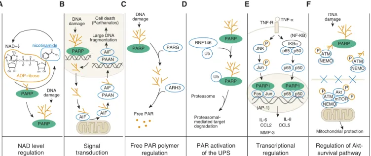
activation (Figure 2). According to the
“classic pathway” (Figure 2A) (in
reperfusion injury, circulatory shock,
various forms of inflammation [as well as in
a variety of other diseases]), reactive
oxidants and free radicals are formed, for
instance, as a consequence of a multitude of
biochemical pathways, including reduced
NAD
1phosphate oxidase, and/or
infiltration of the tissues with activated
immune cells and the consequent release of
various reactive species. These species, in
turn, produce DNA damage (primarily in
the form of single-strand breakage), which
is, in turn, recognized by the zinc
fingers of
PARP1, which, in turn, activate this
enzyme. When the DNA damage is
widespread (because the oxidant burden is
high), the extent of PARP1 activation can
be so pronounced, that a subsequent
cellular energetic deficiency (NAD
1depletion, followed by mitochondrial
inhibition and depletion of cellular ATP)
can produces cell dysfunction (1, 14–17).
For instance, in a rat model of endotoxic
shock, peritoneal macrophages exhibit
reduced NAD
1and ATP concentrations
and suppressed mitochondrial respiration;
these effects are suppressed by inhibition of
PARP by nicotinamide (18). Moreover,
PARP activity (assessed by the rate of
NAD
1consumption in various tissues)
were found to be significantly higher during
shock in nonsurvivors compared with
survivors in a porcine hemorrhagic-shock
model (19). In addition, administration of
the PARP inhibitor PJ-34 led to decreased
serum HMGB1 concentrations (an
indicator of cell necrosis) in mice subjected
to cecal ligation and puncture (CLP) (20).
Figure 2B depicts an alternative
mechanism, which is generally known as
the
“parthanatos concept.” According to
this mechanism (which has initially been
demonstrated in neuronal models but has
subsequently been also extended to a
variety of other cells, tissues, and disease
conditions), the poly–ADP-ribosylation
(PARylation) of various acceptor proteins
(as a result of PARP activation, as in
NAD level regulation
Signal transduction
Free PAR polymer regulation PAR activation of the UPS Transcriptional regulation Regulation of Akt-survival pathway Mitochondrial protection NEMO ATM P NEMO NEMO ATM ATM P P P P Akt mTOR DNA damage nicotinamide ADP-ribose NAD+↓ PARP PARP
A
B
C
D
E
F
N NH2 O NH2 N N N N O OHOH O O O O O O-O OHOH P P AIF PAAN AIF AIF AIF PAAN PARP PARP PARP PARP1 PARP1 PARP Cell death (Parthanatos) Large DNA fragmentation DNA damage DNA damage DNA damage PARG ARH3 Free PAR RNF146 Ub Ub Proteasome Proteasomal-mediated target degradation PARP JNK Jun Fos Jun IKBα (NF-KB) TNF-R TNF-α p65 p65 p65 p50 p50 p50 (AP-1) IL-6 IL-8 CCL2 CCL5 MMP-3 P PFigure 2. Mechanisms responsible for the cytoprotective and antiinflammatory effects of PARP inhibitors in nononcological diseases. (A) PARP activation and consequent NAD1depletion (the “Berger Hypothesis”). These processes can lead to a cellular energetic deficit and cell dysfunction; inhibition of PARP prevents these processes and exerts cytoprotective effects (inhibition of cell necrosis). (B) Role of PARP activation and free PAR polymers in inducing mitochondrial release of AIF (apoptosis-inducing factor), which, in turn induces cell death (parthanatos). Inhibition of PARP suppresses these processes and inhibits parthanatos. (C) The role of PARP in liberating free PAR polymers, which, on their own, exert cytotoxic effects; inhibition of PARP prevents free PAR polymer formation and suppresses cell death. (D) PARylation contributes to activation of the proteasome through an interaction with RNF146; PARP inhibitors suppress these processes. (E) Role of PARP in contributing to proinflammatory signal transduction via enhancing JNK-mediated (left sequence) and NF-kB–mediated (right sequence) activation of multiple genes and gene products. By inhibiting PARP, these processes are attenuated and inflammatory signaling can be attenuated. (F) PARP regulates the activation of the cytoprotective Akt pathway. Under normal conditions, PARylation anchors the ATM–NEMO complexes, which are retained in the nucleus. However, after PARP inhibition, the ATM–NEMO complex translocates to the cytoplasm, where Akt and mTOR are recruited to form the ATM–NEMO–Akt–mTOR cytoprotective signalosome, which, in turn, activates various mitochondrial protective and cell-survival pathways. Adapted by permission from Reference 17. ARH3 = ADP-ribosylhydrolase 3; ATM = ataxia telangiectasia mutated; NEMO = NF-kB essential modulator; P = phosphate group; PAAN = PARP-1–dependent AIF-associated nuclease; Ub = ubiquitin group; UPS = ubiquitin-proteasome system.
degradation of PAR. Free PAR, in turn,
leaves the nucleus and translocates into the
cytosolic compartment of the cell. It also
reaches the mitochondria, where it binds to
specific mitochondrial receptors, resulting
in the release of AIF (apoptosis-inducing
factor). AIF, in turn, diffuses back to the
nucleus, where it induces large-scale
nuclear fragmentation and cell death
(parthanatos). This process has been
primarily indicated in
central-nervous-system pathologies (21); its potential role in
critical illness remains to be explored.
In addition, free PAR polymer can
have independent roles as a pathogenetic
factor (Figure 2C) because it can bind
to various protein acceptors intracellularly
(or even, in some cases, extracellularly).
These PARylation reactions (a form of
posttranslational modifications) have
been shown to contribute to various
pathophysiological processes ranging from
neurodegeneration to vascular injury (21).
Another aspect of PARP relates to the fact
that it can also regulate ubiquitylation-mediated
protein-degradation reactions (Figure 2D).
The process of ubiquitin-mediated protein
degradation is involved in various
cell-signaling and protein-quality-control
processes. Iduna/RNF146, a constitutively
expressed ubiquitin E3 ligase is activated by
PARylation. In turn, proteosomally mediated
degradation of various proteins can ensue,
which may have various adverse effects on the
cells affected by it (22, 23).
A significant further role of PARP1
relates to its regulatory role on gene
transcription (Figure 2E). A general
mechanism involved in this process relates
to the modulation of chromatin structure
(in principle, due to the fact that the PAR
polymer is negatively charged), affecting
the availability of the DNA to the enzymes
involved in gene transcription. A secondary
mechanism relates to transcriptional
coregulation, and a third mechanism relates
to the modulation of DNA methylation. In
this context, PARP1 does not
“need” to be
activated by DNA-strand breaks; resting
(constitutive) PARP can confer these
actions, in some cases because of its
scaffolding (protein–protein interaction)
roles. Pharmacological PARP inhibitors
in many (but not all) instances can
significantly modulate the above
processes, with the end result being the
suppression of gene transcription in a
semispecific manner. In many experimental
models, the generation of proinflammatory
cytokines and chemokines can be
suppressed by PARP inhibitors, producing
an antiinflammatory and/or
immunomodulatory result (15, 23).
However, in complex situations
(e.g., in vivo models of disease), the
antiinflammatory effects of PARP
inhibitors may also be related to the
interruption of various positive-feedback
cycles of disease (1) (Figure 3). In the
context of critical illness, PARP inhibitors
have been demonstrated to suppress the
activation of NF-kB and the subsequent
production of various proinflammatory
cytokines (e.g., TNF-a) (24) and various
chemokines (e.g., MIP-1a and MIP-2) (25).
The models used initially employed murine
models of endotoxemia (24, 25) but were
subsequently extended into in various
rodent and large-animal models of sepsis,
septic shock (16, 17), and ALI (see below).
Finally, PARP inhibition has been
shown to activate the cytoprotective Akt
pathway (Figure 2F). The
first step in this
process is that PARP inhibition increases
the interaction between p-ATM and
NEMO proteins, thereby facilitating the
translocation of this complex from the
nucleus into the cytoplasmic compartment.
In turn, a cytoprotective signalosome
(p-ATM–NEMO–Akt–mTOR) is formed,
which induces the activation of Akt.
Akt is a
“master regulator” of various
cell-survival pathways; its activation produces
a cytoprotective phenotype (26).
The Therapeutic Ef
ficacy of
PARP Inhibitors in ALI
PARP Inhibitors Exert Bene
ficial
Effects in Preclinical Animal Models
of Lung Injury
Multiple lines of in vivo experiments have
demonstrated that pharmacological PARP
inhibitors or PARP1 deficiency can
significantly improve the outcomes of various
animal models of acute and chronic lung
injury, including endotoxin- or sepsis-induced
lung injury, pancreatitis-induced lung injury,
lung inflammation elicited by various agents
(e.g., zymosan, carrageenan or elastase),
ventilator-induced lung injury, environmental
agent– or drug-induced lung injury, or lung
fibrosis and allergy/asthma–associated lung
inflammation and dysfunction (26–84).
Table 1 focuses on the
findings related to the
effect of PARP inhibitors in various forms of
ALI and lung inflammation.
inhibitors include the correction of
the hyperinflammatory response
(i.e., suppression of cytokine and chemokine
production), reduction of the infiltration of
the lung tissue and the alveolar space with
inflammatory cells, reduced oxidative and
nitrosative stress (most likely due to the
interruption of the positive-feedback cycles
outlined in Figure 3), improved pulmonary
gas exchange, and improved histological
status of the lung tissue. Importantly, the
beneficial effects of PARP inhibition not
only have been shown in rodent models but
also hve been extended to several clinically
more-relevant large-animal models of ALI
(31, 40, 49, 50, 65).
However, it must be emphasized that
the studies summarized in Table 1 have a
number of limitations. For instance, many
studies (especially the studies in the 90s)
used
first-generation PARP inhibitors, such
as 3-aminobenzamide or nicotinamide;
these agents have many additional
pharmacological actions in addition to
PARP inhibition, including antioxidant
effects and inhibition of mono-ADP
ribosylation (85–87). Although the use of
such agents was acceptable when the
available tools were limited, current and
future work should use third-generation
inhibitors, preferably in combination with
PARP1-deficient animal models, as these
animals are viable and commercially
available. Another common limitation of
many of the published studies is that they
used only one sex of animals (typically
male). Since the mid-2000s, it has become
more and more obvious that the effect of
PARP inhibitors in various rodent models
of shock, inflammation, and reperfusion
injury is sex-specific; in most (but not all)
cases, male animals benefit more (as well as
aged females and ovariectomized females),
whereas the protective effect of PARP
inhibitors in females is less pronounced (88,
89). However, it should also be mentioned
that in some models (e.g., burn/smoke
inhalation–associated lung injury), PARP
inhibitors show significant therapeutic
benefit in female large-animal models as
well (e.g., References 40, 48). Moreover, in
pancreatitis and pancreatitis-associated
lung injury, both male and female mice
were found to respond comparably
well to PARP inhibition (56, 63, 90). We
recommend that future studies, especially
ones that focus on translationally relevant
(i.e., clinically approved) PARP inhibitors,
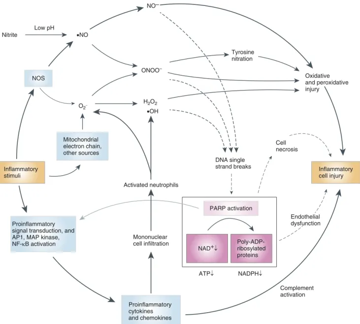
Inflammatory stimuli
Proinflammatory signal transduction, and AP1, MAP kinase, NF-κB activation Mitochondrial electron chain, other sources Proinflammatory cytokines and chemokines Mononuclear cell infiltration Activated neutrophils PARP activation Poly-ADP-ribosylated proteins Complement activation Endothelial dysfunction Inflammatory cell injury DNA single strand breaks Cell necrosis Low pH NOS ONOO– NO– H2O2 •NO O2– •OH Tyrosine nitration Oxidative and peroxidative injury Nitrite NAD+↓ ATP↓ NADPH↓
Figure 3. Pathophysiological triggers of PARP activation and interacting pathways of injury. Various pathophysiological conditions lead to the formation of various reactive oxygen species from various sources (such as the mitochondria, xanthine oxidase, or reduced NAD1phosphate [NADPH] oxidase). In inflammatory states, various proinflammatory pathways are stimulated in response to autoimmune responses and/or proinflammatory microbial components. The corresponding isoforms of NOS (nitric oxide [NO] synthase; brain NOS in the central nervous system, endothelial NOS in the cardiovascular system, and inducible NOS under inflammatory conditions) produce NO (but under conditions ofL-arginine depletion, NOS can also produce superoxide). Under low-pH conditions (such as tissue hypoxia/acidosis), nitrite can also be converted to NO. Superoxide (which is produced from various cellular sources, including mitochondria) and NO react to yield peroxynitrite. Peroxynitrite and hydroxyl radical induce single-strand breaks in DNA, which, in turn, activate PARP. This can deplete the cellular NAD1and ATP pools. Cellular energy exhaustion triggers the further production of reactive oxidants. PARP activation leads to cellular dysfunction via the energetic mechanism as well as via several other pathways outlined in Figure 2. Oxidative and nitrative stress can cause endothelial-cell dysfunction, at least in part though the depletion of NADPH concentrations, which, in turn, leads to reduced endothelial NO formation. The cellular dysfunction is further enhanced by the promotion of proinflammatory gene expression by PARP, through the promotion of NF-kB, AP1 (activator protein-1), and MAP (mitogen-activated protein) kinase activation. PARP can also promote complement activation. The oxidant-induced proinflammatory-molecule and adhesion-molecule expression, along with the endothelial dysfunction, induce neutrophil recruitment and activation, which initiates positive-feedback cycles of oxidant generation, PARP activation, and cellular injury. For instance, tissue-infiltrating mononuclear cells produce additional oxidants and free radicals. PARP is also involved in triggering the release of mitochondrial cell-death factors, such as AIF. There are many oxidative and nitrosative injury pathways that are triggered by oxygen- and nitrogen-centered oxidants and free radicals, which act in parallel or in synergy with PARP-mediated pathways of cell injury. Although most of the pathways shown in the figure have been demonstrated in acute lung injury (ALI), the relative contribution of cell necrosis versus inflammatory cell injury, as well as the relative role of the various pathways shown in the figure, depends on the specific form of ALI and the stage of the disease. Reprinted by permission from Reference 1.
Table
1.
Effects
of
PARP
Inhibition
in
Preclinical
Models
of
ALI
and
Lung
In
fl
ammation
Experimental Model Animal Species Effective Dose of the PARP Inhibitor Effect of PARP Inhibition Notes, Caveats, Limitation s o f the Study Reference Endotoxin-in duced or sepsis-associated ALI LPS-induced ALI Pig 3-AB (20 mg/kg i.v. pretreatment ) Protection against the LPS-induced, delayed suppression of lung compliance; prevention of the LPS-induced increase of nitrotyrosine immunoreactivity* in the lung Many parameters were unaffected by 3-AB (e.g., the recruitment of in flammatory cells into the BAL fluid); no effect of 3-AB was noted on the LPS-induced changes in systemic hemodynamics. Limitations: [A], [B], [C], [D], and [J]. 31 LPS-induced ALI BALB/c mouse PARP1 genetic de ficiency or PJ34 (20 mg/kg i.p., applied as a 1 h after treatment) Inhibition of cell in fi ltration into the BAL fl uid; inhibition of protein extravasation into the alveoli; suppression of TNF a , IL-1 b , and MIP-1 a production; reduced NO metabolite concentratio ns in the BAL; inhibition of lipid peroxidation (MDA) in the lung tissue; reduced MPO †activity in the lung tissue; improved lung histopathology with less alveolar hemorrhaging and fewer in fl ammatory cells Limitations: [C], [E], and [J]. 35 LPS-induced ALI Wistar rat PJ-34 (10 mg/kg i.p. pretreatment) Decreased MDA and MPO †activity in the lung tissue; reduced TNF a , IL-1 b , and nitrite/nitrate serum concentratio ns, improved survival Limitations: [B], [C], [E], and [J]. 37 LPS-induced ALI C57BL/6 3 129/Sv mouse PARP1 genetic de ficiency Attenuated iNOS and ICAM-1 expression in the lung Limitations: [D], [I], and [J]. 39, 46 LPS-induced ALI Rabbit 3-AB (10 mg/kg i.v. bolus 1 5 mg/kg/h infusion, applied as a pretreatment) Inhibition of iNOS expression in the lungs. Decreased pulmonary edema; reduced nitrotyrosine immunoreactiv ity* in the lung; inhibition of systemic NO overproducti on. Reduced lipid peroxidation (TBARS) in the plasma (with a similar trend in the lung tissue). Inhibition of albumin extravasation into the alveoli. Improved lung histology. Reduction of ICAM-1 expression in the lungs and reduction of cell in fi ltration into the lung tissue. Inhibition of the enlargement of the alveolar septa. Reduced leukocyte adhesion to the pulmonary arterioles, capillaries, and venules. Normalization of the effect of LPS on the alveolar –arterial oxygen difference 3-AB did not affect NF-kB activation in the lung tissue. No effect of the inhibitor on systemic hemodynamics or on red-blood-cell velocity. No effect on granulocyte ROS production ex vivo . Limitations: [A], [B], [E], and [J]. 39, 46 LPS-induced ALI Rabbit 3-AB (10 mg/kg i.v. bolus 1 2.5 mg/kg/h infusion, applied as a pretreatment) Restoration of the pulmonary architecture; maintenance of the gas exchange barrier; attenuation of the pulmonary edema; inhibition of the enlargement of the alveolar septa. Decreased oxidative stress (TBARS) in plasma; inhibition of nitrotyrosine* immunoreactivit y in the lungs. Normalization of the effect of LPS on the alveolar –arterial oxygen difference Limitations: [A], [B], [E], and [J]. 47 (ContinuedTable 1. (Continued ) Experimental Model Animal Species Effective Dose of the PARP Inhibitor Effect of PARP Inhibition Notes, Caveats, Limitation s o f the Study Reference LPS-induced ALI BALB/c mouse 4-HQN (100 mg/kg i.p.) pretreatment and delayed-treatment protocols were both used The PARP inhibitor improved survival in pretreatment and also when its administration was delayed to be given 1 h after the LPS challenge. It suppressed the hyperintensity of the MRI signal in the thoracic region (suggesting an inhibition of pulmonary in fl ammation and edema); decreased TNF a plasma concentratio ns; increased Akt phosphorylatio n and activation; attenuated ERK1/2, p90RSK activation; suppressed MAPK p38 activation; prevented NF-kB activation; inhibited c-Fos activation in the lung and in other tissues as well When the administration of the PARP inhibitor was delayed to 6 h , the survival bene fit was no longer seen. Limitations: [C], [D], [G], [I], and [J]. 51 LPS-induced ALI Sprague-Da wley rat Nicotinamide (100 mg/kg i.v. pretreatment) Systemic effects: protection against hypotension, erythropenia, and leukopenia. Protection against the increases in plasma markers of liver and kidney injury. Inhibition of NO, TNF a , and IL-1 b production; prevention of pulmonary edema and pulmonary hemorrhage, protection against the in fi ltration of in fl ammatory cells into the lung; reduced protein extravasation into the alveoli; restoration of tissue ATP content Limitations: [A], [B], [E], and [J]. 58 LPS-or TNF-induced ALI C57BL/6 mouse PARP1-genetic de fi ciency Reduced recruitment of in flammatory cells into the lung; suppressed lung in fl ammation; reduced expression of MCP-1, MIP-1 a , KC, MIP-2; reduced CXCR2 expression; completely suppressed DARC expression Limitations: [I] and [J]. 64 LPS-induced ALI Sprague-Da wley rat 3-AB (20 mg/kg i.v. pretreatment ) Systemic effect: protection against LPS-induced hypotension; prevention of acidosis and lactatemia; improved blood oxygenation. Improved pulmonary edema (evidenced by lung wet/dry weight ratio); decreased TNF a , IL-1 b , and IL-6 expression in the lung; inhibition of NF-kB activation in the bronchial epithelial cells 3-AB inhibited the expression of PARP1 protein in pulmonary epithelial cells (as opposed or possibly in addition to its expected effect on PARP1 activity; perhaps this effect is related to the antioxidant effect of this inhibitor). Limitations: [A], [B], [C], [E], and [J]. 66 (Continued )
Translational Review
577
Table 1. (Continued ) Experimental Model Animal Species Effective Dose of the PARP Inhibitor Effect of PARP Inhibition Notes, Caveats, Limitation s o f the Study Reference LPS-induced ALI C57BL/6 mouse DPQ (10 m g/kg i.p. pretreatment) Inhibition of neutrophil in fi ltration into the lung (evidenced by histological analysis; also con fi rmed by measurement of lung MPO † activity); inhibition of lung vascular hyperpermeabilit y; inhibition of the upregulation of TNF a , IL-1 b , IL-6, MIP-2, iNOS, and CXCL-1 mRNA concentratio ns in the lung tissue and peritoneal macrophage s); inhibition of cell death; maintenance of vascular permeability; inhibition of the activation of the NF-kB pathway; reduction in the number of apoptotic cells in the lung The stated dose of the PARP inhibitor is much lower than generally used effective doses in the literature. A lower doses of the inhibitor (1 m g/kg) was reported as not effective. Limitations: [B], [C], [E], [G], and [J]. 67 LPS-induced multiorgan-fa ilure ALI LACA mouse Olaparib (5 or 20 mg/kg i.p. in 30-min post-treatment protocol relative to LPS) PARP inhibition resulted in a suppression of macrophage and neutrophil in filtration into the BAL fl uid; restoration of the pulmonary redox balance; decreased TNF a , IL-1 b , and VCAM-1 mRNA expression. The LPS-induced oxidative stress in the lung (increase in MDA and decrease in GSH content) was also suppressed by the inhibitor. There were also various bene ficial effects on renal function A lower dose of olaparib (1 mg/kg) was not effective. The experimental group size is not stated in the article. Limitations: [C], [E], [G], and [J]. 70 Hydrochloric acid 1 LPS (“ two-hit model ”)-induced ALI and neuroin fl ammation BALB/c mouse Olaparib (5 mg/kg i.p.). Both a pretreatment protocol in which the inhibitor was given before each “hit ” and a post-treatment protocol (start of olaparib administration 1 w k later and repeated three more times, 2 d apart each) were employed Systemic effects: inhibition of cytokine concentratio ns (TNF a , IL-1 b , IL-6) in the plasma. Inhibition of neutrophil in filtration into the lung; reduction in alveolar capillary damage; reduction in oxidative stress markers in the lungs; protection against the breakdown of blood –brain barrier; reduction of the extent of neuroin fl ammation; protection against cognitive impairment. Olaparib also inhibited HIF-1 a protein expression in the lung and brain. The bene fi cial effects on cognitive impairment were maintained in the post-treatment protocol Limitations: [C], [E], [G], and [J]. 83 CLP-induced septic shock and ALI 129/Sv X C57BL/6 mouse PARP1 genetic de ficiency Decreased TNF a , IL-6, and IL-10 concentratio ns in plasma; inhibition of MPO †in the lung tissue; improved survival; suppression of circulating markers of liver and kidney injury Circulating NO concentrations and lipid peroxidation (MDA content) in the lungs was comparably elevated in wild-type and PARP1 2 /2 animals after CLP. Limitations: [C], [E], [I], and [J]. 36 (Continued
Table 1. (Continued ) Experimental Model Animal Species Effective Dose of the PARP Inhibitor Effect of PARP Inhibition Notes, Caveats, Limitation s o f the Study Reference CLP-induced septic shock and ALI C57BL/6 mouse Olaparib (6 –20 mg/kg total daily dose i.p., starting at 30 min after CLP and repeated 8 h and 24 h later) Increased survival rate; attenuation of TNF a , IL-1 a , IL-1 b , IL-2, IL-4, IL-6, and IL-12p40 plasma concentratio ns reduced the Th17/Treg ratio in the blood. The PARP inhibitor also reduced bacterial CFUs, consistent with an enhancement of antibacterial immune defense Although the CLP model was severe, no signi fi cant increases in lung MPO or MDA concentrations were observed after CLP, and olaparib did not affect these values. Histological analysis of the lung tissue revealed a slight emphysema in all groups. Limitations: [G] and [I]. 81 Pneumonia P. aeruginosa instillation into the lungs Sheep INO-1001 (10 mg/kg i.v. bolus, followed by 0.3 mg/kg/h infusion. The start of the bolus inhibitor was delayed 1 h relative to the injury) Decreased pulmonary edema and hemorrhage; inhibition of neutrophil in fi ltration into the lung tissue (histological analysis); improved gas exchange; protection against the obstruction of bronchi and bronchioles; decreased lipid peroxidation (MDA content in lung tissue); reduced nitrotyrosine* immunoreactiv ity; inhibition of iNOS mRNA expression. There was also a trend for improved survival Limitations: [B], [C], [F], and [J]. 48 Sepsis-induced pneumonia Rabbit PJ34 (10 mg/kg i.v. pretreatment ) Gut wet/dry ratios were improved in the sepsis/PJ34 group. There was a trend for reduced bacteremia No hemodynamic effects of the PARP inhibitor were observed. Although a trend was noted, no statistically signi ficant differences observed in lung wet/dry ratios between the sepsis and sepsis/PJ34 group. No signi ficant differences were found in serum, peritoneal, and luminal gut lactate concentrations or in P CO 2 gap between experimental groups. Limitations: [B], [C], D], [I], and [J]. 55 Lung in flammation Zymosan-induced systemic in flammation Swiss Albino mouse; Wistar rat 3-AB (10 –20 mg/kg i.v. pretreatment) Attenuated MPO †in the lung tissue Limitations: [A], [B], [C], [E], [I], and [J]. 29 Zymosan-induced systemic in flammation 129/SV 3 C57BL6 mouse PARP1 genetic de ficiency Improved pulmonary histology (reduction in interstitial hemorrhagin g and mononuclear-cel l accumulation ) Limitations: [C], [E], [I], and [J]. 29 Zymosan-induced systemic in flammation Long-Evans rat GPI 6150 (40 mg/kg i.p., after treatment: two doses applied at 1 and 6 h after zymosan) PARP inhibition decreased neutrophil migration and MPO †in the lung tissue; decreased pulmonary edema and improved histological picture of the lung. Hyperbilirubi nemia and the increased alkaline-phosphat ase concentratio ns were attenuated; survival rate was improved Limitations: [C], [D], [I], and [J]. 33 (Continued )
Translational Review
579
Table 1. (Continued ) Experimental Model Animal Species Effective Dose of the PARP Inhibitor Effect of PARP Inhibition Notes, Caveats, Limitation s o f the Study Reference Zymosan-induced systemic in flammation BALB/c mouse 5-AIQ (3 mg/kg i.p, after treatment: two doses applied at 1 and 6 h after zymosan) PARP inhibition attenuated pulmonary edema; reduced neutrophil in fi ltration into the lung; and lowered levels a lipid peroxidation marker (MDA) in the lung tissue; it also decreased pulmonary ICAM-1 and P-selectin expression; ameliorated the zymosan-induc ed increases in TNF a and IL-1 b serum concentratio ns Limitations: [E] and [J]. 34 Carrageenan-indu ced lung in flammation Long-Evans rat 3-AB (1 –30 mg/kg i.p. pretreatment) Inhibited plasma extravasation and PMN accumulation in the lungs; reduced plasma nitrite/nitrate concentratio ns; decreased nitrotyrosine* immunoreactivit y in the lung tissue Limitations: [A], [B], [C], [D], [I], and [J]. 30 Carrageenan-indu ced lung in flammation BALB/C mouse 5-AIQ (1.5 mg/kg i.p. pretreatment ) Reduced recruitment of in flammatory cells into the lung tissue; decreased IL-6, TNF a , and IL-1 b plasma concentratio ns; increased IL-10 expression; reduced CD11a, CD62L, ICAM-1, and MCP-1 concentratio ns; decreased iNOS and COX-2 expression; suppressed the activation of the NF-kB system; reduced STAT3 expression; increased IL-4 expression; improved pulmonary histology Treatment of carrageenan-injecte d animals with the inhibitor reduced CD25 1-, GITR 1-, and CD25 1GITR 1-expressing cell subsets in the pleural exudate; it also prevented the changes Foxp3 1 and IL-17 1 (the decrease or increase, respectively) in the peripheral blood and in the pleural exudate, suggesting an immunomodulato ry effect of the PARP inhibitor. Limitations: [B], [C], [F], [G], and [J]. 71 Elastase-induced lung in fl ammation and emphysema BALB/c mice Olaparib 5 o r 1 0 mg/kg i.p., either daily or every 48 h, starting as a pretreatment Decreased neutrophil and macrophage counts in the lung tissue; reduced concentrations of TNF a , IL-6, GCSF, KC, and MCP-1 in the BAL fl uid; restored the pulmonary redox balance (increased ROS production, increased MDA concentrations, reduced GSH concentrations); inhibited NF-kB activation in the lung with consequent reduction of ICAM-1 and VCAM-1 expression. Protection against emphysema (air space enlargement) Lower doses of olaparib (1 or 2.5 mg/kg) were not effective. Although this is principally a COPD model, there are several aspects of the study (effect of olaparib on in flammation and oxidative stress) that are relevant to the focus of the review (ALI). Limitations: [B], [C], [E], and [J]. 80 Elastase-induced lung in fl ammation and emphysema C57BL/6 mice PARP1 genetic de ficiency Decreased neutrophil and counts in the lung tissue; reduced concentratio ns of TNF a , IL-6, KC, and GCSF in the BAL fl uid; improved lung histology Although this is principally a COPD model, there are several aspects of the study (role of PARP1 in pulmonary in flammation) that are relevant to the focus of the review (ALI). Limitations: [C], [D], [I], and [J]. 80 Pancreatitis-a ssociated ALI (Continued
Table 1. (Continued ) Experimental Model Animal Species Effective Dose of the PARP Inhibitor Effect of PARP Inhibition Notes, Caveats, Limitation s o f the Study Reference Pancreatitis-a ssociated ALI Wistar rat 3-AB (10 mg/kg i.v.) Prevention of the increase of P-selectin expression in the lungs; prevention of the increase in MPO † in the lung tissue; inhibition of PAF production; decrease of the in fi ltration of PMNs into the alveoli; prevention of alveolar wall thickening; improvement in lung histology The PARP inhibitor also reduced the severity of pancreatitis. Limitations: [A], [B], [C], [E], and [J]. 32 129/Sv 3 C57BL/6 mouse PARP1 or PARP2 genetic de ficiency or PJ34 (10 mg/kg i.p.) or 3-AB (30 mg/kg i.p.) The two PARP inhibitors and PARP1 de ficiency suppressed lung in fl ammation; improved lung histological picture; lowered levels of MPO †in the lung tissue; decreased IL-6 and IL-1 b plasma concentratio ns The PARP inhibitors and PARP1 de fi ciency also markedly reduced the severity of pancreatitis. PARP2 de fi ciency reduced macrophage in filtration into the pancreas but did not affect any other pancreatic or pulmonary parameter investigated. Limitations: [B], [C], [E], and [J]. 56 Swiss-Webst er mouse PJ34 (15 mg/kg i.p. every 12 h) or KU0058684 ‡(15 mg/kg i.p. every 12 h) PARP inhibitor administration started 36 h after the initiation of the insult to induce pancreatitis (CMDE diet) The two PARP inhibitors were comparable in their effectivenes s. They produced a reduction of alveolar capillary thickening, protected against in flammatory cell in fi ltration into the lung, protected against apoptosis, and decreased IL-6 and IL-1 b plasma concentratio ns Both PARP inhibitors also reduced the severity of pancreatitis and improved survival. Limitations: [C], [F], and [J]. 63 I/R-associated ALI I/R-associated ALI Long-Evans rat INO-1001 (3 mg/kg i.v.) Reduction in the I/R-induced vascular hyperpermeabilit y; inhibition of the I/R-induced increase in MPO †in the lung; reduction of leukocyte in fi ltration into the BAL fl uid; decrease of TNF a , MIP-1 a , MIP-2, and CINC concentratio ns in the BAL fl uid; inhibition of NF-kB activation in lung homogenates; inhibition of caspase-3 immunopositivit y and protection against the increase in TUNEL immunoreactiv ity ‡in the lung Limitations: [B], [C], [E], and [J]. 52 I/R-associated ALI Long-Evans rat INO-1001 (3 mg/kg -i.t.) The PARP inhibitor protected against the developmen t o f vascular hyperpermeabilit y, attenuated MPO †in lung homogenates, suppressed neutrophil in fi ltration into the BAL fl uid, and inhibited the activation of the AP-1 and NF-kB systems; reduced secretion of MCP-1 and CINC into the BAL fl uid The sex of the animals was not stated in the report. Limitations: [B], [C], [D], and [J]. 41 (Continued )
Translational Review
581
Table 1. (Continued ) Experimental Model Animal Species Effective Dose of the PARP Inhibitor Effect of PARP Inhibition Notes, Caveats, Limitation s o f the Study Reference Hindlimb ischemia – reperfusio n-associated remote ALI Wistar rat 3-AB (10 mg/kg i.p. 30 min before the start of the lung reperfusion) 3-AB prevented the increase in MDA and concentratio ns in the lung tissue and plasma; it prevented the increase in lung MPO †; protected against the development of lung edema, prevented in flammatory cell in fi ltration into the lung; protected against the suppression of Na 1/K 1 ATPase activity in the lung tissue. Nitrotyrosine* immunoreactiv ity was also suppressed Limitations: [A], [B], [C], [F], and [J]. 54 I/R-associated ALI Sprague-Da wley rat Nicotinamide (100 mg/kg i.v. after treatment, starting 30 min after the beginning of lung reperfusion) Nicotinamide suppressed pulmonary edema and vascular hyperpermeabilit y. It reduced protein in flux into the BAL fl uid. It prevented the depletion of lung ATP, and it decreased NO, TNF a , and IL-1 b production PARP activation was demonstrated in the lung tissue after reperfusion; the study also con fi rmed the inhibitory effect of nicotinamide on lung PARP activation. Limitations: [A], [E], and [J]. 60 I/R-associated ALI Wistar rat PJ34 (10 mg/kg i.v.) Inhibition of pulmonary edema; suppression of lung in flammation; restoration of cellular ATP concentratio ns decreased TUNEL ‡ staining in the lungs, together with suppression of TNF a and IL-6 production. Inhibition of oxidative stress markers in the lung tissue Instead of PARP activation, the study focused on PARP1 cleavage; this process was apparent in the lung tissue after reperfusion; PJ34 inhibited this process. The PARP inhibitor also protected against hepatic and renal injury. A lower dose (5 mg/kg) of the inhibitor was less effective; a higher dose (20 mg/kg) produced lung edema on its own. Limitations: [B], [C], [E], and [J]. 68 Organ allograft –associated ALI Tracheal allograft –induced obliterative bronchiolitis Brown-Norwa y into Lewis rat INO-1001 (5 mg/kg i.p. twice per day for 2 wk, starting immediately after transplantation) Reduced luminal obstruction; decreased peritracheal in fl ammation; preservation of the integrity of the respiratory epithelium; attenuation of NF-kB activation; decreased TNF a expression; protection against caspase-3 activation and suppression of the developmen t o f TUNEL ‡immunoreactiv ity Limitations: [C], [E], and [J]. 42 Renal allograft –induced remote lung injury Brown-Norwa y into Lewis rat 3-AB (10 mg/kg i.v.) Decreased Rip1 expression; decreased hemorrhage and leukocyte in filtration into the lung; prevention of TUNEL ‡positivity in the lung tissue; suppression of TNF a production, improved lung histological pictures and injury scores PARP1 expression and PARP activation (PAR immunostaining) over the course of the immune rejection was demonstrated. The ef fi cacy of 3-AB was enhanced when it was combined with a necroptosis inhibitor. The interventions also protected against the primary (renal) injury. Limitations: [A], [C], [E], and [J]. 73 (Continued
Table 1. (Continued ) Experimental Model Animal Species Effective Dose of the PARP Inhibitor Effect of PARP Inhibition Notes, Caveats, Limitation s o f the Study Reference Lung transplantation: reconditioning of marginal donors via ex vivo lung perfusion Sprague-Da wley rat 3-AB (perfusion for 3 h with 1 mg/ml of the inhibitor before transplantation) Attenuated nitrosative and oxidative stress (protein carbonyls, nitrotyrosine*) in the transplanted lungs; reduced LDH release indicative of protection against cell necrosis; and IL-6 concentratio ns protect against the increase in lung compliance; improved lung histopathology and protection against perivascular edema in the transplanted lungs PARP activation (PAR staining) and the inhibitory effect of 3-AB on this response was con fi rmed in the study. Limitations: [A], [E], and [J]. 76 Lung transplantation: reconditioning of marginal donors via ex vivo lung perfusion Sprague-Dawley rat 3-AB (perfusion for 3 h with 1 mg/ml of the inhibitor before transplantation) The change in the IL-6/IL-10 ratio in the BAL fl uid was attenuated by the PARP inhibitor. It also protected against the increase in lung compliance; reduced MPO †; reduced MDA and 3-nitrotyrosine* concentratio ns after transplantation; decreased cell in filtration after lung transplantation; improved lung histopathology after transplantation, together with a suppression of P-selectin and VCAM-1 expression The inhibitory effect of 3-AB on PARP activation (PAR staining) was con firmed in the lungs. Limitations: [A], [C], [D], and [J]. 82 Cardiopulmona ry bypass –induced lung injury Cardiopulmonary bypass –induced ALI Dog PJ34 (5 mg/kg i.v.) or INO-1001 (1 mg/kg i.v.) Improved pulmonary function and gas exchange The PARP inhibitors also improved coronary vascular function and exerted bene fi cial systemic hemodynamic effects. Limitations: [A], [C], [D], and [J]. 49, 50 Cardiopulmonary bypass –induced ALI Piglet INO-1001 (1 mg/kg/h i.v.) Protection against the injury-associ ated increase of alveolar wall thickness; decreased TNF a and HSP70 concentrations in the lung Intraalveolar erythrocyte or granulocyte counts were not signi ficantly affected by the PARP inhibitor. Hemodynamic or oxygenation parameters were only slightly impaired or were unaffected by the bypass model, and the PARP inhibitor did not signi fi cantly affect them. Limitations: [A], [C], [D], and [J]. 61 Burn-or thermal injury –associated ALI Burn and smoke inhalation –associated ALI Sheep INO-1001 (3 mg/kg i.v. bolus starting 1 h after injury, followed by 0.3-mg/kg/h infusion) Improved respiratory mechanics; protection against the dysregulation of lymph flow; protection against the developmen t of microvascul ar hyperpermeabilit y; decreased pulmonary edema; decreased oxidative stress markers; improved histological picture of the lungs, prevention of the increase in pulmonary arterial pressure Inhibition of PARP activation (PAR staining) in the lung by INO-1001 was con fi rmed by immunohistochemi cal analysis. The inhibitor also exerted bene fi cial hemodynamic effects (pulmonary vascular resistance, cardiac index, stroke volume index) in this model. Limitations: [F] and [J]. 40 Thermally induced ALI Wistar rat 3-AB (10 mg/kg i.p.) Decreased MPO †and MDA in the lung tissue; decreased nitrotyrosine* immunoreactiv ity in the lungs Bene ficial effects were also noted in other organs (gut, kidney). Limitations: [A], [B], [C], [D], [I], and [J]. 53 (Continued )
Translational Review
583
Table 1. (Continued ) Experimental Model Animal Species Effective Dose of the PARP Inhibitor Effect of PARP Inhibition Notes, Caveats, Limitation s o f the Study Reference Smoke inhalation –associated ALI Sheep PJ-34 (0.003 and 0.03 mg/kg/h i.v. infusion, starting 1 h after injury) Improved respiratory mechanics; prevented the increased lung lymph flow and microvascula r permeability; protected against the development of pulmonary edema The bene fi cial effects were more pronounced in the group receiving the higher dose of the inhibitor. Systemic hemodynam ic parameters were not signi fi cantly affected by the procedure, and PARP inhibition did not have any effect on them. Limitations: [F] and [J]. 65 Scald-burn injury BALB/c mouse Olaparib (10 mg/kg/d i.p.) Reduced pulmonary MPO †7 d after injury; reduced pulmonary MDA concentratio ns 24 h after injury; suppressed the expression of TNF a and IL-1 a , IL-1 b , IL-12, and VEGF in the plasma At the early time point (24 h after injury) olaparib did not protect against the elevation of pulmonary MPO †. Also, olaparib was not protecting the changes in MPO †in liver and kidney. No effect of olaparib was noted on MDA concentrations in lung 7 d after injury. Olaparib also attenuated the increases in plasma markers of pancreatic and renal injury. It affected one liver injury marker (ALT) but not ALP. Limitations: [B], [C], [E], [I], and [J]. 84 VILI VILI C57BL/6 mouse PJ34 (20 mg/kg i.p.) Decreased in fl ammation; protected against the development of lung edema; improved lung histological scores and histological pictures; protected against the loss of lung compliance; inhibited the increase in MPO †in the lung tissue; suppressed NF-kB activation; inhibited NO, TNF a and IL-6 production The article does not specify the fi nal tidal volume delivered to the mice. The ventilation time was only 2 h , suggesting that the injury was primarily mechanical because of overstretching (“ barotrauma ”), rather than being secondary to stretch-induced in fl ammation and cell death. Limitations: [B], [E], and [J]. 59 miR-223 2 /1 mouse PARP1 shRNA Decreased in fl ammatory-media tor (TNF a , IL-1 b , IL-6, CXCL-1) content in the lung; protected against protein extravasation into the BAL fl uid; decreased epithelial injury marker (RAGE expression); reduced TUNEL ‡staining in the lung, inhibited lung edema This study primarily focused on miR-223 and discovered that it regulated PARP1 expression. Thus, the functional role of PARP1 was evaluated in a VILI model. No pharmacological PARP inhibitors were used. The ventilation time was only 3 h , suggesting that the injury was primarily mechanical because of overstretching (“ barotrauma ”). Limitations: [E] and [J]. 79 Environmenta l toxin –associated lung injury Paraquat-induc ed lung injury Sprague-Da wley rat 3-AB (10 mg/kg i.p. twice a day for 4 d ; first dose given in a 1 h after treatment) 3-AB decreased the paraquat-induc ed increases in LDH and neopterin concentrations; decreased TGF-b expression; reduced tissue oxidative stress markers; decreased pulmonary edema and hemorrhaging; inhibited leukocyte in filtration and mesothelial proliferation Limitations: [A], [C], [F], and [J]. 77
Definition o f abb reviations : 3-A B = 3-ami nobenzamide; 4-HQN = 4-hyd roxyquinaz oline; 5-A IQ = 5-ami noiso quinol ine; ALI = acute lung injury; AL P = alkaline phosp hatase; ALT = alani ne aminot ransferase; AP-1 = activ ator protein-1; CFU = colony-forming unit; CINC = cytokine-induced n eutrophil chemo attractant; CLP = c ecal ligation and puncture; CMDE = chol ine/methion ine-deficient/ethionine-su pplemented; COPD = chr onic obstructive pul mona ry disease; COX -2 = inducible isoform of cycloox ygena se; DARC = duf fy ant igen receptor for chemo kine s; DPQ = 3,4-d ihydro-5[4-( 1-piperindinyl)butox y]-1(2H )-isoquino line; GCSF = granulocyte colony stimu lating fac tor; GIT R = gluco c orticoid-induced TNFR -related; GPI 6150 = 1,11b-d ihydro-[2H]b e n zo p yr a n o [4 ,3 ,2 -d e ]is o q u in o lin -3 -o n e ); G S H = g lu ta th io n e (g -l -glu tamy l-l-c yst ein yl -gl yci n e tri pepti d e); H IF = h yp ox ia-inducib le factor; H S P 70 = h eat s h ock p ro tein 70 ; I/R = is chemi a– repe rfu s io n; ICAM = interce llula r adh e si on m o lecul e ; INO-1001 = N -( 3-morp holin-4-ylprop yl)-5-ox o-6, 11 -dihy d ro indeno [1, 2 -c]i soqu inoli n e-9-sul fona m id e; iN OS = indu c ib le NO s ynt hase ; K C = k e ra tino cyt e -d eri ved chemokine; K U 00 5868 4 = re sear ch compo und that is equiv a lent to the c ur rently clinically ap pro ved anti cancer d rug n ir apar ib or (S )-2-( 4 -( pi perid in-3-yl) p h e n yl )-2 H -i n d a zo le -7 -c a rb o x a m id e , marketed a s Zej ula; LACA = Laboratory Animal Centre A-stra in; LDH = lactate dehy drogenase; MCP = monocy te che mo attractant pro tein; MDA = malon dialdehyde; MIP-1 a = macro phage inflam matory protein 1 a , also know n a s CCL3; MIP-2 = macro phage inflam matory protein 2, also know n a s CXCL2 ; M PO = myelop eroxidase; MRI = magn etic reso nance imaging ; N O = nitric o xi d e ;P. = P seu domonas ; PA F = plat elet activa ting fac tor; P A R = p oly(ADP -ribose), th e e n zyma tic pr oduct o f P A R P; P A R P = PA R p olyme rase ; PJ 34 = N -(5,6-dihydro-6-ox o-2-phen an th ridin yl)-2-acet am id e ; P M N = p o lymorphonu clear neutrophil; ROS = re active oxyg en spe cies; TBA RS = thiobarbituric acid re active sub stances; TGF = tum or gro wth factor; Th17 = T-hel per cell type 17; Treg = T regulatory cell; VILI = venti lation-ind uced lung injury. List of comm on limi tations: [A]: F irst-gen eration PARP inhibitor used ; these inhibitors have limited cell uptak e and a poorl y characterized pharma co kinetic pro file in vivo ; they also tar get sever al other enzym es in add ition to PARP 1; they can also exert nonspecific antio xidant effects. [B]: Pre treatment or cotreatm ent with the PARP inhibitor use d onl y; no post-treatment (i.e., therapeutic window) wa s assessed. (This is not cons idered a w e akness in th e transplantation and bypass stud ies in which pretreatmen t o r coadm inistration is clin ically fea sible.) [C]: No target enga geme nt confirmed (i.e., no evidence that the pharmacolo gical age nt, ind eed, inhibited the activity of its target, PARP 1, or that the model pro duces suita ble plasma conc entrations of the inhibitor so that PARP 1 inhibition can be expected). [D] : The study used animals of “either sex, ” o r the sex of th e animals was not stated . In eithe r case, n o separate analysis o f sex effect was conducted. [E ]: All-male study; no comp arison of poten tial sex differences. [F]: All-fe male stud y; n o comp arison of potential sex diff erences. [G] : Althou gh the study is a m ouse stud y, the pharmacolo gical appr oach wa s not combined with a gene tic approach (PAR P1-deficient mice) . [H ]: Group size is not exp lained. [I]: The numbe r o f the mea su red lung injury parameters wa s limited. [J]: Efficacy parameter s were stud ied only; no inve stigation of the saf ety of the inhibitor (e.g ., on DNA integrity or repai r) wa s conducted. *Inhibitory effect of pharmaco logical age nts on tissue nitro tyros ine immunor eactivity was frequ ently equated with re duced pro duction of the ROS p ero xynitrite, which (simil ar to hydr oxyl radical) is an endog enou s trigger of DNA -strand break age and sub sequen t PAR P activatio n. How ever, subsequent stud ies demonstrat ed that other bioc hemical reaction s (includ ing reactions involving MPO) may also produce nitro tyrosine. † Inhibitory effect of pharmacolo gical agen ts on tissue MP O conc entratio ns (in fac t, the assa y most commonl y used measu res MPO enzym atic activity and not MP O conte nt) may also ind icate effects on neut rophil degr anulation, and/o r neut rophil extracellular net formatio n and/o r direct effects on th e activ ity of th e M P O enzyme. In the table, therefore, we disting uish “MPO conte nt” from “ne utroph il lung count ” (evid enced by histo logical or flow cyto metric analy sis). ‡TUNEL po sitivity in tissue sli des is often equated to apopto sis, but in fact it actually show s DNA-s trand break age, which can occ ur in conj unction of v arious form s o f cell death or cell dysfu nction.
![Figure 1. Overview of key biological functions of PARP1. The top part depicts the various domains of PARP (poly[ADP-ribose] [PAR] polymerase), including its DNA-binding domain, with its zinc fingers (ZnI, ZnII, ZnIII), which are essential for recognition o](https://thumb-eu.123doks.com/thumbv2/123doknet/14316402.496200/2.877.54.556.118.588/figure-overview-biological-functions-polymerase-including-essential-recognition.webp)