biology data grid supports live analysis
The MIT Faculty has made this article openly available.
Please share
how this access benefits you. Your story matters.
Citation
Meyer, Peter A.; Socias, Stephanie; Key, Jason; Ransey, Elizabeth;
Tjon, Emily C.; Buschiazzo, Alejandro; Lei, Ming, et al. “Data
Publication with the Structural Biology Data Grid Supports Live
Analysis.” Nature Communications (March 2016): 10882.
As Published
http://dx.doi.org/10.1038/ncomms10882
Publisher
Nature Publishing Group
Version
Final published version
Citable link
http://hdl.handle.net/1721.1/108692
Terms of Use
Creative Commons Attribution
Received 16 Oct 2015
|
Accepted 28 Jan 2016
|
Published 7 Mar 2016
Data publication with the structural biology data
grid supports live analysis
Peter A. Meyer et al.
#
Access to experimental X-ray diffraction image data is fundamental for validation and
reproduction of macromolecular models and indispensable for development of structural
biology processing methods. Here, we established a diffraction data publication and
dissemination system, Structural Biology Data Grid (SBDG; data.sbgrid.org), to preserve
primary experimental data sets that support scientific publications. Data sets are accessible
to researchers through a community driven data grid, which facilitates global data access. Our
analysis of a pilot collection of crystallographic data sets demonstrates that the information
archived by SBDG is sufficient to reprocess data to statistics that meet or exceed the quality
of the original published structures. SBDG has extended its services to the entire community
and is used to develop support for other types of biomedical data sets. It is anticipated that
access to the experimental data sets will enhance the paradigm shift in the community
towards a much more dynamic body of continuously improving data analysis.
Correspondence and requests for materials should be addressed to P.S. (email: [email protected]). #A full list of authors and their affiliations appears at the end of the paper.
A
s one of the most powerful tools in structural biology,
X-ray crystallography allows determination of the
structure (atomic coordinates) of proteins, nucleic acids,
small molecule compounds and macromolecular complexes to
atomic-level resolution. Crystallographic data continue to be a
primary source of mechanistic understanding of macromolecules,
the implications of which extend from basic research to
translational studies and the rational design of therapeutics.
Reflecting the significance of the technique, the number of
published macromolecular crystal structures has rapidly grown to
4100,000 and numerous investigators within structural biology
have been awarded the Nobel Prize, including Drs. Kendrew,
Perutz, Watson, Crick, Wilkins, Hodgkin, Klug, Deisenhofer,
Michel, Huber, Walker, MacKinnon, Kornberg, Ramakrishnan,
Steitz, Yonath and Kobilka.
To support the needs of a growing structural biology
community, a global network of synchrotron beamlines
1has
been established and made available to researchers. These
facilities remain the predominant source for crystallographic
data collection. While the data collection process has become
increasingly streamlined, deployment of a data management
infrastructure to archive original diffraction images has been slow
and uncertain
2. With the exception of a modest number of
data storage systems dedicated to the support of individual
synchrotron beamlines
3, or specific structural genomics projects
4,
storage of diffraction image data sets is typically the responsibility
of primary investigators. Access to these original experimental
data sets is therefore dependent on the policies of individual
laboratories, which vary in storage organization, institutional
resources, and researcher turnover. There is no universal
archiving system to store X-ray diffraction data sets, and raw
data sets are rarely made publicly available. In the cases where
data sets are available, their distribution format can vary
significantly. A typical data set of 360 images collected on
modern detectors is 5 GB, and structure determination can
involve one to tens of data sets, making the logistics of storing
diffraction data for many protein structures a daunting task.
The benefits of easy and public access to experimental data are
numerous
5. Access to primary data would support community
efforts to continuously improve existing models and identify new
features through complete reprocessing of experimental data
6–8with modern software tools and improved criteria
9. Further,
original data may provide a basis for validating questionable
existing structures while mistakes in structure determination may
be identified earlier
10–12. Additionally, access to a diverse volume
of raw data can be used to develop improved software to address
limitations of existing programs. Finally, access to a collection of
varied experimental data will undoubtedly benefit the training
and education of practitioners. The Worldwide Protein Data
Bank
13,14(wwPDB) has illustrated how these achievements can
be realized with the collection of reduced experimental data, in
the form of structure factor amplitudes. Complementing this
resource by preserving raw experimental data and making it
available to a broad community promises a profound scientific
impact in structural biology and other biomedical disciplines that
face the challenges of preserving large data sets.
While the primary role of the SBGrid Consortium (www.
sbgrid.org) has been to curate and support a collection of data
processing software applications and to organize
community-wide computing support
15, SBGrid has also been active in the
management of raw, experimental data sets. In 2012, SBGrid
prototyped a system based on Globus technology
16–19to move
diffraction data between Harvard, The Advanced Photon Source,
and the Stanford Synchrotron Radiation Light source
19.
To support the outstanding needs of the global structural
community, we have established a publication system for
experimental diffraction data sets that supports published
structural coordinates: the Structural Biology Data Grid (SBDG).
The SBDG project was initiated with a collection of X-ray
diffraction image data sets as well as a few additional data set
types contributed by many SBGrid Consortium laboratories. The
collection supports a diverse subset of over 68 peer-reviewed
publications and represents a sampling of numerous structure
determination approaches. To evaluate the utility of such a data
grid, we reprocessed all published diffraction data sets in this
initial collection with modern software and compared the derived
statistics against those reported in the original publications. We
also demonstrate that by integrating the storage resources of
multiple research groups and institutions, the data grid is poised
to deliver a novel community driven data preservation system
to support various types of structural biology and biomedical
data sets.
Results
Structural biology data grid. The SBDG is a centralized data
publication service—a repository for discovering, downloading
and depositing large structural biology data sets. We developed
the SBDG to support the need of the SBGrid community to
archive and disseminate X-ray diffraction image data sets, that is,
images recorded on X-ray detectors, which support published
structures. More than 90% of SBGrid laboratories use X-ray
crystallography in their research, and SBGrid investigators have
contributed over 11,000 X-ray structures to the PDB. The SBDG
complements the PDB, which archives derived data—merged and
post-refined data from diffraction images and the resulting
refined coordinates of macromolecular structural models. The
data grid has been developed in collaboration with the Data
Science team at Harvard’s Institute for Quantitative Social
Science, and it conforms to progressive data science standards
(Table 1). The SBDG limits its collection to data sets that support
journal publications, referred to as ‘primary data’. For X-ray
diffraction data, this primary data consists of experimental
diffraction images supporting a derived structural model and
journal publication. Release of this primary data by the SBDG
coincides with publication of the resulting manuscript and for the
structural biology data sets of related PDB files. As of 1 September
2015, the SBDG stores a diverse collection of 117 data sets,
including 111 X-ray diffraction data sets and a handful of other
data types including computational decoys and data sets from
MicroED, lattice light-sheet microscopy and molecular dynamics
(Supplementary Table 1). These published data sets, contributed
by 50 laboratories with diffraction data sets collected at 11
synchrotron facilities (Fig. 1) and several home sources,
originated 94 structures and 68 journal publications. The X-ray
diffraction data sets range in size from 126 MB (ref. 20) to 20 GB
(ref. 21) with a mean of 4.9 GB and a total of 573 GB of storage.
Extrapolating from this initial collection, which is quite diverse
and registers at just over 0.5 TB, our current 100 TB file system
could immediately support roughly twenty thousand X-ray
diffraction data sets (Fig. 2).
The SBDG’s collection of data sets can be accessed from the
data.sbgrid.org website. On the home page, deposited data sets are
organized into laboratory and institutional collections (Fig. 3a).
Hyperlinked collection pages provide a list of selected data sets
along with the data set’s corresponding data Digital Object
Identifier (DOI), a link to the journal publication, the PDB ID, a
link to the PDB entry, and a link to the depositors’ laboratory
website. The website molecular viewer, PV
22, offers visitors
an option to view structures in a manipulatable cartoon
representation (Fig. 3b). With multiple high-quality viewing
options and flexible search functionality, users of the SBDG
website can easily identify a small subset of relevant data sets.
Persistent data set pages are an important element for any
research data repository because they typically provide a landing
URL, which resolves from a given DOI
23. The SBDG does not
advertise unique codes, but instead distinguishes data sets by fully
qualified DOIs. From each SBDG collection page or viewer page a
user can access those unique Data set Pages (Fig. 4), which offer
Table 1 | Data science standards.
Disclosure Software tools developed under this program will be incorporated into open source software and released to the community. Manuscripts and white papers describing various phases of the project will be released on a regular basis.
Adoption All biomedical image data will be converted to the master formats, such as OME-TIFF or HDF5. Community tools to create, analyse, and manipulate diffraction images will be extended to include support for these formats. All biomedical data are assigned Digital Object Identifiers through the CDL EZID system, and follow modified DataCite and Dataverse metadata schemas. Associated metadata are registered with the International DOI Foundation, making it virtually permanent and independent of SBGrid and Harvard computing infrastructure. All data sets published through the SBDG will be citable using Force 11 recommendations.
Transparency Files within individual data sets will be deposited in their original format (no archives or encryption allowed). Self-documentation: The majority of diffraction data sets are self-documented and include the basic information required for reprocessing in the images themselves. Additional information will be collected during deposition and will include data set representation (the ability to use the data to be processed), reference (relation to PDB files, publications, and other data sets), context (for example, a native data set or a derivative used for phasing), fixity (checksums), and provenance (typically the data collection facility and the project member who deposits the original data set). With conversion to master formats, all secondary information will be appended to the image metadata along with all original headers.
External dependencies
The ability to reprocess some older data sets and verify master format conversions could depend on access to a specific version of data processing software. As data sets enter our repository, they will be reprocessed with our Data Reprocessing Pipeline (one of several we will develop as part of our Data Mining Pipelines). Data Reprocessing Pipelines will be archived within our system, issued DOIs, and interlinked with the data sets. It is worth noting that, since 2002, SBGrid has been archiving structural biology applications and, therefore, has access to previous software versions that might be required to reprocess older data sets. Licensing Biomedical data sets will be deposited under the Creative Commons Zero licence, supporting future development of data
validation services and database replications and migrations. Technical protection
mechanism
The security of the deposited data will be maintained by the DAA. The DAA will join with the Library of Congress sponsored NDSA and the data architect working on the project will ensure that NDSA recommendations are being followed.
NDSA, National Digital Stewardship Alliance; SBDG, Structural Biology Data Grid.
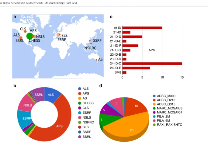
21-ID 19-ID 21-ID-D 21-ID-E 21-ID-F 21-ID-G 23-ID-B 24-ID-C 24-ID-E BM8 23-ID-D 0 5 10 15 APS ALS APS AS CHESS CLS NSLS ESRF NSRRC SLS SSRF SSRL ADSC_M300 ADSC_Q210 ADSC_Q315 MARC_MOSAIC3 MARC_MOSAIC4 PILA_2M PILA_6M RAXI_RAXISHTC 25 APS ALS SSRL NSLS ESRF 10 5 5 6
a
c
b
d
Figure 1 | Data collection statistics for the pilot subset of 112 data sets. (a,b) Data sets were collected from synchrotrons on four continents (in addition to laboratory sources, which are not broken down geographically) and originate from eleven synchrotron facilities: Advanced Light Source, Advanced Photon Source, Australian Synchrotron, Cornell High Energy Synchrotron Source, Canadian Light Source, European Synchrotron Radiation Facility, National Synchrotron Light Source, National Synchrotron Radiation Research Center, Swiss Light Source, Shanghai Synchrotron Radiation Facility, and Stanford Synchrotron Radiation Lightsource. World map image courtesy of the U.S. Geological Survey. (c) Breakdown of data sets collected at the Advanced Photon Source beamlines. (d) Data sets cover a range of detector types, including Area Detector Systems Corporation M300, Q210 and Q315, Rayonix MarMosaic, Dectris Pilatus 2M and 6M, R-AXIS HTC, and MAR345.
additional information for each data set including download
instructions and the fully formatted data set citation for inclusion
in manuscripts, following best practices set by the Joint
Declaration of Data Citation Principles
24. A Data set Page can
also be located by searching the SBDG for a PDB code, although
often several related data sets are used to determine a single set of
macromolecular coordinates. As the Data Grid is developed, the
Data set Pages will include additional functionality, with more
information on how to reprocess data sets, extended data
statistics, and discussion forums allowing users to annotate data
sets after publication. Taken together, the uniquely defined Data
set Pages provide a comprehensive and persistent location for
individual data sets.
Data set access. All data sets in the SBDG are readily and freely
accessible to the community. Access rights were formalized with
adoption of the creative commons zero licence (CC0), which
supports dedication of research results to the public domain and
is used by many open-data projects. This licence allows use and
redistribution of data for both commercial and non-commercial
purposes without requiring additional agreements. The CC0
licence does not affect patents or trademark rights of
con-tributors, and is similar to the licensing terms that are used for
macromolecular models released by the wwPDB.
Although data sets can be downloaded individually, their size
can make this cumbersome. Physical access to SBDG data sets is
facilitated through a data grid infrastructure that is supported by
members of the data access alliance (DAA; Fig. 5a). The DAA
is a voluntary and open organization of research-data-storage
providers and is being developed in collaboration with the Globus
Project. The DAA has two aims: (1) to minimize the chance of
data loss by replicating SBDG data sets, and (2) to facilitate global
data access through its members. Although it is expected that
DAA membership and architecture will evolve rapidly, in its
current state the DAA framework already provides a global
solution for data dissemination. DAA centres in Europe, Asia,
North America and South America replicate the entire SBDG
collection and provide local access to members of regional
communities. There are four DAA centres: Harvard Medical
School in the USA, Uppsala University in Sweden, Shanghai
Institutes for Biological Sciences in China, and Institut Pasteur de
Montevideo in Uruguay. As a secondary service, DAA centres can
provide local, direct access to data sets for their institutional
research groups. For example, Harvard Medical School hosts the
entire collection and provides direct access to all data from its
computing center. The DAA infrastructure is further extended by
the DAA satellites, which replicate fractions of SBDG data sets in
their local storage for direct access by members of individual
institutions. This mode of participation provides an attractive
option for research institutions to develop local archives of all
primary data generated by the local community. For example, the
NE-CAT (Northeastern Collaborative Access Team; sector 24-ID)
synchrotron beamline at the Advanced Photon Source, in
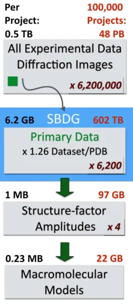
Figure 2 | Estimation of storage requirements for different stages of thestructural biology pipeline, based on the SBDG pilot collection. For structure factor amplitudes and PDB models file sizes were obtained from a subset of 96 PDB depositions derived from the pilot data sets. On average, SBDG stores 1.26 data sets per PDB file. Numbers in red indicated the estimated storage requirements to accommodate data sets for 100,000 structures. We estimate that for each primary data set, additional 100 data sets are collected at national facilities. Primary data refers to original experimental diffraction images supporting the derived structural model, as distinguished from all experimental data (screening images, inferior quality data sets, and so on). For crystallographic experiments, reduced data refers to the integrated intensities (or amplitudes, which do not materially affect storage requirements).
a
b
Figure 3 | Organized display of data collections at SBDG. (a) Graphical view of Laboratory and Institutional Collections within the SBDG; (b) PV structure viewer, displaying a published model with links to its two primary deposited data sets.
Argonne, IL, replicates all SBDG data sets that originate from
NE-CAT beamlines and makes them available to beamline staff and
users. Another SBGrid member and DAA Satellite, Yale
University, replicates all data sets from Yale laboratories on its
institutional storage and makes them accessible to structural
biology workstations through the Network File System. We
expect that, as research storage infrastructure catches up with the
capacities required to archive larger collections of diffraction data
sets, some DAA satellites will elect to replicate a larger fraction of
SBDG archives and make them available to the general
community.
While the DAA offers a variety of data access options that will
support growth of the repository, members of the community can
also download individual data sets directly from SBGrid servers at
Harvard using an rsync protocol. Instructions for downloading
individual data sets are provided on the Data set View Pages, and
effectively all data sets can be downloaded using the following
command: ‘rsync -av rsync://data.sbgrid.org/DOI.’, where DOI is
the digital object identifier for a particular data set. The rsync
utility, which is native to Linux and OS X systems, is particularly
suitable for downloading large data files and can be restarted in
case of interruption. After download, the data integrity of
individual data sets can be verified by following instructions on
the Data Grid website. With a well defined and permissive CC0
access licence and multiple channels for accessing data (four
DAA sites and the rsync download mechanism) our initial
infrastructure is well suited to support expansion of the data
collection.
Data publication cycle. For many SBGrid laboratories, interest
in data deposition is driven by a desire to better organize
research data and comply with institutional, federal, and
project-specific data preservation requirements. During the pilot phase,
data deposition privileges were limited to SBGrid member
laboratories. With recent funding to further support the project,
the Data Grid is now open to the entire structural biology
community. Non-SBGrid groups would first need to register with
the SBDG to obtain proper deposition credentials.
Wide adoption of data preservation systems is often hindered
by the complexities involved in the data deposition process itself.
To mitigate this problem, SBDG deposition involves two simple
steps: registration and uploading (Fig. 5b). To register a data set,
the depositor completes a web form with basic information about
the sample, data collection facility, related objects (for example,
publication, PDB code), and authorship; this information is
mapped to the DataCite schema (Fig. 6). Many details necessary
for data set reprocessing—beam center, distance, wavelength, and
so on—are automatically included with most data sets in the form
of an image header generated by the data collection software
at the time of collection, simplifying the registration process.
A principal investigator is authorized to sponsor depositions as a
recognized member of the community and must approve each
deposit. This system allows maximum flexibility when accepting
data for deposition, facilitating the upload of complex data sets
that otherwise could be challenging to validate. Following
registration, a DOI is reserved for the data set and the user is
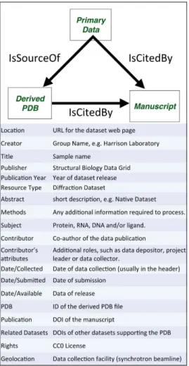
Figure 4 | SBDG persistent data set landing page (the target of a DOIresolver for a published data set). Data set metadata are displayed, as are instructions for downloading and verifying the data set.
a
b
Figure 5 | Experimental data flow and publication. (a) Flow of Primary Experimental Data. Data sets collected at synchrotrons are moved to end-users’ computers for processing and structure determination. Subsequently refined macromolecular models are deposited at PDB and primary data is uploaded to SBDG. From SBDG, data sets are replicated to DAA centres and eventually copied to DAA Satellites. End-users can access data sets by downloading from DAA centres and by direct access from Satellites. (b) Flowchart for data publication.
provided with data transfer instructions. Data deposition is
handled by an automated script provided by SBDG and run on
the depositor’s computer, which uploads the data and checks for
data integrity after upload. Upon verification, the primary data
are either released in the bi-weekly SBDG release or placed on
hold. As with the PDB, release of data placed on hold will
coincide with publication.
The two-step publication process is complemented by
behind-the-scenes data replication, DOI registrations, and data analysis.
All X-ray diffraction images are currently post-processed using
data processing pipelines that provide a post-publication data
review that will be shared with depositors and the community in
the next phase of the SBDG project. We are building additional
tools to help increase data deposition rates, including automatic
reminders sent to consortium members to encourage them to
deposit data for previously published work.
Data citation. Research data are the legitimate and citable
product of research
24,25and, therefore, the SBDG recommends
that depositors and data users cite all data deposited with the
SBDG in the standard reference section of their manuscripts
following well established community standards
24,26,27. Data
citation examples are provided on individual data set pages
(Fig. 4). The SBDG complements our AppCiter application
28,
which facilitates citation of research software. Both services are
now presented to users in a unified publication support workflow
(Fig. 7a). In step 1, the user deposits research-related data that are
put on hold until publication. A set of DOIs and corresponding
data citations are then generated and provided to the end-user.
Users can also use AppCiter to generate a list of software citations
for all scientific software used in the project. In step 2, all research
data and scientific software citations are included in the
References section of the manuscript. In step 3 the user,
anticipating manuscript publication, contacts relevant databases
to request release of the primary and supporting data. This
process should, ideally, take place before manuscript publication
and be timed to coincide with the publication date, allowing the
community to access the data when the manuscript is released.
When preparing future publications that refer to completed
structures, scientists should reference the relevant publications
and macromolecular models, unless they are referring to a specific
data set. For specific data sets, authors should explicitly reference
experimental data using the corresponding data citation (Fig. 7b).
Citation metrics for published data sets will be comparable to
those obtained for journal publications.
Data grid content. Ease of data deposition and community-wide
interest facilitated growth of the initial collection of X-ray
dif-fraction data sets when it opened to the SBGrid community in
May 2015. The data sets deposited during the pilot collection
phase represent a wide cross-section of structures and a diverse
subset of journal articles and structure determination methods.
For example, 68 structures derived from data deposited in the
SBDG have been determined by molecular replacement, while 4
have been solved by Multiple-wavelength Anomalous Diffraction,
4 by Single Isomorphous Replacement with Anomalous
Scatter-ing and 15 by SScatter-ingle-wavelength Anomalous Diffraction. The
highest resolution data set
29extended to 1.04 Å, and the lowest
resolution data set
30to 5.5 Å. The structures ranged in molecular
weight from 8.1 (ref. 31) to 426 kDa (ref. 32). The solvent content
of these structures ranged from 32 (ref. 33) to 85% (ref. 34) and
the longest unit cell edge was reported to be 525.29 Å (ref. 35).
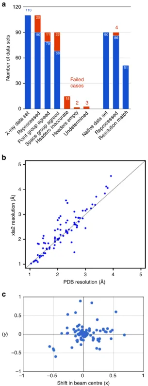
For a proof of concept, released data sets in the SBGridDB were
reprocessed with xia2 (refs 36–42) in a fully automated manner
(Fig. 8a). In all, 90 of the 110 released data sets with a
corresponding PDB ID were successfully reprocessed. In all,86 of
those 90 data sets represented high-resolution, native data and for
51 of those xia2 decision making determined a high-resolution
limit within 0.1 Å of the published structure (Fig. 8b). The point
a
b
Figure 7 | Data publication guidelines. (a) Flowchart illustrating publication guidelines incorporating software and data citations. (b) Data Citation guidelines, adapted from Dataverse Best Practices Guidelines that were developed based on Force 11 Joint Declaration of Data Citation Principles.
Figure 6 | DataCite metadata schema used for primary data sets within the SBDG. Information associated with the DOI record for a primary data set through the EZID system.
group determined by reprocessing agreed with that of the
published structure in 79 cases; for 65 of these the space groups
agreed. The lower degree of recovery of space groups, in
comparison to point groups, is attributed to ambiguity in screw
axis determination at this stage of data processing. To provide
insight into the most common failure modes, data sets for which
xia2 did not produce a set of integrated intensities were investigated
using iMOSFLM
43. Twelve of the failure cases could be attributed to
absent or inaccurate information in the image headers: while
accuracy of the beam center annotation varied within the pilot
collection (Fig. 8c), 10 data sets had visually incorrect beam center
information, two had missing header information. The cause of
failure for the eight remaining data sets was not definitively
determined from the data sets alone; however, consulting the
reprocessing instructions provided by depositors clarified this for
five of these data sets. The reprocessing instructions also suggested
that many of the data sets for which xia2 was able to produce
integrated intensities, but with resolution or symmetry disagreeing
with the deposited structure, could be attributed to incorrect header
information. One outlying reprocessing case for which a
significantly higher resolution was determined than originally
reported was also investigated. For this case, one of four
reprocessing attempts for the data set reported a resolution higher
than that supported by merging statistics. This discrepancy was
resolved by a software update.
In addition to estimates of the Bragg intensities, diffraction
images
can
also
be
analysed
for
additional
features
44.
A well-known example is the isotropic solvent ring that generally
appears
B3–4 Å resolution
45. However, diffraction images also
contain anisotropic diffuse scattering signals under and between the
Bragg peaks that derive from two-point correlations of electron
density fluctuations
7. Analysis of this diffuse scattering could
therefore provide information about protein, nucleic acid, and lipid
structural dynamics and correlated motions, potentially leading to
new mechanistic insights
46or to validating sampling schemes and
energy functions for molecular dynamics simulations
47. One data
set on the model enzyme Cyclophilin A is currently deposited
(Table 2) to be used as ‘gold-standard’ to compare the influence of
temperature on data collection
48and to assess consistency between
X-Ray Free-Electron Laser (XFEL) and synchrotron data
49. This
data set can now also be analysed for diffuse scattering features,
which could distinguish between models of correlated motion
suggested by NMR experiments.
X-ray diffraction reference subset and other collections. To take
advantage of data grid diversity, we have selected a small subset
of cases that could be used to support software development and
teaching of data processing and diverse structure determination
techniques (Table 2). This subset includes high-resolution (1.2 Å),
low resolution (4.5 and 7.0 Å), anisotropic and twinned data sets.
Additionally data sets that supported a variety of experimental
phasing approaches (for example, phasing with selenium, zinc,
uranium, barium/potassium) and molecular replacement cases
(for example, with a 9 Å electron microscopy (EM) envelope) are
included. The subset also incorporates diffraction data for crystals
grown in lipidic cubic phase and an example of multi-crystal
averaging.
Additionally SBDG is suited to support various other primary
data types that are being generated by members of the
consortium, and those pilot collections will seed development
of community-wide data analysis systems. MicroED is a
promising new technique
50,51and inclusions of the early
microcrystal data sets might stimulate the community to
explore this technique and to fine-tune data processing
software. Examples of MicroED data sets that are included in
the pilot collection include three MicroED data sets that were
used to determine structures of the toxic core of a-synuclein
52,
catalyse
53and lysozyme
54. Other types of data sets in our pilot
collection include a 55 GB computational decoy data set for
110 20 90 11 79 22 68 15
Number of data sets
90 86 51 Failed cases 2 3 4 X-ray data set Reprocessed Point g roup ag reed Space g roup ag reed Headers inaccur ate
Headers emptyUndeter mined Nativ e data set Reprocessed Resolution match 5 4 3 2 1 1 1 0.5 –0.5 –1 0 2 PDB resolution (Å)
Shift in beam centre (x) (y) xia2 resolution (Å) 3 4 5 120 90 60 30 0 1 0.5 –0.5 –1 0
a
b
c
Figure 8 | Reprocessing of X-ray diffraction data sets. (a) Analysis of 110 X-ray diffraction data sets that supported previously published PDB coordinates. Most of the failures (represented in red) were due to inaccurate or incomplete image-header information. In several of these cases, depositors provided annotations correcting this information; (b) Comparison of resolution determined by automated xia2 reprocessing with published resolution. Includes data sets not used for final refinement of published structures; (c) Shift in direct beam position from image headers and refined value following successful reprocessing with xia2.
Table 2 | Reference subset.
Data set Description
10.15785/SBGRID/5 Boggon Laboratory Reference Case 1:
MR/Multi-crystal averaging.
Data sets from 5 crystals of SNX17 FERM domain in complex with a peptide corresponding to KRIT1’s NPxY2 motif. Separate integration of the data sets and scaling together allows a complete 3.0 Å data set for molecular replacement solution (original paper used 4GXB as a search model) and structure refinement.
10.15785/SBGRID/117 Baxter Laboratory Reference Case 2:
MR/Low resolution, twinned with rotational pseudosymmetry.
3.70 Å data set collected on a crystal of thioester-containing protein 1 *S1 allele (TEP1*S1). Initial data processing suggested P43212, but one of the two molecules (B1300 aa. each) in the ASU overlapped with
its symmetry-mate. Comparison of alternative scenarios in refinement identified the true space group as P43with twinning and rotational pseudosymmetry. Refinement was completed with TLS, NCS (local) and
external restraints derived by ProSMART65using TEP1*R1 (PDB 4D94) as reference. 10.15785/SBGRID/62
Modis Laboratory Reference case 3: U SAD/Low resolution.
4.5 Å data set of a uranyl acetate derivative used for a challenging structure determination by SAD. Certain images had streaky features and were excluded from data reprocessing. The height and definition of peaks in anomalous difference Patterson maps was improved by omitting certain images near the end of the data collection run.
10.15785/SBGRID/111 Ferre´-D’Amare´ Laboratory
Reference Case 4:
Ba/K SAD; 91 nt RNA-chromophore complex.
2.5 Å data set collected at ALS BL 5.0.2 using 6.0 keV X-rays from a crystal of ’Spinach’ a fluorescent RNA analogue of GFP. Although anomalous signal was very weak, a heavy atom substructure comprised of one barium and six potassium ions resulted in good quality SAD electron density maps.
10.15785/SBGRID/3 Sliz Laboratory Reference Case 5: Zn SAD; 4 Zn/ASU protein/RNA complex.
2.9 Å Zn SAD data set was sufficient to determine a crystal structure of Lin28/let-7d protein-microRNA complex. X-ray beam size was adjusted to maximize flux and minimize radiation damage. One swapped-dimer is located in each asymmetric unit. Two native zinc atoms are located in each tandem CCHC zinc knuckles domain. 10.15785/SBGRID/123 Heldwein Laboratory Reference Case 6: 3.29-Å SeMet SAD 9 Se/ASU
This 3.29-Å selenomethionine SAD data set, collected at 0.9789 Å wavelength at BNL X25 beamline, was sufficient to determine the phases and to trace the structure of HSV-2 gH/gL complex66. There are 9 Se sites in the ASU. During integration in HKL2000, w2appeared very large for some sectors of the data set. These correlated with crystal orientation and likely resulted from a large difference in cell edges (a¼ b ¼ 88 Å versus c ¼ 333 Å).
10.15785.SBGRID/179 Schwartz Laboratory Reference Case 7: MR-SAD at 7.0 Å
Contaminating E.coli protein 4FCC_A, acting as a crystallization chaperone, was found readily by MR. Using these MR phases seven (Ta6Br12)2þ-positions could be found in the 8.8 Å derivative data set 180. The
combined MR-SAD phases were sufficient to position two copies of Nup37 (4FHL) and two copies of Nup120 in the asymmetric unit.
10.15785/SBGRID/218 10.15785/SBGRID/78 Rudenko Laboratory Reference Case 8: MR-SAD at 2.65 Å (44 Se atoms/ASU)
3.25 Å data set (#218) from a crystal of the selenomethionyl neurexin 1alpha ectodomain and 2.65 Å higher resolution native data set (#78), both collected at APS using multiple settings. The structure has 2 molecules/ASU with a total of 14 ordered domains andB2,000 residues. Molecular replacement successfully placed 8 LNS domains (using a single LNS domain as a search model, i.e.B9% of the scattering mass) generating phases which could be used to reveal 37 out of 44 Se atoms/ASU in the 3.25 Å SeMet SAD data set. Refinement was completed using data set #78.
10.15785/SBGRID/9 Tao Laboratory Reference case 9:
3.25 Å data set used for MR with a 9-Å cryo-EM envelope
A 3.25-Å resolution data set was collected at APS LS-CAT. The structure was determined by molecular replacement using a 9-Å resolution cryo-EM reconstruction as a phasing model. Solvent flattening and 15-fold noncrystallographic symmetry averaging were applied during phase extension.
10.15785/SBGRID/83 Drennan Laboratory Reference Case 10:
MR/large unit cell, anisotropic.
Diffraction data from different regions of a crystal of Isobutyryl-coenzyme A mutase fused, a 250 kDa dimeric enzyme. This crystal had a large unit cell (a,b¼ 319 Å, c ¼ 344 Å) and the data were anisotropic. Separate integration of the 6 wedges with individually adjusted resolution limits and scaling together yields a complete 3.35 Å data set that can be used for molecular replacement.
10.15785/SBGRID/125 Kruse Laboratory
(data collected in Kobilka Laboratory) Reference Case 11:
MR, lipidic cubic phase
Diffraction data for lipidic cubic phase crystals of human M2muscarinic acetylcholine receptor bound to
the agonist iperoxo, the allosteric modulator LY2119620, and the conformationally-selective nanobody Nb9-8.
DOI:
10.15785/SBGRID/68 Fraser Laboratory Reference case 12: X-ray diffuse scattering
1.2 Å data set collected at SSRL provides a high-resolution standard data set of the enzyme Cyclophilin to examine the influence of data collection temperature to compare with XFEL data, and to measure X-ray diffuse scattering.
MR, molecular replacement; SAD, Single-wavelength Anomalous Diffraction.
12 X-ray diffraction data sets from the SBDG pilot collection were identified as particularly suitable for software testing and teaching activities. In addition, data sets from molecular dynamics, lattice light-sheet microscopy and MicroED represent an invaluable subset.
55 complexes with associated HADDOCK scores
55, a 2 ms
Desmond
56MD trajectory
57, and a recently collected Lattice
Light-Sheet Microscopy
58,59data set with in-vivo imaging of
zebrafish embryos
60. Here the engagement with domain experts
and respective communities will be also required to establish data
validation pipelines and effective DAA distribution models.
Discussion
We have developed a flexible data publication system, the SBDG,
to support deposition of a variety of large primary data sets. The
data repository complements the wwPDB efforts by preserving
the raw data that supports PDB-deposited structure models.
The pilot phase of the project, which was limited to SBGrid
laboratories, demonstrated both feasibility and strong
participa-tion, with the deposition and publication of 117 data sets (as of 1
September 2015, collected over 3 months). To support annotated
data collection, we have established data processing pipelines that
will evolve the post-deposition data-analysis process. For
example, the pipeline presented in the results section allows
depositors and SBDG curators to quickly identify image-header
problems, and parameters that are refined or corrected will be
included in the expanded Dataverse schema
61–64. The outliers
and failures of the current reprocessing pipeline illustrate areas of
potential improvement to metadata accuracy and the pipeline
itself. Data depositors and other community members will be able
to provide data annotations to assist with the convergence of this
process. Access to this growing collection of X-ray diffraction
data sets will support the proposed paradigm shift in the
community
6from the static archive towards a much more
dynamic body of continuously improving refined models.
Despite being in the age of ‘big data science’, universal storage
of large, biomedical data sets is an issue that has not yet been
resolved, as infrastructure and support responsibilities have not
been well defined. Shifting the burdens of data management from
individual research groups and institutions to global
infrastruc-tures is an effective and economical strategy to address this issue
that has previously been proven successful by the wwPDB and
would now be demonstrated by the SBDG. By virtue of the
consortium’s global presence, SBDG is well positioned to
stimulate community-wide participation. SBGrid may facilitate
integration of the data grid with regional projects and
facility-related efforts to preserve primary diffraction data sets. This data
distribution model is similar to those established in other fields.
For example, the Data Preservation Alliance (www.data-pass.org)
replicates and indexes quantitative data for the social sciences.
Data collected at the Large Hadron Collider are made available
under a multi-tier processing and storage framework. As a large
international consortium backed by diverse funding mechanisms
and DAA storage contributions of its members, SBGrid is
uniquely capable of bypassing grant limitations that would
otherwise deter such a long-term global infrastructure effort.
Given recently secured funding to support data curation and
technology integration under the Dataverse research data
management, and with gradual community investment, SBDG
is poised to scale up to support the entire community.
While access to experimental data is critical to ensuring
research reproducibility, metadata quality is also crucial. Data sets
that are poorly annotated have limited use to the research
community. With a focus on deployment of a sustainable and
flexible data management infrastructure, the SBDG takes a
unique approach on metadata preservation. The repository
employs an accommodating DataCite schema, which preserves
basic information about experiments. The depositions are
self-moderated by contributing laboratories, with data publication
subject to approval of the principal investigators. As our results
demonstrated, this approach worked well for the vast majority of
data sets deposited in the SBDG, 82% of which were
automatically reprocessed with current data processing software
and the majority of the remaining data sets could be easily
reprocessed manually. This success rate for reprocessing
diffrac-tion data sets was achieved without any explicit quality control to
ensure that the data sets contained sufficient information for
reprocessing—in other words, using image headers as the only
source of experimental (geometry and detector) parameters. Two
possibilities under consideration for maintaining and improving
this success rate are allowing depositors to annotate updated
experimental parameters (for example, beam center) and explicit
checks for metadata required for reprocessing prior to data
publication. To facilitate interoperability with other projects and
further stimulate uniform data evaluations, we will work in
parallel to develop tools that will support download of archived
data sets in community accepted master formats supporting
intrinsic metadata, such as OME-TIFF or HDF5. This process will
allow annotation of downloadable data sets with additional
information from analysis pipelines, and will be guided by
feedback from projects that interface with SBDG. Ideally,
publication of data sets will encourage the communities to adopt
standardized formats and ensure complete population of
experi-mental metadata with adequate accuracy to support reprocessing.
While the SBDG immediately serves the well-defined area of
X-ray crystallography, our pilot project has demonstrated that
our infrastructure can preserve additional data types, such as
decoy data sets for NMR computations or MicroED data sets.
SBDG will duplicate XFEL data sets that are currently accessible
through the Coherent X-ray Imaging Data Bank (http://
www.cxidb.org/) and support their distribution by DAA. In
addition, SBDG will collaborate with MicroED and XFEL
collection curators who will moderate development of
commu-nity driven efforts to automate data analysis pipelines to parallel
automatic processing of X-ray diffraction data sets with packages
like DIALS or xia2. We envision that the tools and technologies
that arise from this project will ultimately lead to the
development of a fully featured, primary data publication system.
Features of such a system would include the capability of
supporting a variety of experimental data types and automatic
incorporation of pertinent data set information during data
collection at local, regional and national facilities. The integration
of primary data management with a base set of scientific software
enables repositories to progress towards dynamically improving
sources of knowledge, as well as providing an integrated
computing environment for ongoing research.
In summary, we have presented the SBDG, a new system for the
preservation and publication of large experimental data sets. The
system is the latest product of SBGrid’s mission to maintain a
community-wide research-software infrastructure. Through
disclo-sure, adoption, transparency, management of external
dependen-cies, permissible licensing, and technical protection mechanisms,
the SBDG is committed to compliance with evolving community
standards of data preservation. We expect that the widespread
sharing of experimental data will support methods development
and will ultimately lead to better quality of structural models that
are subject to continuous methods improvement.
Methods
Current implementation
.
The databank deposition process involves five stages: (1) recording associated metadata, (2) local checksum calculation, (3) data transfer, (4) post-transfer verification and (5) public identifier registration.A publicly accessible web frontend is used for handling user interactions with the databank. Built using the Python-based Django web framework, this frontend runs on an Ubuntu 14 LTS Server with a PostgreSQL 9.3 database. It collects the necessary metadata during deposition and informs the backend systems about deposition requests. A cryptographic checksum (SHA1 SUM, FIPS 180-4) is calculated before
data transfer. This ensures that the data set is unchanged. Data transfer is handled by rsync over ssh. Once data transfer is complete, the databank verifies that the data set has been transferred uncorrupted, or reports a problem with the data set. If necessary, extraneous files (intermediate data files, processing or transfer scripts) are removed, data files are uncompressed and checksums re-computed. In the event of any modifications to the data set, an unmodified copy is stored in an offline file system. Upon data set release, the DOI reserved during data set registration is registered using the recorded metadata, and the data set (including checksum information) is made available for download over anonymous rsync.
Metadata schema
.
DOIs are issued through EZID, through the Harvard University Library and the California Digital Library. Metadata are organized following the DataCite schema (Fig. 6).Reprocessing details
.
Data sets that had been publicly released by 1 September 2015 were reprocessed by xia2 in a fully automated manner. For each data set, four attempts were made to reprocess using options ‘-2d’, ‘-3d’, ‘-3dii’ and ‘-dials’, using MOSFLM, XDS, XDS (indexing with peaks from all images) and DIALS, respectively. AIMLESS37and POINTLESS38were used by xia2 for space group determination. A data set was considered successfully reprocessed if any of these attempts succeeded, and comparisons to the originally published structure were done with the best matching result. Investigation of unsuccessfully reprocessed data sets was performed using iMOSFLM41. This investigation was performed ‘blinded’ to the reprocessing instructions provided by depositors, in order to better investigate the limits of relying solely on diffraction images.Data alliance
.
Released data sets are distributed to Data Alliance mirror sites using the same mechanism as individual data set distribution. Data set checksums enable accurate data transfer. Users can select a mirror site by picking an appropriate rsync URL for data download.References
1. Bilderback, D. H., Elleaume, P. & Weckert, E. Review of third and next generation synchrotron light sources. J. Phys. B: At. Mol. Opt. Phys. 38, S773–S797 (2005).
2. Guss, J. M. & McMahon, B. How to make deposition of images a reality. Acta Crystallogr. D Biol. Crystallogr. 70, 2520–2532 (2014).
3. Meyer, G. R. et al. Operation of the Australian Store.Synchrotron for macromolecular crystallography. Acta Crystallogr. D Biol. Crystallogr. 70, 2510–2519 (2014).
4. Elsliger, M.-A. et al. The JCSG high-throughput structural biology pipeline. Acta Crystallogr. Sect. F Struct. Biol. Cryst. Commun. 66, 1137–1142 (2010). 5. Kroon-Batenburg, L. M. J. & Helliwell, J. R. Experiences with making
diffraction image data available: what metadata do we need to archive? Acta Crystallogr. D Biol. Crystallogr. 70, 2502–2509 (2014).
6. Terwilliger, T. C. & Bricogne, G. Continuous mutual improvement of macromolecular structure models in the PDB and of X-ray crystallographic software: the dual role of deposited experimental data. Acta Crystallogr. D Biol. Crystallogr. 70, 2533–2543 (2014).
7. Wall, M. E., Adams, P. D., Fraser, J. S. & Sauter, N. K. Diffuse X-ray scattering to model protein motions. Structure 22, 182–184 (2014).
8. Joosten, R. P. et al. PDB REDO: automated re-refinement of X-ray structure models in the PDB. J. Appl. Crystallogr. 42, 376–384 (2009).
9. Karplus, P. A. & Diederichs, K. Linking crystallographic model and data quality. Science 336, 1030–1033 (2012).
10. Tanley, S. W. M., Diederichs, K., Kroon-Batenburg, L. M. J., Schreurs, A. M. M. & Helliwell, J. R. Experiences with archived raw diffraction images data: capturing cisplatin after chemical conversion of carboplatin in high salt conditions for a protein crystal. J. Synchrotron Radiat. 20, 880–883 (2013). 11. Matthews, B. W. Five retracted structure reports: inverted or incorrect? Protein
Sci. 16, 1013–1016 (2007).
12. Janssen, B. J., Read, R. J., Brunger, A. T. & Gros, P. Crystallography: crystallographic evidence for deviating C3b structure. Nature 448, E1–E2 (2007).
13. Berman, H., Henrick, K. & Nakamura, H. Announcing the worldwide protein data bank. Nat. Struct. Biol. 10, 980–980 (2003).
14. Berman, H., Kleywegt, G., Nakamura, H. & Markley, J. The protein data bank archive as an open data resource. J. Comput. Aided Mol. Des. 28, 1009–1014 (2014).
15. Morin, A. et al. Collaboration gets the most out of software. eLife 2, e01456 (2013).
16. Foster, I. in Network and Parallel Computing (eds Jin, H., Reed, D. & Jiang, W.) volume 3779, 2–13 (Springer, 2005).
17. Foster, I. Globus Online: Accelerating and democratizing science through cloud-based services. IEEE Internet Computing 15, 70–73 (2011). 18. Chard, K. et al. in 2015 IEEE 11th International Conference on e-Science
(e-Science), 401–410 (Munich, 2015).
19. Stokes-Rees, I. et al. Adapting federated cyberinfrastructure for shared data collection facilities in structural biology. J. Synchrotron Radiat. 19, 462–467 (2012).
20. Lee, D. & Raman, C. X-Ray Diffraction data for: Escherichia coli DOS Br complex. PDB Code 1V9Z, Structural Biology Data Grid, volume V1, http://dx.doi.org/10.15785/SBGRID/137 (2015).
21. Rudenko, G. X-Ray Diffraction data for: neurexin 1alpha extracellular domain. PDB Code 3QCW, Structural Biology Data Grid, volume V1, http://dx.doi.org/ 10.15785/SBGRID/78 (2015).
22. Biasini, M. PV-WebGL-based protein viewer. Zenodo doi:10.5281/ zenodo.12620 (2014).
23. Starr, J. et al. Achieving human and machine accessibility of cited data in scholarly publications. PeerJ Comput. Sci. 1, e1 (2015).
24. Martone, M. (ed). Data citation synthesis group: Joint declaration of data citation principles. FORCE11; https://www.force11.org/datacitation (2014). 25. Bourne, P. E. et al. Improving the future of research communications and
e-Scholarship (Dagstuhl Perspectives Workshop 11331). Dagstuhl Manifestos 1, 41–60 (2011).
26. Altman, M. & King, G. A proposed standard for the scholarly citation of quantitative data. D-lib Mag. 13 (2007).
27. Altman, M. & Crosas, M. The evolution of data citation: from principles to implementation. IASSIST Q. 37, 62 (2013).
28. Socias, S., Morin, A., Timony, M. & Sliz, P. AppCiter: a web application for increasing rates and accuracy of scientific software citation. Structure 23, 807–808 (2015).
29. Hunter, J. C. & Westover, K. D. X-Ray Diffraction data for: Human GT- Pase KRAS G12R bound to GDP. PDB Code 4QL3, Structural Biology Data Grid, volume V1, http://dx.doi.org/10.15785/SBGRID/160 (2015).
30. Gilman, M. S. A. & McLellan, J. S. X-Ray diffraction data for: Motavizumab and AM14 in complex with prefusion RSV f. PDB code 4ZYP, Structural Biology Data Grid, volume V1, http://dx.doi.org/10.15785/SBGRID/155 (2015).
31. Feldkamp, M. D. & Chazin, W. J. X-Ray diffraction data for: human RPA32C. PDB code 4OU0, Structural Biology Data Grid, volume V1, http://dx.doi.org/ 10.15785/SBGRID/92 (2015).
32. Tolia, N. H. X-Ray diffraction data for: erythrocyte binding antigen 140. PDB code 4GF2, Structural Biology Data Grid, volume V1, http://dx.doi.org/ 10.15785/SBGRID/115 (2015).
33. Hunter, J. C. & Westover, K. D. X-Ray diffraction data for: human GTPase KRAS G12C bound to GDP. PDB code 4LDJ, Structural Biology Data Grid, volume V1, http://dx.doi.org/10.15785/SBGRID/158 (2015).
34. Corbett, K. D. & Harrison, S. X-Ray diffraction data for: S. cerevisiae Csm1-Mam1 complex. PDB code 4EMC, Structural Biology Data Grid, volume V1, http://dx.doi.org/10.15785/SBGRID/24 (2015).
35. Gajadeera, C. S. & Tsodikov, O. V. X-Ray diffraction data for: Inorganic pyrophosphatase from staphylococcus aureus in complex with mn2 þ . PDB code 4RPA, Structural Biology Data Grid, volume V1, http://dx.doi.org/ 10.15785/SBGRID/22 (2015).
36. Winter, G., Lobley, C. M. C. & Prince, S. M. Decision making in xia2. Acta Crystallogr. D Biol. Crystallogr. 69, 1260–1273 (2013).
37. Evans, P. R. & Murshudov, G. N. How good are my data and what is the resolution? Acta Crystallogr. D Biol. Crystallogr. 69, 1204–1214 (2013). 38. Evans, P. Scaling and assessment of data quality. Acta Crystallogr. D Biol.
Crystallogr. 62, 72–82 (2006).
39. Kabsch, W. XDS. Acta. Cryst. 66, 125–132 (2010).
40. Winn, M. D. et al. Overview of the CCP4 suite and current developments. Acta Crystallogr. D Biol. Crystallogr. 67, 235–242 (2011).
41. Leslie, A. G. W. Integration of macromolecular diffraction data. Acta Crystallogr. D Biol. Crystallogr. 55, 1696–1702 (1999).
42. Waterman, D. G. et al. The DIALS framework for integration software. CCP4 Newslett. Protein Crystallogr. 49, 13–15 (2013).
43. Battye, G. T., Kontogiannis, L., Johnson, O., Powell, H. R. & Leslie, A. G. iMOSFLM: a new graphical interface for diffraction-image processing with MOSFLM. Acta Crystallogr. D Biol. Crystallogr. 67, 271–281 (2011). 44. Helliwell, J. R. & Mitchell, E. P. Synchrotron radiation macromolecular
crystallography: science and spin-offs. IUCrJ 2, 283–291 (2015).
45. Welberry, T. Diffuse X-Ray Scattering and Models of Disorder (OUP Oxford, 2004).
46. Wall, M. E., Clarage, J. B. & Phillips, Jr G. N. Motions of calmodulin characterized using both bragg and diffuse X-ray scattering. Structure 5, 1599–1612 (1997).
47. Wall, M. E. et al. Conformational dynamics of a crystalline protein from microsecond-scale molecular dynamics simulations and diffuse X-ray scattering. Proc. Natl. Acad. Sci. 111, 17887–17892 (2014).
48. Wall, M. Methods and software for diffuse X-ray scattering from protein crystals. In Micro and Nano Technologies. in Bioanalysis Methods in Molecular Biology (eds Foote, R. S. & Lee, J. W.) volume 544, 269–279 (Humana Press, 2009).
49. Fraser, J. S. X-Ray diffraction data for: Cyclophilin a. PDB code 4YUO, Structural Biology Data Grid, volume V1, http://dx.doi.org/10.15785/SBGRID/ 68 (2015).
50. Shi, D., Nannenga, B. L., Iadanza, M. G. & Gonen, T. Three-dimensional electron crystallography of protein microcrystals. Elife 2, e01345 (2013). 51. Nannenga, B. L., Shi, D., Leslie, A. G. & Gonen, T. High-resolution structure
determination by continuous-rotation data collection in MicroED. Nat. Methods 11, 927–930 (2014).
52. Reyes, F., Rodriguez, J. & Gonen, T. Micro-Electron diffraction data for: alpha-synuclein. PDB code 4RIL, Structural Biology Data Grid, volume V1, http://dx.doi.org/10.15785/SBGRID/193 (2015).
53. de la Cruz, J., Shi, D. & Gonen, T. Micro-Electron diffraction data for: bovine catalase. PDB code 3J7B, Structural Biology Data Grid, volume V1, http:// dx.doi.org/10.15785/SBGRID/186 (2015).
54. Shi, D. & Gonen, T. Micro-Electron diffraction data for: Hen egg white lysozyme. PDB code 3J6K, Structural Biology Data Grid, volume V1, http:// dx.doi.org/10.15785/SBGRID/185 (2015).
55. Vangone, A. & Bonvin, A. M. HADDOCK docking models, Structural Biology Data Grid, volume V1, http://dx.doi.org/10.15785/SBGRID/131 (2015). 56. Bowers, K. et al. in Proceedings of the ACM/IEEE SC 2006 Conference, 43–43
(Tampa, FL, USA, 2006).
57. Sliz, P. Molecular dynamics trajectory of human O-GlcNAc transferase. PDB code 3PE4, Structural Biology Data Grid, http://dx.doi.org/10.15785/SBGRID/ 190 (2015).
58. Chen, B.-C. et al. Lattice light-sheet microscopy: Imaging molecules to embryos at high spatiotemporal resolution. Science 346, 1257998 (2014).
59. Kural, C. et al. Asymmetric formation of coated pits on dorsal and ventral surfaces at the leading edges of motile cells and on protrusions of immobile cells. Mol. Biol. Cell 26, 2044–2053 (2015).
60. Upadhyayula, S. & Kirchhausen, T. Lattice Light-Sheet microscopy data for: developing zebrafish embryo, Structural Biology Data Grid, V1, http:// dx.doi.org/10.15785/SBGRID/187 (2015).
61. Crosas, M., Honaker, J., King, G. & Sweeney, L. Automating open science for big data. Ann. Am. Acad. Polit. Soc. Sci. 659, 260–273 (2015).
62. Crosas, M. A data sharing story. J. eScience Librariansh. 1, 7 (2013). 63. Crosas, M. The dataverse network: an open-source application for sharing,
discovering and preserving data. D-lib Mag. 17, 2 (2011).
64. King, G. An introduction to the Dataverse Network as an infrastructure for data sharing. Sociol. Meth. Res. 36, 173–199 (2007).
65. Nicholls, R. A., Fischer, M., Stuart, M. & Murshudov, G. N. Conformation-independent structural comparison of macromolecules with ProSMART. Acta Crystallogr. D Biol. Crystallogr. 70, 2487–2499 (2014).
66. Chowdary, T. K. et al. Crystal structure of the conserved herpesvirus fusion regulator complex gH-gL. Nat. Struct. Mol. Biol. 17, 882–888 (2010).
Acknowledgements
Development of the Structural Biology Data Grid is funded by The Leona M. and Harry B. Helmsley Charitable Trust 2016PG-BRI002 to PS and MC. Development of citation workflows is supported NSF 1448069 (to PS). DAA is being developed as a pilot project of the National Data Service, with additional funds to support storage and technology development, including NIH P41 GM103403 (NE-CAT) and 1S10RR028832 (HMS) and DOE DE-AC02-06CH11357; NIH 1U54EB020406-01, Big Data for Discovery Science Center; and NIST 60NANB15D077 (Globus Project). AB acknowledges Ariel Chaparro for assistance with the DAA setup (Inst Pasteur Montevideo). Collections of pilot data sets were supported by various grants (see Supplementary Table 1).
Author contributions
All authors contributed to the current study, including intellectual input and editing of the manuscript. P.A.M., S.S. and P.S. developed the data grid system and A.B., M.L., C.B., J.W., D.N., K.R., J.K., F.R.N.C.M., I.F., MC and P.S. implemented the Data Access Alliance infrastructure. P.A.M, K.D. and P.S. analysed the data. K.S.A, R.H.B., S.C.B., T.J.B., D.B., T.J.B., A.C., C.I.C., W.J.C., K.D.C., M.S.C., S.C., S.D.P., E.D.C., C.L.D., M.J.E., B.F.E., Q.R.F., A.R.F., J.S.F., J.C.F., K.C.G., R.G., P.G., S.C.H., E.E.H., Z.J., R.J.K., A.C.K., M.K., J.S.M., Y.M., Y.N., Z.O., E.F.P., P.J.B.P., C.P., C.S.R., T.A.R., A.R., M.K.R., G.R., J.S., T.U.S., Y.S., H.S., Y.J.T., N.H.T., O.V.T., K.D.W., H.W. and P.S. contributed X-ray diffraction data sets. A.M.J.J.B contributed the HADDOCK docking decoys data set. T.G., T.K., and P.S. contributed MicroED, Lattice Light-Sheet Microscopy and Molecular Dynamics data sets, respectively. P.A.M., E.R and P.S. Analysed the data and wrote the paper.
Additional information
Supplementary Informationaccompanies this paper at http://www.nature.com/ naturecommunications
Competing financial interests:The authors declare no competing financial interests. Reprints and permissioninformation is available online at http://npg.nature.com/ reprintsandpermissions.
How to cite this article:Meyer, P. A. et al. Data publication with the structural biology data grid supports live analysis. Nat. Commun. 7:10882 doi: 10.1038/ncomms10882 (2016).
This work is licensed under a Creative Commons Attribution 4.0 International License. The images or other third party material in this article are included in the article’s Creative Commons license, unless indicated otherwise in the credit line; if the material is not included under the Creative Commons license, users will need to obtain permission from the license holder to reproduce the material. To view a copy of this license, visit http://creativecommons.org/licenses/by/4.0/
Peter A. Meyer
1
, Stephanie Socias
1
, Jason Key
1
, Elizabeth Ransey
1
, Emily C. Tjon
1
, Alejandro Buschiazzo
2,3
,
Ming Lei
4
, Chris Botka
5
, James Withrow
6
, David Neau
6
, Kanagalaghatta Rajashankar
6
, Karen S. Anderson
7
,
Richard H. Baxter
8
, Stephen C. Blacklow
1
, Titus J. Boggon
7
, Alexandre M.J.J. Bonvin
9
, Dominika Borek
10
,
Tom J. Brett
11
, Amedeo Caflisch
12
, Chung-I Chang
13
, Walter J. Chazin
14
, Kevin D. Corbett
15,16
,
Michael S. Cosgrove
17
, Sean Crosson
18
, Sirano Dhe-Paganon
19
, Enrico Di Cera
20
, Catherine L. Drennan
21
,
Michael J. Eck
1,19
, Brandt F. Eichman
22
, Qing R. Fan
23
, Adrian R. Ferre
´-D’Amare
´
24
, J. Christopher Fromme
25
,
K. Christopher Garcia
26,27,28
, Rachelle Gaudet
29
, Peng Gong
30
, Stephen C. Harrison
1,31,32
,
Ekaterina E. Heldwein
33
, Zongchao Jia
34
, Robert J. Keenan
18
, Andrew C. Kruse
1
, Marc Kvansakul
35
,
Jason S. McLellan
36
, Yorgo Modis
37
, Yunsun Nam
38
, Zbyszek Otwinowski
10
, Emil F. Pai
39,40
,
Pedro Jose
´ Barbosa Pereira
41
, Carlo Petosa
42
, C.S. Raman
43
, Tom A. Rapoport
44
, Antonina Roll-Mecak
45,46
,
Michael K. Rosen
47
, Gabby Rudenko
48
, Joseph Schlessinger
49
, Thomas U. Schwartz
50
, Yousif Shamoo
51
,
Holger Sondermann
52
, Yizhi J. Tao
51
, Niraj H. Tolia
53
, Oleg V. Tsodikov
54
, Kenneth D. Westover
55
, Hao Wu
1,56
,
Ian Foster
57
, James S. Fraser
58
, Filipe R.N.C. Maia
59,60
, Tamir Gonen
61
, Tom Kirchhausen
62,63
, Kay Diederichs
64
,
Merce
` Crosas
65
& Piotr Sliz
1
1Department of Biological Chemistry and Molecular Pharmacology, Harvard Medical School, Boston, Massachusetts 02115, USA.2Laboratory of Molecular & Structural Microbiology, Institut Pasteur de Montevideo, Montevideo 11400, Uruguay.3Department of Structural Biology & Chemistry, Institut Pasteur, 75015