HAL Id: inserm-00581808
https://www.hal.inserm.fr/inserm-00581808
Submitted on 31 Mar 2011
HAL is a multi-disciplinary open access
archive for the deposit and dissemination of
sci-entific research documents, whether they are
pub-lished or not. The documents may come from
teaching and research institutions in France or
abroad, or from public or private research centers.
L’archive ouverte pluridisciplinaire HAL, est
destinée au dépôt et à la diffusion de documents
scientifiques de niveau recherche, publiés ou non,
émanant des établissements d’enseignement et de
recherche français ou étrangers, des laboratoires
publics ou privés.
pulmonary hypertension.
Marie Billaud, Diana Dahan, Roger Marthan, Jean-Pierre Savineau, Christelle
Guibert
To cite this version:
Marie Billaud, Diana Dahan, Roger Marthan, Jean-Pierre Savineau, Christelle Guibert. Role of the
gap junctions in the contractile response to agonists in pulmonary artery from two rat models of
pulmonary hypertension.. Respiratory Research, BioMed Central, 2011, 12 (1), pp.30.
�10.1186/1465-9921-12-30�. �inserm-00581808�
R E S E A R C H
Open Access
Role of the gap junctions in the contractile
response to agonists in pulmonary artery from
two rat models of pulmonary hypertension
Marie Billaud
1,2†, Diana Dahan
1,2†, Roger Marthan
1,2,3, Jean-Pierre Savineau
1,2, Christelle Guibert
1,2*Background: Pulmonary hypertension (PH) is characterized by arterial vascular remodelling and alteration in vascular reactivity. Since gap junctions are formed with proteins named connexins (Cx) and contribute to vasoreactivity, we investigated both expression and role of Cx in the pulmonary arterial vasoreactivity in two rat models of PH.
Methods: Intrapulmonary arteries (IPA) were isolated from normoxic rats (N), rats exposed to chronic hypoxia (CH) or treated with monocrotaline (MCT). RT-PCR, Western Blot and immunofluorescent labelling were used to study the Cx expression. The role of Cx in arterial reactivity was assessed by using isometric contraction and specific gap junction blockers. Contractile responses were induced by agonists already known to be involved in PH, namely serotonin, endothelin-1 and phenylephrine.
Results: Cx 37, 40 and 43 were expressed in all rat models and Cx43 was increased in CH rats. In IPA from N rats only, the contraction to serotonin was decreased after treatment with37-43Gap27, a specific Cx-mimetic peptide blocker of Cx 37 and 43. The contraction to endothelin-1 was unchanged after incubation with40Gap27 (a specific blocker of Cx 40) or37-43Gap27 in N, CH and MCT rats. In contrast, the contraction to phenylephrine was decreased by40Gap27 or37-43Gap27 in CH and MCT rats. Moreover, the contractile sensitivity to high potassium solutions was increased in CH rats and this hypersensitivity was reversed following37-43Gap27 incubation.
Conclusion: Altogether, Cx 37, 40 and 43 are differently expressed and involved in the vasoreactivity to various stimuli in IPA from different rat models. These data may help to understand alterations of pulmonary arterial reactivity observed in PH and to improve the development of innovative therapies according to PH aetiology.
Background
Gap junctions are clusters of intercellular channels resulting from the connection of two hexameric assem-bly of membrane proteins termed connexins (Cx) [1]. Each hexameric assembly is also known as a hemichan-nel or connexon, localized on the membrane of two adjacent cells and arranged with identical Cx (homo-meric connexon) or different Cx (hetero(homo-meric con-nexon) with various possible combinations [2]. Such process has functional consequences and provides an efficient cellular strategy to finely regulate cell-to-cell communication. In the vascular wall, the most common
connexins are Cx37, Cx40 and Cx43 in endothelial and smooth muscle cells [3]. Gap junctions allow cell-to-cell coupling in between vascular cells of the same type, namely endothelial or smooth muscle cells but they are also present in between endothelial and smooth muscle cells (myoendothelial gap junctions). Gap junctions allow direct diffusion of ions and small molecules between adjacent cells in almost all animal tissues. As a consequence, gap junctions are vital components in the coordination of vascular response and are therefore essential for the control of vascular functions including vasoreactivity and cell proliferation [3]. There is now accumulating evidence indicating that Cx may play a role in a variety of vascular diseases including systemic arterial hypertension [4]. For instance, elevated pressure has been shown to increase the expression of Cx 43 in * Correspondence: christelle.guibert@u-bordeaux2.fr
† Contributed equally
1INSERM, U1045, 146 rue Léo Saignat, F-33076 Bordeaux, France
Full list of author information is available at the end of the article
© 2011 Billaud et al; licensee BioMed Central Ltd. This is an Open Access article distributed under the terms of the Creative Commons Attribution License (http://creativecommons.org/licenses/by/2.0), which permits unrestricted use, distribution, and reproduction in any medium, provided the original work is properly cited.
cultured cells from aorta [5]. However, the role of gap junctions in pulmonary hypertension (PH) remains lar-gely unknown.
PH is a multifactorial disease characterized by a pro-gressive increase in pulmonary vascular resistance caused by vasoconstriction, vascular cell proliferation and obliteration of pulmonary microvasculature. These processes lead to right heart failure and ultimately to death [6]. PH occurs in a variety of clinical situations and is associated with a broad spectrum of histological patterns and molecular abnormalities. Because of this diversity, early diagnosis is difficult and efficient treat-ments are still lacking. The recent revision of the classi-fication of PH distinguishes five groups [7]. Among these groups, the category 1 PH also known as pulmon-ary arterial hypertension (PAH) includes idiopathic PAH, familial PAH and acquired PAH, the latter of which being associated with other diseases such as HIV or connective tissue diseases. The non-category 1 PH previously known as secondary PH includes the category 3 which is a widely distributed PH secondary to alveolar hypoxia as a result of lung disease such as chronic obstructive pulmonary disorder (COPD). Although, PH has progressively evolved from a fatal to a chronic dis-ease, none of the currently available therapies is curative [8]. Despite intensive research, PH remains an important medical challenge and a better knowledge of the under-lying molecular and cellular mechanisms remains crucial for the development of new or additional innovative therapies.
To comprehensively address the issue of the role of gap junction in PH, we have used two different rat models, the hypoxia and monocrotaline-induced models that share pathophysiological characteristics with category 3 and category 1 PH, respectively. Like category 1 and 3 PH patients, monocrotaline- and the chronic hypoxia-treated rats (MCT and CH rats respectively) exhibit high circulating concentrations of serotonin (5-HT), endothe-lin-1 (ET-1) and norepinephrine (an adrenoceptor ago-nist) [9-14]. These increased concentrations of 5-HT, ET-1 and norepinephrine participate to the increase in pulmonary vascular tone [14-16]. Moreover, reduced expression of a variety of potassium channels and mainly voltage-gated and calcium-activated potassium channels leads to membrane depolarisation, voltage-gated calcium channel opening and cytosolic calcium increase in rat and patients with PH [15,17]. The resulting intracellular calcium increase participates to the high pulmonary arterial vascular tone observed in PH.
In the normal pulmonary arterial wall, we have recently demonstrated a functional role of gap junctions in contractile and calcium responses to 5-HT [18]. The aim of the present study was thus to examine the role of gap junctions in the abnormal pulmonary arterial
wall in PH. We have demonstrated that Cx 37, 40 and 43 are expressed in intrapulmonary arteries (IPA) from the normoxic (N), chronic hypoxia (CH) and MCT rats and that Cx 43 is overexpressed in CH rats. By using Cx-mimetic peptides as blockers of gap junctions [18,19], we also highlight the role of Cx in the contrac-tile responses to stimuli, already known to be involved in PH, namely 5-HT, ET-1, phenylephrine (an a1-adrenoceptor agonist, Phe) and depolarising solutions (high potassium solutions).
Methods
Animal models of PH
Male Wistar rats (300-400 g) were separated into 3 groups: the first group (control or normoxic rats - N rats) was housed in ambient room air, the second group (chronic hypoxic rats - CH rats) was exposed to chronic hypoxia for 3 weeks in a hypobaric chamber (50 kPa) and the third group was injected with a single intraperi-toneal dose of monocrotaline (60 mg.kg-1) (MCT rats) and used 4 weeks later. MCT (Sigma, St Quentin Falla-vier, France) was dissolved in an equal volume of HCl (1 M) and NaOH (1 M). We checked that an injection of an equal volume of isotonic saline solution did not modify the pulmonary arterial reactivity to agonists. All animal care and experimental procedures complied with the recommendations of the Federation of European Laboratory Animals Science Association, and were approved by the local ethics committee (Comité d’éthi-que régional d’Aquitaine - referenced AP 2/11/2005).
PH was assessed by measuring both the mean pul-monary arterial pressure (mean PAP) and right ventricle hypertrophy. To measure PAP, N, CH and MCT rats were anesthetized with xylazine 10 mg.kg-1
and keta-mine 50 mg.kg-1by intraperitoneal injection and mean PAP was measured, in closed-chest rats, through a catheter inserted in the right jugular vein, then through the right atria and the right ventricle into the pulmonary artery, and attached to a Baxter Uniflow gauge pressure transducer. Pressure was recorded with an automatic monitor (Physiogard SM 785, ODAM, Wissembourg, France) [20]. Right ventricle hypertrophy was estimated by the ratio of right ventricle (RV) to left ventricle plus septum (LV+S) weight (Fulton’s index).
Tissue Preparation
Rats were sacrificed using CO2asphyxia according to the
animal care and use local committee (Comité d’éthique régional d’Aquitaine - referenced AP 2/11/2005). The left lung was rapidly removed and rinsed in Krebs solution containing (in mM): 118.4 NaCl, 4.7 KCl, 1.2 MgSO4, 25
NaHCO3, 1.2 KH2PO4, 2.5 CaCl2, and 11.1 D-glucose,
pH 7.4 with NaOH. Intrapulmonary arteries with an external diameter of 300-350μm (intrapulmonary artery
of the third order - IPA3) and 1.5-2 mm (intrapulmonary artery of the first order - IPA1) were then dissected free from surrounding connective tissues under binocular control.
Quantitative RT-PCR RNA Extraction
IPA from one rat was homogenized using 600 μl of Trizol (Invitrogen, Cergy Pontoise, France), then, 120μl of chloroform (Sigma, St Quentin Fallavier, France) was added. RNA was extracted from the aqueous phase after centrifugation at 15,000 g for 15 min. RNA was precipi-tated in the presence of isopropanol (Sigma, St Quentin Fallavier, France) at 20°C overnight. Pure RNA was obtained by centrifugation at 15,000 g for 15 min and was washed with 80% ethanol (Sigma, St Quentin Falla-vier, France). The concentration of RNA was measured spectrophotometrically by GeneQuant RNA/DNA calcu-lator (Amersham Pharmacia, Orsay, France). Total RNA (1 μg) was reverse transcribed into cDNA by using AMV reverse transcriptase (Promega, Charbonnières-les-bains, France), RNase inhibitor, and oligo d(T) as a primer at 42°C for 60 min followed by heating at 94°C for 3 min.
Real-time Quantitative Polymerase Chain Reaction (PCR)
Real-time quantitative PCR was performed with a Rotor-Gene 2000 (Corbett Research, Cortaboeuf, France) as previously described [21]. cDNA from 10 ng of total RNA were added to 0.2μl of 50X Titanium Taq DNA Polymerase combined to its buffer (Clontech Labora-tories, Saint Germain-en-Laye, France), 1 mM dNTP, each of the appropriate primer (Sigma Genosys; see table 1 for concentrations and sequences, St Quentin
Fallavier, France), and 0.5X SYBR Green (Molecular Probes, Cergy Pontoise, France).
PCR conditions used were identical to those used pre-viously [18]. PCR negative controls were systematically made by using water instead of cDNA. All specific pri-mers were designed by using the primer analysis soft-ware (Oligo 6.6, Molecular Biology Insights, Cascade, USA). The efficiency of PCR reactions was always more than 90%. Specificity of the amplified PCR products was checked with melting curve analysis and by electrophor-esis analysis on a 2% agarose gel containing SYBR Green.
Western Blot
IPA3 from 4 rats were homogenized and proteins were extracted as previously described [22]. Such protein extraction from IPA3 was performed from 4 different pools of 4 rats each. Protein extracts were subjected to electrophoresis on a 10% acrylamid reducing gel, and transferred to polyvinylidene fluoride (PVDF) mem-branes (Immobilon-P, Millipore, Molsheim, France). The immunoblots were then incubated using rabbit anti-Cx 37, rabbit anti-Cx 40 or mouse anti-Cx 43 (Zymed, Paris, France) overnight at 4°C. After incuba-tion with appropriate secondary antibodies coupled to horseradish peroxydase (HRP, Santa Cruz, Heidelberg, Germany) for 2 h at room temperature, immunoblots were then revealed by enhanced chemiluminescence acquired using Kodak Image Station 4000 MM. Band densities were quantified using GeneTool software (SynGene, Cambridge, United Kingdom). Immunoblots were then stripped and revealed with mouse anti b-actin.
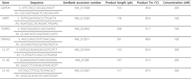
Table 1 Sequences of the primer pairs (S: sense; AS: antisense) for housekeeping genes (GAPDH, HPRT, PLRPO and YWHAZ) and genes of interest (Cx 37, Cx 40 and Cx 43) are shown as well as GenBank accession number, product length, product Tm and concentrations
Gene Sequence GenBank accession number Product length (pb) Product Tm (°C) Concentration (nM)
GAPDH S: ATTCTACCCACGGCAAGTT NM_017008 153 89.4 200 AS: CGCCAGTAGACTCCACGACATA HPRT S: TGTTGGATATGCCCTTGACTA NM_012583 178 85.6 100 AS: AGATGGCCACAGGACTAGAAC PLRPO S: AGGTGGGAGCCAGCGAAGC NM_022402 208 91.7 100 AS: GCAACAGTCGGGTAGCCAATC YWHAZ S: AGCCGAGCTGTCTAACGAG NM_013011 291 88.4 100 AS: GCCAAGTAGCGGTAGTAGTCA Cx 37 S: GGTGGCAGAGGACGGTCGTCT NM_021654 133 85.3 200 AS: CCATGGTCCAGCCGTAGAGA Cx 40 S: GGAAAGAGGTGAACGGGAAG NM_01280 197 91.3 200 AS: GGGCCTCGAGACATAACAGTT Cx 43 S:TCTGCCTTTCGCTGTAACACT NM_012567 117 87.5 200 AS: GGGCACAGACACGAATATGAT
Immunofluorescence
Immunolabelling of IPA3 isolated from N, MCT and CH rats was performed as previously described [18]. IPA3 were fixed and cut in 10μm sections with a cryo-stat. Sections were first incubated in blocking buffer containing 0.1% Triton-X100 (Sigma, St Quentin Falla-vier, France) and 1% bovine serum albumin (BSA) in PBS for 1 h at room temperature and then in the pri-mary antibodies (same antibodies as the ones used for Western Blot) overnight at 4°C. Sections were then incubated with the appropriate secondary antibodies coupled to Alexa 546 (Molecular Probes, Cergy Pon-toise, France). Nuclei were labeled with 45μM Hoechst 33342 (Molecular Probes, Cergy Pontoise, France). Sec-tions were then observed with a laser scanning confocal microscope TE2000 (Nikon, Champigny-Sur-Marne, France) with a × 60, 1.40 NA plan apochromat oil-immersion objective. Excitation was obtained with a diode laser at 408 nm to observe nuclei, an argon laser at 488 nm to observe autofluorescence of the internal and external elastic lamina and a helium-neon laser at 543 nm to observe Cx labelling. The emitted light was filtered as appropriate: 450 ± 35 nm for nuclei (blue), 515 ± 30 nm for internal elastic lamina (green), and 605 ± 75 nm for Cx labelling (red).
Isometric tension measurements
IPA1 were divided into short tubular segments with an external diameter of 1.5-2 mm and used for isometric contraction measurement as reported previously [23]. Arterial rings were mounted in isolated organ bath sys-tems, containing Krebs solution at 37°C and bubbled continuously with either 15% O2, 5% CO2 for IPA1
iso-lated from N and MCT rats, or with 9% O2, 5% CO2for
IPA1 isolated from CH rats. As previously determined [22], an initial load of 0.8 g was applied to arterial rings isolated from control rats, and 1.6 g to arterial rings iso-lated from CH and MCT rats. Tissues were allowed to equilibrate for 1 h in Krebs solution and washed out every 15 min. A high KCl solution (80 mM) was applied in order to obtain a reference contraction used to nor-malize subsequent contractile responses. 80 mM high KCl solution induced similar contractions in pulmonary arterial rings from the three rat models (1036 ± 50 mg, n = 67; 1205 ± 143 mg, n = 64; 953 ± 54 mg, n = 68 in N, CH and MCT rats respectively; P > 0.05). Contractile responses to different agonists were then tested by con-structing a cumulative concentration-response curve (CCRC) to 5-HT (10 nM to 100 μM), ET-1 (0.1 to 100 nM), Phe (0.1 nM to 10 μM) or high KCl solutions (4.7 to 100 mM). We inhibited gap junction communi-cations through Cx 37 and Cx 43 or Cx 40 with 300 μM synthetic connexin-mimetic peptides:37-43Gap 27 (SRPTEKTIFII; Peptide 2.0 Inc, Shirley, USA),40Gap
27 (SRPTEKNVFIV; Genscript, Piscataway, USA) respectively [18,19]. When indicated, peptides were pre-incubated during 1 h, and then CRC to agonist was per-formed in the presence of the peptide. Specificity of these peptides was checked in a previous study [18]. High potassium solutions were obtained by substituting an equimolar amount of KCl for NaCl from Kreb’s solu-tion. To determine the role of the gap junctions in con-ditions close to physiological concon-ditions, we studied the vessels with intact endothelium. Endothelial function was tested on each ring by examining the relaxation induced by 10 μM carbamylcholine on 0.3 μM Phe-induced preconstricted pulmonary arterial rings. Passive and active mechanical properties were assessed using transducer systems, coupled to IOX software (EMKA technologies, Paris, France) in order to facilitate data acquisition and analysis [23].
Data analysis and statistics
Results are expressed as mean ± SEM; n indicates the number of rats for quantitative RT-PCR, Fulton ratio and mean PAP whereas it indicates the number of experiments from 4 pools of rats (4 rats per pool) for Western Blot. For isometric tension measurements, n indicates the number of pulmonary arterial rings. CCRC to agonists were fitted using Origin 6 software (Originlab, Paris, France) to the logistic equation: T = ((T0-Tmax)/(1+(X/EC50)p)) + Tmax
where T, Tmax and T0 are, respectively, the amplitude of tension developed, the relative maximum and minimal tensions for a given agonist concentration normalized to the 80 mM KCl responses, X is the concentration of ago-nist used, EC50is the concentration of agonist which
pro-duces half maximal tension, and p is the slope of the curve. Global comparisons of the CCRC were performed using ANOVA. When global comparisons of the curves were significantly different, unpaired Student’s t tests were performed on maximal effect (Emax) and/or EC50. EC50
was determined only when the maximal contraction was achieved. Quantitative RT-PCR results were analyzed with the GeNorm method [18]. Statistical analyses were per-formed on all other data using a non parametric test for unpaired samples (Mann-Whitney test). A value of P < 0.05 was considered significant.
Results
Assessment of pulmonary hypertension in two rat models
The mean PAP value was significantly increased from 15 ± 2 mmHg in N rats (n = 5) to 30.4 ± 1.03 mmHg in CH rats (n = 5) or 29.8 ± 1.3 mmHg in MCT rats (n = 5). The Fulton’s index was also significantly increased in each pulmonary hypertensive rat model indicating a right ventricle hypertrophy in CH and MCT rats com-pared to N rats (0.63 ± 0.04, n = 20; 0.54 ± 0.03, n = 20 and 0.27 ± 0.03, n = 20 respectively).
To differentiate MCT from CH rat models, we examined the endothelial NO-dependent function by inducing a relaxation with 10μM carbamylcholine on 0.3 μM Phe-induced preconstricted pulmonary arterial rings. Relaxation to carbamylcholine 10μM was not significantly changed in CH compared to N rats whereas it was almost abolished in MCT rats (58.1 ± 3.0% and 3.3 ± 0.9% of the precontrac-tion to 0.3μM Phe in CH and MCT rats respectively, n = 48 and 44 vs 64.1 ± 2.9% in N rats, n = 48).
Cx 37, 40 and 43 are expressed in the pulmonary arterial wall from N, CH and MCT rats
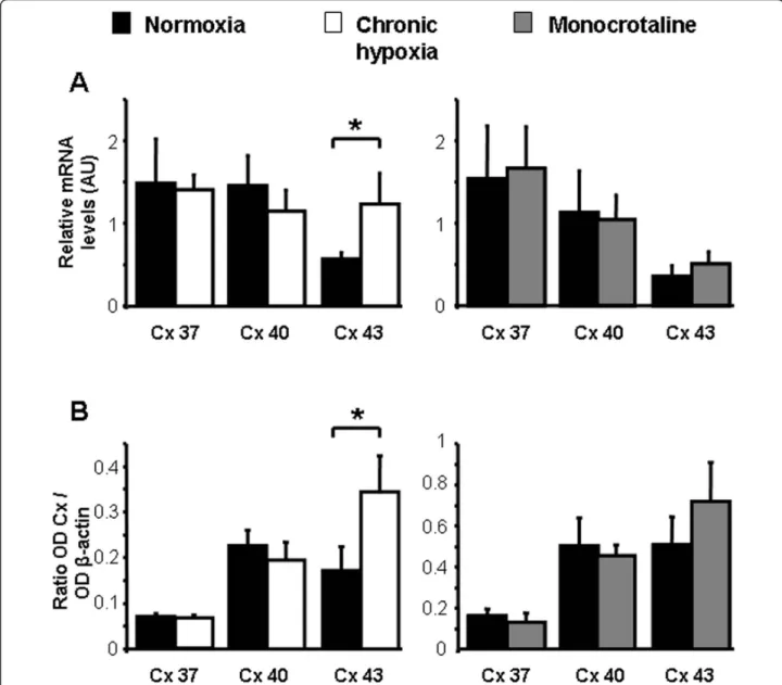
Quantitative RT-PCR and Western Blot experiments have demonstrated the presence of the mRNA and proteins, respectively for Cx 37, 40 and 43 in IPA3 from N, CH and MCT rats (Figure 1A and 1B respectively). Interestingly, a significant increase in both mRNA and protein levels was observed for the Cx 43 in IPA3 from CH rats compared to IPA3 from N rats (Figure 1A and 1B, left panels).
Immunofluorescent labelling studies confirmed the presence of the Cx 37, 40 and 43 proteins (Figure 2). The autofluorescence of the external and internal elastic lamina in green allowed us to delimit the smooth mus-cle layers from the endothelial cells and the adventitia as previously observed [18]. The three connexins were mainly present in the endothelial and possibly in the myoendothelial junctions (Figure 2). Some punctuate labelling, characteristic of the Cx staining, was also spar-sely present for Cx 37 and 40 in the smooth muscle of IPA3 from N and MCT rats (Figure 2). Since identical antibodies were used for Western Blot and immuno-fluorescent labelling experiments, the typical punctuate labelling observed demonstrate the specificity of these Cx antibodies and strengthen our Western Blot results.
Comparison of the pulmonary arterial reactivity to various stimuli in N, CH and MCT rats
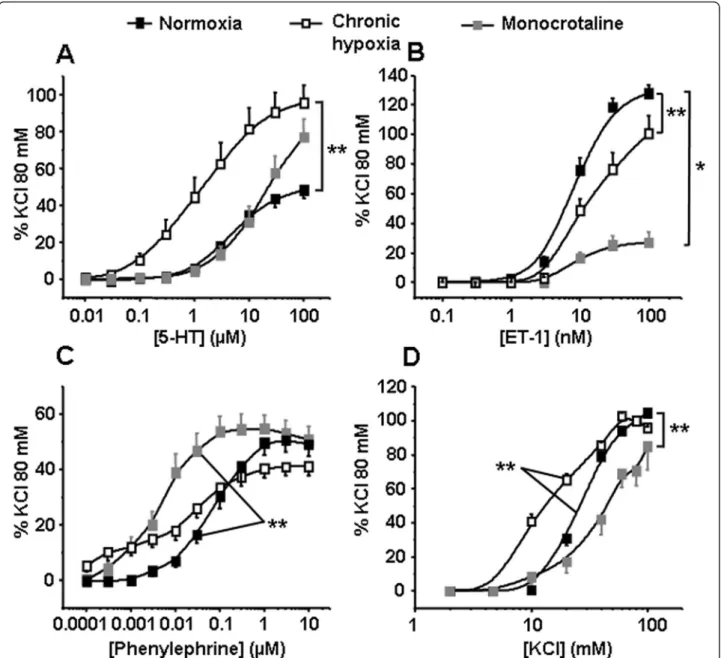
We then compared contractile responses to receptor-dependent or -inreceptor-dependent stimuli such as (1) 5-HT, (2) ET-1, (3) Phe, an agonist of thea1-adrenoceptor and (4) a depolarising agent such as high potassium solutions in IPA from N, CH and MCT rats. The contractile responses to 5-HT and high potassium solutions were significantly increased in CH rats (Figure 3A and 3D) whereas those to ET-1 were significantly decreased in both pulmonary hypertensive rat models (CH and MCT rats, Figure 3B). Finally, contractile responses to Phe were significantly increased in MCT rats (Figure 3C).
Role of Cx 37, 40 and 43 in the reactivity to various stimuli in intrapulmonary arteries from N, CH and MCT rats
Since, (1) Cx 37, 40 and 43 are all expressed (Figure 1 and 2) and (2) 5-HT, ET-1, Phe and depolarisation
induce a mechanical activity (Figure 3) in intrapulmon-ary arteries from CH and MCT rats, we systematically addressed the role of Cx 37, 40 and 43 in the isometric contraction to 5-HT, ET-1, Phe and high potassium solutions by using specific blockers of Cx 40 or Cx 37 and 43 (namely 40Gap27 and 37-43Gap27 respectively, two Cx-mimetic peptides) in IPA from N, CH and MCT rats.
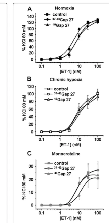
Contractile responses to 5-HT were significantly decreased by 300 μM 37-43Gap27 only in IPA from N rats (Figure 4A). In IPA from either rat model, the con-tractile responses to ET-1 were unchanged following one hour incubation with 300 μM 37-43Gap27 or
40
Gap27 (Figure 5). Interestingly, and inversely to 5-HT, contractile responses to Phe were decreased following 300μM37-43Gap27 or40Gap27 treatment in IPA from CH and MCT rats (Figure 6 B, C and 6D, E respec-tively). In N rats, contractile responses to Phe were unchanged by37-43Gap27 or40Gap27 incubation (Figure 6A). The effects of the37-43Gap27 and40Gap27 on con-tractile changes from N to CH rats and from N to MCT rats are demonstrated in figure 6C and 6E respectively.
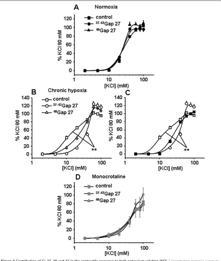
When using a receptor-independent depolarising agent (high potassium solutions), contractile responses were significantly decreased in the presence of 300μM
37-43
Gap27 only in IPA from CH rats (Figure 7B). EC50
was significantly increased in the presence of37-43Gap27 in IPA from CH rats (40.2 ± 0.5 mM vs 15 ± 1.1 mM in the absence of37-43Gap27) indicating a decrease in the sensitivity to high potassium solutions. 300μM40Gap27 had no effect, under identical conditions, in any of the hypertensive rat models used (Figure 7B and 7D). In the presence of the 37-43Gap27, the contraction to KCl in CH rats was identical to the contraction to KCl in N rats (Figure 7C).
Discussion
Underlying cellular mechanisms as well as targets, gov-erning the pathogenesis of PH, still remain to be fully elucidated. This may be due, at least in part, to the fact that the pathogenesis of PH differs depending on the type of aetiology of this vascular disorder. For this rea-son, we conducted the present study in two different PH models: MCT induced PH (non hypoxic model) and CH-induced PH (hypoxic model).
Two major excitation-contraction coupling mechan-isms are involved in regulating pulmonary vascular tone: electromechanical and pharmacomechanical coupling. We thus adressed agonist-mediated pulmonary contrac-tion (pharmacomechanical coupling induced by either 5-HT, ET-1 or Phe) and depolarisation-mediated con-traction (electromechanical coupling induced by high potassium solution). In the present study, IPA from CH rats exhibited hyperreactivity to 5-HT (Figure 3A),
which is consistent with previous studies [22,24,25]. Regarding ET-1, vasoconstriction to this agent was only slightly decreased in hypoxic PH whereas it was drama-tically decreased in MCT-induced PH. In CH rats, pul-monary arterial contractile responses to ET-1 have been shown to be increased in resistant IPA but decreased in extra pulmonary arteries [16]. Consequently, the slight decrease in the contraction to ET-1 observed in CH rats may be explained by the intermediate size of the pul-monary artery (PA) used for the present contractile experiments (namely PA of the first order or IPA1). In MCT rats, a recent study has shown that although the
contraction to ET-1 was not altered, ET-B receptors were decreased in resistance PA [26]. Since the contrac-tion to ET-1 is linked to ET-A receptors in extra-PA and to ET-B receptors in IPA [26,27], the present decreased ET-1 contraction may result from the reported decrease in ET-B receptors in MCT rats. Regarding Phe and in contrast to ET-1, we found that Phe-induced contraction was increased in PA from MCT rats. A recent report from Mam et al. (2010) showed a reduced contraction to Phe in CH and MCT rats [28]. This difference may be explained by different animal strain (Sprague Dawley vs Wistar rats) and/or Figure 1 Expression of Cx 37, 40 and 43. A shows mean values ± S.E.M. of relative mRNA expression levels measured by quantitative RT-PCR in pulmonary arteries from N rats (normoxia, black column, n = 4 - 5), chronic hypoxic rats (chronic hypoxia, white column, n = 9) and monocrotaline-injected rats (monocrotaline, grey column, n = 5 - 7). B shows protein expression measured by Western Blot and normalized tob actin expression level in normoxic rats (black column, n = 6 - 12), chronic hypoxic rats (white column, n = 9 - 12) and monocrotaline-injected rats (grey column, n = 5 - 7). * means a significant difference for P < 0.05.
Figure 2 Localization of Cx 37, 40 and 43 in the pulmonary arterial wall. Immunofluorescent labelling experiments shows the presence of Cx (red) 37 (top row), 40 (middle row) and 43 (bottom row) in transverse sections of pulmonary arteries from normoxic (normoxia, left column), chronic hypoxic (chronic hypoxia, middle column) and monocrotaline-injected rats (monocrotaline, right column). Green shows the
autofluorescent signal of the vessels and blue shows the nuclei. SMC means smooth muscle cells and EC means endothelial cells. 7-8 normoxic rats have been used and the number of IPA3 sections, used for the labelling of Cx 37, 40 and 43, was 21, 22 and 14 respectively. 4-5
monocrotaline-injected rats have been used and the number of IPA3 sections, used for the labelling of Cx 37, 40 and 43, was 11, 17 and 14 respectively. Finally, we have used 3 chronic hypoxic rats and the number of IPA3 sections used for the labelling of the Cx 37, 40 and 43, was 4, 5 and 7 respectively. Scale bars are 15μm.
different vascular preparation (extra-PA vs IPA) and/or different experimental conditions (normobaric vs hypo-baric hypoxia). Finally, regarding the receptor indepen-dent depolarizing agent KCl, pulmonary vascular reactivity to membrane depolarization was increased in CH rats, whereas it was reduced in MCT rats. These results are consistent with previous findings from our laboratory indicating that PA smooth muscle cells (PASMC) from CH rats exhibit a higher resting mem-brane potential and a higher basal intracellular calcium concentration [22,29]. Moreover, the decreased expression
of potassium channels previously observed in CH rats [17] could also contribute to increase the sensitivity to depolarisation.
We have previously shown that Cx 37, 40 and 43, three gap junction proteins commonly found in the vas-culature, are expressed in rat PA endothelial cells, while Cx 37 and 40 only are found in PASMC [18]. In the current study, Cx 37, 40 and 43 were also expressed in pulmonary arterial wall from both hypoxic and non-hypoxic PH rats. However, PASMC from CH rats no longer expressed Cx 37 and 40 isoforms, whereas they Figure 3 Contraction induced by 5-HT, ET-1, Phe and high potassium solutions (KCl). Isometric tension measurements were recorded on intrapulmonary arteries in response to cumulative concentrations of 5-HT (A, n = 19 - 31), ET-1 (B, n = 17 - 26), Phe (C, n = 21 - 29) and KCl (D, n = 10 - 20) in normoxic, chronic hypoxic and monocrotaline-injected rats (black, white and grey squares respectively). Data are means ± SEM and are expressed as a percentage of the high potassium solution (80 mM)-induced response. ** means a significant difference for P < 0.01.
Figure 4 Contribution of Cx 37, 40 and 43 in the contractile response to 5-HT. Concentration-response curves to 5-HT were performed in the absence (control, squares) or in the presence of connexin-mimetic peptides targeted against Cx 37 and 43 ( 37-43Gap27, circles) and 40 (40Gap27, triangles). Each set of experiments
was performed on intrapulmonary arteries from normoxic (normoxia, panel A, black symbols, n = 12 - 24), chronic hypoxic (chronic hypoxia, panel B, white symbols, n = 8 - 19) and monocrotaline-injected rats (monocrotaline, panel C, grey symbols, n = 9 - 20). Data are means ± SEM and are expressed as a percentage of the high potassium solution (80 mM)-induced response. * means a significant difference for P < 0.05.
Figure 5 Contribution of Cx 37, 40 and 43 in the contractile response to ET-1. Concentration-response curves to ET-1 were performed in the absence (control, squares) or in the presence of connexin-mimetic peptides targeted against Cx 37 and 43 (
37-43
Gap27, circles) and 40 (40Gap27, triangles). Each set of experiments was performed on intrapulmonary arteries from normoxic
(normoxia, panel A, black symbols, n = 10 - 26), chronic hypoxic (chronic hypoxia, panel B, white symbols, n = 8 - 17) and monocrotaline-injected rats (monocrotaline, panel C, grey symbols, n = 8 - 20). Data are means ± SEM and are expressed as a percentage of the high potassium solution (80 mM)-induced response.
Figure 6 Contribution of Cx 37, 40 and 43 in the contractile response to Phe. Concentration-response curves to Phe were performed in the absence (control, squares) or in the presence of connexin-mimetic peptides targeted against Cx 37 and 43 (37-43Gap27, circles) and 40 (40Gap27, triangles). Each set of experiments was performed on intrapulmonary arteries from normoxic (normoxia, panel A, black symbols, n = 11 - 21), chronic hypoxic (chronic hypoxia, panel B, white symbols, n = 14 - 23) and monocrotaline-injected rats (monocrotaline, panel D, grey symbols, n = 7 - 13). Panels C and E summarize the effects of the Gap27 on the contractile changes to Phe from N to CH or from normoxic to MCT rat models respectively. Data are means ± SEM and are expressed as a percentage of the high potassium solution (80 mM)-induced response. * means a significant difference for P < 0.05.
Figure 7 Contribution of Cx 37, 40 and 43 in the contractile response to high potassium solution (KCl). Concentration-response curves to KCl were performed in the absence (control, squares) or in the presence of connexin-mimetic peptides targeted against Cx 37 and 43 (
37-43
Gap27, circles) and 40 (40Gap27, triangles). Each set of experiments was performed on intrapulmonary arteries from normoxic (normoxia, panel A, black symbols, n = 8 - 20), chronic hypoxic (chronic hypoxia, panel B, white symbols, n = 8 - 20) and monocrotaline-injected rats
(monocrotaline, panel D, grey symbols, n = 4 - 12). Panels C summarizes the effect of the37-43Gap27 on the contractile changes to KCl from
normoxic to CH rat model. Data are means ± SEM and are expressed as a percentage of the high potassium solution (80 mM)-induced response. ** means a significant difference for P < 0.01.
were sparsely expressed in MCT rats (Figure 2). Inter-estingly, Cx 43 expression was upregulated in CH rats but not in MCT rats (Figure 1). We cannot exclude that Cx 43 is expressed at the myoendothelial junctions and possibly on the smooth muscle side of these heterocellu-lar structures. Cowan et al. have indeed demonstrated that hypoxia (2.2% O2 during 6 h) increased Cx 43
expression in cultured smooth muscle cells from rat thoracic aorta [5]. Consequently, we can hypothesize that the increase in Cx 43 in the IPA from CH rat could be due to the effect of hypoxia on Cx 43 localized on the smooth muscle side of the myoendothelial gap junc-tions. Interestingly, we have previously demonstrated that Cx 43 present between PASMC and endothelial cells plays an important role in the vasoreactivity to 5-HT in IPA from N rats [18].
Since (i) the three Cx 37, 40 and 43 are expressed in CH and MCT rats (Figure 1 and 2) and (ii) Cx 37 and 43 are involved in the contractile and calcium responses to 5-HT in IPA from N rats [18], we addressed the role of these Cx in the contractile response to various ago-nists known to contribute to PH (5-HT, ET-1 and Phe) in IPA from CH and MCT rats. In the present study, the effect of Gap27 varied according to the agonists used and the rat model considered. Since we have pre-viously demonstrated that the role of the gap junctions in the contraction depended on the amount of superox-ide anion (O2●) level in smooth muscle, it can be
hypothesized that the modulation of O2●level in IPA
from hypertensive rat models may differ according to the agonist. In this respect, we can suggest that, unlike 5-HT, Phe increases O2●levels in pulmonary
hyperten-sive models and not in N rats although this hypothesis would require further experimental investigation.
Moreover, 5-HT, ET-1 and Phe are pulmonary arterial vasoconstrictors with different modes of actions. Smooth muscle contraction is well known to be both dependent and/or independent on cytosolic calcium increase depend-ing on the vasoconstrictor. When smooth muscle contrac-tion is calcium independant, the contraccontrac-tion is due to calcium sensitization of the contractile proteins. On the one hand, ET-1 and 5-HT increase intracellular calcium by acting on receptors localised in both smooth muscle and endothelial cells (namely 5-HT2A, 5-HT1B/D, ETAand
ETBin PASMC and 5-HT2Band ETBin endothelial cells)
whereas Phe acts ona1-adrenoceptors on PASMC only
[14,21,30,31]. On the other hand, although 5-HT, ET-1 and Phe activate inositol 1,4,5-triphosphate-induced intra-cellular calcium release, 5-HT and Phe also stimulate vol-tage-independent calcium permeable channels (namely receptor- and/or store-operated channels) whereas ET-1 rather stimulates voltage-dependent calcium channels [32-34]. Finally, contraction to ET-1 and Phe are strongly
dependent on calcium sensitization of contractile proteins whereas contraction to 5-HT is only slightly dependent on this process [21,35-37]. Altogether, such differences in the contractile mechanisms might explain why the effects of the gap junction blockers differ according to the agonist used.
Regarding receptor-independent depolarisation, we have observed a significant decrease in the contraction to KCl in IPA from MCT rats but an increase in CH rats (Figure 3D). Moreover,37-43Gap27 decreased the sensitiv-ity to high potassium solutions in IPA from CH rats only, thus inducing a contraction similar to the one observed in the N rats (Figure 7C). These results suggest that Cx 37 and/or 43 were involved in the hypersensitivity to KCl in CH rats. This result is thus in good agreement with the expected role of Cx in the conduction of the electrical activity observed in various systemic vessels [38].
Conclusions
In conclusion, we have evidenced the presence of func-tional Cx 37, 40 and 43 not only in normal but also in pathological pulmonary vessels. These three Cx partici-pate to pulmonary arterial vascular contractility in response to both receptor-dependent and receptor-inde-pendent stimuli in N, CH and MCT rats. However, the role of these Cx in the vasoreactivity of pulmonary artery varies according to the stimulus and the rat model. These results suggest that Cx may act as specific therapeutic targets for different type of PH.
List of used abbreviations
CH: chronic hypoxia; COPD: chronic obstructive pulmonary disorder; CRC: concentration-response curve; Cx: connexins; ET-1: endothelin-1; 5-HT: serotonin; IPA: intrapulmonary arteries; IPA1: intrapulmonary artery of the first order; IPA3: intrapulmonary artery of the third order; LV+S: left ventricle plus septum; MCT: monocrotaline-treated; mean PAP: mean pulmonary arterial pressures; N: normoxic; O2●: superoxide anion; PA: pulmonary artery; PAH:
pulmonary arterial hypertension; PASMC: pulmonary arterial smooth muscle cells; PH: pulmonary hypertension; Phe: phenylephrine; RV: right ventricle.
Acknowledgements and funding
We thank Huguette Crevel for her technical assistance in performing isometric contraction experiments.
This work was funded by the Fondation de France (2008002719), the Agence Nationale de la Recherche (ANR06-Physio-015-01) and the Société de Pneumologie de Langue Française (SPLF) - Boerhinger du Fonds de Dotation Recherche en Santé Respiratoire.
The authors declare that they have no competing interests.
Author details
1
INSERM, U1045, 146 rue Léo Saignat, F-33076 Bordeaux, France.2Université Bordeaux Segalen, Centre de Recherche Cardio-Thoracique de Bordeaux, F-33076 Bordeaux, France.3CHU de Bordeaux, F 33076 Bordeaux, France.
Authors’ contributions
MB, DD, RM, JPS and CG contributed to the conception and design of the study. MB, DD and CG performed experiments, evaluated results and interpreted data. MB, DD, RM, JPS and CG were involved in drafting and revising the manuscript. All authors read and approved the final manuscript.
Received: 19 October 2010 Accepted: 17 March 2011 Published: 17 March 2011
References
1. Goodenough DA, Paul DL: Gap junctions. Cold Spring Harbor Perspect Biol 2009, 1:a002576.
2. Cottrell GT, Burt JM: Functional consequences of heterogeneous gap junction channel formation and its influence in health and disease. Biochim Biophys Acta 2005, 1711:126-141.
3. Brisset AC, Isakson BE, Kwak BR: Connexins in vascular physiology and pathology. Antioxid Redox Signal 2009, 11:267-282.
4. Rummery NM, Hill CE: Vascular gap junctions and implications for hypertension. Clin Exp Pharmacol Physiol 2004, 31:659-667. 5. Cowan DB, Jones M, Garcia LM, Noria S, del Nido PJ, McGowan FX Jr:
Hypoxia and stretch regulate intercellular communication in vascular smooth muscle cells through reactive oxygen species formation. Arterioscler Thromb Vasc Biol 2003, 23:1754-1760.
6. Humbert M, Sitbon O, Simonneau G: Treatment of pulmonary arterial hypertension. N Engl J Med 2004, 351:1425-1436.
7. Simonneau G, Robbins IM, Beghetti M, Channick RN, Delcroix M, Denton CP, Elliott CG, Gaine SP, Gladwin MT, Jing ZC, Krowka MJ, Langleben D, Nakanishi N, Souza R: Updated clinical classification of pulmonary hypertension. J Am Coll Cardiol 2009, 54:S43-54. 8. Humbert M: Update in pulmonary hypertension 2008. Am J Respir Crit
Care Med 2009, 179:650-656.
9. Giaid A, Yanagisawa M, Langleben D, Michel RP, Levy R, Shennib H, Kimura S, Masaki T, Duguid WP, Stewart DJ: Expression of endothelin-1 in the lungs of patients with pulmonary hypertension. N Engl J Med 1993, 328:1732-1739. 10. Herve P, Launay JM, Scrobohaci ML, Brenot F, Simonneau G, Petitpretz P, Poubeau P, Cerrina J, Duroux P, Drouet L: Increased plasma serotonin in primary pulmonary hypertension. Am J Med 1995, 99:249-254. 11. Hosoda Y: Pathology of pulmonary hypertension: a human and
experimental study. Pathol Int 1994, 44:241-267.
12. Kanai Y, Hori S, Tanaka T, Yasuoka M, Watanabe K, Aikawa N, Hosoda Y: Role of 5-hydroxytryptamine in the progression of monocrotaline induced pulmonary hypertension in rats. Cardiovasc Res 1993, 27:1619-1623.
13. Miyauchi T, Yorikane R, Sakai S, Sakurai T, Okada M, Nishikibe M, Yano M, Yamaguchi I, Sugishita Y, Goto K: Contribution of endogenous endothelin-1 to the progression of cardiopulmonary alterations in rats with monocrotaline-induced pulmonary hypertension. Circ Res 1993, 73:887-897.
14. Salvi SS: Alpha1-adrenergic hypothesis for pulmonary hypertension. Chest 1999, 115:1708-1719.
15. Humbert M, Morrell NW, Archer SL, Stenmark KR, MacLean MR, Lang IM, Christman BW, Weir EK, Eickelberg O, Voelkel NF, Rabinovitch M: Cellular and molecular pathobiology of pulmonary arterial hypertension. J Am Coll Cardiol 2004, 43:13S-24S.
16. Shimoda LA, Sham JS, Sylvester JT: Altered pulmonary vasoreactivity in the chronically hypoxic lung. Physiol Res 2000, 49:549-560.
17. Guibert C, Marthan R, Savineau JP: Modulation of ion channels in pulmonary arterial hypertension. Curr Pharm Des 2007, 13:2443-2455. 18. Billaud M, Marthan R, Savineau JP, Guibert C: Vascular smooth muscle modulates endothelial control of vasoreactivity via reactive oxygen species production through myoendothelial communications. PLoS One 2009, 4:e6432.
19. Chaytor AT, Evans WH, Griffith TM: Peptides homologous to extracellular loop motifs of connexin 43 reversibly abolish rhythmic contractile activity in rabbit arteries. J Physiol 1997, 503(Pt 1):99-110. 20. Tual-Chalot S, Guibert C, Muller B, Savineau JP, Andriantsitohaina R,
Martinez MC: Circulating Microparticles from Pulmonary Hypertensive Rats Induce Endothelial Dysfunction. Am J Respir Crit Care Med 2010, 182:261-268. 21. Rodat-Despoix L, Crevel H, Marthan R, Savineau JP, Guibert C:
Heterogeneity in 5-HT-induced contractile and proliferative responses in rat pulmonary arterial bed. J Vasc Res 2008, 45:181-192.
22. Rodat L, Savineau JP, Marthan R, Guibert C: Effect of chronic hypoxia on voltage-independent calcium influx activated by 5-HT in rat intrapulmonary arteries. Pflugers Arch 2007, 454:41-51.
23. Guibert C, Savineau JP, Crevel H, Marthan R, Rousseau E: Effect of short-term organoid culture on the pharmaco-mechanical properties of rat extra- and intrapulmonary arteries. Br J Pharmacol 2005, 146:692-701.
24. Keegan A, Morecroft I, Smillie D, Hicks MN, MacLean MR: Contribution of the 5-HT(1B) receptor to hypoxia-induced pulmonary hypertension: converging evidence using HT(1B)-receptor knockout mice and the 5-HT(1B/1D)-receptor antagonist GR127935. Circ Res 2001, 89:1231-1239. 25. MacLean MR, Sweeney G, Baird M, McCulloch KM, Houslay M, Morecroft I:
5-Hydroxytryptamine receptors mediating vasoconstriction in pulmonary arteries from control and pulmonary hypertensive rats. Br J Pharmacol 1996, 119:917-930.
26. Sauvageau S, Thorin E, Villeneuve L, Dupuis J: Change in pharmacological effect of endothelin receptor antagonists in rats with pulmonary hypertension: role of ETB-receptor expression levels. Pulm Pharmacol Ther 2009, 22:311-317.
27. MacLean MR, McCulloch KM, Baird M: Endothelin ETA- and ETB-receptor-mediated vasoconstriction in rat pulmonary arteries and arterioles. J Cardiovasc Pharmacol 1994, 23:838-845.
28. Mam V, Tanbe AF, Vitali SH, Arons E, Christou HA, Khalil RA: Impaired vasoconstriction and nitric oxide-mediated relaxation in pulmonary arteries of hypoxia- and monocrotaline-induced pulmonary hypertensive rats. J Pharmacol Exp Ther 2010, 332:455-462.
29. Bonnet S, Dubuis E, Vandier C, Martin S, Marthan R, Savineau JP: Reversal of chronic hypoxia-induced alterations in pulmonary artery smooth muscle electromechanical coupling upon air breathing. Cardiovasc Res 2002, 53:1019-1028.
30. Hyvelin JM, Guibert C, Marthan R, Savineau JP: Cellular mechanisms and role of endothelin-1-induced calcium oscillations in pulmonary arterial myocytes. Am J Physiol 1998, 275:L269-282.
31. Ivy D, McMurtry IF, Yanagisawa M, Gariepy CE, Le Cras TD, Gebb SA, Morris KG, Wiseman RC, Abman SH: Endothelin B receptor deficiency potentiates ET-1 and hypoxic pulmonary vasoconstriction. Am J Physiol Lung Cell Mol Physiol 2001, 280:L1040-1048.
32. Guibert C, Marthan R, Savineau JP: 5-HT induces an arachidonic acid-sensitive calcium influx in rat small intrapulmonary artery. Am J Physiol Lung Cell Mol Physiol 2004, 286:L1228-1236.
33. McDaniel SS, Platoshyn O, Wang J, Yu Y, Sweeney M, Krick S, Rubin LJ, Yuan JX: Capacitative Ca(2+) entry in agonist-induced pulmonary vasoconstriction. Am J Physiol Lung Cell Mol Physiol 2001, 280:L870-880. 34. Shimoda LA, Sylvester JT, Sham JS: Mobilization of intracellular Ca(2+) by
endothelin-1 in rat intrapulmonary arterial smooth muscle cells. Am J Physiol Lung Cell Mol Physiol 2000, 278:L157-164.
35. Barman SA: Vasoconstrictor effect of endothelin-1 on hypertensive pulmonary arterial smooth muscle involves Rho-kinase and protein kinase C. Am J Physiol Lung Cell Mol Physiol 2007, 293:L472-479. 36. Rodat-Despoix L, Aires V, Ducret T, Marthan R, Savineau JP, Rousseau E,
Guibert C: Signalling pathways involved in the contractile response to 5-HT in human pulmonary artery. Eur Respir J 2009, 34:1338-1347. 37. Sauzeau V, Le Jeune H, Cario-Toumaniantz C, Smolenski A, Lohmann SM,
Bertoglio J, Chardin P, Pacaud P, Loirand G: Cyclic GMP-dependent protein kinase signaling pathway inhibits RhoA-induced Ca2+ sensitization of contraction in vascular smooth muscle. J Biol Chem 2000, 275:21722-21729. 38. de Wit C, Griffith TM: Connexins and gap junctions in the EDHF
phenomenon and conducted vasomotor responses. Pflugers Arch 2010, 459:897-914.
doi:10.1186/1465-9921-12-30
Cite this article as: Billaud et al.: Role of the gap junctions in the contractile response to agonists in pulmonary artery from two rat models of pulmonary hypertension. Respiratory Research 2011 12:30.