HAL Id: hal-01337125
https://hal.archives-ouvertes.fr/hal-01337125
Submitted on 24 Jun 2016
HAL is a multi-disciplinary open access
archive for the deposit and dissemination of
sci-entific research documents, whether they are
pub-lished or not. The documents may come from
teaching and research institutions in France or
abroad, or from public or private research centers.
L’archive ouverte pluridisciplinaire HAL, est
destinée au dépôt et à la diffusion de documents
scientifiques de niveau recherche, publiés ou non,
émanant des établissements d’enseignement et de
recherche français ou étrangers, des laboratoires
publics ou privés.
SpinalCOM: Ambulatory monitoring of the Spinal Cord
Olivier Tsiakaka, Mehdi Terosiet, Olivier Romain, H Benali, V Marchand, P.
F. Pradat, Sylvain Feruglio
To cite this version:
Olivier Tsiakaka, Mehdi Terosiet, Olivier Romain, H Benali, V Marchand, et al.. SpinalCOM:
Am-bulatory monitoring of the Spinal Cord. Colloque du GDR SoC-SiP, Jun 2016, Nantes, France.
�hal-01337125�
SpinalCOM: Ambulatory monitoring of the Spinal Cord
O. Tsiakaka
1, M. Terosiet
2, O. Romain
2, H. Benali
3, V. Marchand
3, P.F. Pradat
3,4, S. Feruglio
11
Sorbonne University, UPMC Univ Paris 06, CNRS UMR 7606, LIP6, F-75005, Paris, France 2
Cergy Pontoise University, ENSEA, CNRS UMR 8051, ETIS, F-95004, Cergy Pontoise, France 3
Sorbonne University, UPMC Univ Paris 06, CNRS UMR 7371/INSERM UMR S 1146, LIB, F-75005, Paris, France 4
CHU Paris-GH La Piti´e Salpˆetri`ere-Charles Foix - Hospital Piti´e-Salpˆetri`ere , Paris, France
Abstract
Little is known about the processes occurring after Spinal Cord (SC) damage. Whether permanent or recov-erable, those processes have not been precisely charac-terized. Because usual imaging techniques (MRI, Echo-Doppler, ...) provide only limited information on this or-gan, there is an unmet need for methods to assess the func-tional consequences of the Spinal Cord Injuries (SCI) and the effect of therapeutic interventions. To meet this need, the SpinalCOM project aims to investigate a new imag-ing approach of the SC, through the realization of a mul-timodal communicating device for measuring the activity of the SC. A first proof of concept was validated with cus-tom made prototypes through in vivo experiments on ani-mals. This approach will establish locally and specifically the functional state of the SC, which will provide a break-through in the care and therapeutic trials in SCI.
1. Introduction
Trauma of the Spinal Cord (TSC) and pathologies of the spine are major causes of mortality and disability. Accounting thousands of new cases per year in Europe, vertebro-medullar injuries are common surgical proce-dure. After the acute phase, secondary mechanisms, in-cluding vascular, impact the functional prognosis. Traditional noninvasive imagery methods, such as Mag-netic Resonance Imaging (MRI), do not provide func-tional information, which is crucial to judge the con-sequences of secondary injury mechanisms and the ef-fectiveness of spontaneous recovery mechanisms or after therapeutic interventions.
Although interesting, functional MRI (fMRI) of the SC is a method under development and faces significant limita-tions. The temporal resolution is modest and the spatial resolution is limited by many artifacts [12]. Moreover, it does not provide continuous monitoring information. Fi-nally, it imposes a total immobility of the patient. We are in remote conditions from everyday living conditions of the subject.
Also on the functional assessment plan,
electrophysiolog-ical techniques, such as EEG (ElectroEncephaloGram), SSEP (Somatosensory Evoked Potentials) or MEP (Mo-tor Evoked Potentials), are useful for intraoperative mon-itoring. However, they show very limited contribution to the monitoring of TSC. Indeed, these weakly inva-sive techniques can’t identify early metabolic dysfunction before neurological functions are permanently affected. Echo-Doppler is also an interesting non-invasive tech-nique. However, the obtained data are rather qualitative: only plethysmograph on a large zone is obtained. Thus, there is an unmet need for methods to assess the functional consequences of TSC and, as part of clinical trials, the effect of surgical strategies, pharmacologic, or appropriate rehabilitation [7, 10, 12].
2. Aim of the project
The SpinalCOM1 project aims to establish the proof
of concept of an innovative tool for the continuous am-bulatory collection of electrophysiological and metabolic parameters simultaneously at the SC level in the big an-imal. The device employs the Diffuse Optical Imaging (DOI) principle and will make use of a PhotoDetector (PD) and a minimum of 2 pulsed light sources at differ-ent wavelengths, for acquisition of blood oxygen levels in real-time. This first embedded system is coupled with a tailor-made instrumentation to acquire bio-potentials, due to the SC electrical activity (ESG - ElectroSpinoGram). After processing, the obtained data will be transmitted by radiofrequency outwardly to be operated. The evaluation of metabolic activity is based on the neuro-hemodynamic coupling, an approach validated by brain imaging optic which is currently applied in humans. The SC is also stud-ied by near infrared spectroscopy, typically, but with clas-sical intraoperative monitoring equipment, which does not allow chronic measurement [4, 9]. In addition, in current experimental works [5, 11], it is the small animal which has been used as a model (mouse, cat). The relevance of measuring nerve activity by implanted electrodes (ESG)
1This work was performed within the Labex SMART
(ANR-11-LABX- 65) supported by French state funds managed by the ANR within the Investissements d’Avenir programme under reference ANR-11-IDEX-0004- 02
is, in turn, supported by numerous experimental studies [1, 8, 13].
To our knowledge, there is no system or project providing a device for continuously measuring both metabolic and electrophysiological parameters at the SC level. More-over, the proposed project presents no equivalent measure-ment system for comparison/reference.
In fine, this project finds its clinical application in the field of TSC through the implementation of a multimodal de-vice at the vertebra during orthopedic surgery, performed in the usual care of TME. This medical device is likely to produce a breakthrough, especially for monitoring pa-tients during the surgical operation and also after and as part of the evaluation of new therapeutic approaches.
3. Scientific Results
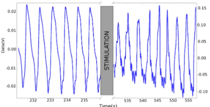
This project, located at the intersection of engineering and medicine, builds on the significant results achieved by the various partners’ projects, including a patent [6] and several articles in international journals and presenta-tions at scientific conferences [2, 3, 5, 14–16]. Previously, in 2010, an elementary wired probe was developed and tested on 6 cats for about 10 hours [5]. This study has provided proof of concept for the visualization of neural processes by DOI in small animal. For 2 years, starting in September 2013, the Labex SMART has supported this project. Various experiments on big animals (closer to the human being) have been performed on FBM pig model in collaboration with veterinaries (subcontractor). With a spectrophotometer, the in vivo optical characterization of the SC was performed (see Fig. 1) and proof of feasibility of this type of measure has been made (see Fig. 2) and published [14–16]. Hard points have been identified and are actually addressed through prototyping.
Figure 1. In vivo optical transmittance ver-sus wavelength of the pig’s SC before laminectomy (Case 1) and after (Case 2).
The measurements were performed with veterinaries of the XP-MED society, after obtaining permission from the local ethical committee of Cr2i (INRA, Jouy-en-Josas, France) under authorization number 03-1405.
Figure 2. PPG of the SC with IR LED before and after chemical stimulation.
4. Futur works
The SpinalCOM project concerns the development of a new tool for doctors, which purpose is mainly to propose a new functional approach to exploration of the SC after TSC. At long term, the aim of this translational work is to develop a miniaturized device that can be implemented during decompressive surgery, carried out under the usual TSC in human being. At short term, we will focus on ver-tebrate animals in order to propose a tool for researchers and veterinaries. Then, the potential transfer to human be-ing will be studied.
References
[1] E. T. Cunningham and P. E. Sawchenko. Journal of Com-parative Neurology, 417(4):448–466, 2000.
[2] S. Feruglio et al. Key Engineering Materials, 605:470– 473, 2014.
[3] S. Feruglio et al. IEEE Sensors Journal, 2015.
[4] M. Fok et al. In International Conference on Sensing Tech-nology, 2014.
[5] A. Goguin et al. Biomedical Circuits and Systems, IEEE Transactions on, 4(5):329–335, 2010.
[6] A. Goguin et al., May 19 2011. WO Patent App. PCT/EP2010/006,832.
[7] D. Lammertse et al. The Journal of Spinal Cord Medicine, 30(3):205–214, 2007.
[8] P. Lu, Y. Wang, L. Graham, K. McHale, M. Gao, D. Wu, J. Brock, A. Blesch, E. S. Rosenzweig, L. A. Havton, et al. Cell, 150(6):1264–1273, 2012.
[9] A. J. Macnab et al. Spine, 27(1):17–20, 2002.
[10] F. Miyanji et al. Radiology, 243(3):820–827, June 2007. [11] J. P. Phillips et al. Journal of biomedical optics,
18(3):037005–037005, 2013.
[12] P. W. Stroman et al. NeuroImage, 84:1070–1081, Jan. 2014.
[13] K.-D. Sthmeier et al. The Journal of the American Society of Anesthesiologists, 79(6):1170–1176, Dec. 1993. [14] O. Tsiakaka et al. In Engineering in Medecine and
Biol-ogy Conference, Proceedings of 37th IEEE EMBC Con-ference, page to appear, Milan, Italy, Aug. 2015.
[15] O. Tsiakaka et al. In 7th International IEEE EMBS Neural Engineering Conference, Montpellier, France, Apr. 2015. [16] O. Tsiakaka et al. In JETSAN, Compi`egne, France, May
2015.