Journal of Dairy Research (1978), 45, 259-265 2 5 9
Effect of incubation temperature on the development of lactic
acid bacteria and their phages
B Y TOMASO SOZZI, J.-MARC POULIN AND ROGER MARET
Department of Technological, Development, Nestle" Products Technical Assistance Co. Ltd, Lausanne, Switzerland
{Received 20 May 1977)
SUMMARY. Thirty-one strains of mesophilic and thermophilic lactic acid bacteria and their respective phages were tested for their minimum, optimum and maximum multiplication temperatures. Culture growth was strongly influenced by temperature during the first few hours of incubation, but less so after 24 h. Most of the phages showed the same pattern of development as their hosts, but one phage lysing a thermophilic lactobacillus and 3 phages lysing mesophilic streptococci proved temperature-sensitive, having a lower maximum temperature than that of their hosts. One phage was unusual in that its minimum development temperature was 7 °C above that of its host. Differences in temperature sensitivity were insufficient to reduce risk of phage infection by temperature control in industrial processes.
Certain bacteriophages are unable to multiply at temperatures at which their host bacteria still grow actively. This characteristic is widely used for studies of microbial genetics and especially those concerned with phages of certain strains of
Escherichia coli (Epstein et al. 1963; Spiegelmann et al. 1968) and Bacillus subtilis
(Hemphill & Whiteley, 1975).
A similar phenomenon may sometimes be observed with the lactic acid bacteria. For instance, certain wild-type bacteriophages of Streptococcus cremons do not develop at 37 °C although their host grows well at this temperature (Hunter, 1943; Zehren & Whitehead, 1954). Temperature-sensitive phages of both Leuconostoc mesenteroides (Kaneko, Iwano & Kitahara, 1955) and Lactobacillus casei (Murata, 1971) have also been isolated. The latter is not replicated above 40 °C although its host, which has an optimum growth temperature of 37-41 °C, grows well at 43-44 °C.
Recently Keogh (1973), in a study on the latent period and burst size of 19 phages tested at 30 and 37 °C found that for 6 of them, no phage was produced at 37 °C while a burst size of 2-50 was observed at 30 °C.
The aim of our study was to improve our knowledge of lactic acid bacteria and their phages and to see whether production losses caused by phages in the dairy industry could be prevented by careful choice of incubation temperatures.
Table 1. Origin of the strains tested
Organisms Strains Obtained from*
Streptococcus cremoris C13, BK5, Z8, P2, E8, TB, H2 Pearce, NZDBI
Z8(2), HP, ML1, CU Keogh, CSIBO
Str. lactis 11, Lille, Sal, 264, 274, 280 Sozzi, LINOB
ML3 Galloway, WSAC
Str. diacetylactis 26-2 Sandine, OSU Str. thermophilus S265, L12, S133 Sozzi, LINOB
440 Acoolas, INBA 19987 Kiuru-Tybeok, ATCC
LactobaciUu8 bulgaricus 448,449 Accolas, INBA L. helveticus LI 12 Sozzi, LINOB
450 Acoolas, INBA
L. lactis A, F l Sozzi, LINOB
15808 Kiuru-Tybeck, ATCC * NZDBI, New Zealand Dairy Besearch Institute; CSIBO, Commonwealth Scientific and Industrial Besearch Organisation, Australia; LINOB, Laboratoire Industriel Nestl6, Orbe, Switzerland; WSAC, West of Scotland Agricultural College, Dept. of Dairy Technology; OSU, Oregon State University, Dept. of Microbiology, U.S.A.; INBA, Institut National de la Becherche Agronomique, Jouy-en-Josas, France; ATCC, American Type Culture Collection.
MATERIALS AND METHODS
Phages and host bacteria. These are listed in Table 1. The taxonomic name Str. lactis subsp. diacetylactis will hereafter be abbreviated to Str. diacetylactis.
Maintenance of cultures. The strains and their phages were transferred and stored
using the routine method described previously (Sozzi, 1972).
Culture media. The following 2 media were used: MRS broth (de Man, Rogosa &
Sharpe, 1960) for the lactobacilli; Hogg & Jago's (1970) medium for the streptococci.
Temperature gradient. The temperature gradients were obtained by means of an
apparatus of our own construction, 'Graditherm', based on Oppenheimer's principle (Oppenheimer & Drost-Hansen, 1960). I t provides a linear gradient of 25 temperatures for up to 6 different samples (i.e. 150 tubes in all), with a stability better than 0-2 °C/24 h and ± 0-15 °C fluctuations (after a 4-h equilibration time).
A gradient from 7 to 55 °C (AT = 2 °C) for mesophiles and from 20 to 56 °C (AT = 1-5 °C) for thermophiles was used.
Experimental technique. The tubes containing the medium (10 ml) were placed
in the Graditherm at least 4 h before inoculation to allow temperature to stabilize. Each tube was inoculated (1%) with a 6-h culture of the test strain and cultured in the same medium, at 30 °C for mesophiles and 40 °C for thermophiles. Two series of tubes were inoculated for each strain, the first with the bacterial strain only, the second with the strain and its phage. After careful mixing the tubes were incubated in the Graditherm for 6 h. The growth in each tube was then determined by measuring the optical density of the suspension at 540 nm in a Spectronic 20 spectrophotometer (Baush and Lomb, 14625-Rochester N.Y. U.S.A.). In cases of slow growth, this determination was repeated after 24-h incubation.
Phage count. The phages were estimated by counting the plaques obtained by the
Growth temperature of lactic acid bacteria and phages 261 2 0 0-7 0 4 0 2 0-1 20 r 07 04 02 01 -(a) / L// 100 80 60 40 £ i-20 11 15 19 23 27 31 35 39 Temp., °C (b) _ fTTTT / / / / .WW W / /
v\
/
\/
//
//
//
/
ww
w
//
/
/
^
//
//
V
//
///
//
///
///
/
/
\
///
/
in
./
/
Y \y I / / / / yI
7 /I
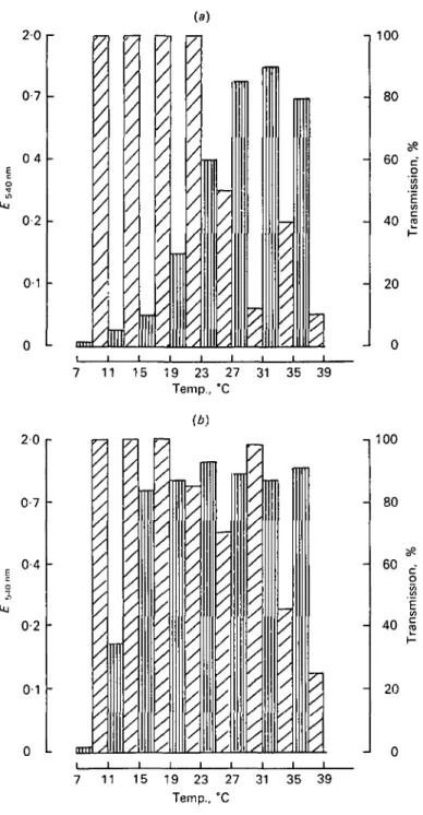
_ y / -,100 - 80 - 60 - 40 7 11 15 19 23 27 31 35 39 Temp., °CFig. 1. Effect of incubation temperature on the growth of Streptococcus cremoris CSIRO C 11 and its phage. In a temperature gradient: (a) 6-h incubation; (6) 24-h incubation. QD, Absorbance 540 nm phage free host; £3, transmission phage infected host.
20 r ia\ -I 100 0-7 0-4 0-2 0-1 IM S 80 60 40 20 24 28 32 36 40 44 48 52 Temp., °C 20 0-7 0-4 02 0-1 (b) / / 100 80 60 c o 40 2 20 24 28 32 36 40 44 48 52 Temp., °C
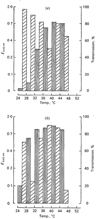
Fig. 2. Effect of inoubation temperature on the growth of Streptococcus thermophilua ATCC 19987 and its phage. In a temperature gradient: (a) 6-h incubation; (6) 24-h incubation. QJ Absorbance 540nm phage free host; 0 , transmission phage infected host.
Growth temperature of lactic acid bacteria and phages 263
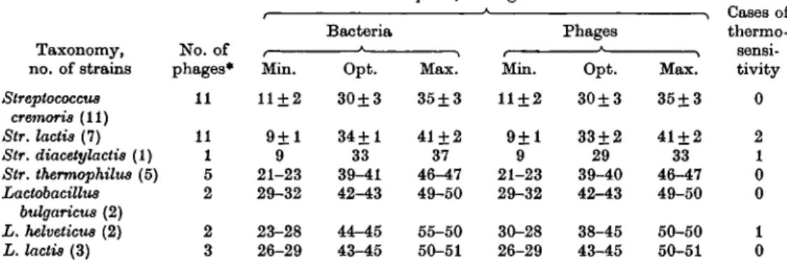
Table 2. Minimum, optimum and maximum growth temperatures for the host bacteria
and their bacteriophages
Temp. °C, average values
Taxonomy, no. of strains Streptococcus cremoris (11) Str. lactis (7) Str. diacetylactis (1) Str. thermophilus (5) Lactobacillus bulgaricus (2) L. helveticus (2) L. lactis (3) No. of phages* 11 11 1 5 2 2 3 Min. 11 + 2 9 + 1 9 21-23 29-32 23-28 26-29 Bacteria Opt. 30±3 34+1 33 39-41 42-43 44-45 43-45 Max. 35 + 3 41 + 2 37 46-47 49-50 55-50 50-51 Min. 11±2 9 + 1 9 21-23 29-32 30-28 26-29 Phages Opt. 30±3 33 + 2 29 39-40 42-43 38-45 43-45 Max. 35+3 41 + 2 33 46-47 49-50 50-50 50-51 thermc fiflnsi tivity 0 2 1 0 0 1 0 * Each bacterial strain had 1 specific phage except Str. lactis 274, which was the host for 5 phages. Table 3. Minimum, optimum and maximum temperatures of multiplication of some
temperature-sensitive phages and their hosts
Temp., °C Host and phage
Streptococcus lactis 274 Phage 21 Str. lactis 280 Phage 280 Str. diacetylactis 26-2 Phage 26-2 Lactobacillus helveticus L 112 Phage 1 112 Min. 9 9 9 9 9 9 23 30 Opt. 33 33 33 33 33 29 44 38 Max. 41 37 41 35 37 33 55 50
estimating the number of phages 'were withdrawn -with those for the first spectro-photometric reading, i.e. after 6 h of incubation.
RESULTS
The effect of temperature on the growth of the microorganisms after 6 and 24 h of incubation is shown in Figs 1 and 2. Bacterial growth was strongly influenced by temperature in the first few hours of incubation (Figs la and 2a). However, after 24 h, this effect diminished considerably and the precise optimum temperature was replaced by a range of temperatures permitting maximum growth (Figs 16 and 26). The multiplication of the phages followed almost the same pattern as that of the host strain. The maximum degree of bacterial lysis is attained after 24 h of incubation, but in some cases the bacterial culture was well clarified after 6 h and dense again after 24 h (Fig. 1, 31-35 °C, Fig. 2, 44-48 °C). This is a secondary culture of the host, no doubt due to the proliferation of phage-resistant mutants. This secondary growth was generally greatest close to the optimum temperature of the host strain.
The minimum, optimum and maximum temperatures for development of phages and their host bacteria are grouped in Table 2. The parallel between the phages and
Table 4. Behaviour of the phages ofmesophilic streptococci incubated for
6 h at different temperatures
No. of phage cultures
Phages of Streptococcus cremoris (11 strains) Str. lactis and diacetylactis (12 strains) Approx. (pfu/ml) 10s 10' 104 10s 10* 10s 108 10' 10« 105 101 10s Initial — — 5 4 2 — — 4 4 4 7 — — 4 5 1 1 — — 5 4 3 After 17 — 8 2 1 — 8 2 2 — 6 h incubatioi 27 3 6 2 — — — 6 5 1 — 37 — 2 1 4 4 — 4 3 4 1 — i at CC 47 — — 1 5 3 2 — — 10 2 1 4 3 3 5 6 1 (Distribution of the cultures according to their approximate concentration in pfu/ml.)
their hosts is evident and there are very few temperature-sensitive phages. In general, the bacteriophages multiplied whenever the bacteria grew.
Table 3 shows in more detail the differences between the 4 temperature-sensitive phages which we found. All of them differed from their hosts in maximum tem-perature, but one only in minimum temtem-perature, and two in optimum temperature. To supplement the photometric measurements, we estimated the rate of phage multiplication after 6 h of incubation at different temperatures using the plaque count method. The results are grouped in Table 4. Each strain was tested for the number of active phages at the time of inoculation, and after 6 h of incubation at 6 different temperatures. The Table shows, for each temperature, the number of cultures containing the concentration of phages (pfu/ml) indicated in the margin.
These results are in agreement with those obtained photometrically, and it is interesting to note that the phages are not killed by exposure at 55 °C for 6 h: their multiplication is blocked, but their numbers are not reduced.
DISCUSSION
In this study we have only used short incubation times - 6 and 24 h - to in-vestigate the effect of temperature on the growth of phages and their host bacteria. Although the results may be of little interest to taxonomists, who generally work with much longer incubation periods, they should be valuable to the food industry, for in any fermentation process the quality of the end-product is greatly dependent on what happens in the first few hours of fermentation. This is illustrated by the temperature profiles which we obtained. Indeed, although quite a wide range of temperatures favour growth after 24 h incubation, after only 6 h incubation there is a precise optimum temperature and deviations from this may cause serious delays in growth. The importance of strictly controlling the optimum temperature for
Growth temperature of lactic acid bacteria and phages 265 development of lactic acid bacteria is thus obvious, especially during the milk coagulation process in cheese production.
The knowledge acquired from this study may be valuable for the future pre-paration of starters and especially mixed cultures. Attention must be paid to the fact that the optimum temperature is very near to the maximum one, above which bacterial growth decreases extremely rapidly. Thus, temperature regulation cannot be used to prevent phage attacks because, with few exceptions, the phages exhibit the same temperature curves as their hosts.
We are grateful to Mr O. Depierraz for his excellent technical assistance and to Miss A. Walker for her help in writing this paper in English.
REFERENCES
DE MAD-, J. C , KOOOSA, M. & SHARPE, M. E. (1960). Journal of Applied Bacteriology 23, 130. EPSTEIN, R. H., BOIXE, A., STEINBERG, C. M., KELLENBERGER, E., BOY DE LA TOUR, E., CHEVALLEY,
R., EDGAR, R. S., SUSMAN, M., DENHARDT, G. H. & LIELAUSIS, A. (1963). Cold Spring Harbor Symposia
on Quantitative Biology 28, 375.
HEMTTTTTJ., H. E. & WHITELEY, H. R. (1975). Bacteriological Reviews 39, 257. HOGG, D. MCC. & JAGO, G. R. (1970). Journal of Dairy Research 37, 199. HUNTER, G. J . E. (1943). Journal of Dairy Research 13, 136.
KANBKO, T., IWANO, S. & KITAHARA, K. (1955). Journal Agricultural Chemical Society of Japan 29, 788. KEOGH, B. P. (1973). Journal of Dairy Research 40, 303.
MURATA, A. (1971). Agricultural and Biological Chemistry 35, 667.
OPPENHEIMER, C. H. & DROST-HANSEN, W. (1960). Journal of Bacteriology 80, 21. Sozzi, T. (1972). Lait 52, 454.
Sozzi, T., MARET, R. & POULIN, J. M. (1976). Applied and Environmental Microbiology 32, 131. SPIEGELMAN, S., PACE, N. R., MILLS, D. R., LEVISOHN, R., EIKHOM, T. S., TAYLOR, M. M., PETERSON
R. L. & BISHOP, D. H. L. (1968). Cold Spring Harbor Symposia on Quantitative Biology 33, 101. ZEHREN, V. L. & WHITEHEAD, H. R. (1954). Journal of Dairy Science 37, 209.