OATAO is an open access repository that collects the work of Toulouse
researchers and makes it freely available over the web where possible
Any correspondence concerning this service should be sent
to the repository administrator:
[email protected]
This is an author’s version published in:
http://oatao.univ-toulouse.fr/22997
To cite this version:
Tormo, Hélène and Ali Haimoud Lekhal, Djamila and Roques, Christine
Phenotypic and genotypic characterization of lactic acid bacteria isolated from
raw goat milk and effect of farming practices on the dominant species of lactic
acid bacteria. (2015) International Journal of Food Microbiology, 210. 9-15.
ISSN 0168-1605
Phenotypic and genotypic characterization of lactic acid bacteria isolated
from raw goat milk and effect of farming practices on the dominant
species of lactic acid bacteria
Hélène Tormo
a,⁎
, Djamila Ali Haimoud Lekhal
a, C. Roques
baUniversité de Toulouse, Ecole d'Ingénieurs de Purpan, 75, voie du TOEC, BP 57611, F-31076 Toulouse Cedex 03, France bUniversité de Toulouse, UPS, Lab Genie Chim, F-31 062 Toulouse, France
a b s t r a c t
a r t i c l e
i n f o
Keywords: Lactococcus lactis Enterococcus subsp Goat milk REP-PCR16S rRNA gene sequencing Farming practices
Lactic acid bacteria, in particular Lactococcus lactis, play a decisive role in the cheese making process and more particularly in lactic cheeses which are primarily produced on goat dairy farms. The objective of this study was therefore to identify the main lactic acid bacteria found in raw goats' milk from three different regions in France and evaluate if certain farming practices have an effect on the distribution of species of lactic acid bacteria in the various milk samples. Identification at genus or species level was carried out using phenotypic tests and genotypic methods including repetitive element REP-PCR, species-specific PCR and 16S rRNA gene sequencing. The distribution of the main bacterial species in the milk samples varied depending on farms and their charac-teristics. Out of the 146 strains identified, L. lactis was the dominant species (60% of strains), followed by Entero-coccus (38%) of which EnteroEntero-coccus faecalis and EnteroEntero-coccus faecium. Within the species L. lactis, L. lactis subsp lactis was detected more frequently than L. lactis subsp cremoris (74% vs. 26%). The predominance of L. lactis subsp cremoris was linked to geographical area studied. It appears that the animals' environment plays a role in the balance between the dominance of L. lactis and enterococci in raw goats' milk. The separation between the milking parlor and the goat shed (vs no separation) and only straw in the bedding (vs straw and hay) seems to promote L. lactis in the milk (vs enterococci).
1. Introduction
The sensorial particularity of farmhouse goats' cheese is partly linked to the use of raw milk of which the properties vary according to farming practices. The physico-chemical characteristics of milk depend on the breed of goat and the feed, which in turn influence the technological and sensorial characteristics of the cheeses. The microbial flora in raw milk is also a key characteristic in cheese quality as it in-creases the diversity of flavors (Steele and Ünlu, 1992; Fox et al., 1996; Lynch et al., 1997; Monteil et al., 2014). Due to their acidifying ca-pacity, lactic acid bacteria play a key role in the acidification of the curd essential to cheese making, but they also contribute to cheese aroma and texture as they possess endo and exopeptidases which are involved in the production of sapid molecules; they generate precursors of aro-matic compounds (Mauriello et al., 2001; Herreros et al., 2003). Lactic acid bacteria are also very important in the manufacturing of farmhouse raw goats' milk cheese as the coagulation at low temperature (20 °C) lasts approximately 24 h. The whey, rich in Lactococcus lactis (Tormo
and Talliez, 2000), is used as a natural lactic starter. Raw milk is often described as a major source of lactic acid bacteria in the whey (Bachmann et al., 1996; Centeno et al., 1996; Manopoulou et al., 2003; Duthoit et al., 2005) and so it is important, particularly for these cheeses, to control the microbiological quality of the milk as the success of the whey and the cheese depends on it. The dominance of L. lactis in the whey is a factor of success (Demarigny et al., 2006). Raw milk, rich in L. lactis may therefore be very interesting, particularly for this type of cheese making. Certain studies have shown that the microbiological characteristics of milk depend on the farm and the farming practices (Michel et al., 2001; Verdier Metz et al., 2009; Tormo et al., 2011; Mallet, 2012, Mallet et al., 2012). However, to date, no studies have been undertaken which look at the relationships between the species of lactic acid bacteria found in raw goat milk and the farming practices. The objective of this study was to (i) identify the major lactic acid bacteria in raw goats' milk that potentially have the capacity to acidify raw milk. The bacteria were identified using phenotypic tests, analyses and genotypic methods including repetitive element REP-PCR, species-specific PCR techniques, and 16S rDNA sequencing; and (ii) to evaluate the relationship between farm practices and the distribution of dominant species of lactic acid bacteria in raw goats' milk. These prac-tices concerned: general management, monitoring of flock, bedding management practices, environmental conditions during and after ⁎ Corresponding author at: Laboratoire de microbiologie, Ecole d'Ingénieurs de Purpan,
75, voie du TOEC, BP 57611, F-31076 Toulouse Cedex 03, France. Tel.: +33 5 61 15 29 94; fax: +33 5 61 15 30 60.
E-mail address:[email protected](H. Tormo).
milking, cleaning of teats and milking machine, handling and character-istics of the milking machine.
2. Materials and methods 2.1. Choice and monitoring of farms
The 21 farms selected were all farms producing farmhouse goats' cheese from the three French geographical areas: PDO Rocamadour (11 farms from department of Lot), Pélardon (6 farms from departments
of Herault and Gard) and Franche-Comté (4 farms). They were chosen on the basis of their diverse farming practices and methods of milk produc-tion and were representative of their region. The management and the farming practices were reflected inTable 1. The practices were monitored in May and June 2006. For each farm, milk samples were collected after milking; the samples (once sample of milk from one milking per farm) were cooled to 10 °C and frozen without cryoprotectant at −25 °C for a maximum of one month.
2.2. Numeration, isolation and purification of lactic acid bacteria Elliker medium modified according toChamba et al. (1981)was cho-sen for numeration, isolation and culture of lactic acid bacteria. This se-lective medium is used to count acidifying bacteria of which the majority is lactic acid bacteria.
After inoculation in the mass of dilutions of milk in sterile buffered peptone water (Biomérieux, France) and incubation of 72 h at 20 °C, approximately ten acidifying bacterial colonies (colonies with yellow halo) were isolated from the suitable dilution and incubated in Elliker broth overnight at 30 °C. The isolates (50 μL of the culture) were puri-fied by subculture on Elliker agar (48 h, 30 °C) and one colony was incu-bated overnight at 30 °C in Elliker broth. After centrifugation (5000 rpm during 5 min at 4 °C), the bacterial pellets were dispersed in skim milk, frozen and stored at −80 °C in reconstituted sterile semi-skimmed milk (150 g/L) with 20% glycerol (500 μL of pellet in 500 μL of broth). 2.3. General procedure for identification of lactic acid bacteria
Firstly the bacterial isolates were characterized using phenotypic tests in order to verify that the isolates were lactic acid bacteria and to have phenotypic profiles. Strains belonging to groups of lactic acid bacteria with different phenotypic profiles and from milk samples from different farms were selected for the continuation of the character-ization: PCR to confirm the Lactococcus lactis subspecies and the genus of the other lactic acid bacteria completed by REP-PCR, a molecular tool which is useful for elucidating relationships within and between bacterial species (Mancuso et al., 2007). Finally, the strains with differ-ent profiles (phenotypic, Rep-PCR) were sequenced and subsequdiffer-ently assigned at species or subspecies level.
2.4. Phenotypic characterization of the isolates
The Gram positive, catalase negative isolates were analyzed at genus level. The growth of the isolates in Elliker broth (DIFCO, France) at 10 °C for a week, at 45 °C, pH 9.6 and 6.5% (P/V) salt for 96 h as well as growth in “litmus milk” (Becton Dickinson and Company, USA) were tested. Subsequently, the isolates were characterized using the following tests: growth in Elliker broth (DIFCO, France) with 4% salt and at 40 °C, growth at pH 9.2, ability to ferment maltose, ribose, sorbitol and raffinose in MRS broth (DIFCO, France). The presence of an arginine dihydrolase was in-vestigated in BHI broth with 0.3%L-arginine (SIGMA, France). After incu-bation 24 h at 30 °C, 2 to 3 drops of Nessler reagent were added. An orange precipitate indicates the presence of the NH3.
2.5. PCR-based method 2.5.1. DNA extraction
Strains were incubated at 30 °C for 24 h in MRS broth and genomic DNA was extracted using the NucleoSpin tissue kit (Macherey Nagel, 67 722 Hoerdt, France).
2.5.2. PCR amplification
The strains were confirmed to belong to Lactococcus lactis subsp lactis, Lactococcus lactis subsp cremoris or enterococci by means of a PCR-based method. Lactococcus lactis subsp lactis or subsp cremoris were identified using primers His 1 and His 2 (Corroler et al., 1998). Table 1
Groups of variables describing the management practices and number of farms per practice.
Variable label Level Number
of farms 1. General management
Size of flock 116 ± 83 21
Pasture in spring and summer Yes 18
No 3
Level of production (kg/goat/year) 639 ± 196 21
Area PDO Rocamadour 11
PDO Pélardon 6
Franche-Comté 4
2. Monitoring of flock
Milk testing No milk testing 9
Milk testing 12
Monitoring of somatic cells counts No monitoring 15
Monitoring 6
Antiparasitic treatment No 4
Yes 17
Antibiotic treatment during drying off
No 10
Yes 11
Homeopathic treatment during drying off
No 10
Yes 11
3. Bedding management practices
Bedding Straw 12
Straw + hay 9
Additive in the bedding Yes 3
No 18
4. Environmental conditions during and after milking
Mulching during milking Yes 4
No 17
Frequency of cleaning milking platforms
Frequently: after each milking 11 Not frequently: less frequently than after each milking
10 Position of milking parlor No separation with the bedding
area
9 Physical separation with the bedding area
12 Method of cleaning milking
platform
Dry method 17
With water 4
5. Practices concerning the teats
Disposal of premilking Yes 16
No 5
Desinfecting of teats after milking Yes 3
No 18
6. Cleaning of milking machine Maximal temperature (°C) of cleaning of milking machine
60,5 ± 12 21
Intercleaning with alkaline and acid products
Frequently: change of product every day
13 Not frequently: change less frequently than every day
8 Residue of water in the MMa Yes 13
No 8
Washable sanitary trap Yes 5
No 16
7. Handling and characteristics of the MMa
Length of pipeline b 120 m 15
≥120 m 6
Number of elbows and fittings b 4 9
≥4 12
Enterococcal DNA was amplified using primers Conrev 23 and Genter according toFrahm et al. (1998). In all cases, amplification reactions were performed in a final volume of 25 μL containing 1× reaction PCR buffer (Qiagen, France), 0.3 μM of each opposing primer, 2.5 mM of MgCl2, 0.2 mM of each deoxynucleoside triphosphate, 0.5 U Taq
polymerase and 5 μL of DNA.
Inter-repetitive extragenic sequences were amplified by means of two 18-mer primers in combination (REP 1R-Dt, REP 2-D) (Invitrogen, Cergy Pontoise, France) as described in other publications (Versalovic et al., 1991; Berthier et al., 2001). The final PCR reaction mixture was 25 μL: 2 μM of each of the PCR primers REP-1 and REP-2, 200 μM of each of the desoxyribonucleotides (dNTP) (Invitrogen, Cergy Pontoise, France), 0.4 units of Taq DNA Polymerase (Invitrogen, Cergy Pontoise, France), PCR buffer 1× with MgCl2(1/10th of total volume) (SIGMA,
France) and 5 μL of extracted DNA.
The primer sequences and the PCR amplification conditions are recapitulated inTable 2. The amplification cycles were performed with a thermal cycler (Gene Amp PCR System 9700, Perkin Elmer), then 25 μL of PCR product was electrophoresed in a 10 g·l−1Seakem
GTG agarose gel (Sigma) in TBE (Tris-Borate-EDTA pH 8) at 100 V for 3 h. The 123-pb DNA ladder (Invitrogen, 95613 Cergy Pontoise, France) was used as a size standard. The DNA fragments were stained with ethidium bromide (Sigma), examined under UV light (312 nm) and photographed (G-Box, SYNGENE). Digitized gel pic-tures of Rep-PCR were normalized by comparison with reference bands (G-Box, SYNGENE). The similarities among profiles were cal-culated using the Pearson correlation. Dendrograms were construct-ed using the unweightconstruct-ed pair group method with Arithmetic Mean (UPGMA).
2.5.3. Partial 16S rDNA sequencing
Fifty-one strains with different profiles (phenotypic, Rep-PCR) were sequenced and subsequently assigned at species or subspecies level. The REP-PCR profiles of the non-sequenced strains were compared to the profiles of the strains for which the DNA fragments have been sequenced in order to assign these strains at species or subspe-cies level. The 16S rRNA gene (V1–V4) was amplified by PCR using primers E8F and E807R (Baker et al, 2003). DNA was amplified in 50 μL volumes containing 50 ng of template, 500 μM dNTPs, 5 μM of the respective primers, 2.5 U of Taq DNA polymerase (Invitrogen, Cergy Pontoise, France) and Thermopol buffer 10 ×. PCR products were cleaned using QUIAquick columns (Qiagen, France) according to the manufacturer's instructions and subsequently commercially sequenced (Eurofins MWG biotech, 91967 Les Ulis, France) using primer E8F. The analysis of the chromatograms and the multiple
alignments were carried out using MEGA 4.1 software (Tamura
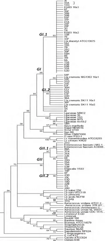
et al, 2007). Subspecies identification was carried out by construc-tion of a phylogenetic tree (using MEGA 4.1, Neighbor Joining method, bootstrap 1000) using the sequences obtained from the se-quencing and the sequences of Firmicutes species acquired on the NCBI website (http://www.ncbi.nlm.nih.gov/).
The primer sequences and the PCR amplification conditions are reca-pitulated inTable 2.
2.6. Statistical analysis
For each farm, a dominant species or genus was assigned (70% of the isolates for each farm) with a dominant species or genus. The relation-ships between the dominant species or genus on the farm and the farming practices were studied using Pearson's chi-square test (SPAD ver-sion 5.5, Pantin, France).
3. Results
3.1. Phenotypic characterization of isolates
There was a high variability of the level of acidifying bacteria accord-ing to the farm: the size of the populations ranged from 44 to 54 000 CFU per mL of raw milk and the average was 640 CFU·mL−1.
A total of 204 out of the 206 isolates had a Lactococcus or Enterococcus phenotype. For these 204 isolates, 6 phenotypic profiles were distin-guished (Table 3). Profiles A and B correspond to the phenotype Enterococcus (47.5% of the strains). These two profiles differ in their ca-pacity to coagulate milk: the isolates in group A do not coagulate milk, whereas those in group B do. Profiles C, D, E and F correspond to the phenotype Lactococcus lactis (52.5% of the strains). The strains belong-ing to profiles D, E and F have the phenotype L. lactis subsp lactis as they can grow in the presence of 4% NaCl, at 40 °C, hydrolyze arginine and produce acid in the presence of maltose. The strains in groups E and F can grow at pH 9.2. The strains in group E can grow in the presence of 6.5% NaCl. The strains in group C have an unusual phenotypic profile as they do not grow at 40 °C and do not hydrolyze arginine. They have a phenotype similar to that of L. lactis subsp cremoris even though they grow in the presence of 4% salt and most grow at pH 9.2. A total 146 strains out of the 206 were selected according to their different phe-notypic profiles (belonging to the different groups A, B, C, D, E and F) and genotypic characterized.
3.2. Genus, species and subspecies specific PCR
Genus and species specific PCR confirmed that L. lactis and Enterococ-cus subsp were the dominant lactic acid bacteria isolated from Elliker agar modified accordingChamba et al. (1981). So, 73 strains were iden-tified as L. lactis subsp lactis (50% of strains tested), 17 strains as L. lactis subsp cremoris (12%) and 56 strains as enterococci (38%).
3.3. REP-PCR
Fig. 1shows the profiles obtained from the 146 isolates. Four main groups can be distinguished. Isolates representative of the diversity of each group were sequenced (seeSection 3.4). Then, profiles were suc-cessfully assigned to bacteria species. Profile 1 was assigned to Entero-coccus faecium, 2 to L. lactis subsp lactis, 3 to EnteroEntero-coccus subsp faecalis and 4 to L. lactis subsp cremoris. Only two of the 51 sequenced strains were not Lactococcus and Enterococcus and had similar finger-print to those cited species (14H, 37G).
For each group, we were able to distinguish different clusters (85% similarity to the obtained profiles). Except for the farm numbers Table 2
Primer sequences, amplification and application of PCR reactions.
Primer sequences Amplification conditions Application
His1: 5′-CTTCGTTATGATTTTACA-3′ 5 min at 94 °C, 30 cycles of: 1 min 94 °C, 2 min at 45 °C, 2 min at 72 °C, final step 5 min at 72 °C
Detection of Lactococcus lactis lactis and Lactococcus lactis cremoris His2: 5′-AATATCAACAATTCCATG-3′
Conrev 23: 5′-GGTGGATGCCTTGGCACT-3′ 5 min at 94 °C, 30 cycles of: 30 s 94 °C, 30 s at 52 °C, 30 s at 72 °C, final step at 5 min at 72 °C
Detection of the genus Enterococcus Genter: 5′-CTCTACCTCCATCATTCT-3′
REP1R-Dt: 5′-IIINCGNCGNCATCNGGC-3′ 5 min at 94 °C, 30 cycles of: 1 min at 94 °C, 1 min at 40 °C,
6 min ramping to 72 °C and 1 min at 72 °C Rep-PCR fingerprinting REP2-D: 5′-NCGNTTATCNGGCCTAC-3′
E8F: 5′ –AGAGTTTGATCCTGGCTCAG-3′ 3 min at 94 °C, 30 cycles of: 45 s 94 °C, 2 min at 55 °C, 1 min at 72 °C, final step 5 min at 72 °C
Amplification of the V1–V4 region of 16s rDNA
26 and 53, there was a different Rep-PCR fingerprint inside the same farm and, in an obvious way, between farms.
3.4. Partial 16s rDna sequencing
Fifty-one strains representative of the diversity of the phenotypic, genotypic and protein profiles were classified genetically into 5 species
by partial 16s rDNA sequencing (Fig. 2). The two main groups contain the genus Enterococcus (group II) divided into 2 sub-groups containing the species E. subsp faecalis (II.1) and E. subsp faecium (II.2) and the spe-cies L. lactis subsp cremoris (I.2). Two strains did not belong to these groups and were identified as Aerococcus viridans (14H) and Streptococ-cus parauberis (37G). The correlation between the results of the pheno-typic tests, the PCR tests and the sequencing is reported inTable 4. Table 3
Phenotypic characteristics of strains isolated from milks. +: all strains positive; −: all strains négative; (../..): number of positive/negative strains. All strains grew at 10 °C. Clusters Number of strains Number of farm Growth at 4% of salt Growth at 6.5% of salt Growth at pH 9,2 Growth at pH 9,6 Growth at 40 °C Growth at 45 °C Coagulation of litmus milk NH3 from arginine
Maltose Ribose Phenotype
A 13 5 + + + + + + − − + + Enterococcus subsp B 84 17 + + + + + + + + + + Enterococcus subsp C 12 6 + − 9/3 − − − + − 10/2 + Lactococcus Lactis D 28 10 + − − − + − + + + + Lactococcus Lactis E 30 11 + + + 17/13 + − + + + + Lactococcus Lactis F 37 17 + − + 11/26 + − + + + - Lactococcus Lactis
Fig. 1. Rep-PCR patterns showing the representative fingerprints of the different clusters. The numbers followed by letters to the right of the figure are the codes of the strains analyzed. The number corresponds to the farm, the letter to the clone isolated. The numbers in bold represent the sequenced isolates.
From the phenotypic tests it was possible to predict the species L. lactis as 26 strains out of the 33 were identified as L. lactis. The 7 re-maining strains had the phenotypic characteristics of enterococci (grow at 45 °C, 6.5% salt, pH = 9.6).
Similar results were also observed for the genus Enterococcus: 13 strains out of the 15 identified had an Enterococcus phenotype.
A good correlation between the results of the PCR and the DNA se-quencing was observed for Enterococcus subsp and L. lactis subsp lactis (Table 4). So, 13 strains out of 15 (87%) identified as enterococci by
DNA sequencing were characterized as enterococci by PCR and 22 strains out of 25 (82%) identified as L. lactis subsp lactis were also iden-tified as such by PCR. However, the correlation is not as good concerning L. lactis subsp cremoris as only 6 strains out of 9 (67%) identified as L. lactis subsp cremoris were also identified as such by PCR.
3.5. Dominant species in milk and relationship with farming practices Out of the 146 strains identified isolated from the medium modified according toChamba et al. (1981), L. lactis was the dominant species (87 strains). The subspecies L. lactis subsp lactis was the most common (68 strains) in comparison with L. lactis subsp cremoris (19 strains). E. faecalis (37 strains) and E. faecium (20 strains) were the two species of Enterococcus isolated.Table 5presents the distribution of the species according to the farms. A farm was removed from the analysis as only one isolate out of the five isolates could be cultured. When more than 2/3 of the strains identified from each of the farms belonged to the same species or genus, this species or genus was considered as domi-nant. The distribution of the species varied according to the farms. Twelve farms were characterized by the dominance of L. lactis and eight by a dominance of Enterococcus. For one farm (no. 37), the distri-bution between L. lactis and Enterococcus was similar; no dominant group was allocated. The groups of dominant species were significantly discriminated (p-value ≤ 5%) by certain farming practices (Table 6). Most of the farms putting hay in the bedding were associated with enterococci group (6 farms among 8) and most farms with only straw on the bedding were associated with lactococci group (10 farms among 12). Most of the farms which have no separation between bedding area and milking parlor was associated with enterococci group (6 farms among 9) and farms with separation between bedding area and milking parlor was rather associated with lactococci group (9 farms among 11).
The presence of L. lactis subsp cremoris seems to be linked to the geo-graphic area. Indeed, 2 farms out of the 4 in Franche-Comté were char-acterized by a dominance of L. lactis subsp cremoris, while in the other two regions, L. lactis subsp lactis was largely dominant. Only one farm out of the 6 farms in the Pélardon PDO had milk in which L. lactis subsp cremoris was dominant. No L. lactis subsp cremoris were detected in the Rocamadour PDO area.
4. Discussion
L. lactis, E. faecalis and E. faecium are the three species most common-ly isolated. These species were identified using a combination of pheno-typic and genopheno-typic methods. Subspecies specific PCR using primers targeting the Histidine operon biosynthesis region carried out according to the protocol developed byCorroler et al. (1998)correctly discrimi-nated L. lactis subsp lactis but seems to be less effective for L. lactis subsp cremoris (3 false positives out of 9 strains in total). REP-PCR pro-files enabled the Enterococcus species and the L. lactis subspecies studied to be discriminated correctly, as previously reported byJurkovic et al. (2006)andJan et al. (2007).
For the species L. lactis, all the phenotypes corresponded to the spe-cies L. lactis subsp lactis, no specific phenotype was associated with L. lactis subsp cremoris. This result is not surprising as the strains of phe-notype L. lactis subsp cremoris are isolated in a dairy environment where lactic starters are regularly used in the manufacture of fermented prod-ucts (Klijn et al., 1995). The majority of L. lactis strains had a L. lactis subsp lactis genotype (68 strains out of the 87 belonging to the species L. lactis). The 19 remaining strains corresponded to the genotype L. lactis subsp cremoris. The dominance of L. lactis and Enterococcus in milk has already been underlined by numerous authors including
Zamfir et al. (2006)in cows' milk andBadis et al. (2004)in goats' milk. Certain strains from different farms belonging to the L. lactis phe-notype and gephe-notype can grow in the presence of 6.5% salt and at pH 9.6. This result is not surprising as these environmental microorganisms
GI
GI.1
GI.2
GII
GII.1
GII.2
Fig. 2. Phylogenic tree obtained by partial 16s rDNA sequencing of the strains isolated from goat milk and the reference strains obtained by the Neighbour-Joining (boostrap 1000). Goat milk strains' codification: Number, letter (ex: 3A).
undergo significant stress. These phenotypic particularities have already been underlined byCorroler et al. (1998)for strains isolated from cows' milk.
The distribution of the species in the milk depends on the farms. This farm-specific characteristic has already been underlined by
Corroler et al (1998)in cows' milk from Normandy. Certain farms are characterized by a dominance of L. lactis subsp lactis (9 farms out of the 21 farms studied), others by E. faecalis (5 farms out of the 21) or E. faecium (3 farms out of the 21). The presence of a major-ity of L. lactis subsp cremoris in the milk (3 farms out of the 21) seems to depend on the geographic area as it is found preferentially on farms located in Franche-Comté. It is important to underline that the farms in Franche-Comté are equipped with bucket milking sys-tems contrary to the other regions where milk is recovered via a milk line. The methods of cleaning and the materials being different, it can be assumed that the bacteria are transported in different ways (Laithier et al., 2004; Marchand et al., 2012).
The dominance of Enterococcus (E. faecalis, E. faecium) or L. lactis seems to be due in part to certain farm-specific characteristics. The direct contact between the milking parlor and the bedding area or the presence of hay in the bedding seems to promote inoculation of milk with E. faecalis or E. faecium. The natural habitat of Enterococcus is the intestines of humans and animals (Facklam et al., 2002).
Gelsomino et al. (2001)showed that enterococci were present in the cow feces, human feces and milk. In goat farm, in the same areas of our study,Detomi (2009)showed that enterococci were
found as a dominant lactic acid bacteria in goat bedding (50 from 51 strains were enterococci). In the same study, in which 20 farms were monitored, enterococci were improved in the air of the milking parlor when there was no separation between milking parlor and bedding area (p ≤ 5%). Then, we could supposed that aerosol in the goat shed was contaminated by the enterococci of the bedding (probably contaminated by feces). When the milking parlor was not separated to the goat shed, the air was contaminated by the air of the goat shed. During the milking, the milk was more con-taminated by enterococci than in a milking parlor separated to goat bedding. Concerning the relationship between the dominance of En-terococcus in milk and the presence of hay in the straw-based bed-ding, no scientific studies have been undertaken on this subject as far as we know. However, we can assume that the hay from fodder is less absorbent than straw, therefore favoring the development of bacteria on the surface of the bedding. This waste food may also be more contaminated as it is put in the troughs before being used for bedding.
These first results concerning the possible association between cer-tain farming practices and the main bacteria isolated are worth confirming by analyzing the sources of contamination of Enterococcus and Lactococcus, L. lactis in particular. Continuing this study in this direc-tion would permit to identify the farming operadirec-tions that would enable a decrease in the contamination of Enterococcus and promote the devel-opment of L. lactis in milk. The technological and sensorial quality of lac-tic cheeses could therefore be improved.
Table 4
Correlation between the sequencing results of the 16s rDNA V1–V4 region and the results of the phenotypic tests and PCR. The numbers correspond to the number of strains. Analyses
Sequencing
L. lactis lactis L. lactis cremoris Enterococcus subsp Aerococcus viridans Streptococcus parauberis
Number of strains sequenced 25 9 15 1 1
Typology
Phenotype L. lactis lactis 14 8 2 1 0
L. lactis cremoris 3 0 0 0 1 Enterococcus subsp 7 1 13 0 0 n.i 1 0 0 PCR L. lactis lactis 19 1 2 1 1 L. lactis cremoris 3 6 0 0 0 Enterococcus subsp 2 2 13 0 0 Table 5
Distribution of number of strains per farm identified as being L. lactis or Enterococcus and allocation to a dominant group. 1 : total number of strains, 3 : Dominant species group. E: Enterococcus dominant, L: L.lactis dominant.
No. farms Area L. lactis lactis L. lactis cremoris L. lactis E. faecalis E. faecium Enterococcus subsp Other species nT1 %lactis Groups3
1 PDO Rocamadour 2 – 2 – 5 5 – 7 29 E 3 PDO Rocamadour 6 – 6 – – 0 – 6 100 L 6 PDO Rocamadour 1 – 1 – 3 3 – 4 25 E 7 PDO Rocamadour 5 – 5 – – 0 – 5 100 L 8 PDO Rocamadour 3 – 3 1 – 1 – 4 75 L 13 PDO Rocamadour 6 – 6 – – 0 – 6 100 L
14 PDO Rocamadour – – 0 6 – 6 S. parauberis 7 0 E
19 PDO Rocamadour 3 – 3 6 – 6 – 9 33 E 20 PDO Rocamadour 6 – 6 2 – 2 – 8 75 L 22 PDO Rocamadour 1 – 1 3 1 4 – 5 20 E 25 PDO Rocamadour 10 – 10 – – 0 – 10 100 L 26 PDO Pélardon 2 – 2 – 8 8 – 10 20 E 29 PDO Pélardon 1 – 1 9 – 9 – 10 10 E 31 PDO Pélardon 1 6 7 – – 0 – 7 100 L 33 PDO Pélardon 4 – 4 – – 0 – 4 100 L
37 PDO Pélardon 3 – 3 – 3 3 A. viridans 7 43 –
43 PDO Pélardon 1 – 1 9 – 9 – 10 10 E 52 Franche-Comté 4 1 5 – – 0 – 5 100 L 53 Franche-Comté 1 6 7 1 – 1 – 8 88 L 55 Franche-Comté 7 – 7 – – 0 – 7 100 L 58 Franche-Comté 1 6 7 – – 0 – 7 100 L Total 68 19 87 37 20 57 146
This study was supported by the Midi-Pyrénées Conseil Régional (CCRDT 07005477) and the association GALA (Janzé, France). Acknowledgments
The authors would like to thank the technicians and farmers from the Pelardon, Rocamadour and Franche-Comté regions for their implica-tion in this study.
References
Bachmann, H.P., McNulty, D.A., McSweeney, P.L.H., Ruegg, M., 1996.Experimental designs for studying the influence of the raw milk flora on cheese characteristics: a review. J. Soc. Dairy Technol. 49, 53–56.
Badis, A., Guetarni, D., Boudjema, B.M., Henni, D.E., KIHAL, M., 2004.Identification and technological properties of lactic acid bacteria isolated from raw goat milk of four Algerian races. Food Microbiol. 21, 579–588.
Baker, G.C., Smith, J.J., Cowan, D.A., 2003.Review and re-analysis of domain-specific 16S primers. J. Microbiol. Methods 55, 541–555.
Berthier, F., Beuvier, E., Dasen, A., Dufrene, F., Grappin, R., 2001.Origin and diversity of mesophilic lactobacilli in Comté cheese, as revealed by PCR with repetitive and species-specific primers. Int. Dairy J. 11, 293–305.
Centeno, J.A., Menendez, S., Rodriguez-Otero, J.L., 1996.Main microflora present as natu-ral starters in Cebreiro raw cow's-milk cheese (Northwest Spain). Int. J. Food Microbiol. 33, 307–313.
Chamba, J.F., Bonnaz, G., Bourg, P., 1981.Comparaison de diverses méthodes de dénombrement de la flore acidifiante du lait cru. Lait 61, 555–567.
Corroler, D., Mangin, I., Desmasures, N., Gueguen, M., 1998.An ecological study of lactococci isolated from raw milk in the camembert cheese registered designation of origin area. Appl. Environ. Microbiol. 64, 4729–4735.
Demarigny, Y., Sabatier, C., Laurent, N., Prestoz, S., Rigobello, V., Blachier, M.J., 2006. Micro-bial diversity in natural whey starters used to make traditional Rocamadour goat cheese and possible relationships with its bitterness. Ital. J. Food Sci. 18, 261–276.
Detomi, C., 2009.Environmental Microflora Which Could Contamined the Milk. Trainee-ship report. Engineering school of Purpan, Toulouse, France (54 pp.).
Duthoit, F., Tessier, L., Montel, M.C., 2005.Diversity, dynamics and activity of bacterial populations in ‘Registered Designation of Origin’ Salers cheese by single-strand con-formation polymorphism analysis of 16SrRNA genes. J. Appl. Microbiol. 98, 1198–1208.
Facklam, R., Carvallo, M.G., Teixeira, L., 2002.History, taxonomy, biochemical characteris-tics and antibiotic susceptibility testing of enterococci. In: Gilmore, M. (Ed.), The En-terococci: Pathogenesis, Molecular Biology and Antibiotic Resistance. ASM Press, Washinghton, DC (20036–2904).
Fox, P.F., Wallace, J.M., Morgan, S., Lynch, C.M., Niland, E.J., Tobin, J., 1996.Acceleration of cheese ripening. Antonie Leuewenhoek 70, 271–297.
Frahm, E., Heiber, I., Hoffmann, S., Koob, C., Meier, H., Ludwig, W., Amann, R., Schleifer, K.H., Obst, U., 1998.Application of 23S rDNA-targeted oligonucleotide probes specific for enterococci to water hygiene control. Syst. Appl. Microbiol. 21, 450–453.
Gelsomino, R., Vancanneyt, M., Condon, S., Swings, J., Cogan, T.M., 2001.Enterococcal di-versity in the environment of an Irish Cheddar-type cheese making factory. Int. J. Food Microbiol. 71, 177–188.
Herreros, M.A., Fresno, J.M., Gonzales Prieto, M.J., Tornadijo, M.E., 2003.Technological characterization of lactic acid bacteria isolated from Armada cheese (a Spanish goat's milk cheese). Int. Dairy J. 13, 469–479.
Jan, L., Rademaker, W., Herbet, H., Starrenburg, M., Naser, J.C., Gevers, S.M., Kelly, D., Hugenholtz, W.J., Swings, J., Van Hyckama Vlieg, J., J.E.T., 2007.Diversity analysis of
dairy and nondairy Lactococcus lactis isolates, using a novel multilocus sequence analysis scheme and (GTG) 5-PCR fingerprinting. Appl. Environ. Microbiol. 73, 7128–7137.
Jurkovic, L., Krizkova, M., Sojka, A., Belicova, R., Dusinsky, J., Krajcovic, C., Snauwaert, S., Naser, P., Vandamme, M., Vancanneyt, 2006.Molecular identification and diversity of enterococci isolated from Slovak Bryndza cheese. J. Genet. Appl. Microbiol. 52, 329–337.
Klijn, N., Weerkamp, A.H., De Vos, W.M., 1995.Detection and characterization of lactose-utilizing Lactococcus spp. in natural ecosystems. Appl. Environ. Microbiol. 61, 788–792.
Laithier, C., Chatelin, Y.M., Tormo, H., Lefrileux, Y., 2004.Biofilms in farms producing goat cheese: localisation, nature and role on products quality. Proceedings of the 11th Rencontres Recherche Ruminant Conference 11. INRA, Paris, France, p. 112.
Lynch, C.M., McSweeney, P.L.H., Fox, P.F., Cogan, T.M., Drinan, F.D., 1997.Contribution of starter lactococci and non lactobacilli to proteolysis in cheddar cheese with a con-trolled microflora. Lait 77, 441–459.
Mallet, A., 2012.Diversité microbienne des laits crus: états des lieux, reservoirs et expres-sion en transformation fromagère: exemple de Camembert de Normandie. (Thesis). Université Caen Basse Normandie.
Mallet, A., Guéguen, M., Kauffmann, F., Chesneau, C., Sesboué, A., Desmasures, N., 2012.
Quantitative and qualitative microbial analysis of raw milk reveals substantial diver-sity influenced by herd management practices. Int. Dairy J. 27, 13–21.
Mancuso, M., Avendano-Herrera, R., Zaccone, R., Toranzo, A., Magarinos, B., 2007. Evalua-tion of different DNA-based fingerprinting methods for typing Photobacterium damselae ssp. Piscicida, Biol Res 40, 85–92.
Manopoulou, E., Sarantinopoulos, P., Zoidou, E., Aktypis, A., Moschopoulou, E., Kandarakis, I.G., Anifantakis, E.M., 2003.Evolution of microbial population during traditional feta cheese manufacturing and ripening. Int. J. Food Microbiol. 82, 153–161.
Marchand, S., De Block, J., De Jonghe, V., Coorevits, A., Heyndrickx, M., Herman, L., 2012.
Biofilm formation in milk produduction and processing environments; influence on milk quality and safety. Compr. Rev. Food Saf. 11, 133–147.
Mauriello, S., Moio, L., Moschetti, G., Piombino, P., Addeo, F., Coppola, S., 2001. Character-ization of lactic acid bacteria strains on the basis of neutral volatile compounds pro-duced in whey. J. Appl. Microbiol. 82, 153–161.
Michel, V., Hauwuy, A., Chamba, J.F., 2001.Raw cowmilk microflora: diversity and influ-ence of conditions of production. Lait 81, 575–592.
Monteil, M.-C., Buchin, S., Mallet, A., Delbes-Paus, C., Vuitton, D., Desmasures, N., Berthier, F., et al., 2014.Traditional cheeses: rich and diverse microbiota with associated ben-efits. Int. J. Food Microbiol. 177, 136–154.
Steele, J.L., Ünlu, G., 1992.Impact of lactic acid bacteria on cheese flavor development. Food Technol. 1992, 128–135.
Tamura, K., Dudley, J., Nei, M., Kumar, S., 2007.MEGA4: molecular evolutionary genetics analysis (MEGA) software version 4.0. Mol. Biol. Evol. 24, 1596–1599.
Tormo, H., Talliez, P., 2000.Contribution d'un levain naturel à la spécificité des fromages fermiers de chèvre (Natural starter contribution to the typicity of goat farm cheese). In: Gruner, L., Chabert, Y. (Eds.), 7th International Conference on goats II, pp. 583–585.
Tormo, H., Agabriel, C., Lopez, C., Ali Haimoud Lekhal, D., Roques, C., 2011.Relationship between the production conditions of goat's milk and the microbial profiles. Int. J. Dairy Sci. 6, 13–28.
Verdier Metz, I., Michel, V., Delbès, C., Montel, M.-C., 2009.Do milking practices influence the bacterial diversity of raw milk? Food Microbiol. 26, 305–310.
Versalovic, J., Koeuth, T., Lupski, J.R., 1991.Distribution of repetitive DNA sequences in eubactaria and application of fingerprinting genomes. Nucleic Acids Res. 19, 6823–6831.
Zamfir, M., Vancanneyt, M., Makras, L., Vaningelgem, F., Lefebvre, K., Pot, B., Swings, J., De Vuyst, L., 2006.Biodiversity of lactic acid bacteria in Romanian dairy products. Syst. Appl. Microbiol. 29, 487–495.
Table 6
Farming practices discriminating the dominant groups of species.
Practices Number of farms per practice Number of farms according to
groups of dominant specie
p value (%) E (na= 8) L (na= 12)
Addition of reject hay in straw-based bedding 8 6 2 1,5
No addition of rejet hay in straw base bedding 12 2 10
No separation of the milking parlor with the bedding area 9 6 3 2.8
Physical separation of the milking parlor with the bedding area 11 2 9


![Lactic Acid Bacteria against [i]Staphylococcus aureus[/i] in the milk production chain: applications from farm to fork](data:image/gif;base64,R0lGODlhAQABAIAAAP///wAAACH5BAEAAAAALAAAAAABAAEAAAICRAEAOw==)