This is an author-deposited version published in:
http://oatao.univ-toulouse.fr/
Eprints ID: 5632
To link to this article: DOI:10.1016/J.ANAEROBE.2010.07.005
URL:
http://dx.doi.org/10.1016/j.anaerobe.2010.07.005
To cite this version:
Grimoud, Julien and Durand, Henri and Courtin,
Céline and Monsan, Pierre and Ouarné, Françoise and Theodorou, Vassilia
and Roques, Christine. (2010) In vitro screening of probiotic lactic acid
bacteria and prebiotic glucooligosaccharides to select effective synbiotics.
Anaerobe pp. 493-500. ISSN 1075-9964
O
pen
A
rchive
T
oulouse
A
rchive
O
uverte (
OATAO
)
OATAO is an open access repository that collects the work of Toulouse researchers and
makes it freely available over the web where possible.
Any correspondence concerning this service should be sent to the repository
administrator:
[email protected]
Clinical Microbiology
In vitro screening of probiotic lactic acid bacteria and prebiotic
glucooligosaccharides to select effective synbiotics
Julien Grimoud
a,b,*, Henri Durand
b, Céline Courtin
a,b, Pierre Monsan
c, Françoise Ouarné
d,
Vassilia Theodorou
e, Christine Roques
aaLaboratoire de Bactériologie, Virologie et Microbiologie Industrielle, Faculté des Sciences Pharmaceutiques e Université Paul Sabatier, 35 chemin des Maraîchers,
31062 Toulouse Cedex 09, France
bDépartement Nutrition Humaine, Lallemand SAS 19 rue des Briquetiers, BP 59, 31072 Blagnac Cedex, France
cUMR 792 Laboratoire d’Ingénierie des Systèmes Biologiques et des Procédés, INSA INRA CNRS, 165 avenue de Rangueil, 31077 Toulouse Cedex 4, France dCRITT Bio Industries, INSA INRA CNRS, 165 avenue de Rangueil, 31077 Toulouse Cedex 4, France
eLaboratoire de Neurogastroentérologie et Nutrition, INRA ESAP UPS, 180 chemin de Tournefeuille, St-Martin-du-Touch, BP 93173, 31027 Toulouse Cedex 03, France
a b s t r a c t
Probiotics and prebiotics have been demonstrated to positively modulate the intestinal microflora and could promote host health. Although some studies have been performed on combinations of probiotics and prebiotics, constituting synbiotics, results on the synergistic effects tend to be discordant in the published works. The first aim of our study was to screen some lactic acid bacteria on the basis of probiotic characteristics (resistance to intestinal conditions, inhibition of pathogenic strains). Bifido-bacterium was the most resistant genus whereas Lactobacillus farciminis was strongly inhibited. The inhibitory effect on pathogen growth was strain dependent but lactobacilli were the most effective, especially L. farciminis. The second aim of the work was to select glucooligosaccharides for their ability to support the growth of the probiotics tested. We demonstrated the selective fermentability of oligo-dextran and oligoalternan by probiotic bacteria, especially the bifidobacteria, for shorter degrees of polymerisation and absence of metabolism by pathogenic bacteria. Thus, the observed characteristics confer potential prebiotic properties on these glucooligosaccharides, to be further confirmed in vivo, and suggest some possible applications in synbiotic combinations with the selected probiotics. Furthermore, the distinctive patterns of the different genera suggest a combination of lactobacilli and bifidobacteria with complementary probiotic effects in addition to the prebiotic ones. These associations should be further evaluated for their synbiotic effects through in vitro and in vivo models.
1. Introduction
The gut microbiota is made up of diverse and complex microbial communities including bacteria, fungi and protozoa. This micro-biota plays a key role in the host’s overall health[1]through its metabolic activities and physiological regulation such as promotion of nutrient absorption, synthesis of bioactive compounds, improvement of intestinal barrier function, motility, resistance to pathogens or modulation of the immune system. Alteration of the microbiota may cause some direct or indirect digestive pathologies like infectious diseases and chronic inflammation[2,3], metabolic
disorders[4]or atopic diseases[5]. This has led to the development of strategies aiming to restore or to maintain the intestinal ecosystem through probiotics and prebiotics. Probiotics were defined by the FAO/WHO in 2001 as “live microorganisms which, when administered in adequate amounts, confer a health benefit on the host”[6]. Prebiotics were defined as non-digestible food ingredients that beneficially affect the host by selectively stimu-lating the growth and/or the activity of one or a limited number of bacteria in the colon, and thus improve host health[7,8]. Most of the probiotic strains belong to the Lactobacillus and Bifidobacterium genera, which are health promoting bacteria forming part of the balanced intestinal microbiota[9]. The main prebiotics used are fibres and carbohydrates, such as resistant starch, wheat bran, inulin or oligosaccharide, which are short polymers of glycosidic residues such as fructose in fructooligosaccharides or galactose in galactooligosaccharides and have been demonstrated to selectively * Corresponding author. Département Nutrition Humaine, Lallemand SAS, 19 rue
des Briquetiers, BP 59, 31072 Blagnac Cedex, France. Tel.: þ33 5 62 25 68 60; fax: þ33 5 61 25 95 72.
increase bifidobacteria and lactobacilli among the colic microflora
[10]. Glucooligosaccharides, with glucose residues linked by specific glycosidic bonds are more recently described oligosaccha-rides but have shown some prebiotic properties concerning phys-iological effects on glucose metabolism or vaginal microflora
[11,12]. A novel approach combines probiotics and prebiotics in an association defined as synbiotic[13,14]in an attempt to obtain synergistic effects of the two compounds by an improvement of the probiotic colonisation or metabolic effect. More recently, synbiotics have been shown to be more effective than probiotics or prebiotics alone in improving the quality of life in patients suffering from ulcerative colitis[15], in colorectal cancer prevention[16]or in very general positive regulation of the microbiota[17].
Therefore, in the present study, we aimed to i) investigate in vitro probiotic properties for some bacterial strains: their resistance to artificial gastric and intestinal juices and their ability to inhibit intestinal pathogenic strains and ii) select some potential prebiotic glucooligosaccharides, synthesised in biofermenters by maltose acceptor reaction providing short polymers, which can support the growth of some probiotic strains, and to demonstrate their speci-ficity through their resistance to metabolisation by intestinal pathogenic strains in an attempt to select some pertinent synbiotics for potential further use in intestinal applications. Thus, we aimed to propose some synbiotics on the basis of in vitro probiotic and prebiotic properties.
2. Materials and methods 2.1. Strains and culture conditions
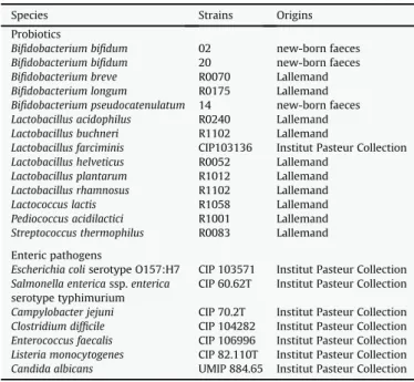
All the strains tested are listed inTable 1. Lactobacillus farciminis CIP 103136 was obtained from Institut Pasteur Collection (Paris, France). Bifidobacterium bifidum 02, 20 and Bifidobacterium pseudo-catenulatum 14 were previously described new-born faeces clinical isolates[18]. Other probiotic strains were obtained from the Lalle-mand Company collection. The strains were stored at "80#C in
Eugon broth (AES, Rennes, France) with 20% (v/v) glycerol (Fluka,
Butch, Switzerland) and reactivated at 37 #C under anaerobic
conditions (Anoxomat System WS 80; Mart Microbiology, Drachten, the Netherlands) on agar plates as indicated: (a) on Reinforcement Clostridial Medium for the bifidobacteria (Oxoid, Cambridge, UK), (b) on MRS (AES, Rennes, France) for the lactobacilli, Lactococcus lactis R1058 and Pediococcus acidilactici R1001 and (c) on M17 (AES, Rennes, France) for Streptococcus thermophilus R0083. Before the experiments, overnight cultures were prepared in MRS broth (AES, Rennes, France) at 37#C under anaerobic conditions.
Potential or opportunistic pathogenic strains were obtained from Institut Pasteur Collection (Paris, France). The strains were stored at "80#C in Eugon broth (AES, Rennes, France) with 20% (v/v)
glycerol (Fluka, Butch, Switzerland) and reactivated at 37#C on agar
plates as indicated: (i) on trypcase soy (Biomérieux, Marcy l’Etoile, France) under aerobic conditions for Escherichia coli serotype O157:H7 CIP 103571, Salmonella enterica subsp. enterica serotype tiphymurium CIP 60.62T and Enterococcus faecalis CIP 106996, (ii) on Columbia with 5% (v/v) sheep blood (Biomérieux, Marcy l’Etoile, France) under aerobic conditions for Listeria monocytogenes CIP 82.110T or (iii) under anaerobic conditions for Clostridium difficile CIP 104282 and (iv) under microaerophilic conditions (GENbox Microaer; Biomérieux, Marcy l’Etoile, France) for Campylobacter jejuni CIP 70.2T, (v) on Sabouraud (Biomérieux, Marcy l’Etoile, France) under aerobic conditions for Candida albicans UMIP 884.65. 2.2. Glucooligosaccharides
The glucooligosaccharides tested and synthesised by CRITT-Bio Industries (Toulouse, France) were: oligoalternan (OA), which consists of alternate
a
-(1,3)/a
-(1,6)-linked glucosyl residues (DP 3to DP 6) and is synthesised by maltose acceptor reaction catalysed by Leuconostoc mesenteroides NRRL B-1355 alternansucrase [19], oligodextran (OD), which consists of
a
-(1,6)-linked glucosylresi-dues (DP3 to DP 9) and is synthesised by maltose acceptor reaction catalysed by L. mesenteroides NRRL B-512F dextransucrase [20], resistant oligodextran (rOD), which consists of
a
-(1,6)-linkedglu-cosyl residues (DP3 to DP 9) with a
a
-(1,2)-linked glucosyl residuelocated at the non-reducing end or constituting a branching point of each oligodextran and is synthesised by maltose acceptor reac-tion catalysed by L. mesenteroides NRRL B-1299 dextransucrase[21]. 2.3. Investigation for probiotic properties
2.3.1. Resistance to simulated gastric and intestinal fluids
The simulated gastric and intestinal fluids used have been described previously[22]. Simulated gastric juice (NaCl 125 mM, KCl, 7 mM, NaHCO3, 45 mM, pepsin, 3 g/L) at a final pH adjusted to
2.5 was inoculated with probiotic strain overnight cultures at a final concentration of 1 $ 107CFU/mL. Suspensions were incubated at 37#C with stirring at 200 rpm. After 3 h of incubation, bacteria were
washed with PBS (Lonza) by centrifugation at 3800 rpm for 10 min and the pellets were resuspended in simulated intestinal fluid (pancreatin 0.1% w/v, bovine bile salts 0.15% w/v) at a final pH adjusted to 8.0. Then, suspensions were incubated at 37#C for 3 h
with stirring at 200 rpm. Cell viability was assayed by plating samples collected after incubation in simulated gastric fluid and after incubation in simulated intestinal fluid. The percentage of bacterial survival was calculated as follows:
CFUassay
CFUcontrol$ 100
where CFUassayrepresents CFU after incubation in simulated gastric
or intestinal fluids and CFUcontrolthe CFU after incubation in PBS as
a control. Separate experiments were performed three times. Table 1
Strains tested.
Species Strains Origins
Probiotics
Bifidobacterium bifidum 02 new-born faeces Bifidobacterium bifidum 20 new-born faeces Bifidobacterium breve R0070 Lallemand Bifidobacterium longum R0175 Lallemand Bifidobacterium pseudocatenulatum 14 new-born faeces Lactobacillus acidophilus R0240 Lallemand Lactobacillus buchneri R1102 Lallemand
Lactobacillus farciminis CIP103136 Institut Pasteur Collection Lactobacillus helveticus R0052 Lallemand
Lactobacillus plantarum R1012 Lallemand Lactobacillus rhamnosus R1102 Lallemand Lactococcus lactis R1058 Lallemand Pediococcus acidilactici R1001 Lallemand Streptococcus thermophilus R0083 Lallemand Enteric pathogens
Escherichia coli serotype O157:H7 CIP 103571 Institut Pasteur Collection Salmonella enterica ssp. enterica CIP 60.62T Institut Pasteur Collection serotype typhimurium
Campylobacter jejuni CIP 70.2T Institut Pasteur Collection Clostridium difficile CIP 104282 Institut Pasteur Collection Enterococcus faecalis CIP 106996 Institut Pasteur Collection Listeria monocytogenes CIP 82.110T Institut Pasteur Collection Candida albicans UMIP 884.65 Institut Pasteur Collection
2.3.2. Growth inhibition of pathogenic strains
Growth inhibition of pathogenic strains by probiotics was eval-uated using an agar spot test[23]. Briefly, 8
m
L of each probioticsuspension at 1 $ 108CFU/mL was spotted onto the surface of MRS agar plates and incubated anaerobically at 37#C for 48 h. Pathogenic
strains were then inoculated in 5 mL of soft agar (containing 0.7% agar) in an appropriate medium, described previously, at a final concentration of 1 $ 107CFU/mL, and poured onto MRS agar with
probiotic spots. The plates were incubated in conditions suitable for each pathogen for 24e48 h. Growth inhibition was then measured. Considering the diameter of inhibition in mm, significant inhibition was defined for inhibition areas having a diameter of at least 50 mm, intermediate inhibition when the diameter was between 20 mm and 50 mm, and inability to inhibit the pathogen for diameters of less than 20 mm. Separate experiments were performed three times. 2.4. Evaluation of glucooligosaccharides for use as carbon source 2.4.1. Probiotic growth on glucooligosaccharides
Glucooligosaccharide consumption by probiotics was evaluated in microtitre plates. MRS broth was reconstituted without glucose according to the composition previously described [24] and completed by filter-sterilised (0.2
m
m) glucooligosaccharide solutionor glucose solution as positive control at a final concentration of 10 g/L. Each well was inoculated with overnight cultures at a final concen-tration of 1 $ 107CFU/mL. Chemically induced anaerobic conditions were obtained with ascorbate at a final concentration of 1 mg/mL. The microtitre plates were sealed with a plastic film in sterile conditions. Growth parameters were kinetically monitored using a POLARstar Galaxy automated microplate reader (BMG Labtech, Offenburg, Germany). Turbidity was assayed at 640 nm every 15 min during 36 h of incubation at 37#C pH values were monitored at the beginning and
end of the cultures. The growth parameters were calculated by stan-dard methods. Separate experiments were performed three times. 2.4.2. High performance liquid chromatography analyses
As the glucooligosaccharides tested were mixtures of different degrees of polymerisation (DP), high performance liquid chroma-tography analyses were performed to quantify the preferentially consumed DP of each glucooligosaccharide when the probiotic culture reached the stationary phase. Samples were prepared as follows: supernatants from probiotic strain growth assays on glu-cooligosaccharides were collected by centrifugation (3600 g/10 min) and filtered (0.2
m
m) for residue removal. Standards were glucose,fructose and saccharose. The solutions were then analysed by HPLC (Ultimate 3000; Dionex, Sunnyvale, USA) on a C18 Synergi 4
m
mhydro-RP 80A column (Phenomenex, Torrance, USA). Elution was performed with ultrapure water at a flow rate of 1 mL/min at 50#C.
Glucooligosaccharide content was determined by refractive index detection (RI-101; Shodex, Kawasaki, Japan). The results were expressed as percentages of consumption of each DP of the glu-cooligosaccharides, corresponding to the following ratio:
½CS&0"½CS&t
½CS&0
$ 100
where [CS]t represents the concentration of each DP at time t of
culture and [CS]0 the concentration measured at time 0. Peaks
related to media components were monitored and subtracted from the area of the corresponding DP. Separate experiments were per-formed three times.
2.4.3. Pathogen growth on glucooligosaccharides
The growth of pathogenic strains on oligosaccharide carbon source was assayed in appropriate reconstituted broth without
glucose and completed with filter-sterilised (0.2
m
m)glucooligo-saccharide solution or glucose solution, as the positive control, at a final concentration of 10 g/L. Five mL of each medium was inoc-ulated with overnight cultures at a final concentration of 1 $ 107CFU/mL. Strains were cultivated at 37#C for 48 h as follows:
(i) in reconstituted trypcase soy broth under aerobic conditions with stirring for E. coli serotype O157:H7 CIP 103571, S. enterica subsp. enterica serotype tiphymurium CIP 60.62T and E. faecalis CIP 106996, (ii) in reconstituted Schaedler broth under aerobic condi-tions for L. monocytogenes CIP 82.110T or (iii) under anaerobic conditions for C. difficile CIP 104282 and (iv) under microaerophilic conditions for C. jejuni CIP 70.2T, (v) on Sabouraud under aerobic conditions with stirring for C. albicans UMIP 884.65. Turbidity at 640 nm was determined at the end of the log phase. The growth parameters were calculated by standard methods. Separate experiments were performed three times.
3. Results
3.1. Investigation for probiotic properties
3.1.1. Resistance to simulated gastric and intestinal fluids
Probiotic survival rates are presented inFig. 1. The bifidobacteria had high survival rates in both simulated gastric and intestinal conditions except for Bifidobacterium longum, which showed a loss of viability >50% after treatment by intestinal fluid. Lactobacillus helveticus and Lactobacillus plantarum were also strongly resistant to the various conditions. Lactobacillus rhamnosus, L. lactis, P. acid-ilactici and S. thermophilus were resistant to the artificial gastric juice but were inhibited to a greater extent by simulated intestinal fluid. P. acidilactici was the least sensitive among the latter strains, while the two strains of Lactobacillus acidophilus, Lactobacillus buchneri and S. thermophilus where much less resistant. L. farciminis had very poor resistance properties in both gastric and intestinal conditions.
3.1.2. Growth inhibition of pathogenic strains
The inhibitory activity exerted by probiotics against pathogens is presented in Table 2. Among the bifidobacteria, only Bifido-bacterium breve and B. bifidum 02 presented a high inhibitory effect against Gram positive bacteria, respectively C. difficile and E. fae-calis, whereas B. bifidum 20 significantly inhibited E. coli serotype O157:H7 as well as S. thermophilus. L. farciminis was the most effective strain for inhibiting pathogen growth with strong effects against the Gram negative and Gram positive bacteria: S. typhi, C. jejuni and L. monocytogenes. L. heleveticus was also able to inhibit growth of C. jejuni and C. difficile, and L. buchneri had a strong activity against C. jejuni. Concerning the other genera, P. acidilactici exerted strong inhibitory activities against C. jejuni and C. difficile and S. thermophilus had antimicrobial effects against E. coli O157:H7. The other strains exerted only intermediate inhibition effects. None of the strains was able to significantly inhibit the growth of C. albicans.
3.2. Evaluation of glucooligosaccharides for use as a carbon source 3.2.1. Probiotic growth on glucooligosaccharides
Kinetic analyses were used to monitor consumption of glucoo-ligosaccharides by probiotics. The experimental conditions were validated by checking with positive and negative control experi-ments performed respectively with modified MRS broth com-plemented with glucose or without any carbon source. Table 3
presents the growth parameters for all the strains tested in pres-ence of the different carbohydrates.
Turbidity, generation time and pH values showed significant growth on OA for all the six bifidobacteria strains and no significant growth rates for lactobacilli. All the bifidobacteria were also able to grow on OD whereas only three of the lactobacilli and L. lactis used this carbohydrate. Microbial development on resistant rOD did not reach significant levels in any strain.
3.2.2. High performance liquid chromatography analyses
Supernatants from cultures supporting a significant growth in presence of OD and OA were collected at the stationary phase of culture and subjected to HPLC analyses in order to determine the degradation rate of each oligosaccharide DP during bacterial growth. The results are listed inTable 4.
Concerning OD, all the strains tested consumed most of the DP3 and DP4 ('80%) except B. bifidum 02, L. helveticus and L. rhamnosus for DP4. DP5 were strongly consumed by B. bifidum 01, B. breve, B. longum and L. farciminis whereas L. lactis was the only strain able to consume DP6 at a similar rate. DP7 and DP8 were poorly hydrolysed in our conditions. Concerning OA, DP3 were consumed
by the six bifidobacteria at some intermediate rates (between 50% and 65%). DP ' 3 were poorly metabolised.
These results suggest the potential use of OD and, to a lesser degree, OA in synbiotic combination with the selected probiotic strains. These carbohydrates were investigated to determine their resistance to pathogenic strains.
3.2.3. Pathogen growth on glucooligosaccharides
OD and OA were subjected to assays with intestinal pathogenic bacteria. Positive and negative controls were conducted in recon-stituted media with glucose or no carbon source.Fig. 2presents the turbidity reached by each strain at the stationary phase of culture. For all pathogenic microorganisms, growth on glucooligo-saccharides did not exceed the negative control average value and remained markedly below the glucose control. These data demonstrate that the intestinal pathogens tested were unable to metabolise OD and OA and suggest a potential use of glucooligo-saccharides as safe prebiotics or, combined with probiotics, in synbiotics.
Fig. 1.Survival rates of probiotic strains after 3 h of exposure to simulated gastric fluid (black bars) and after a consecutive 3 h of exposure to simulated intestinal fluid (white bars). Results are given as the average value of three different experiments ( standard deviation.
Table 2
Pathogen growth inhibition by probiotics expressed in diameter of inhibition expressed in mm. ND means no significant results. Probiotics Pathogenic strains
Gram negative bacteria Gram positive bacteria Yeast
E. coli serotype O157:H7 CIP 103571
S. enterica ssp. enterica serotype typhimurium CIP 60.62T C. jejuni CIP 70.2T C. difficile CIP 104282 E. faecalis CIP 106996 L. monocytogenes CIP 82.110T C. albicans UMIP 884.65 B. bifidum 02 27 ((4) 31 ((2) 46 ((4) 40 ((4) 70 ((8)a 25 ((2) ND B. bifidum 20 53 ((4)a 38 ((5) 33 ((2) ND 22 ((3) 33 ((3) ND B. breve R0070 29 ((2) 23 ((2) 35 ((1) 58 ((1)a 24 ((1) 26 ((3) 19 ((1) B. longum R0175 27 ((3) 36 ((3) 37 ((3) 35 ((3) 25 ((4) 29 ((4) 17 ((2) B. pseudocatenulatum 14 30 ((4) 35 ((3) 45 ((1) 36 ((1) 28 ((2) 35 ((4) ND L. acidophilus R0240 35 ((3) 35 ((3) 40 ((2) 41 ((2) 21 ((3) 46 ((4) 16 ((2) L. buchneri R1102 31 ((2) 33 ((4) 56 ((4)a 46 ((1) 19 ((2) 31 ((3) 17 ((1) L. farciminis CIP 1031136 36 ((4) 58 ((7)a 50 ((1)a 39 ((1) 26 ((4) 53 ((6)a 16 ((1) L. helveticus R0052 44 ((4) 22 ((2) 53 ((1)a 52 ((2)a 32 ((2) 22 ((2) 26 ((2) L. plantarum R1012 34 ((4) 35 ((4) 17 ((1) 29 ((4) 31 ((3) 21 ((3) 21 ((1) L. rhamnosus R0011 44 ((5) 22 ((4) 50 ((1) 41 ((2) 31 ((4) 38 ((3) 19 ((3) L. lactis R0058 28 ((3) 25 ((2) 36 ((1) 35 ((1) ND 21 ((3) ND P. acidilactici R1001 39 ((4) 38 ((4) 50 ((4)a 53 ((2)a 35 ((3) 28 ((4) 21 ((2) S. thermophilus R0083 50 ((5)a 26 ((2) 32 ((3) 32 ((6) 23 ((3) 29 ((2) 15 ((2) aMeans inhibitions considered as significant (diameter > 50 mm).
4. Discussion
In this study, we first aimed to evaluate some probiotic char-acteristics of a panel of lactic acid bacteria. To be considered as probiotic, microorganisms had to meet some selection criteria. Among all the in vitro parameters defined (human origin for human use, survival in gastrointestinal conditions, pathogen inhibition, adhesion to intestinal epithelial cells, etc.) we chose to test resis-tance to acidic and bile salt conditions along the digestive tract and antimicrobial activity as defined previously [25]. Exposure to gastric and intestinal fluids along the digestive tract is the main stress that could decrease the viability of ingested probiotics[26]. Optimum delivery of viable microorganisms to the distal gut is critical for intestinal probiotic effects[27]and acid resistance is required for food applications. Thus we investigated the probiotics’ resistance through a protocol simulating gastric and intestinal conditions as already described [22]. Most of the strains were resistant to gastric conditions but had more variable resistance profiles to artificial intestinal fluid. This decrease in viability after the transition to intestinal fluid from gastric fluid has been described previously and may be due to the rapid pH shift from acidic to basic conditions [28]. The observed variations of such resistance could be explained by strain dependent responses as
reported previously[29]and provide us with a first-level relevant selection criterion, highlighting the strongest resistance effects of the bifidobacteria in the present study. Pathogen inhibition is also a major probiotic selection criterion and this mechanism is involved in the restoration of gut microbiota balance[30]. It thus has significant positive effects in various physiological functions and in the reduction of pathologies such as inflammatory bowel disease or colorectal cancer[31e33]. Here, the antimicrobial effects exerted by the lactic acid bacteria tested were strain-specific. Three strains among the Gram positive and Gram negative intestinal pathogens were inhibited significantly by three of the six bifido-bacteria. Lactobacilli showed the most effective antimicrobial properties overall, with a noteworthy effect of L. farciminis and a lesser but significant effect of L. helveticus. P. acidilactici also showed significant antimicrobial properties. C. albicans resistance to antagonist activities exerted by probiotics has been described previously[12]and explained by yeast resistance to acidic condi-tions, oxidative stress or bacteriocins, which are among the main mechanisms involved in probiotic antibacterial activities [34]. These variations could be used as a second screening criterion. Some of the strains displaying interesting antimicrobial properties were active against only one or a few pathogenic strains. A broader antibacterial activity could be obtained by combining probiotics, Table 3
Growth parameters of probiotic cultures on selected oligosaccharides or glucose as positive control after 48 h of incubation. Results are given as average values of three samples ( standard deviation. ND means no significant results.
Tested strains Growth parameters
DmaxTa Gb DmaxpHc
Glucose OA OD Glucose OA OD Glucose OA OD
B. bifidum 02 1.37 ((0.20) 0.71 ((0.38) 0.77 ((0.03) 05h20 ((01h47) 10h40 ((00h24) 05h02 ((01h59) 1.7 ((0.2) 0.3 ((0.1) 1.2 ((0.4) B. bifidum 20 1.28 ((0.08) 0.58 ((0.09) 0.72 ((0.10) 03h39 ((00h40) 08h07 ((01h35) 05h52 ((01h18) 1.6 ((0.3) 0.4 ((0.1) 1.6 ((0.1) B. breve R0070 1.39 ((0.13) 0.67 ((0.04) 0.95 ((0.07) 03h14 ((00h28) 11h36 ((03h54) 06h24 ((01h33) 1.8 ((0.1) 1.1 ((0.2) 1.6 ((0.1) B. longum R0175 1.31 ((0.04) 0.86 ((0.15) 0.71 ((0.12) 03h03 ((00h39) 08h38 ((01h58) 03h22 ((00h46) 1.7 ((0.4) 1.4 ((0.2) 1.9 ((0.1) B. pseudocatenulatum 14 1.11 ((0.09) 0.57 ((0.24) 0.75 ((0.11) 06h08 ((01h03) 06h40 ((01h47) 04h52 ((01h31) 1.7 ((0.2) 0.3 ((0.1) 1.1 ((0.2) L. acidophilusR0240 0.64 ((0.01) ND ND 03h16 ((01h00) ND ND 1.5 ((0.4) ND ND L. buchneri R1102 1.09 ((0.13) ND ND 05h39 ((01h33) ND ND 1.7 ((0.2) ND ND L. farciminis CIP 103136 1.20 ((0.04) ND 0.95 ((0.19) 02h03 ((00h36) ND 05h27 ((01h55) 1.7 ((0.2) ND 1.6 ((0.6) L. helveticus R0052 1.28 ((0.27) ND 0.79 ((0.08) 04h18 ((00h27) ND 05h26 ((01h22) 1.7 ((0.3) ND 1.3 ((0.8) L. plantarum R1012 1.31 ((0.03) ND ND 04h46 ((00h08) ND ND 1.8 ((0.2) ND ND L. rhamnosus R0011 1.34 ((0.09) ND 0.67 ((0.22) 02h32 ((00h02) ND 06h41 ((00h46) 1.8 ((0.1) ND 1.2 ((0.5) L. lactis R1058 0.55 ((0.07) ND 0.59 ((0.23) 01h39 ((00h04) ND 02h32 ((00h48) 1.6 ((0.2) ND 1.7 ((0.4) P. acidilactici R1001 0.99 ((0.11) ND ND 02h03 ((00h36) ND ND 1.8 ((0.5) ND ND S. thermophilus R0083 0.92 ((0.20) ND ND 01h59 ((00h41) ND ND 0.7 ((0.3) ND ND a D
maxT ¼ maximal variation of turbidity at 640 nm compared to the initial value. b G ¼ generation time (h:min).
c D
maxpH ¼ maximal variation of pH compared to the initial value.
Table 4
Percentage of consumption by probiotics of each degree of polymerisation (DP) of selected oligosaccharides after 48 h of incubation. Results are given as average values of three samples ( standard deviation. ND means no significant results.
Tested strains Carbohydrates
OD OA DP3 DP4 DP5 DP6 DP7 DP8 DP3 DP > 3 B. bifidum 02 97 ((2) 86 ((5) 71 ((10) 55 ((25) 30 ((4) 15 ((6) 49 ((11) 15 ((7) B. bifidum 20 96 ((1) 38 ((6) 39 ((3) 21 ((4) 18 ((3) 18 ((1) 61 ((4) 21 ((7) B. breve R0070 95 ((2) 94 ((1) 84 ((3) 35 ((25) 19 ((10) 12 ((8) 66 ((3) 22 ((1) B. longum R0175 97 ((3) 96 ((3) 86 ((1) 48 ((4) 44 ((3) 2 ((1) 65 ((1) 40 ((3) B. pseudocatenulatum 14 98 ((2) 78 ((9) 48 ((10) 39 ((11) 31 ((16) 23 ((1) 50 ((8) 9 ((7) L. acidophilus R0240 ND ND ND ND ND ND ND ND L. buchneri R1102 ND ND ND ND ND ND ND ND L. farciminis CIP 103136 98 ((1) 83 ((14) 84 ((7) 62 ((23) 22 ((4) 20 ((7) ND ND L. helveticus R0052 97 ((3) 39 ((25) 32 ((12) 29 ((15) 23 ((3) 13 ((8) ND ND L. plantarum R1012 ND ND ND ND ND ND ND ND L. rhamnosus R0011 75 ((11) 33 ((27) 26 ((16) 25 ((22) 19 ((13) 16 ((7) ND ND L. lactis R1058 94 ((5) 85 ((2) 60 ((6) 81 ((7) 38 ((9) 14 ((9) ND ND P. acidilactici R1001 ND ND ND ND ND ND ND ND S. thermophilus R0083 ND ND ND ND ND ND ND ND
especially among the lactobacilli tested strains that were the most effective against the different pathogens.
The second aim of this study was to select some probiotic strains able to grow on glucooligosaccharides, which are potential prebi-otic carbohydrates, in order to propose a rational selection of original synbiotic combinations in vitro. Some works have previ-ously demonstrated glucooligosaccharide fermentability charac-teristics with an induction of bifidobacteria and lactobacilli populations of human colonic microflora [35] or their potential prebiotic effects on vaginal lactobacilli [12]. The microoganisms tested did not use rOD. The absence of significant growth on rOD may be due to the chemical structure of this carbohydrate including an
a
-1,2 glycosidic bond at the non-reducing end or at branch point[21], which is very uncommon. This glucooligosaccharide was demonstrated to be more resistant than dextran with only
a
-1,6glycosidic bonds in pH-controlled fermenters and was more significantly hydrolysed in in vivo conditions [36]. This suggests that the lack of growth on rOD could also be due to our experi-mental conditions, which were characterised by batch conditions, with no pH control and no renewal of nutrients in the culture medium. This is very different from an intestinal ecosystem with a great diversity of microorganisms, where more favourable conditions could lead to the expression of some enzymes involved in specific glycosidic bond hydrolyses. In consistency with probiotic bacteria growth parameters, we confirmed some fermentability characteristics here for two glucooligosaccharides, displaying some potential synbiotic uses: OD and OA. OD was metabolised by a larger number of lactic acid bacterial strains and promoted bacterial growth more efficiently than OA. Bifidobacterium was more effective than the other genus in metabolising glucooligo-saccharides as a carbon source. Bifidobacteria possess a wide range of genes involved in carbohydrate catabolism[37]and have been described for their ability to grow on several short-chain oligo-saccharides, such as fructooligosaccahrides and xylooligo-saccahrides [38,39]. Among these short-chain carbohydrates, bifidobacteria are able to metabolise OD in batch cultures[40,41]
and to hydrolyse OA[42]. OA can also support specific intestinal bacterial growth[43]. Moreover, the absence of significant growth of intestinal pathogens on OD and OA showed the specificity of the metabolisation by probiotics and thus conferred potential prebiotic characteristics on them. This finding is important as some potential prebiotics have been demonstrated to induce the development of pathogenic bacteria in animal models[44]. These properties need to be confirmed in gut models in vitro or in assays in vivo in order to assess the selective growth effects of the tested glucooligo-saccharides on positive bacteria in the context of a diversified microbial community.
OD were more effective than OA to promote development of some lactic acid bacteria when generation time and turbidity at stationary phase culture were considered. This suggests a faster metabolisation of OD than OA by bifidobacteria.
Five of the six bifidobacteria that grew on OD and OA were resistant to the artificial gastric and intestinal conditions but only three of the four lactobacilli using OD were resistant. Some of these glucooligosaccharide-fermenting strains were also able to inhibit intestinal pathogenic bacteria, especially L. farciminis, which had antimicrobial characteristics against one Gram positive and two Gram negative pathogens. However, this tested strain was the most affected by the digestive tract conditions. Considering the good probiotic characteristics displayed in vitro by L. farciminis CIP 103136 in this study and its anti-inflammatory and anti-nociceptive effects
[45,46], this strain should be assayed in formulations enhancing protection against damage induced by gastrointestinal transit, such as microencapsulation in gel beads [47] or ingestion with milk, soymilk or dairy products which buffer the pH variation that can occur along the digestive tract[48,49]. The same techniques could be used to protect other interesting strains before an in vivo evaluation of probiotic or synbiotic properties. HPLC analyses showed a pref-erential consumption of shorter OD DP and only OA DP3 were significantly consumed. Short DP preferential metabolisation could be explained by a faster consumption of these polymers or could be induced by batch conditions with a lack of some nutriments after Fig. 2.Maximal variation of turbidity at 640 nm of pathogenic cultures on glucose as positive control (white bars), without carbohydrates as negative control (black bars) or selected oligosaccharides, oligoalternan (hatched bars) and oligodextran (dotted bars) after 48 h of incubation. Results are given as the average value of three different experiments ( standard deviation. * Different from negative control with no carbohydrates, P < 0.05.
a certain period of bacterial growth. Extended time of analysis or continuous conditions could help to check whether bacteria (lac-tobacilli, Pediococcus and Streptococcus) are able to metabolise larger DP or not, or if it is possible to improve bifidobacteria growth on OA. It would be interesting to test these two glucooligosaccharides enriched in preferentially consumed DP in an attempt to optimise lactic acid bacteria growth. Bifidobacteria were the most effective in using glucooligosaccharides as a carbon source but displayed low antimicrobial characteristics, whereas lactobacilli, P. acidilactici and S. thermophilus significantly inhibited pathogenic strains but were generally less resistant to digestive tract conditions and were unable to grow on glucooligosaccharides. These observations suggest the use of associations of strains with complementary properties. Some studies have already highlighted the interest of combinations of different probiotic strains, especially bifidobacteria and lactobacilli, to improve their intrinsic properties[50,51].
In conclusion, some of the strains tested displayed interesting probiotic characteristics and two potential prebiotic glucooligo-saccharides, OD and OA, were selected for their ability to support growth of specific probiotic bacteria. These carbohydrates need to be assayed in models in vivo to demonstrate their potential prebi-otic properties before proceeding to clinical trial. Bifidobacteria were the most resistant to the gastrointestinal conditions and the most effective in metabolising glucooligosaccharides, and lactoba-cilli displayed the strongest inhibitory effects on enteric pathogens. A combination of a glucooligosaccharide-fermenting bacterium with a non-fermenting one could also improve the glucooligo-saccharides metabolisation for the latter and extend the antibac-terial spectrum. Thus this study has identified some potential synbiotics to be further evaluated for beneficial effects through interaction with human cells in assays in vitro and to extend the screening to in vivo models before potential use in human applications.
Acknowledgements
This work was supported by a grant from Région Midi-Pyrénées. The authors would like to thank Sarah de Souza for her contribution to the present work during her internship and Carine Saux for her technical assistance with HPLC analyses at CRITT-Bio Industries. We are also grateful to Susan Becker and Andrew Swift for correcting the English of this article.
References
[1] Possemiers S, Grootaert C, Vermeiren J, Gross G, Marzorati M, Verstraete W, et al. The intestinal environment in health and disease e recent insights on the potential of intestinal bacteria to influence human health. Curr Pharm Des 2009;15:2051e65.
[2] Packey CD, Sartor RB. Commensal bacteria, traditional and opportunistic pathogens, dysbiosis and bacterial killing in inflammatory bowel diseases. Curr Opin Infect Dis 2009;22:292e301.
[3] Spiller R, Garsed K. Infection, inflammation, and the irritable bowel syndrome. Dig Liver Dis; 2009.
[4] Cani PD, Delzenne NM. The role of the gut microbiota in energy metabolism and metabolic disease. Curr Pharm Des 2009;15:1546e58.
[5] Penders J, Stobberingh EE, van den Brandt PA, Thijs C. The role of the intestinal microbiota in the development of atopic disorders. Allergy 2007;62:1223e36. [6] Bergonzelli GE, Blum S, Brussow H, Corthesy-Theulaz I. Probiotics as a
treat-ment strategy for gastrointestinal diseases? Digestion 2005;72:57e68. [7] Gibson GR, Roberfroid MB. Dietary modulation of the human colonic
micro-biota: introducing the concept of prebiotics. J Nutr 1995;125:1401e12. [8] Teitelbaum JE, Walker WA. Nutritional impact of pre- and probiotics as
protective gastrointestinal organisms. Annu Rev Nutr 2002;22:107e38. [9] Ventura M, O’Flaherty S, Claesson MJ, Turroni F, Klaenhammer TR, van
Sinderen D, et al. Genome-scale analyses of health-promoting bacteria: pro-biogenomics. Nat Rev Microbiol 2009;7:61e71.
[10] Gibson GR. Dietary modulation of the human gut microflora using prebiotics. Br J Nutr 1998;80:S209e12.
[11] Boucher J, Daviaud D, Simeon-Remaud M, Carpene C, Saulnier-Blache JS, Monsan P, et al. Effect of non-digestible gluco-oligosaccharides on glucose sensitivity in high fat diet fed mice. J Physiol Biochem 2003;59:169e73. [12] Rousseau V, Lepargneur JP, Roques C, Remaud-Simeon M, Paul F. Prebiotic
effects of oligosaccharides on selected vaginal lactobacilli and pathogenic microorganisms. Anaerobe 2005;11:145e53.
[13] Roberfroid MB. Prebiotics and synbiotics: concepts and nutritional properties. Br J Nutr 1998;80:S197e202.
[14] Ziemer CJ, Gibson GR. An overview of probiotics, prebiotics and synbiotics in the functional food concept: perspectives and future strategies. Int Dairy J 1998;8:473e9.
[15] Fujimori S, Gudis K, Mitsui K, Seo T, Yonezawa M, Tanaka S, et al. A randomized controlled trial on the efficacy of synbiotic versus probiotic or prebiotic treatment to improve the quality of life in patients with ulcerative colitis. Nutrition 2009;25:520e5.
[16] Liong MT. Roles of probiotics and prebiotics in colon cancer prevention: postulated mechanisms and in-vivo evidence. Int J Mol Sci 2008;9:854e63. [17] Saulnier DM, Gibson GR, Kolida S. In vitro effects of selected synbiotics on the
human faecal microbiota composition. FEMS Microbiol Ecol 2008;66:516e27. [18] Vaugien L, Prevots F, Roques C. Bifidobacteria identification based on 16S rRNA and pyruvate kinase partial gene sequence analysis. Anaerobe 2002;8:341e4. [19] Lopezmunguia A, Pelenc V, Remaud M, Biton J, Michel JM, Lang C, et al.
Production and purification of alternansucrase, a glucosyltransferase from Leuconostoc-mesenteroides Nrrl B-1355, for the synthesis of oligoalternans. Enzyme Microb Technol 1993;15:77e85.
[20] Remaud M, Paul F, Monsan P, Heyraud A, Rinaudo M. Molecular-weight characterization and structural-properties of controlled molecular-weight dextrans synthesized by acceptor reaction using highly purified dex-transucrase. J Carbohydr Chem 1991;10:861e76.
[21] Remaud-Simeon M, Lopez-Munguia A, Pelenc V, Paul F, Monsan P. Production and use of glucosyltransferases from Leuconostoc mesenteroides NRRL B-1299 for the synthesis of oligosaccharides containing alpha-(1e>2) linkages. Appl Biochem Biotechnol 1994;44:101e17.
[22] Zarate G, Chaia AP, Gonzalez S, Oliver G. Viability and beta-galactosidase activity of dairy propionibacteria subjected to digestion by artificial gastric and intestinal fluids. J Food Prot 2000;63:1214e21.
[23] Schillinger U, Lucke FK. Antibacterial activity of Lactobacillus sake isolated from meat. Appl Environ Microbiol 1989;55:1901e6.
[24] De Man JD, Rogosa M, Sharpe ME. A medium for the cultivation of Lactobacilli. J Appl Bacteriol 1960;23:130e5.
[25] Kalliomaki M, Salminen S, Isolauri E. Positive interactions with the micro-biota: probiotics. Adv Exp Med Biol 2008;635:57e66.
[26] Liong MT, Shah NP. Acid and bile tolerance and cholesterol removal ability of lactobacilli strains. J Dairy Sci 2005;88:55e66.
[27] Sanders ME. Probiotics: considerations for human health. Nutr Rev 2003;61:91e9.
[28] Takahashi N, Xiao JZ, Miyaji K, Yaeshiima T, Hiramatsu A, Iwatsuki K, et al. Selection of acid tolerant bifidobacteria and evidence for a low-pH-inducible acid tolerance response in Bifidobacterium longum. J Dairy Res 2004;71:340e5. [29] Charteris WP, Kelly PM, Morelli L, Collins JK. Development and application of an in vitro methodology to determine the transit tolerance of potentially probiotic Lactobacillus and Bifidobacterium species in the upper human gastrointestinal tract. J Appl Microbiol 1998;84:759e68.
[30] Isolauri E, Kirjavainen PV, Salminen S. Probiotics: a role in the treatment of intestinal infection and inflammation? Gut 2002;50(Suppl. 3). III54-9. [31] Kirpich IA, Solovieva NV, Leikhter SN, Shidakova NA, Lebedeva OV, Sidorov PI,
et al. Probiotics restore bowel flora and improve liver enzymes in human alcohol-induced liver injury: a pilot study. Alcohol 2008;42:675e82. [32] Heilpern D, Szilagyi A. Manipulation of intestinal microbial flora for
thera-peutic benefit in inflammatory bowel diseases: review of clinical trials of probiotics, pre-biotics and synbiotics. Rev Recent Clin Trials 2008;3:167e84. [33] Davis CD, Milner JA. Gastrointestinal microflora, food components and colon
cancer prevention. J Nutr Biochem 2009;20:743e52.
[34] Bogovic-Matijasic B, Rogelj I, Nes IF, Holo H. Isolation and characterization of two bacteriocins of Lactobacillus acidophilus LF221. Appl Microbiol Biotechnol 1998;49:606e12.
[35] Wichienchot S, Prasertsan P, Hongpattarakere T, Gibson GR, Rastall RA. In vitro fermentation of mixed linkage glucooligosaccharides produced by Glu-conobacter oxydans NCIMB 4943 by the human colonic microflora. Curr Issues Intest Microbiol 2006;7:7e12.
[36] Djouzi Z, Andrieux C, Pelenc V, Somarriba S, Popot F, Paul F, et al. Degradation and fermentation of alpha-gluco-oligosaccharides by bacterial strains from human colon: in vitro and in vivo studies in gnotobiotic rats. J Appl Bacteriol 1995;79:117e27.
[37] van den Broek LA, Hinz SW, Beldman G, Vincken JP, Voragen AG. Bifido-bacterium carbohydrases-their role in breakdown and synthesis of (potential) prebiotics. Mol Nutr Food Res 2008;52:146e63.
[38] Kaplan H, Hutkins RW. Fermentation of fructooligosaccharides by lactic acid bacteria and bifidobacteria. Appl Environ Microbiol 2000;66:2682e4. [39] Palframan RJ, Gibson GR, Rastall RA. Carbohydrate preferences of
Bifido-bacterium species isolated from the human gut. Curr Issues Intest Microbiol 2003;4:71e5.
[40] Rycroft CE, Jones MR, Gibson GR, Rastall RA. A comparative in vitro evaluation of the fermentation properties of prebiotic oligosaccharides. J Appl Microbiol 2001;91:878e87.
[41] Olano-Martin E, Mountzouris KC, Gibson GR, Rastall RA. In vitro ferment-ability of dextran, oligodextran and maltodextrin by human gut bacteria. Br J Nutr 2000;83:247e55.
[42] Holt SM, Teresi JM, Cote GL. Influence of alternansucrase-derived oligosac-charides and other carbohydrates on alpha-galactosidase and alpha-glucosi-dase activity in Bifidobacterium adolescentis. Lett Appl Microbiol 2008;46:73e9.
[43] Sanz ML, Cote GL, Gibson GR, Rastall RA. Influence of glycosidic linkages and molecular weight on the fermentation of maltose-based oligosaccharides by human gut bacteria. J Agric Food Chem 2006;54:9779e84.
[44] Petersen A, Heegaard PM, Pedersen AL, Andersen JB, Sorensen RB, Frokiaer H, et al. Some putative prebiotics increase the severity of Salmonella enterica Serovar Typhimurium infection in mice. BMC Microbiol 2009;9:245. [45] Lamine F, Eutamene H, Fioramonti J, Bueno L, Theodorou V. Colonic responses
to Lactobacillus farciminis treatment in trinitrobenzene sulphonic acid-induced colitis in rats. Scand J Gastroenterol 2004;39:1250e8.
[46] Ait-Belgnaoui A, Eutamene H, Houdeau E, Bueno L, Fioramonti J, Theodorou V. Lactobacillus farciminis treatment attenuates stress-induced overexpression of
Fos protein in spinal and supraspinal sites after colorectal distension in rats. Neurogastroenterol Motil 2009;21:567e73. e18-9.
[47] Ainsley Reid A, Vuillemard JC, Britten M, Arcand Y, Farnworth E, Champagne CP. Microentrapment of probiotic bacteria in a Ca(2þ)-induced whey protein gel and effects on their viability in a dynamic gastro-intestinal model. J Microencapsul 2005;22:603e19.
[48] Wang J, Guo Z, Zhang Q, Yan L, Chen W, Liu XM, et al. Fermentation charac-teristics and transit tolerance of probiotic Lactobacillus casei Zhang in soymilk and bovine milk during storage. J Dairy Sci 2009;92:2468e76.
[49] Sharp MD, McMahon DJ, Broadbent JR. Comparative evaluation of yogurt and low-fat cheddar cheese as delivery media for probiotic Lactobacillus casei. J Food Sci 2008;73. M3M3775-M3M3777.
[50] Dunne C, O’Mahony L, Murphy L, Thornton G, Morrissey D, O’Halloran S, et al. In vitro selection criteria for probiotic bacteria of human origin: correlation with in vivo findings. Am J Clin Nutr 2001;73:386Se92S.
[51] Collado MC, Isolauri E, Salminen S. Specific probiotic strains and their combinations counteract adhesion of Enterobacter sakazakii to intestinal mucus. FEMS Microbiol Lett 2008;285:58e64.