ةيروهمجلا ةيرئازجلا ةيطارقوميدلا ةيبعشلا ةرازو ميلعتلا يلاعلا و ثحبلا يملعلا
People’s and Democratic Republic of Algeria Ministry of Higher Education and Scientific Research
ةعماج
دمحم
قيدصلا
نب
ىيحي
-لجيج
-University Mohammed Saddik Benyahia -Jijel-
Faculty of Nature and Life Sciences Department: Applied Microbiology and Food Sciences ةيلك مولع ةعيبطلا و ةايحلا مسق ايجولويبوركيملا ةيقيبطتلا و مولع ةيدغتلا
Thesis submitted to obtain master in Biology
Option:
Applied Microbiology
Effect of some conditions on the viability and the
cell release of encapsulated lactic acid bacteria
Examiners: Presented by:
Amel BELOUCIF Chairman :Dr.Sonia BENAILI
Examiner : Mr.Yazid RAHMOUNE Supervisor:Miss.Samiya AMIRA
Academic year 2016-2017
List of figures
Part I.Literature review
Introduction ... 1
I.1.Lactic Acid Bacteria ... 2
I.2.Probiotic ... 2
I.3.Microencapsulation ... 2
I.3.1.Definition ... 2
I.3.2.Materials used to encapsulate cells ... 3
I.3.2.1. Alginate ... 3
I.3.2.2.Gellan gum and xanthan gum ... 3
I.3.2.3. K- Carrageenan... 3
I.3.2.4. Chitosan ... 3
I.3.2.5. Starch ... 4
I.3.2.6. Gelatin ... 4
I.3.2.7.Milk proteins ... 4
I.3.2.8. Cellulose acetate phthalate ... 4
I.3.3.Factors affecting microencapsulation effectiveness of probiotic ... 5
I.3.4. Methods of probiotic microencapsulation ... 5
I.3.4.1-Extrusion ... 6
I.3.4.2-Emulsion ... 6
I.3.4.3. Spray Drying ... 6
Summary
Part II. Materials and methods
II.1.Materials ... 9
II.1.1.Bacteriastrains ... 9
II.1.2.Revivication of bacterial strains ... 9
II.1.3.Media and buffers ... 9
II.1.4.Apparatus ... 9
II.2.Methods ... 10.
II.2.1.Preparation of cells for microencapsulation process ... 10
II.2.2.Encapsulation of bacteria in alginate matrix ... 10
II.2.3.Kinetic of cell release when exposed to Ph acide ... 11
II.2.4.Kinetic off cell release when exposed to 0.3% bile salts ... 11
II.2.5.Heat treatement of free and encapulatedcells ... 11
II.2.6.Free and microencapsulated cells in simulated gastric conditions ... 11
II.2.7.Determination of total viable count ... 11
PartIII.Results and discussion : III.1.Microcapsules characteristics and size ... 12
III.2.Kinetic of cell release whe nexposed to acidic pH2.0 ... 13
III.3.Kinetic of cell release whenexposed to 0.3% bile salt ... 13
III.4. Survival of free and encapsulated cells in simulated gastric conditions ... 15
III.5. Survival of free and encapsulated cells in simulated gastric conditions after 40 days of storage III.6.Bacterial count of free and microencapsulated cells after heat treatements ... 20
Conclusion ... 23 References
List of figures
Figure I.1.Representation of the extrusion procedure.………..6
Figure I.2: representation of the emulsion procedure………..7
Figure I.3: spray-drying thecnique………8
FigureIII.1.Aspect of encapsulated lactic acid bacteria………13
FigureIII.2: Kinetic of cell release of encapsulated cells when exposed to acidic pH2.0………...14
FigureIII.3: Kinetic of cell release of encapsulated cells when exposed at 0.3% of bile salt……...15
Figure III.4: Effect of temperatures on the viability of free and encapsulated L.cellubiosus……...16
FigureIII.5: Effect of temperatures on the viability of free and encapsulated L.plantarum………17
Figure III.6. Effect of temperature on the viability of free and encapsulated L.pentosus………..17
Figure III.7 :viability of free and encapsulated L.cellubiosus in simulated gastrointestinal conditions after 40 day of storage………19
Figure III.8 :viability of free and encapsulated L.plantarum in simulated gastrointestinal conditions after 40 day of storage………..19
Figure III.9 :viability of free and encapsulated Enterococcus feacalis in simulated gastrointestinal conditions after 40 day of storage………..20
Figure III.10:viability of free and encapsulated Lactobacillus plantarum(K18) in simulated gastrointestinal conditions after 40 day of storage……….21
Abbreviation
Expansion
CFU Colony Forming Unit
MRS Man-Rogosa Sharp
LAB Lactic Acid Bacteria
PBS Phosphate Buffer Saline
GRAS Generally Recognized as Safe
GI Gastro-Intestinal
Introduction
1
Lactic acid bacteria (LAB) are useful microorganisms in dairy technology and they also contribute to the benefic effects on the health and their utility is reinforced by their demonstrated probiotic properties (Liu et al., 2011).
The survival of microorganism in the human digestive tract is a challenge for development of probiotic product due to the unfavorable conditions during exposure to processing, storage and gastric conditions which can cause loss of viability ( Bernucci et al., 2017). For this reason; protection by techniques such as microencapsulation may help to maintain the required biomass in the product with good survival rates and viability,and many microencapsulation strategies have been examined for the ability to protect probiotic bacteria from enviromental stress such us high acid and bile salt concentration (Sabikhi et al., 2008).
Viability of encapsulated cells can be affected by the physico chemical properties of capsules,type and concentration of the coating material, particle size,initial cell numbers and bacterial strains are some parameters to be considered (Nazzaro et al., 2012).
Several microencapsulation systems have been proposed for the oral delivery of live probiotic bacteria,among these alginate systems have been widely used to encapsulate these live microorganisms (Ariful et al., 2010).This natural polymer pocess several attractive properties such as good biocompatibility,wide availability, low cost and simple gelling procedure under mild conditions (Bkhitet al., 2016).
The cell release test provides information about the capacity of a gel to retain cells wihin the beads (Ouled Heddar et al., 2016). content of microcapsules are released by a variety of mechanisms,the coating may be mechanically ruptured for example by act of chewing (physical release),coating may melt when exposed to heat or dissolve when placed in solvents. Also, changes in pH may alter the permeability of polymer coatings. Protein or lipid coatings may be degrade by the action of proteases and lipases (Kunz et al., 2003).
The main purpose of this work is to investigate the effect of some conditions on the viability and the cell release of encapsulated lactic acid bacteria with sodium alginate and also to test their resistance to heat treatment and simulated gastrointestinal conditions.
I.1. Lactic Acid Bacteria (LAB)
Lactic acid bacteria are Gram- positive, non sporulating, non respiring cocci or rods, which through fermentation of carbohydrates, produce lactic acid as their major end product( Khalissani,2011). The taxonomy of lactic acid bacteria are based on Gram staining and can be classified according to the nature of the products of bacterial metabolism obtained from carbohydrates. The lactic acid bacteria comprises 13 genera of which the most studied are:Lactobacillus,Lactococcus,Streptococcus,Leuconostoc,Oenococcus,Enterococcus,Pedioco
ccus (Givry,2006, Saad,2010). Lactic acid bacteria are chemotrophic, they take the energy
required for their entire metabolism from the oxidation of chemical copounds. The oxidation of sugars constitutes the principle energy producing pathway. Lactic acid bacteria of genera
Lactobacillus,Leuconostoc and Pediococcus,winemaking, assimilate sugars by either a
homofermentative or heterofermentative pathway( Khalissani,2011).
I.2. Probiotics
Probiotics are defined as live microorganisms which when are administered in adequate amounts confer health benefit to the host. According to(FAO/WHO, 2001) probiotics are said to be vital organisms that when exixts in sufficient amount’s(superieur 10-7
CFU/g of finiched product) Probiotic foods and include usually strains of lactic acid bacteria of genera
Lactobacillus and Bifidobacterium.When a new strain of probiotic bacterium is being tested
to be used in foods, viability test to gastrointestinal conditions and to conditions of processing and storage should be performed.However,cell viability in these products is often low and the ability to survive and multiply in the digestive tract strongly influences the benefits that probiotics can produce (Grazila et al., 2011).
I.3. Microencapsulation I.3.1. Definition
Microencapsulation (ME) is defined as a technology of packaging solids, liquids or gaseous materials in matrices,sealed capsules that can release their contents at controlled rates under the influences of specific conditions.It is a physicochemical or mechanical process to entrap a substance in a material in order to produce particles with diameters of a few nanometers to a few millimeters.A microcapsule consists of a semi permeable,spherical, thin, and strong membrane surrounding a solid/liquid core( Burgain, 2011).Microencapsulation is a promising technique to protect bacteria against adverse conditions to which probiotics can be exposed (Grazila et al., 2011 ; Mirzae, 2012).
Part I Literature review
3
I.3.2. Materials of encapsulation I.3.2.1. Alginate
Alginate is a naturally derived polysaccharide extracted from various species of algae and composed of β-D-mannuronic and α-L-guluronic acids. The composition of the polymer chain varies in amount and in sequential distribution according to the source of the alginate and this influences functional properties of alginate as supporting material. Alginate hydrogels are extensively used in cell encapsulation and calcium alginate is preferred for encapsulating probiotics because of its simplicity,non-toxicity,biocompatibility and low cost (Burgain, 2011) with high mechanical stability,high porosity and tolerance to salts and chelating agents. However, some disadvantages are attributed to the use of alginate. For example, alginate beads are sensitive to the acidic environment which is not compatible for the resistance of the microparticles in the stomach conditions and the scaling-up of the process that is very difficult. (Kaila, 2002 ;Burgain, 2011).
I.3.2.2.Gellan gum and xanthan gum
Gellan gum is a microbial polysaccaride derived from Pseudomonas elodea which is constituted of a repeating units of four monomers that are glucose, glucuronic acid, glucose and rhamnose.A mixture of xanthan - gelan gum has been used to encapsulate probiotic cells and contrary to alginate,the mixture presents high resistance towards acid conditions (Sultana et al., 2000 ;Burgain, 2011).
I.3.2.3. K- Carrageenan
K-Carrageenan is a natural polymer which is commonly used in the food industry. The technology using the compound requires a temperature comprised between 40° and 50° C at which the cells are added to the polymer solution. By cooling the mixture to room temperature, the gelation occurs and then, the microparticles are stabilised by adding potassium ions (Krasaekooptet al., 2003). The encapsulation of probiotic cells in k-carrageenan beads keeps the bacteria in a viable state but the produced gels are brittle and are not able to withstand stresses (Chen and Chen, 2007).
I.3.2.4. Chitosan
Chitosan is a linear polysaccharide composed of glucosamine units which can polymerise in the presence of anions and polyanions. This component has not shown a good efficiency for increasing cell viability by encapsulation (Mortazavian et al., 2007). In fact, encapsulation of probiotic bacteria with alginate and chitosan provides protection in simulated gastrointestinal
conditions and therefore, it is a good way of delivery of viable bacterial cells to the colon; however, chitosan has some disadvantages and it seems to have inhibitory effects on LAB (Chávarri et al ., 2010).
I.3.2.5. Starch
Starch is a polysaccharide constituing of a large number of glucose units joined together by glucosidic bonds. Starch consists mainly of amylose, a linear polymer of D-glucopyranose joined by 1-4 glucosidic bond and amylopectin, a branched polymer of glucose joined by α-1-4 glucosidic bond and α-1-6 glycosidic bond for ramification (Sajilata et al., 2006). Resistant starch is the starch which is not digested by pancreatic enzymes (amylases) in the small intestine. Resistant starch can reach the colon where it will be fermented (Sajilata et
al.,2006 ; Anal andSingh, 2007).This specificity provides good enteric delivery characteristic
that is specificity provides good enteric delivery characteristic that is a better release of the bacterial cells in the large intestine. Moreover, by its prebiotic functionality, resistant starch can be used by probiotic bacteria in the large intestine (Mortazavian et al., 2007).
I.3.2.6. Gelatin
Gelatin is a protein gum used for probiotic encapsulation, alone or in combination with other compounds. Due to its amphoteric nature,it is an excellent candidate for cooperation with anionic polysaccharides such as gellangum.These hydrocolloids are miscible at a pH higher than 6, because they both carry net negatives charges and repel each other.However, the net charge of gelatin becomes positive when the pH is adjusted below the isoelectric point and this causes the formation of a strong interaction with the negatively charged gellan gum (Krasaekoopt et al., 2003 ; Anal and Singh, 2007).
I.3.2.7.Milk proteins
Milk proteins are natural vehicles for probiotics cells and because of their structural and physico-chemical properties, they can be used as a delivery system (Livney,2010). For example, the proteins have excellent gelation properties and this specificity has been exploited by (Heidebach et al.,2009) to encapsulate probiotic cells.The results of these studies are promising and using milk proteins is an interesting way because of their biocompatibility (Livney, 2010).
I.3.2.8. Cellulose acetate phthalate
Because of having a safe nature,cellulose acetate phthalate is used for controlling drug release in the intestine (Mortazavian et al., 2007).The advantage of this component is that it is not soluble at acidic pH (less than 5) but it is soluble at pH higher than 6. The encapsulation
Part I Literature review
5
of probiotic bacteria using cellulose acetate phthalate provides good protection for microorganisms in simulated gastrointestinal conditions (Fávaro-Trindade, 2002).
I.3.3.Factors affecting the effectiveness of microencapsulation
Different parameters can be considered for evaluating the effectiveness of the probiotic encapsulation process such as viability maintenance after encountering detrimental environmentalconditions,cell release/recovery ability and hardening time (Murtazavian et al 2007):
-Capsule characteristics against the surrounding environment:
Good selection of capsule materials is very important. Alginate should be avoided from environment containing high acidity and chelating agents,
-Coating of the capsules:
is an efficient way to improve the physico-chemical characteristics. Shell coating makes the alginate capsules more resistant to chelating agents such as calicium ions.
-Concentration of capsules making solution and beads diameter :
increasing beads diameter, their protecting effects againt the violent environmental factors increase.
-Effect of bacteria on the capsules :
There is a report regarding the digestion of starch capsules by encapsulated bacteria. Therefore prior to select material for encapsulation.
-Modification of capsule materials :
Chemical modification of capsule is a common practise to improve encapsulation effectiveness. Structural modification of the capsule materials might be done by direct structural changes and /or addition of special additives.
-Initial concentration of microbial cells:
A concentration of microbial cells in the encapsulation solution increases, the number of entrapted cells in each bead (cell load) and as a result, quantitative efficiency of encapsulation increases.
I.3.4. Methods of probiotic microencapsulation
Microencapsulation can be achieved by different methods, the principal ones are the, extrusion and emulsion techniques, which have also been called droplet and two phase system methods respectively,are two basic ways for encapsulation of probiotic microorganisms (Krasaekoopt et al., 2003).
I.3.4.1-Extrusion
Extrusion method is the oldest and the most common procedure of producing hydrocolloid capsules. In general,it is a simple and cheap method with gentle operations which makes cell injuries minimal and causes relatively high viability of probiotic cells.Biocompatibility and flexibility are some of the other specification of this method (king, 1995).However, the most important disadvantage of this method is that it can not be feasibly used for large-scale production due to slow formation of the microbeads. In other words, it is difficult to be scaled up. Generally, the diameter of beads formed in this method (2-5 mm) is larger than those formed in the emulsion method. Extrusion method in the case of alginate capsules consists of the following stages:
preparation of hydrocolloid solution, addition of probiotic cells into the mentioned solution in order to form cell suspension and extrusion of the cell suspension through syringe needle in a way that the resulting droplets directly drip into the hardening solution (Klien
et al., 1983;Tanaka et al.,1984; Martinsen et al., 1989 ; Jankowski et al., 1997).
Fig I.1.Representation of the extrusion procedure (Kailasapathy, 2002).
Part I Literature review
7
I.3.4.2-Emulsion
Emulsion technique has been successfully applied for the microencapsulation of lactic acid bacteria.Contrary to the extrusion technique, it can be easily scaled up and the diameter of beads produced is considerably smaller (25 µm-2 mm). However, this method is more expensive compared to the extrusion method due to need of using vegetable oil for emulsion formation (Audet et al., 1988 ; Lacroix et al.,1990 ; Groboillot et al., 1993).
Emulsion produces beads with smaller diameters,because the emulsifiers decrease interfacial tension of the water and oil phases(Adamson, 1982)It has been claimed that by applying emulsifiers of tween 80 and lauryl sulphate together,beads with a range of 25-35 µm in diameter can be produced.Microbeads produced by emulsion method are usually recovered by the membrane filtration technique (Sheu and Marshall,1993 ; Jankowski et
al., 1997).
Figure I.2: Representation of the emulsion procedure( Burgain et al.,2011) I.3.4.3. Spray Drying
Drying of the encapsulated mixture in order to produce cell powders/granules can be achieved by different methods. The most important of these methods are freeze drying, spray drying and fluidized bed drying (Dimantov et al., 2003).
In general, the drying process causes some injuries to the microbeads, release of some cells and reducing viability of the cells.In the freeze drying technique, heat injuries to the cells are minimal compared with other techniques. However, this method is relatively
expensive and difficult to be performed on the industrial scale. Spray drying has been recommended for this reason because it is a relatively cheap method and large volumes of solutions can be processed by this technique.However, viability loss of the cells is high due to presence of both dehydration and heating factors, simultaneously (Fu and Etzel, 1995).In food industry, spray drying is a commonly applied encapsulation method producing large amounts of microcapsules in one continuous process step.This method is very suitable when microencapsulated probiotics need to be dried in order to allow strorage over a long period ( Kailasapathy, 2002 ; Picot and Lacroix, 2004).
Figure I.3: Spray-drying technique (Bilancetti et al.,2010).
I.4 The effect of prebiotic on the viability of encapsulated probiotic:
prebiotic are defined as non digestible ,but fermentable foods that beneficialy affect the host by selectively stimulating the groth and the activity of bacteria.(Quigli et al., 2010) the combination of probiotics and prebiotics is known as a symbiotic combination and is used in food products to take avantages the synergic effets of probiotics and prebiotics.Diffrents starch,including modified starches from diverse botanical sources have been used to protect probiotics(Peredo et al., 2016).
The adding of prebiotics as HI-maize starch,Raftiline and Raftilose to alginate beads improve protection of Lactobacillus acidophilus under in vitro acidic and bile salt conditions and also in stored yogurt ,Lyer and Kailasapathy demonsrated.and also Lotfipour et al.,2012 have demonstrated that the addition of psyllium to alginate beads increases significantly the protection of Lactobacillus acodophilus(Atia,2016).
Materials and Methods
9
This work was realised in the Laboratory of Microbiology,Departement of Applied Microbiology and Food Sciences,University of Jijel, between April and June 2017.
II.1.Materials
II.1.1.Bacterial strains:
Five strains of lactic acid bacteria isolated and identified by Samiya Amira from different origins have been used: Lactobacillus pentosus isolated from Dried camel meat,and
Enterococcus fecalis isolated from dried camel meat and Lactobacillus plantarum isolated
from Qlila,Traditional Algerian Cheese, Lactobacillus cellobiosus from Camel milk II.1.2.Revivication of bacteria strains :
fivestrainschosen for the presentstudyweremaintained as frozen stocks in MRS medium at 4°C, All bacterialcellswererevitalized in MRS broth 37°C for 24h before use.thesestrains have been checked for theirpurity by the pourlpate of MRS and by coloration of Gram.
II.1.3.Media and buffers :
-MRS Medium (Man RogosaScharp) -Buffer solution (PBS)
-Sodium alginate analytical grade manufactured at 2% -Calcium chloride Cacl2 0.5M
-Bile salt 0.3%
-Ethanol solution (20%) -Distilled water
-Normale Saline solution (0.9%) -Tween80
II.1.4.Apparatus :
-pH meter(HANNA instrument) -Balance(Scout Pro)
-Spectrophotometer(Amersham Biosciences) -Precission optical microscope(Paralux)
-Autoclave(Slli AVX electronic) -Syringes(2.5ml)
-Vortex(Minishaker IKA)
-Centrifuge( HETTICH ZENTRFUGEN) -Water bath
- Incubator’s(Mammert) -Colony counter
-Magnetic stirrer II.2.Methods :
II.2.1.Preparation of cells for microencapsulation process:
Five strains chosen for the present workhave activated on MRS broth and the purty was confirmed by the inoculation in MRS agar.Colonies were inoculated in Man Rogosa Sharp broth and incubated at 37C° for 24h. they were centrifuged at (6000 g for 10 min). The pellet containing the cells was washed with normal saline and finally suspended in 10 ml of distilledl water,to reach a final density of 1.6 at 660 nm to the method described by De Prisco et al., (2015).
II.2.2.Encapsulation of bacteria in sodium alginate:
The extrusion technique was adopted, as explained by(Chaikham et al., 2012), with some modifications, 40 ml of 2% sodium alginate solution previously autoclaved mixed with 10 ml of the bacterial cells and aseptically homogenized with a magnetic stirrer, the mixture thus obtained was introduced into a sterile syringe. The solution was dispersed into 200 ml of CaCl2 previously autoclaved and cooled. The beads formed were left under stirring for 30
minutes. All the operation was carried out aseptically. The beads were filtered, and 1 ml of the previous mixture contained about 50 beads, and the number of cells per bead was listed (Chaikham et al., 2012).
II.2.3.Kinetic of cell release when exposed to pH acide.
The rate of cell release from the microcapsules was monitored as function of incubation time by measuring OD 660 nm of the culture as described by Klinkenberg et al.,(2001) with some modifications.The kinetics of cell release was first tested by incubating 15
Materials and Methods
11
microencapsules of diffrent strains in MRS broth (pH=2) for 3h at 37C°,OD 660nm was recorded at 1h intervals over the assay period.
II.2.4.Kinetic off cell release when exposed at 0.3% bile salts :
The study of cell release was realised by incubating of encapsulated cell in MRS broth supplemented with bile salts for 5h,OD was recorded at 3h and 5h intervals.
II.2.5.Heat treatement of free and encapulated cells :
Temperature is one of the important factors that affect the growth of microorganism.Most species have a characteristic range of temperature in which they can grow.Bacterial survival was tested against four time-temperature combinations. (25,40,50,60C°) for 20 minutes of incubation.1 ml of free culture was suspended in 10 ml of sterile distilled water ,also for encapsulated cells.serial dilutions in normal saline and viable counts were carried out before and after the incubation periode(Mandel et al.,2006).
II.2.6.Free and microencapsulated cells in simulated gastrointestinal conditions :
In order tostudy the viability of free an encapsulated strains of lactic acid bacteria in simulated gatric conditions.Simulated gastric juice was prepared by dissolving 0.3%pepsine in 150 ml of stomach solution.50beads of encapsulated cells were incubated in stomach juice after filtration at 37C° fot 2h with gentle movement every 30min then serial dilutions were done and the number of cells was taken.And then they have exposed to intestinal conditions for 4h(Del Piano et al.,2011).
II.2.7.Determination of total viable counts:
The results of viable counts determined by a pour plate method using MRS agar and incubated at 37°C for 48h were expressed in percentage of viability follows(Pacheco and Toro.,2010):
% viability=(log CFUt/logCFUt0).100 Where CFUt= final viable count
III.1.Microcapsules characteristics and size :
Microcapsules were prepared by the emulsion method with alginate at 2%,they appeared as spherical structures in size of about 1.62mm of diameters as the table shows.
Table III.1.General characteristics of the obtained beads
Strains Size of diameter (mm)
form Weight(mg) Number
of cell/bead Number of bead/ml of gel Lactobacillus pentosus 1.62 spherical 7,3 2,5.109 50 Enterococcus faecalis 1.62 spherical 7,5 2,5.109 50 Lactobacillus plantarum 1.62 spherical 7,8 2,87.109 50 Lactobacillus planatarum(K18) 1.62 spherical 6,75 0,05.109 50 Lactobacillus cellubiosis 1.62 spherical 6,9 2,5.109 50
We found 2,5.109 of cells/beads by suspending 4 beads in 9 ml of PBS and vortex, after serial dilutions the number was calculated after the pour plate methode
Results and discusions
13
III.2.Kinetic of cell release when exposed to acidic pH2.0 :
The cell release test provides information about the apacity of a gel to retain cells wihin the beads.the result in figureIII.2 shows that at acidic pH the release rates of five cells was not detectable.the result indicate that the alginate was the stong gel for encapsulation(Ouled heddar et
al.,(2016),our results agree with those found in the study of Ouled heddar et al.,(2016),where they
tested the effect of diffrent types of polymers on the kinetic of the release in same pH that we used,they found that the efficiency of cell entrapment of the gels was as follows starting from the highest capacity to the lowest one :sodium alginate,alginate-agar,alginate –starchand K-carrageenan.
As explanation of those results,the polymer itself,its composition,its texture,its viscosity and the degree of porosity, can affect the cell release(Mortazavian et al.,2007).
FigureIII.2: Kinetic of cell release of encapsulated cells when exposed to acidic pH2.0 III.3.Kinetic of cell release when exposed to 0.3% bile salt :
Cell release from alginate beads of the five srains increased gradually during the incubation period.However the cell release of encapsulated cell of Lactobacillus cellubiosus increased with a low rate after three hours of incubation to reach 0.1,where it remainedstable at this point.for the ell release of Lactobacillus plantarum it increased to reach 0.06 in 3 hour and it was constant after 5h of incubation.for Enterococcus faecalis there is no cell release during the first 3 hours but it increased to reach 0.07 in 5hours.for Lactobacillus pentosus and Lactobacillus plantarum(K3),we
0 0,1 0,2 0,3 0,4 0,5 0,6 0,7 0,8 0,9 1 0 1 2 3 4 5 D O 660nm time of incubation(H)
five strains
five strainsshowed the same release kinetics with an increase to 0.05 after three hours,then a decrease to 0.03 after the hours.
FigureIII.3: Kinetic of cell release of encapsulated cells when exposed at 0.3% of bile salts As observed with acidic pH,bile salts influenced the physical appearance of beads,the diffrent cell release rates obtained with diffrent strains were related to bacterial cells include biomass distribution inside the bead,cell density as well as biomas distribution near the surface of the beads, and the species used .Furthermore interactions between bacterial cells and the polymers were not to be excluded,since they affect the cell release rate (Oulad heddar et al.,2016).
In addition to the previous reasons, bile salt influences the difusion of cells through the membrane biopolymer contained the matrix.(Wijffels,2000).
0 0,02 0,04 0,06 0,08 0,1 0,12 0 1 2 3 4 5 6 D O 6060 N M incubation time (H) Lb5 Lb6 K3 k18 10
Results and discusions
15
III.5. Survival of free and encapsulated cells in simulated gastric conditions
Microencapsulation of lactic acid bacteria not only protects cells from external detrimental enviroment but also ensures the controlled release at the targeted locations (Chen et., al 2017). Viability decreases during gastrointestinal transit due to detrimental conditions such as harsh acidic environment, thus, microencapsulation is considered as an effective approach for their efficient survival under gastrointestinal conditions and during shelf life to maintain their health promoting effects( Cabuk et al., 2015).
In this study, we encapsulated lactic acid bacteria in alginate beads then we tested their viability in gastro intestinal conditions of course comparing to the no encapsulated cells.
Figure III.4 :viability of free and encapsulated L.cellubiosus in simulated gastrointestinal conditions.
As figure III.4 shown the viability of free and encapsulated L.cellubiosus cells decreased by 20% in gactric condition and the microencapsulated cells remained stable after incubated in intestinal condition,for free L.cellubiosus we have a clear decrease in the intestinal incubation. The number of free cells decreased from 33.108 CFU/ml to 1.10 CFU/ml(82%)after treatement in gastric conditions,however after incubation in intestinal condition it reached 0CFU/ml.On the other hand,the encapsulated L.cellubiosus decreased to reach 2,5.108 CFU/ml after incubation in gastric conditions and 1,5.108 after being incubated in intestinal conditions.This indicated that,the viability of L.cellubiosus cells was improved using microencapsulation.
0 20 40 60 80 100 120 0 1 2 3 4 5 vi ab ili ty(% ) Time (h) encapsulated L. cellubiosus free L.cellubiosus
Figure III.5 :viability of free and encapsulated L.plantarum in simulated gastrointestinal conditions.
The result shwen in figureIII.5 The number of free cells decreased from 18.108 CFU/ml
to 1.5.104 CFU/ml(45%)after treatement in gastric conditions,however after incubation in intestinal condition it reached 0.75.10 1 CFU/ml.On the other hand,butter result with the encapsulated
L.platarum ,decreased to reach 2.108 CFU/ml after incubation in gastric conditions and 1.105 after being incubated in intestinal conditions.
Figure III.6 :viability of free and encapsulated Enterococcus feacalis in simulated gastrointestinal conditions.
The result shown in figureIII.6 The number of free cells decreased from 22.108 CFU/ml 0 20 40 60 80 100 120 0 1 2 3 4 5 vi ab ili ty(% ) Time (h) encapsulated L.plantarum free L.plantarum 0 20 40 60 80 100 120 0 1 2 3 4 5 vi ab ili ty(% ) Time (h) encapsulated E.feacalis free E.feacalis
Results and discusions
17
To1.3.108 CFU/ml(86%)after treatement in gastric conditions,however after incubation in intestinal condition it reached 0CFU/ml.On the other hand,butter result with the encapsulated Enterococcus
feacalis,decreased to reach 2.5.108 CFU/ml after incubation in gastric conditions and 1.108 after being incubated in intestinal conditions.
Figure III.7:viability of free and encapsulated Lactobacillus plantarum(K18) in simulated gastrointestinal conditions .
The result shwen in figureIII.7 The number of free cells decreased from 18.108 CFU/ml To 1.108 CFU/ml(86%)after treatement in gastric conditions,however after incubation in intestinal condition it reached 0.5.108 CFU/ml.On the other hand,butter result with the encapsulated Lactobacillus plantarum(K18),decreased to reach 1.5.108 CFU/ml after incubation in gastric conditions and 1.108 after being incubated in intestinal conditions. 0 20 40 60 80 100 120 0 1 2 3 4 5 vi ab ili ty(% ) Time (h) encapsulated L.plantarum(K18) freeL.plantarum
III.2. Survival of free and encapsulated cells in simulated gastric conditions
The results of cells treated with simulated gastric conditions are shown in the figures below
Figure III.8 : viability of free and encapsulated L.plantarum in simulated gastric conditions after 40 days of storage.
The viability of encapsulated cells decreased by 34% in gastric conditions after 2 hours of incubation,on the other hand, the free cells decreased by 53% of the initial count in the same conditions.
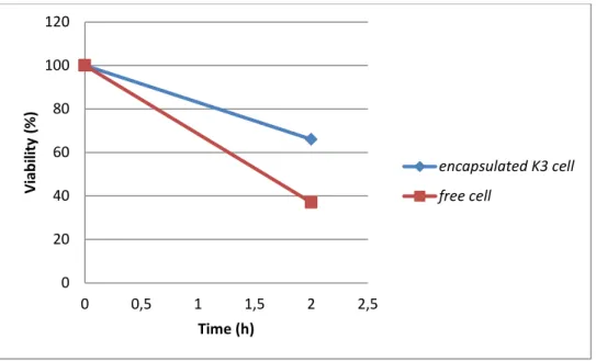
Figure III.9: viability of free and encapsulated L.pentosus in simulated gastric condition after 40 days of storage. 0 20 40 60 80 100 120 0 0,5 1 1,5 2 2,5 Vi ab ili ty (% ) Time (h) encapsulated K3 cell free cell 0 20 40 60 80 100 120 0 0,5 1 1,5 2 2,5 Vi ab ili ty (% ) Time (h) encapsulated Lb5 free cell
Results and discusions
19
As seen in the figure, viability of encapsulated cells was decreased by 40%, however, the count of free cells with the same treatement was 55% lower than the initial count, that means that microencapsulated cells with sodium alginate were better protected to low of acidity.
Figure III.10: viability of free and encapsulated L.cellubiosis in simulated gastric condition after 40 days of storage.
The viability of encapsulated cells in this case decreased to reach 65% ( are viable) in gastric conditions, however the remaining viable free cells were only 40 %.
Protection of probiotics has been proposed with a physical barrier for improving probiotic viability in various food products and also during passage through the humain gastrointestnal trac t(Brinques et al.,2011). Alginate is naturally occuring biopolymer,which is extensively used in bacterial encapsulation because of its biocompatibility good gelling conditions (Cook et al., 2012). It was reported that encapsulation with alginate improved better protection of probiotis in food products(Brinques et al.,2011).
Moreover, In the study of Del piano et al.,( 2011) seven Lactobacillus plantarum probiotic strains were tested for their resistance to both simulated gastric juce and human gastric juice and withdrawn on an empty stomach from healthy individuals. It was noted that less than 20% of the bateria survived after an hour exposure to simulated gastric juice ,while human gastric juice allowed a survival rate between 15% and 45%.
Chandramouli et al.,2004 found significant increase in viable number of L. acidophilus at pH2.0 when encapsulated in alginate. Microencapsulated cells of L.acidophilus in alginate beads survived better after sequencial incubation in simulated gastric juices,and confirm that
0 20 40 60 80 100 120 0 0,5 1 1,5 2 2,5 Vi ab ili ty (% ) Time (h) encapsulated 10 free cell
microencapsulated Lb.acidophilusCSCC2400and Lb.acidophilus CSCC2409 were subjected to low pH and high bile salts concentration under optimal microencapsulation conditions there was a significant increase in viable cells counts compared to the free cells under similar conditions. Higher survival was also reported when Lactobacilli immobilized in alginate beads were incubated in simulated gastric juice (Lee et al.,2004). It was also reported that microencapsulation has been used to increase the survival of probiotics organisms in high-acid-fermented products such as yoghurts ( Sabikhi et al.,2010).
III.4. Viability of free and microencapsulated bacteria cells after heat treatement :
The principal objective of thermal processing is to inactive the spoilage and pathogenic microorganisms and produce a safe product with enhanced shelf life, it is inevitable to kill non-pathogenic organisms that provide health benefits .Therefore, it may be important to search for new methods to selectively control such organisms under thermal processing (Kim et al ., 2008).
To achieve this purpose, this test was carried to study the affect of different temperatures on the viability of Lactobacillus cellubiosus,Lactobacillus pentosus,Lactobacillus plantarum Results are presented in figure III.10.
Figure III.11: Effect of temperatures on the viability of free and encapsulated L.cellubiosus As the figure showed, at 25C° the initial number of about 4.109CFU/ml of free and encapsulated cells was not reduced (100 of viability) with a slight decrease for free cells at 40C° and50C°. However, at 60C°, the viability of both encapsulated and free cells was decreased to 67 and 60 only.
0 20 40 60 80 100 120 25 40 50 60 vi ab ili ty % Temperature c° viability of encapsulated L.cellubiosus viability of free L. cellubiosus
Results and discusions
21
FigureIII.12: Effect of temperatures on the viability of free and encapsulated L.plantarum. As shown in figureIII.5, the survival of free cell show a decrease by the increase of temperaturethey ware : 98%, 73%, 50% after exposure to 40C°,50C°,60C° respectively, however encapsulated cells resist better and showed viability of 100%, 91% and 71% at 25 oC, 40 oC, 50 oC and 60oC respectively.
Figure III.13. Effect of temperature on the viability of free and encapsulated L.pentosus As shown in figure III.12 The viability of free cells showed a decrease by increasing temperature and the viability obtained was 98%,75% , and 46% when exposed to 40C° ,50C°,60C°
0 20 40 60 80 100 120 25 40 50 60 Vi ab ili ty % Temperature C° encapsulated L.p free cell L.p 0 20 40 60 80 100 120 25 40 50 60 Vi ab ili ty % Temperature C° encapsulated L.pentosus free L.pentosus
respectively while encapsulated cells resist better these conditions and viability signaled was 100%,99%,88%.
In general,cell survival after heat treatement indicated that the viable count of free cells were lower than the ancapsulated one,(Kim et al.,2008)in their publications when testing heat resistance of encapsulated L.acidophilus ATCC 431215(1.4-107 CFU/ml) and non encapsulated L.acidophilus ATCC-107CFU/ml) at 65°C for 30min,there was a decreased from (1.4-107 to 3.5-104) and from (1.2-107 to 2.1-105) respectively,there was a higher heat stability of L.acidophilus loaded in alginate than no encapsulated L.acidophilus and it was suggested that microencapsulation using alginate may enhace thermal resistance.
In publication of Ding and Shah,(2007) ,The heat tolerance of free and encapsulated probiotic bacteria incubated at 65C° for up to 1h , was found to be lathal all free probiotic strains tested.Microencapsulated probiotic bacteria survived well at 30 min with an average loss of only 4.17-logCFU/mL compared to free probiotic bacteria with an average loss of 6.74 -logCFU/mL. point.However,after 1h of incubation the survival of free and microencapsulated probiotic was semilair.
Mandal et al.,2006 have studied the viability of microencapsulated Lactobacillus casei in different alginate concentration reported that free cells in distilled water (9.20 log CFU/ml) ware drastically reduced to 5.55,4.93,3.98 log CFU/ml on heat treatement at 55,60,65C° for 20 min, respectively.However,there are no data available at very high temperatures.The survival of the encapsulated probiotic organisms might be due to high concentration 4% of alginate and additional protection given by starch and stearic acid.
Conclusion
23
My work was carried out to test the effect of some conditions on the viability and cell release of encapsulated cells in sodium alginate.
Five strains have been encapsulated in sodium alginate at 2%, they were
L.plantarum(K18),L.plantarum(K3),L.pentosus( Lb5),L.cellubiosis (10),Enterococcus faecalis (Lb6), and gave the following results:
The microencapsulation with sodium alginate exerts a benificial effect on the tested strains in simulated gastro intestinal conditions and the survival of microencapsulated cell in sodium alginate was significatly higher than that free cells where we found:
encapsulated L.plantarum(K3) decreased by 55% however the free cells decreased by20% of the initial count in the same condition.
We are also tested three strains of free and encapsulated lactic acid bacteria for tolerane to heat treatement,obtained result confirm that encapsulated bacteria resist butter: the viability obtained of free L.pentosus( Lb5) was 98%,75% , and 46% when exposed at 40C° ,50C°,60C° respectively,but encapsulated Lb5 resist 100%,99%,88% respectively.
Our results need further studies to prove the use of sodium alginate for encapsulating cells in order to clarify the effectivness of microencapsulation under other conditions which cause damage to alginate.
A-B
Adamson A.W. (1982). Physical Chemistry of surfaces, New York : Wiley Inc.
Adeniyi B.A., Adetoye A. and Ayeni F.A., 2015. Antibacterial activities of lactic acid bacteria isolated from cow faeces against potential enteric pathogens. African Health Sci.15 (3): 888-95. Agrawal R. (2005). Probiotics : an emerging food supplement with health benefits Food
Biotechnology, 19(3) : 227-246.
Anal A.K. and Singh H., 2007. Recent advances in microencapsulation of probiotics for industrial applications and targeted delivery. Trends Food Science & Technology .18: 240-251.
Arena M.P., Caggianiello G., Russo P., Albenzio M., Massa S., Fiocco D., Capozzi V. and SpanoG., 2015. Functional starters for functional yogurt. Foods. 4: 15-33.
Ariful I.M., Yun C.H., Choi Y.J. and Cho C.S. (2010). Microencapsulation of live probiotics bacteria. Journal of Microbiology Biotechnology. 20(10) : 1367-1377.
Atia, A. (2016). Développement d'une matrice prébiotique pour l'encapsulation des probiotiques bactériocinogènes, destinée à l'alimentation animale: de la physicochimie à la biopharmacie (Doctoral dissertation, Université Laval).
Audet P., Paquin C. and Lacroix C. ( 1988). Immobilized growing lactic acid bacteria with K-carrageenan- locust bean gum gel. Appl Microbiol Biotechnol. 29 : 11-18.
Bemucci B.S.P., Loures C.M.G., Lopes S.C.A., Oliveira M.C., Sabino A.P ., Vilela J.M.C., Andrade M.S., Lacerda I.C., Nicoli J.R. and Oliveira E.S. (2017). Effect of microencapsulation conditions on the viability and functionality of Bifidobacterium longum 51A, LWT- Food Science and Technology.
Bekhit, M., Sánchez-González, L., Messaoud, G. B., & Desobry, S. (2016). Encapsulation of Lactococcus lactis subsp. lactis on alginate/pectin composite microbeads: Effect of matrix composition on bacterial survival and nisin release. Journal of Food Engineering, 180, 1-9.
Bilancetti, L., Poncelet, D., Loisel, C., Mazzitelli, S., and Nastruzzi, C. (2010) A statistical approach to optimize the spray drying of starch particles : Application to dry powder coating.
References
25
Borgogna M., Bellich B., Zorzin L., Lapasin R. and Cesáro A. (2010). Food microencapsulation of bioactive compounds: rheological and thermal characterisation of non-conventional gelling system. Food Chemistry 122(2) : 416-423.
Burgain J., Gaiani C., Linder M. and Scher J., (2011). Encapsulation of probiotic living cells : from laboratory scale to industrial applications. Jornal homepage : www. Elsevier. com/ locale/j foodeng.
C-D
ChaikhamP., Apichartsrang koon A., Worametra Chanon S., Supraditarepon W., Chokiatirote E. and Van des Wielet. (2012). Activities of free and encapsulated L. acidophilus LA5 or L.case 01 in processed lonyan juices on exposure to simulated, g intesti tract- journal of science food and agriculture 93 : 2229-2238.
Champagne C. and Fustier P. (2007b). Microencapsulation for delivery of probiotics and other ingredients in functional dairy products. In : Saarela, M. (ED.), Functional Dairy Products, second ed. Woodhead Publishing Ltd., Boca Raton, pp. 404-426.
Chandramouli V., Kalasapathy K., Peiris P. and Jones M. (2004). An improved method of microencapsulation and its evaluation to protect Lactobacillus spp. In simulated gastric conditions. J Microbiol Meth. 56 : 27-35.
Chávarri M., Marañón I., Ares R., Ibáñez F.C., Marzo F and del Carmen Villarán M. (2010). Microencapsultion of a probiotic and prebiotic in alginate- chitason capsules improves survival in simulated gastro-intestinal conditions. Internaional Journal of Food Microbiology. 142(1-2) : 185- 189.
Chen M.J. and Chen K.N. (2007). Applications of probiotic encapsulation in dairy products. In : Lakiss, Jamileh M. (ED), Encapsulation and Controlled Release Technologies in food Systems. Wiley-blackwell,USA, PP.83-107.
Cizeikiene D., Juodeikiena G., Pakevicius A. and bartkiene Elena. (2013). Antimicrobial activity of lactic acid bacteria against pathogenic and spoilage microorganism isolated from food and their control in wheat bread. Food control. 31 (2): 539-545.
Crittenden R., Laitila A., Forsell P., Matto J., Saarela M., Mattila-Sandholm T. and Myllarinen P. (2001). Adhesion of bifidobacteria to granular starch and its implications in probiotic technologies. Applied and Eenvironmental Microbiology 67(8) : 3469-3475.
De prisco A., Maresca D., Ongeng D. and Mauriello D. (2015). Microencapsulation by vibrating technologie of the probiotic strain lactobacillus reuteri DMS 17938 to enhace its survival in foods gastro intestnal enviroment food science and technology 61 : 452-462.
Dimantov A., Greenberg M., Kesselman E. and Shimoni. (2003). Study of high amylase corn starch as food grade enteric coating in a microcapsule model systems. Innov. Food Sci Eng
Technol. 5 : 93-100.
Dinakar P. and Mistry V.V. (1994). Growth ans viability of Bifidobacterium bifidum in cheddar cheese. J Dairy Sci. 77 : 2854-5864.
Ding W.K and Shah N.P., 2009, An improved Method of micro encapsulation of probiotic bacteria for their stability in acidic and bile conditions during storange. Food Microbiol Safety ; 74 : 2.
F-G
FAO/OMS. (2002). Guidelines for the evaluation of probiotics in food.Food and Agriculture Organization of the United Nations and World Health Organization (Organisation Mondiale pour la Santé OMS).Working Group Report. London, Ontario,Canada.
Fávaro- Trindade C.S. and Grosso C.R.F. (2002). Microencapsulation of L. acodophilus (La-05) and B. Lactis (Bb -12) and evaluation of their survival at the pH values of the stomach and in bile. Journal of Microbioloencapsulation 19(4) : 485-494.
Fu W.Y. and Etzel M.R. (1995). Spray drying of Lactococcus Lactis spp. Lactis C2 and cellular injury. J food Sci. 60 : 195-200.
Gevers D. (2002). Tetracycline resistance in lactic acid bacteria isolated from fermented dry sausages. Thèse Doc. Univ. Gent. Fac. Sci. Gent. Belgium.
Gouin S. (2004). Microencapsulation : industrial appraisalof existing technologies and trends. Trends in Food Science and Technology. 15(7-8) : 330-347.
Graziela Brusch Brinques and Marco Zachia Ayub. (2011). Effect of microencapsulation on survival of Lactobacillus plantarum in simulated gastrointestinal conditions, refigeration, and yogurt. Journal of Food Engineering. 103 : 123-128.
Groboillot A.F., Champagne C.P., Darling G.D. and Poncelet D. (1993). Membrane formation by interfacial cross- linking of chitosan for encapsulation of lactobacillus lactis. Biotechnol
References
27
H-J
Heidebatch T., Först P. and Kulozik U. (2009a). Microencapsulation of probiotic cells by means of rennet-gelation of milk proteins. Food Hydrocolloids 23(7) : 1670-1677.
Heidebatch T., Först P. and Kulozik U. (2009b). Transglutaminase-induced caseinate gelation for the microencapsulation of probiotic cells. International Dairy Journal 19(2) : 77-84.
Hogg T., 2005. Essential microbiology. John Wiley & Sons, Ltd. 188-190.
Houria Ouled- Haddar., Mohamed Sifour., Tayeb Idoui., Hamida Bouridane and Somia Arid.( 2016). Lactobacillus plantarum GI Microencapsulation enhanced its Viability during Storage and Gastrointestinal Transit. Sains Malaysiana 45(7) : 1049- 1050.
Jankowski T., Zielinska M. and Wysakowska. (1997). Encapsulation of lactic and bacteria with alginate/ starch capsules. Biotechnol technol. 11: 31-34.
K-L
Kailasapathy K. (2002). Microencapsulation of probiotic bacteria technologie and potentials applications current issues in intestinal microbiology. 3 : 39-48.
Kebary K.M.K., Hussein S.A Badawi R.M.( 1998). Improving viability of Bifidobacterium and ther effect on frozen ice mlik. J Dairy Sci. 26 : 319-337.
Khalissani K. (2011). An overview of lactic acid bacteria, Int.J. Biosci. 1(3) : 1-13.
Kim, W. S., Perl, L., Park, H.J., Tandianus, E. J., and Dunn, W. N. (2001) Assessment of stress response of the probiotic Lactobacillus acidophilus. Current Microbiology. 43(5) :346–350. King A.H. (1995). Encapsulation of Food ingredients : a review of avaible technology, focusing on
hydrocolloids. In : Encapsulation and controlled release of food ingredients, PP. 213-220, S.J. Risch and GA. Reineccius (Eds.). Washington DC. American Chemical Society.
Klien J. Stock J. and Vorlop K.D. (1983). Pore size and properties of spherical calcuim alginate bicatalysts. Eur J APPl Microbiol Biotechnol. 18: 86-91.
Krasaekoopt w ., Bhandari B. and Deeth H. (2004). Comparison of texture of yogurt made from conventionnaly treated milk and UHT milk fortified with low- heat skim milk powder. Journal of Food Science 69 (6) : E276-E280.
chitosan-coated alginate beads in yaghourt from UHT- and conventionally treated milk during storage. LWT – Food Science and technology 39 (2) : 177-183.
Krasaekoopt W., Bhandari B and Deeth H. (2004). The influence of coating materials on some properties of alginate beads and survivability of microencapsulation probiotic bacteria. International Dairy Journal. 14 : 737-743.
Krasaekoopt W., Bhandari B. and Deeth H. (2003). Evaluation of encapsulation techniques of probiotics for yoghurt. Int.Dairy J. 13: 3-13.
Kunz b.and kramer j. (2003). Influence of diffrent capsule materials on the physiological properties of microencapsulated Lactobacilus acidophilus : these de doctorat universite de Bonn allmagne.
Lacroix C., Paquin C. and Arnaud JP. (1990). Batch Fermentation with entraped cells of lactobacillus casei : optimization of the rheological properties of the entrapment gel matrix. APPl
Microbiol Biotechnol. 32 : 403-408.
Lakkis J.M. (2007). Applications of probiotic encapsulation in dairy products in chen MJ et chen KN encapsulated an controlled release technologie in food systems pp.83-112 USA
Lee K.Y. and Heo T.R. (2000). Survival of bifidobacterium longum immobilized in calcium alginate deads in simulated gastric juices and bile salt solution. Applied and Environmental Microbiology 66(2) : 869-973.
Liu C.F., Tseng K.C., Chiang S.S., Lee B.H., Hsua W.H. and Pan T.M. (2011). Immunomodulatory and antioxidant potential of Lactobacillus exopolysaccharides. Journal of Food science and Agriculture. 91: 2284-2291.
Livney Y.D. (2010). Milk proteins as vehicles for bioactives. Current Opinion in Colloid and Interface Science 15(1-2) : 73-83.
M- N-P-Q
Mandel S., Puniya H.K. and Sigh K. (2006). Effet of alginate concentration on survival of microencapsulation L. casei NCDC, International Dairy Journal 16 : 1195-1190.
Martinsen A., Skjak-Braek C. and Smidsrod. (1989). Alginate as immobilization material : Correlation between chemical and physical properties of alginate gel beads. Biotechnol Bioeng. 33 : 79-89.
References
29
Mirzaei, H., Pourjafar, H., & Homayouni, A. (2012). Effect of calcium alginate and resistant starch microencapsulation on the survival rate of Lactobacillus acidophilus La5 and sensory properties in Iranian white brined cheese. Food Chemistry, 132(4), 1966-1970.
Mortazavian A.M. and Sohrabvandi S. (2006c). Probiotics and Food Probiotic products. Eta Publication, Iran, (In Farsi).
Mortazavian A.M., Azizi A., Ehsani M.R., Razavi S.H., Mousavi S.M., Sohrabvandi S. and Reinteimer A.J.(2007). Survival of emcapsulated probiotic bacteria in Iranian yogurt drink ( Doogh) after the product exposure to simulated gastrointestinal conditions. Milchwissenschaft 63(4) : 4276429.
Mozzi F., Raya R.R. et Vignolo G.M. (2010). Biotechnology of lactic acid bacteria: Novel applications. Blackwell. Publishing. 13.
Nazzare F., Oriand P., Fratianni F. and Coppola R. (2011). Microencapsulation in food science and biotechnologique current opinion biotech, 23 : 1-5
Pachaco K.C. and Toro G.V. (2010). Viability of L. dabruekii unde human gastro intestinal conditions simulates in vitro. American Journal of Agricultural and Biological science. 5 (1) : 37-42.
Peredo, A. G., Beristain, C. I., Pascual, L. A., Azuara, E., & Jimenez, M. (2016). The effect of prebiotics on the viability of encapsulated probiotic bacteria. LWT-Food Science and Technology,
73, 191-196.
Picot A. and Lacroix C. (2004). Encapsulation of bifidobacteria in whey protein-based microcapsules and survival in simulated gasrtointestinal conditions and in yoghurt. Int.dairy
J.14 : 505-515.
Quigley, E. M. (2010). Prebiotics and probiotics; modifying and mining the microbiota. Pharmacological research, 61(3), 213-218.
R-S
Rowley J.A., Madlambayan G. and Mooney D.J. (1999). Alginate hydrogels as synthetic extracellular matrix materials. Biomaterials 20(1) : 45-53.
Sabikhi Lath., Babu R., Thopkinson D.K and Suman Kapila. (2008). Resistance of Microecapsulated Lactobacillus acidophilus LA1 to Processing Treatments and Simulated Gut Conditions. Food Bioprocess Technology 3 : 586-593.
Sajilata M.G., Singhal R.S. and Kulkarni P.R. (2006). Resistant starch- a review. Comprehensive Reviews in Food Science and Food Safety 5(1) : 1-17.
Sheu T.Y., Marshall R.T. and Heymann H. (1993). Improving Survival of culture bacteria in frozen desserts by microentrapment. Journalof Dairy Science 76(7) : 1902-1907.
Sheu T.Y.and Marshall R.T. (1993). Microentrapmen of lactobacilli in calcium alginate gels. Journalof Food Science 58 : 557-561.
Streit F. (2008). Acidification improvescryo tolerance of Lactobacillus delbrueckiisubsp.
bulgaricusCFl1. J. Biotechnol. 128 : 659-667.
Sultana K., Godward G., Reynolds N., Arumugaswamy R., Peiris P and Kailasapathy K. (2000). Encapsulation of probiotic bacteria with alginate- starch and evaluation of survival in simulated gastrointestinal conditions and in yaghurt. International Journal Food Microbiology 62 : 47-55.
Sun W. and Griffiths M.W. (2000). Survival of bifidobacteria in yogurt and simulated gastric juice following immobilization in gellan-xanthan beads. International Journal of Food Microbiolgy 61(1) : 17-25.
T-W
Takata I., Tosa T. and Chibata I. (1977). Screening of matrix suitable for immobilization microbal cells. J solid- phase Biochem. 2 : 225-236.
Tanaka H., Masatose M. and Veleky I.A. ( 1984). Diffusion characteristics of substrates in calcium- alginate beads. Biotechnol Bioeng. 26 : 53-58.
Thompson D.B. (2000). Strategies for the manufacture of resistant starch. Trends in Food Sci
Technol. 11 : 245- 253.
Truelstrup-Hansen L., Allan- wojtas P.M., Jin Y.L. and Paulson A.T. (2000). Survival of free and calcium- alginate microencapsulated Bifidobacterium spp. in simulated gastro-intestinal conditions. Food Microbiol. 19 : 35-45.
Annex
MRS broth(Man Rogosa Sharp)Peptone………..10g Yeast extract………..4g Beef extract………8g Glucose ……….20g Dipotassique phosphate……….2g Sodium acetate………5g Ammonium citrate………..2g Manganous citrate………..0.2g Magnesium sulfate ………0.05g Tween 80………1mL pH= 6.2 Eau distillée………1000mL autoclavage 120°C /20min
PBS ( sodium phosphate puffer) :
Na2HPo4………..63.9g/l NaH2Po4………13.8g/l pH=7.4 stomach solution : Nacl ………5g/l Kcl………..2.2g/l NaHCo3……….1.2g/l Cacl2………0.22g/l Autoclavage 120C°/20min
Strains 0h 3h 5h Lb5 0 0.0534 0.0340 Lb6 0 0.0191 0.0718 K3 0 0.0527 0.0364 K18 0 0.0674 0.0765 10 0 0.0996 0.1054
Table2 :OD obtained from kinetic of cell release on pH acid
Strains 0h 2h 3h 4h Lb5 0 0 0 0 Lb6 0 0 0 0 K3 0 0 0 0 K18 0 0 0 0 10 0 0 0 0
Table 3 : effect of heat tratement on free and encapsulated Lb5
Temperature C° 25 40 50 60 Number of free cell 4.109 3,33.109 2.107 3.104 Number of encapsulated cell 4,3.109 4.109 3.108 2.106
Table 4 :effect of heat treatement on free and encapsulated 10
Temperature 25 40 50 60 Number of free cell 4.109 4.109 3.33.109 2.105 Number of encapsulated cell 4.109 4.109 3.109 2.106
Table 5: effect of heat tratement on free and encapsulated K3
Temperature 25 40 50 60 Number of free cell 4.109 3.109 2.107 3.33.104 Number of encapsulated cell 4.109 4.109 3.109 2.107
Annex
Cells time 10 K3 K18 LB6 E F E F E F E F 2 h (₵/ml) 2.5.108 1.108 2.108 1.5.104 1.5.108 1.108 2.5.108 1.33.108 4 h (₵/ml) 1.5.108 0 1.105 0.75.101 1.108 0.5.108 1.108 0 Table 7 :free and encapsulated Lb5 in simulated gastric conditions :Time 0h 2h
Number of free cell 22.109 3.104
Number of encapsulated cell 33.109 17.105
Table 8 : free and encapsulated 10 cell in simulated gastric conditions
Time 0h 2h
Number of free cell 33.108 15.103
Number of encapsulated cell 45.108 34.105
Table 9 : free and encapsulated K3 cell in simulated gastric conditions
Time 0h 2h
Number of free cell 33.108 12.103
صخلملا : تانيجلأ ةدامب ةقيقدلا ةلسبكلا ةيلعاف ةسارد لجأ نم ثحبلا اده سرك زيكرتب مويدوصلا 2 نم تلالاس سمخل ةئملاب يضماح طسو يف اهعضو دنع ايلاخلل ررحت دجوي لا هنأ جئاتنلا ترهظأ ثيح ةفلتخم لوصأ نم ةلوزعم ةينبللا ايريتكبلا زيكرتب ةيوارفصلا حلاملأل ضرعتت امدنع نيح يف 0.3 تكبلا تلالاسلا عيمجل ةلسبكلا نم ايلاخلل ررحت كانه ةئملاب ةيري ثلاثلا تلالاسلا ةمواقم مييقت مت كلدك نم لضفأ جئاتن تطعأ ةفلغلا ايلاخلا نأ ظحول ثيحب ةضفخنملا ةضومحلا لدعمل دنع مويدوصلا تانيجلأ ةدامب ةفلغم ريغ ايلاخلل ةضفخنم ةمواقم كانه ةرارحلل اهضرعت دنع كلدك ةفلغم ريغ ايلاخلا 50.60 .ةيوئم ةجرد ةيحاتفملا تاملكلا مويدوصلا تانيجلأ ةدام ،ةقيقدلا ةلسبكلا، ةينبللا ايريتكبلا : .يمضهلا زاهجلا، Amel BELOUCIF Examiner :Mr. Yazid RAHMOUNE
Supervisor :Miss Samiya AMIRA
Effect of some conditions on the viability and the cell release of encapsulated lactic acid bacteria
Abstract :
Our study has focused on the affirmation of the protective effect of microencapsulation with sodium alginate matrix 2% of five strains of lactic acid bacteria isolated from different origins, the kinetic of cell release at acidic pH2 of L.plantarum, L.pentosus, L.cellubiosus, Enterococcus
faecalis, L.plantarum, cells were not detectable, the results showed that all the strains released
when exposed to 0.3% bile salts.the survival rate under simulated gastric conditions at pH2.0 during 2h was evaluated , encapsulated cells gave better results then free ones heat treatment gave different results at 25,40°C,but at 50,60°C with low resistance of the free cells..
Key words:lactic acid bacteria,microencapsulation,sodium alginate,gastrointestinal
Resumé :
Notre étude a porté sur l'affirmation de l'effet protecteur de la microencapsulation par la matrice d'alginate de sodium 2% de cinq souches de bactéries lactiques isolées de différentes origines, la cinétique de relargage à pH2 de L.plantarum, L.pentosus, L. Cellubiosus,
Enterococcus faecalis, L.plantarum, n'a pas été détectée, les résultats ont montré que toutes
les souches ont un relargage qui augmente legerement lorsqu'elles étaient exposées à des sels biliaires à 0,3%. Le taux de survie dans des conditions gastriques simulées a pH2,0 pendant 2 h a été évalué, les cellules encapsulées ont donné de meilleurs résultats , le traitement thermique a donné des résultats différents à 25,40 ° C, mais à 50,66°C une faible résistance des cellules libres a été mesurée.