ةيبعشلا ةيطارقميدلا ةيرئازجلا ةيروهمجلا
يملعلا ثحبلا و يلاعلا ميلعتلا ةرازو
People’s Democratic Republic of AlgeriaMinistry of Higher Education and Scientific Research
Thesis
Realized by Samiya AMIRA
In Fulfillment of the Requirements for the Degree of Doctor of Sciences
In Biology
Option: Microbial Biotechnology
Under the Supervision of: Pr. Mohammed SIFOUR
Examiners committee
Chairman: Pr. Tayeb IDOUI University Mohamed Seddik Benyahia-Jijel
Supervisor: Pr. Mohammed SIFOUR University Mohamed Seddik Benyahia-Jijel Examiners: Dr. Hani BELHADJ University of Farhat Abbas-Setif 01
Dr. Abdelhakim AOUF University of Farhat Abbas-Setif 01 Dr. Abdelhafid BOUBENDIR University of Abdelhafid Boussouf -Mila
Academic year: 2019/2020
Serial number:
Effect of microencapsulation on the viability of
probiotic bacteria under storage and simulated
gastrointestinal conditions
يحي نب قيدصلا دمحم ةعماج -لجيج ةايحلا و ةعيبطلا مولع ةيلك ةيذغتلا مولعو ةيقيبطتلا ايجولويبوركملا مسقUniversity Mohammed Seddik Benyahia - Jijel Faculty of Nature and Life Sciences
Department of Applied Microbiology and Food Sciences
ii
Acknowledgments
Firstly, I thank Allah for all things given for me,
I would like to express my sincere gratitude to my advisor Prof. Mohammed SIFOUR for the continuous support of my doctorate thesis and related research, for his patience, motivation,
and immense knowledge. His guidance helped me in all the time of research and writing of this thesis. I could not have imagined having a better advisor for my doctorate study. A very
special gratitude goes out to Prof. Houria OULED HADDAR and Prof. Tayeb IDOUI for helping me during the realization of this work.
Besides my advisor, I would like to thank the members of thesis committee, the chairman Prof. Tayeb IDOUI, and the examiners Dr. Hani BELHADJ, Dr. Abdelhakim AOUF and Dr.
Abdehafid BOUBENDIR for their acceptance to evaluate this work.
I am grateful to my mother, who has provided me through moral and emotional support in my life. I am also grateful to my father, brother, sisters and friends who have supported me along
the way.
With a special mention to Dr. Asma Cherbal, for her patience, her help in material provided and thesis redaction. I acknowledge also, Ms. Saliha Hirache and Ms. Moufida Bensam for
their emotional support.
Another person to whom I address my special thanks, for his help in the thesis manuscript, thank you Ahmed.
Big thanks for Ms. Sawsen Hadef, Mr. Tarek Khennouf and Mr.Mohammed Taher Boubezari.
I am grateful to Ministry of Higher Education and Scientific Research to support this research.
My sincere thanks also go to Pr. Gianluigi Mauriello, from the University of Federico II, Naples, Italy, who provided me an opportunity to join his laboratory team: Dr. Diamante Maresca, and Dr. Annachiara De prisco, who gave me the opportunity to access to the
laboratory and research facilities.
I am also grateful to the staff of Laboratory of Applied Microbiology and Laboratory of Molecular Toxicology at the University Mohamed Seddik Benyahia, Jijel.
And finally, I thank everyone who contributed in the realization of this thesis.
List of content
Acknowledgements ... ii
List of abbreviations... iii
List of figures ... iv
List of tables ... vi
Introduction ... 1
I. Literature review I.1. Lactic acid bacteria and probiotics ... 3
I.1.1. Probiotics ... 3
I.1.2. Lactic acid bacteria ... 3
I.1.3. Screening and selection of probiotics ... 5
I.1.4. Health benefits and mechanisms of action of LAB ... 6
I.2. Klila ... 8
I.3. Microencapsulation ... 11
I.3.1. Definition ... 11
I.3.2. Encapsulating materials ... 12
I.3.2.1. Alginate... 12
I.3.2.2. Gellen gum... 12
I.3.2.3. Xanthan gum ... 13
I.3.2.4. Chitosan ... 13
I.3.2.5. Cellulose Acetate Phtalate (CAP) ... 13
I.3.2.6. Starch ... 13
I.3.2.7. K-carrageenan ... 14
I.3.2.8. Gum Arabic (Acacia gum) ... 14
I.3.2.9. Locust bean gum ... 15
I.3.2.10. Gelatin... 15
I.3.2.11.Whey and Milk proteins ... 15
I.3.2.12.Pectin ... 16
I.3.2.13. Chickpea protein ... 16
I.3.3. Encapsulating techniques ... 16
I.3.3.1. Extrusion ... 16
I.3.3.2. Emulsion ... 17
I.3.3.3. Spray drying... 18
I.3.3.4. Coacervation ... 19
I.3.3.5. Spray chilling/cooling/congealing ... 19
I.3.3.6. Freeze-drying ... 19
I.3.4. Factors affecting on microencapsulation effectiveness ... 19
I.3.5. Applications of microencapsulation ... 20
I.3.6. Probiotics in food products ... 20
I.3.6.1. Dairy products ... 20
I.3.7.1.1. Yoghurts ... 21
I.3.6.1.2. Cheese ... 21
I.3.6.1.3. Ice cream and frozen dairy desserts ... 21
I.3.6.2. Non-dairy products ... 21
II. Materials and methods
II.1. Bacterial isolates ... 23
II.2. Media and chemicals ... 23
II.3. Isolation and purification of lactic acid bacteria ... 24
II.4. Phenotypic characteristics ... 24
II.4.1. Gram stain ... 24
II.4.2. Production of gas (CO2)... 24
II.4.3. Fermentation of carbohydrates ... 24
II.4.4. ADH test ... 25
II.4.5. growth at different temperatures ... 25
II.4.6. Growth inpresence of NaCl ... 25
II.5. Moleular characterization ... 25
II.5.1. DNA extraction ... 25
II.5.2. Sequencing of 16S rDNA of bacteria ... 25
II.6. Storage of isolates ... 26
II.7. Characterization of some probiotic and technological traits ... 26
II.7.1. Technological traits ... 26
II.7.1.1. Acidifying ability ... 26
II.7.1.2. Detection of proteolytic activity ... 26
II.7.1.3. Detection of lipolytic activity ... 27
II.7.1.4. Texturing activity ... 27
II.7.2. Probiotic traits ... 27
II.7.2.1. Antibacterial activity ... 27
II.7.2.2. Resistance to antibiotics ... 27
II.72.3. Acid tolerance ... 27
II.7.2.4. Resistance to simulated gastrointestinal conditions ... 28
II.7.2.5. Bile salts tolerance ... 28
II. 7.2.6. Hemolysis test ... 28
II.7.2.7.Cell surface hydrophobicity ... 29
II.7.2.8. Adhesion to Intestinal Epithelial Cells ... 29
II. 7.2.9. Autoaggregation ... 29
II.8. Encapsulation of Lb. casei B1 and Lb.plantarumQ18 ... 30
II.8.1. Encapsulation in sodium Alginate ... 30
II.8.1.1. Procedure of encapsulation ... 30
II.8.1.2. Tolerance to acid pH ... 30
II.8.1.3. Tolerance to NaCl ... 30
II.8.1.4. Storage in strawberry juice ... 30
II.8.2. Encapsulation of Lb. plantarum Q18 in sodium alginate using vibrating technology and coating with chitosan ... 30
II.8.2.1. Viability under simulated gastrointestinal (GI) conditions ... 31
II.8.2.2. Capsules morphology ... 31
II.8.3. Encapsulation of Lb. casei B1 and Lb. plantrum Q18 and using different polymers ... 31
II.8.3.1. Storage at different periods ... 32
II.8.3.2. Viability under simulated gastrointestinal (GI) conditions ... 33
III. Results and Discussion
III.1. Isolation and Identification ... 34
III.2. Technological and probiotic properties ... 37
III.2.1. Technological properties ... 37
III.2.1.1. Acidifying Ability ... 37
III.2.1.2. Proteolytic Ability ... 40
III.2.1.3. Lipolytic Ability ... 41
III.2.1.4. Texturing Ability... 41
III.2.2. Probiotic properties ... 42
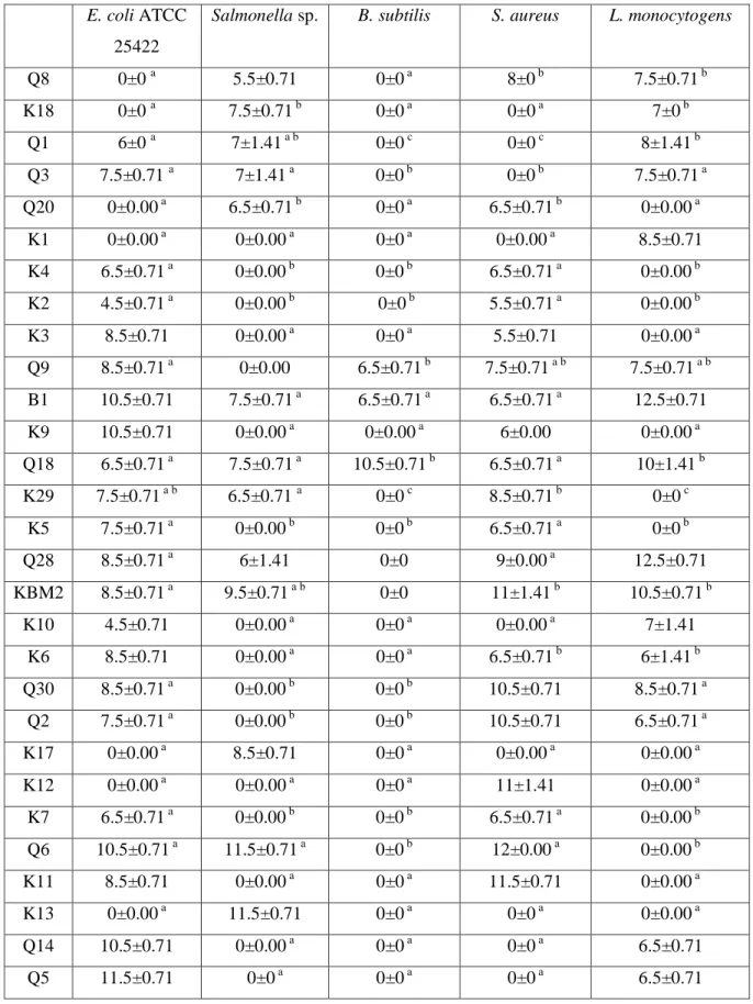
III.2.2.1. Antagonistic activity ... 42
III.2.2.2 Antibiotic Resistance ... 45
III.2.2.3. Resistance to acid pH ... 48
III.2.2.4. Resistance to gastrointestinal conditions ... 49
III.2.2.5. Resistance to bile salts ... 49
III.2.2.6. Hemolytic activity ... 50
III.2.2.7. Adhesion to epithelial cells, autoagregation and hydrophobicity ... 51
III.3. Encapsulation of Lb.casei B1 and Lb. plantarum Q18 ... 53
III.3.1. Encapsulation of Lb.casei B1and Lb. plantarum Q18 in sodium alginate ... 53
III.3.1.1. Tolerance of free and encapsulated bacterial cells to acid stress ... 53
III.3.1.2. Tolerance to NaCl ... 55
III.3.1.3. Storage in strawberry juice ... 58
III.3.1.4. Viability under simulated gastrointestinal (GI) conditions ... 59
III.3.2. Encapsulation of Lb. plantarum Q18 in sodium alginate using vibrating technology and chitosan coating... 59
III.3.2.1.Viability under simulated gastrointestinal (GI) conditions ... 62
III.3.2.2. Visualization of microbeads using the fluorescence microscopy ... 63
III.3.3. Encapsulation of Lb. casei B1 and Lb. plantarum Q18 in different polymers ... 65
III.3.3.1. Bead size and morphology ... 65
III.3.3.2.Viability of free (non encapsulated) and encapsulated bacteria with polymers... 65
III.3.3.2.1.Viability within 4 weeks of storage at 4oC in normal saline ... 69
III.3.3.2.2. Viability of stored bacteria in pineapple beverage for four weeks ... 71
III.3.3.2.3.Viability under simulated gastrointestinal (GI) conditions ... 75
IV. Conclusion ... 81
V. References ... 83
iii
List of abbreviations
ADH: arginine dihydrolase.
CAP: cellulose acetate phtalate.
CFU: colony forming units. GIC: gastrointestinal conditions.
GSJ: gastric simulated juice.
ISJ: intestinal simulated juice.
LAB: lactic acid bacteria.
MRS: Man Rogosa and Sharp medium.
o
D: Dornic degree.
iv
List of Figures
Figure I.1. Mechanisms of pathogen inhibition by the probiotics LAB ... 7
Figure.I.2. Algerian traditional products ... 8
Figure.I.2. Diagram of Klila cheese making ... 10
Figure.I.4. Types of capsules ... 11
Figure I.5. Chemical structure of alginate ... 12
Figure.I.6. Chemical structure of chitosan ... 13
Figure I.7. Structure of k-carrageenan ... 14
Figure I.8. Structure of gum Arabic ... 14
Figure I.9. Chemical structure of locust bean gum, where n indicates the number of galactomannan unit repeats ... 15
Figure I.10. The encapsulation process of probiotics by extrusion technique ... 17
Figure I.11. The encapsulation process of probiotics by emulsion technique ... 18
Figure I.12. Encapsulation process of probiotics by spray drying ... 18
Figure III.1. The phylogenetic trees of the tow selected strains generate by MEGA X program, (a) Lb. plantarum Q18 and (b) Lb. casei B1 ... 36
Figure III.2. Proteolytic activity of the isolates Lb. brevis KBM2, Lb. plantarum Q18, Lb. plantarum (K2), Lb. casei B1 as shown on MRS agar supplemented with 10% skimmed milk. 41 Figure III.3. Viability of isolated bacteria in acidic pH (pH 3) after 24h incubation ... 49
Figure III.4. Viability of isolates in simulated gastric juice (pH2.5, 2h) and in intestinal simulated juice (pH 7.5, 2h).) ... 50
Figure III.5. Adhesion of (a) Lb. plantarum Q18 and (b) Lb. casei B1 to poultry ileum epithelial cells observed with optical microscope (x100). ... 54
Figure III.6. Viability of free and encapsulated Lb. casei B1 at different pH values (pH=2, pH=4, pH=7) after their storage at 4oC for 3 h, 07 and 14 days. ... 52
Figure III.7. Viability of free and encapsulated Lb. plantarum Q18 at different pH values (pH=2, pH=4, pH=7) after their storage at 4oC for 3 h, 07 and 14 days ... 54
Figure III.8. Viability of free and encapsulated Lb. casei B1 at different NaCl concentrations (3%) (a), 6% (b), 9% (c)) after their storage at 4°C for, 3h, 07, 14, 21 and 28 days. ... 56
Figure.III.9. Viability of free and encapsulated Lb. plantarum Q18 at different NaCl concentrations (3%) (a), 6%) (b), 9% (c)) after their storage at 4°C for, 3 h, 07, 14, 21 and 28 days. ... 57
Figure III.10. Viability of free and encapsulated Lb. plantarum Q18 after their storage in strawberry juice at 4°C. ... 59
Figure III.11. Viability of free and encapsulated Lb. casei B1 after their storage in strawberry juice at 4°C. ... 59
Figure III.12. Viability of Lb. plantarum Q18 encapsulated in sodium alginate in gastrointestinal conditions (2h in simulated gastric juice and 4h in intestinal juice). ... 60
Figure III.13. Viability of Lb. casei B1 encapsulated in sodium alginate in simulated gastrointestinal conditions (2h in simulated gastric juice and 4h in intestinal juice) ... 60
Figure III.14. Optical microscopy images of Lb. plantarum Q18 alginate microcapsules at 320 magnification ... 61
v
Figure III.15. Viability of Lb. plantarum Q18 encapsulated in sodium alginate and cocaoted with chitosan in gastrointestinal conditions (2h in simulated gastric conditions and 4h in
intestinal simulated onditions) ... 62 Figure III.16. Fluorescence microscopy images at 400 magnification of stained Lb.
plantarum Q18 in alginate (a) and chitosan-alginate microcapsules (b)... 64
Figure III.17. Macroscopic aspect of the encapsulated Lb. plantarum Q18 with the seven polymers; a: alginate, b: chitosan, c: k-carrageenan, d: glycogen, e: alginate-gum Arabic, f: alginate-locust bean alginate-gum, g: alginate-starch. ... 68 Figure III.18. Viability of free and encapsulated Lactobacillus plantarum Q18 in different polymers and incubated in normal saline for four weeks at 4°C. ... 70 Figure III.19. Viability of free and encapsulated Lactobacillus casei B1 with different
polymers and incubated in normal saline for four weeks at 4°C. ... 70 Figure III.20. Viability of Lactobacillus plantarum Q18 encapsulated in different polymers in pineapple juice for four weeks at 4oC. ... 71 Figure III.21. Viability of Lactobacillus casei B1 encapsulated for four weeks in different polymers in pineapple juice at 4oC. ... 72 Figure III.22. Survival of encapsulated Lb. plantarum Q18 in simulated gastric juice (2h, pH 2.5) and simulated intestinal juice (4h, pH 7.5).. ... 76 Figure III.23. Survival of encapsulated Lb. plantarum Q18 in simulated gastric juice (2h, pH 2.5) and simulated intestinal juice (4h, pH 7.5).. ... 76
vi
List of tables
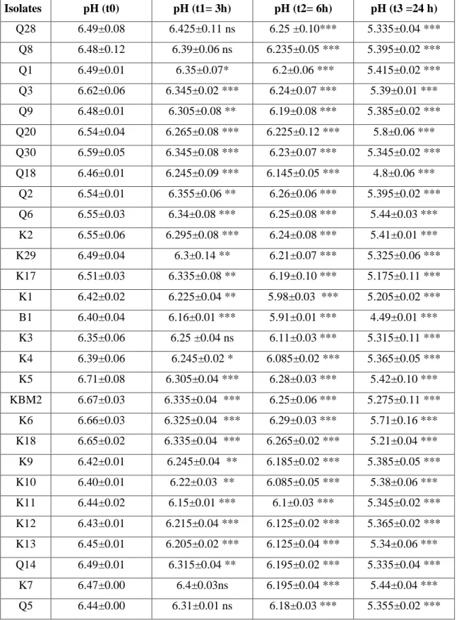
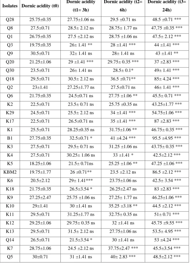
Table I.1. Taxonomy of lactic acid bacteria ... 4 Table III.1. Accession numbers of the isolated and identified LAB from Klila Cheese deposited in GenBank. ... 35 Table III.2. pH variation of different isolates incubated in skimmed milk (12%) for different time………38 Table III.3. Dornic acidity variation of different isolates incubated in skimmed milk (12%) for different time. ... 39 Table III.4. Inhibition zones (mm) of lactic acid bacteria against the tested bacteria ... 43 Table III.5. Succeptibility of lactic acid bacteria towards some antibiotics (diameters in mm) ... 46 Table III.6. Autoaggregation, hydrophobicity, hemolysis and salts resistance of Lb.
plantarum Q18 and Lb. casei B1 ... 52
Introduction
1
Recently, it becomes possible that a food serves as medication, fights diseases and contributes to good health. Hence new concepts with specific terms appeared, i.e. functional foods or super foods. The same meaning was previously proposed by Hippocrates when he said “Let food be the medicine, and let medicine be the food” (Elmalikis et al., 2019). In fact, the denomination of functional foods is related to components they contain, such as natural or added specific minerals, vitamins, fatty acids, dietary fibers and biologically active substances such as phytochemicals, antioxidants, and probiotics and also related to the consequences they have for health enhancement and decrease of diseases risk (Koutelidakis and Dimou, 2016; Chaudhary, 2019).
Lactic acid bacteria (LAB), is an important group of probiotics, well known by their health effects. In fact, they contribute to normal intestinal microflora balance, reduce gastrointestinal and urogenital pathogens number, lower serum cholesterol level and blood pressure and in some cases prevent some cancers (Bron et al., 2012; Khani et al., 2012; Amine et al., 2014). All these properties render lactic acid bacteria successful candidates to be used for functional foods. They are usually added as a concentrated culture to a beverage (fruit juice for example), as a freeze-dried dietary supplement formulated in solid dosage forms (powder, capsules, tablets) or inoculated in prebiotic fibers or in milk based foods (González-Ferrero
et al., 2018).
According to Lee and Salminen (1995), the daily recommended intake of probiotics to be health effective is at least 106-109 living cells, which means that a final processed functional food including stored foods should contain at least this number. Besides, we have to take into consideration the harsh gastrointestinal environment such as acidity of stomach and destructive enzymatic machinery (pepsin), and the intestinal conditions with their basic environment containing pancreatin and bile salts through which probiotics pass to exert their therapeutic properties. At this regard, the challenge of micro-encapsualation appeared as a real potential technology to sustain probiotics viability from their processing (biomass production, lyophilisation, storage, application in food) up to their passage through gastrointestinal tract (Chavarri et al., 2010; Chen et al., 2017).
Microencapsulation of probiotics is a way of covering living cells in order to protect them and release them under appropriate conditions. It uses a variety of materials or polymers, also known as matrices, which are commonly introduced as food additives, and are all originating from nature, like alginate and carrageenan from algae, starch, gum Arabic, soy and pea
Introduction
2
protein from plants, gellan and xanthan from bacteria and milk, gelatin and whey protein from animal origin. These polymers were reported to be biologically compatible and safe for both bacteria and consumer. They are applied with a multitude of techniques such as direct gelation, emulsification or complex coacervation. Characteristics of the final obtained capsules such as morphology, texture, size and porosity and also functional properties are material and technology dependant (Dong et al., 2013; Bosnea et al., 2014; Wang et al., 2014; Eratte et al., 2015). Vesale Pharma (probiotic food supplements producer), Lallemand which developed Probiocap® (a probiotic microencapsulation technology) and the French Capsulae (research and development company that offers customized solutions in the field of microencapsulation) are some examples of research foundations interested in enhancement of microcapsules and selling, in Europe (Deprisco and Maueriello, 2016).
Klila “cheese” is one of the most common Algerian traditional fermented dairy products, consumed in a fresh or dried form. It is a very nutritional product due to its components (high protein and calcium and low fat content). It is important to note that besides its chemical composition, it harbors a multitude of microorganisms considered as indigenous microorganisms (lactic acid bacteria) which influence the nutritional, organoleptic and safety of the final products (Boubekri and Otha, 1996; Mennane et al., 2007; Leksir et al., 2019).
We aim via this work to isolate and identify lactic acid bacteria from an Algerian traditional fermented dairy products “Klila”. The obtained isolates were further characterized for their probiotic and technological aptitudes in order to select the more potent. The aim is also extended to encapsulate the selected bacteria in a variety of polymers and to study the effect of cold storage and the simulated gastrointestinal conditions on the viability of the encapsulated bacteria. Finally, the encapsulated bacteria were introduced in food matrices (pineapple and strawberry beverages) and their viability under storage in cold conditions was studied.
Literature review
3
I.1. Lactic acid bacteria and probiotics I.1.1. Probiotics
Probiotics is a Greek word meaning for life, however, the accepted definition is that given by FAO and WHO (2002), “probiotics are living microorganisms which, when administered in adequate quantities, confer benefits to the host health”. The amount of probiotics that food should contain to be efficient and to be benefic is at least 106 CFU/ml or a daily intake between 108 and 1011 CFU/day (Uriot et al., 2017). Moreover, this number must remain stable, throughout the storage time of the product (Saad et al., 2013). In general, there is a recent agreement that the population requirement of probiotic cultures to exert health beneficial effects to the consumer can vary depending on the strain and the expected beneficial effect (Sireswar et al., 2017).
Probiotics include a variety of microorganisms, however, lactic acid bacteria (LAB) are the most used in foods principally Lactobacillus spp. and Bifidobacterium spp., Enterococcus
faecalis, Enterococcus faecium, Lactococcus and Sporolactobacillus inulinus, and also
non-lactic acid bacteria including Bacillus cereus, Saccharomyces cerevisiae and Saccharomyces
boulardii, Escherichia coli Nissle 1917 and Propionibacterium freudenreichii are also
considered as probiotics (Homayouni, 2014).
I.1.2. Lactic acid bacteria
Lactic acid bacteria are microorganisms sharing the following features: they are Gram-positive, non-spore forming, cocci or rods, catalase-negative, usually non motile, catalase negative, devoid of cytochromes and they are also characterized by their tolerance to low pH (Van Geel-Schuttena et al., 1998; Kaban and Kaya, 2008). Lactic acid bacteria ferment carbohydrates to obtain energy, using endogenous carbon sources as the final electron acceptor instead of oxygen. They are aerotolerant, and are protected against oxygen by-products such as hydrogen peroxide by peroxidases. Phenotypic methods have been most commonly used for the identification of LAB (physiological and biochemical characteristics), however, molecular characterization becomes an efficient tool for identification, it includes: random amplified polymorphic DNA profiling, 16S rRNA gene sequencing, PCR-based fingerprinting and soluble protein patterns (Salminen et al., 1998) and differentiation of species by multiplex PCR assay by using specific recA derived primers (Torriani et al., 2001).
Literature review
4
Cellular morphology, temperature growth range, glucose fermentation mode and also sugar use pattern are the characteristics on which classification of LAB is depending on. LAB are currently belonged to the order Lactobacillales, class of Bacilli and the Firmicutes phylum (Quinto et al., 2014). Six families of LAB belonged to the order of Lactobacillales, they are
Aerococcaceae, Carnobacteriaceae, Enterococcaceae, Lactobacillaceae, Leuconostocaceae, and Streptococcaceae (Sun et al., 2015; Mozzi et al., 2016). Each family includes its corresponding genera as shown in table I.1.
According to the final products of carbohydrates fermentation, LAB are distinguished into two categories homofermentative and heterofermentative microorganisms. If the final product is only lactic acid, LAB are called homofermentative whereas heterofermentative LAB produce lactic acid, acetic acid or alcohol and carbon dioxide (Mokoena et al., 2016). It is important to mention that Bifidobacteria carbohydtates fermentation end products are also lactate and acetate however the pathway followed is fructose-6-phosphate differs from metabolic pathway of LAB. Moreover, C and G percentage is more than 50 mol%, and are not related to LAB but to Actinobacteria (Bjorkroth and Koort, 2011).
Table I.1: Taxonomy of lactic acid bacteria (Mozzi et al., 2016).
Family Genus
Aerococcaceae Abiotrophia, Aerococcus, Dolosicoccus, Eremococcus, Facklamia,
Ignavigranum, Globicatella,
Carnobacteriaceae Alkalibacterium, Allofustis, Alloiococcus, Atopobacter,
Atopococus, Atopostipes, Carnobacterium, Desemzia, Dolosigranulum, Granulicatella, Isobaculum,
Marinilactibacillus, Trichococcus
Enterococcaceae Enterococcus, Melissococcus, Tetragenococcus, Vagococcus
Lactobacillaceae Lactobacillus, Paralactobacillus, Pediococcus
Leuconostocaceae Leuconostoc, Oenococcus, Weissella
Streptococcaceae Lactococcus, Lactovum, Streptococcus
LAB are highly needed in the following field: fermentation of milk, vegetables, sausages, beverages, and bakery products, furthermore, they are responsible of modification of the composition and flavor of the products. LAB can confer the following benefits: they improve the absorption of nutrients (mainly calcium) in the intestinal tract, also, they modulate the immune system, they exhibit antihypertensive effect, they have antimicrobial activity through
Literature review
5
the production of organic acids, carbon dioxide, hydrogen peroxide, ethanol, aromatic compounds and bacteriocins (Calo-Mata et al., 2008).
LAB can be isolated from different sources, showing that their habitats are various. For example Lactococcus and Lactobacillus, Enterococcus, Leuconostoc, Pediococcus, and
Streptococcus are commonly detected in milk and milk products (Stiles and Holzapfel,
1997). LAB are widely distributed in nature and they could be isolated from soils, water, plants, silages, waste products, oral cavity, gastrointestinal tract and genitalia of animals and humans (Konig et al., 2017). Other sources such as traditional fermented foods, traditional fermented drinks, vegetables, and fruit juices are also interesting sources of LAB (Siddiqee et
al., 2013; Schoster et al., 2014).
I.1.3. Screening and selection of probiotics
Probiotics have properties that vary depending on the species or microbial strain. The first criterion is that they must be GRAS (generally recognized as safe), which means being non pathogenic, not allergic and not being able to transfer any antibiotic genes to other bacteria (Sornplang and Piyadeatsoo, 2016). The second important criterion is the resistance to harsh conditions in order to be alive in the target site. In addition to these features, to be considered and used as probiotics, LAB may possess the following criteria: survive, proliferate and colonize their specific locations, have intestinal epithelial adhesion properties, and have the ability to inhibit known pathogens and spoilage organisms (Daliri and Lee, 2015).
The human origin of probiotics is a condition, and despite the diversity of sources of LAB, to use a probiotic strain for the human purpose, it should be isolated from human microflora system which will have the ability to adhere more in the human intestinal cell walls than the others and also must be safe (Gupta et al., 2018).
Another condition is the viability in some conditions of stress and food, storage, and till the end of the shelf life of the products; and must survive and colonize while passing through the gastrointestinal tract (Ali, 2010; Brinti and Shind, 2011; Ravinder et al., 2012). Adhesion to the intestinal epithelial tissue is a primordial criterion for colonization and thus producing health benefits by competing with the other bacteria, and exerting antagonistic effect against harmful pathogens (Tuomola et al., 2001; Marco et al., 2006). The adhesion of LAB to the intestinal epithelial tissue is related to a good adherence ability, to flow-rate of the LAB through the gut and to the presence of mucins to trap, protect and lubricate the intestinal
Literature review
6
surfaces. Furthermore, adhesion of LAB may involve the binding of LAB to a specific cell surface receptor or it may bind to extracellular matrix such as collagen (Howard et al., 2000).
I.1.4. Health benefits and mechanisms of action of LAB
It is well known that the digestive system of humans at birth is empty from microflora, however, the acquisition of microflora appeared through contact to environment and through food consumption. The use of probiotics is valid whenever the balance of the intestinal microflora is broken, the organism itself becomes unable to return to normal. However, the use of probiotics seems to be efficient since they compete with the other bacteria as shown in figure I.1, inhibiting their growth and ultimately leading to the restoration of biological balance (Sanders, 2008; Sornplang and Piyadeatsoo, 2016).
The mechanisms of action of probiotics on the host are complex and some of them are as follow (Chen et al., 2013; Bakirtzi et al., 2016): they serve as supplement to the host microflora and provide protection against various enteric pathogens; they strengthen the gut barrier by competition with pathogenic microbiota for adhesion to the gut; they stimulate and regulate the immune response by initiating the activation of specific genes of localized host cells. They regulate chronic inflammation in intestinal mucosal tissue; this suggests that a direct contact of these probiotics with the various constituents of the intestinal barrier such as endogenous microflora, intestinal mucus and epithelial cells is necessary.
In fact, probiotics affect the immune system by induction of the production of chemokines, cytokines, regulatory T cells, activation of dendritic cells and macrophages and also stimulation of the production of specific antibodies and mucous (Harzallah and Belhadj, 2013; Ranadheera et al., 2014; Shewale et al., 2014). They contribute also in decreasing the duration and preventing intestinal diseases such as Inflammatory Bowel Disease, diarrhea and constipation by colonizing and modulating gut microflora (Van Geel-Schuttená, 2008; Gupta, 2011). It was reported that prevention of diarrhea was related to the consumption of food enriched with probiotic lactic acid bacteria, this food results in the production of lactoferrin, bioactive peptides, flavonoids and many dietary compounds which help in maintaining the gut microflora (shewale et al., 2014), antioxidants and anti-inflammatory, neuropeptides, and polyamines that modulates and also benefits brain health (Harzallah and Belhadj, 2013).
diabetes, obesity and cardiovascular diseases may be also prevented by enhancing gut microbiota, restoring antioxidant system, decreasing insulin resistance and inflammation
Literature review
7
(Parvez et al., 2006); in addition, probiotics may prevent cancer, by detoxification of chemical carcinogens, decreasing the release of toxic metabolites, enhancing antioxidant system, modulating immune response to inhibit self-proliferation of cancer (Kim et al., 2003; Kumar et al., 2010); LAB found to be efficient in lactose intolerance by providing β-galactosidase (Lactase) enzyme (Parvez et al., 2006). They have also benefits in lowering the cholesterol level by precipitating cholesterol with free bile salts into bile acids and thereby reducing cholesterol absorption (Park et al., 2018).
Moreover, harmful pathogens maybe also excluded by probiotic LAB and this by different mechanisms such as adhesion to the active site (Bikila, 2015; Gupta et al., 2018); and production of different substances with antimicrobial effect, (bacteriocins, lactic acid, acetic acid, propionic acid, alcohol, and diacetyl). These compounds interact with the cell membranes of the pathogens and lower the intracellular pH and thus inhibit them or disrupt their membrane permeability through pores formation (Aymerich, 2000; Saraniya and Jeevaratnam, 2014).
Literature review
8
I.2. Klila
Mediterranean countries are characterized by their variety of cheeses produced traditionally. The most popular of them in North Africa are Jben, Lben, Klila and Raib (Mechai and Kiran, 2008). In Algeria, and till now, 10 types of traditional cheeses were characterized, some are consumed fresh while others in a dry form and they are produced throughout the country, from the north to the south and from the east to the west. The well kown ones are Klila and Jben while the less known are Bouhezza, Mechouna and Madeghissa in the east of Algeria (Chaouia region), Takammèrite and Aoules in the south or Igounanes in the middle north (Kabily region). All these cheeses have some common steps, coagulation, draining, salting and for some cheeses also ripening. However, bouhezza seems to be the only ripened traditional cheese (figure I.2) (Mechai and Kiran, 2008; Leksir et al., 2019). Despite this variety in cheeses, industrial ones appeared more popular and consumers still know little about them because their production is generally at a small, local scale.
Figure I.2. Algerian traditional cheese (Leksir et al., 2019)
The cheese Klila has been consumed by the Algerian people for many centuries, probably from as far back as the Antiquity until now (Leksir et al., 2019). It is one of the most well-known traditional fermented dairy products (cheese in Algeria), it is rich with nutritious compounds of varied flavors, aromas, and textures. This cheese is based on the metabolic
Algerian traditionnal cheeses
Fresh cheeses Jben Mechouna(chnina) Ighounane Aghoughlou Kemariya (takemmarite) Oudiouan oulli Fresh klila Hard cheese Ioulsan (Aoules) Takemmarite Dry klila Matured (ripened cheese) Bouhezza Processed cheese) Medghissa
Literature review
9
activity of LAB to ferment sugars, especially glucose and galactose, so to produce lactic acid and aroma substances that give typical flavors and tastes to the fermented products.
The Klila cheese is produced in steppe and mountainous areas. It is traditionally made with milk of ewe, goat or cow (Boubekri and Otha, 1996). In fact, dry Klila is characterized by its long period of storage because it contained more than (> 90%) dry matter which leads to its safety from microbial spoilage (Leksir and Chemmam, 2015).
For denomination, different propositions may be given since the right meaning and the right first use still unknown. According to Leksir and Chemmam (2015), probably, the origin of the term Klila is Berber meaning ikil a curdled milk, or Tiklitt (the milk that curdles spontaneously), the Arabic meaning may be related to the amount of cheese produced compared to the initial amount of milk (it is few), another denomination appeared in the north east of Algeria “Lagta”, the same word used by Berbers but for low-fat cheese made and dried under the sun’s rays. Another denomination is “Lemjeben” in other regions which is also a sun-dried cheese (Leksir et al., 2019).
The cheese fermentation, like many traditional fermenting processes, is spontaneous and uncontrolled and so involves several food microorganisms whose type are influenced by the environmental conditions of the area where the cheese is produced. Microorganisms which are responsible for the acid production in cheese making are LAB (Boubekri and Otha, 1996).
Benamara et al. (2016) reported that the microbiological and biochemical characterization of Klila prepared from the previously mentioned milks showed the presence of Lactobacilli and
Enterococci where Lb. plantarum was the main specie isolated, followed by Pediococcus pentosaceus, Leuconostoc pseudomesenteroïdes and Lb. fermentum. The Enterococcus genus
was dominated by Ec. durans, Ec. faecium and Ec. hirae. Probably, these microbes are representative of the environmental context in which Klila is produced.
Klila is obtained from fermented churned milk called Lben. Lben is heated, drained and pressed to obtain Klila (figure I.3) (Benamara et al., 2016). This popular cheese is still based on a traditional farmhouse production method which contributes to the pleasant sensory attributes and nutritional properties; which explains the increasing consumer demand for
Literature review
10
Klila. Unfortunately only few microbiological, biochemical and technological data are available (Leksir and Chemmam, 2015).
Figure.I.3. Diagram of Klila cheese making (Benamara et al., 2016)
Raw milk
Fermentation (room temperature 24h to 72h)
Rayeb (curd obtention)
Churning (30 min to 45 min) in goatskin (chakoua)
Lben
Skimming
Heating (20 min at 60oC) + draining
Fresh klila
Drying outside (up to 5 days at the sunlight)
Dry klila
Literature review
11
I.3. Microencapsulation I.3.1. Definition
Microencapsulation is defined as a technology of including sensitive ingredients (solid, liquid or gaseous) within several matrices since the ingredients are entrapped or completely surrounded by the protective matrices (Deprisco and Mauriello, 2016). The encapsulated substance is named core material, active agent, filler agent, internal phase, or payload phase. The encapsulating substance is a coating membrane, shell, carrier or wall material, external phase or matrix (Zuidam and Nedovic, 2010).
Hence, microencapsulation of probiotics represents the incorporation of probiotic bacteria into a specific material in order to preserve probiotic viability, reduce cell loss, resist harsh environmental conditions and permit their release under specific conditions (Deprisco and Mauriello, 2016). According to bead size, two types of encapsulation were found: macroencapsulation with beads diameter ranging from millimeters to centimeters and bacterial cells will normally grow on the beads surface due to depletion in nutrient diffusion efficiency in depth of more than 300-500 μm as well as toxic metabolites accumulation in the center of the beads. In case of microencapsulation, beads diameters are between 1 and 1000 μm. Microcapsules are mechanically more robust than macrocapsules (Park and Chang, 2000).
A microcapsule is a sphere with diameter ranging between few microns to 1 mm. Three types of capsules were obtained, reservoir type, matrix and coated matrix type (figure I.4). In the matrix type the active agent dispersed over the encapsulating material. In the reservoir type a layer around the core material was found (also called capsule). The third type is called coated matrix, it is a combination of the previous types, where the active agent is a capsule covered by an additional layer (Lakkis, 2007). The most important properties of microcapsules are the water-insoluble to maintain their integrity in the food matrix and in the upper part of the GI tract and finally, ability of cells release during the intestinal step (Picot and Lacroix, 2004; Ding and Shah, 2007).
Literature review
12
I.3.2. Encapsulating materials
To be used for probiotic encapsulation, the matrix or the biopolymer must have the following criteria: it must be food grade, and it must possess adequate chemical and physical characteristics ensuring protection of bacteria inside. It is important to note that the final morphological and functional characteristics of probiotics are dependent on the type of matrix and on the technique used (Deprisco and Mauriello, 2016).
Wide varieties of polymers were used for encapsulation by researchers, thus, it is important to choose the appropriate polymer according to the objective of encapsulation. The most commonly used food-grade biopolymers include proteins (whey proteins and caseins and gelatin) and carbohydrates (alginate, starch, gums, carrageenan and xanthan) (Etchepare et
al., 2015).
I.3.2.1. Alginate
It is a natural polymer from algal or bacterial origin, structurayl speaking, it is composed of unbranched (1→4)-linked ß-D-mannuronic acid (M) and α -L-guluronic acid (G) residues (figure I.5), it is widely used for bacterial encapsulation, generally as sodium or calcium alginate in the concentration of 0.5-4 %. It has many advantages, mainly it is safe, gel formation is simple and rapid, do not require hard conditions to occur and furthermore, high release in intestinal conditions, however, porosity of gels is the major inconvenient of this gel. (Krasaekoopt et al., 2004; Gouin, 2004).
Figure I.5. Chemical structure of alginate (Lee and Mooney, 2012).
I.3.2.2. Gellan gum
A microbial derived polysaccharide (from Pseudomonas elodea), it is a result of 4 monomers combination (glucose, glucuronic acid, glucose and rhamnose). Upon cooling, gellan gum produces thermo-reversible gel and gelation temperature depends upon polymer concentration, ionic strength and cation type in the solution (Vivek, 2013).
Literature review
13
I.3.2.3. Xanthan gum
Isolated from the bacterium Xanthomonas campestris, the basic units of this polysaccharide are glucose, mannose and glucoronic acid. Due to its high gel setting temperature (80-90oC/1h), it is not compatible with viable cells (Garcia et al., 2000).
I.3.2.4. Chitosan
It is a polysaccharide derived from insect cuticles, membranes of fungi and crustacean shells, the basic units are glucosamine, as presented in figure I.6 (Zargar et al., 2015).It is well known for film forming, this is why it is preferred as coating material surrounding a capsule and avoided as encapsulating one since it affected bacterial cells viability negatively with direct contact (Krasaekoopt et al., 2004).
Figure I.6. Chemical structure of chitosan (Mahapatro and Singh, 2011).
I.3.2.5. Cellulose Acetate Phtalate (CAP)
It is a chemically inert polysaccharide, the reason behind its use in bacterial encapsulation, it is also non toxic and highly resistant to acid environment. CAP is widely used as a coating agent. It is used for controlling drug release in the intestine due to its safe nature and because it is physically inert (mortazavian et al., 2008). The encapsulation of probiotic bacteria using CAP provides good protection for microorganisms in simulated GI conditions (Chopde et al., 2014).
I.3.2.6. Starch
Polysaccharide where monomers are glucose units, molecules linked together with α -D-(1-4) and/or α -D-(1-6) linkage. The specific feature of starch (resistant starch) is its ability to be fermented in colon since it resists pancreatic enzymes and as a result released in large intestine. Hence, resistant starch has a double function; it is used as prebiotic by probiotic cells and serves as a carrier for them (Mortazzavian et al., 2008).
Literature review
14
I.3.2.7. K-carrageenan
It is a natural polysaccharide associated to sulphate groups as showing figure I.7 (Kariduraganavar et al., 2014). It is used as a thickener and as a stabilizer agent in foods; nevertheless, it is not assimilated by the human body (fiber). It is used for cells encapsulation at temperature of 40-50oC, however, it is not suitable to be used for gastrointestinal stress resistance (Krasaekoopt et al., 2003; Chen and Chen 2007).
Figure I.7. Structure of k-carrageenan (Kariduraganavar et al., 2014).
I.3.2.8. Gum Arabic (Acacia gum)
It is a natural polysaccharide extracted from acacia trees and it is composed of various monomers (figure I.8) (Mariod, 2018) in a complex manner with branched chains, it is known by its low viscosity and high water solubility. Gum Arabic is a complex highly branched polysaccharide, consist of mixed calcium, magnesium and potassium salt of polysaccharides acid. Its main chain is composed of 1,3-linked β-ᴅ-galactopyranosyl units and the side chain (2 to 5 1,3- linked β-D-galactopyranosyl) units are joined to the main chain by 1,6- linkages. Both the main and side chains comprise α-ʟarabinofuranosyl, α-ʟ-rhamnopyronosyl, β-ᴅ-glucuronopyranosyl and 4-O-methyl-β-ᴅ-glucuronopyranosyl (Ali et
al., 2009).
Literature review
15
I.3.2.9. Locust bean gum
The structure of this polysaccharide is presented in figure I.9, the principal units are galactomannan while mannose and galactose contents have been reported to be about 73-86% and 27-14%, respectively (mannose:galactose ratio are of approximately 4:1) joined to form a linear chain of (1 → 4)-linked β-D-mannopyranosyl units (mannopyranose) with(1 → 6) linked α-D galactopyranosyl residues (galactopyranose) as side chains (Menieur et al., 2014).
It is extracted from the seed of the locust/carob tree (Ceratonia siliqua (L.) Taub) of the family Leguminosae (Menieur et al., 2014). Locust bean gum is a cationic, natural polysaccharide and used to improve the stability of alginate beads (Cheow et al., 2014).
Figure I.9. Chemical structure of locust bean gum, where n indicates the number of galactomannan unit repeats (Menieur et al., 2014).
I.3.2.10. Gelatin
Gelatin is another type of polymers used for bacterial cells encapsulation, it is a protein with amphoteric property which has the ability to combine with polysaccharides and form efficient capsules (Krasaekoopt et al., 2003). Gelatin, a biodegradable protein material derived from the partial collagen hydrolysis, was the primary commercial choice as a wall material due to its excellent water solubility, emulsifying and thickening capacity, and high crosslinking activity due to the presence of primary amino groups (Shu et al., 2006).
I.3.2.11. Whey and Milk proteins
Whey proteins are a mixture of globular proteins isolated from whey, the liquid material created as a result of the production of cheese. All of them are characterized by their high ability to form gels and to probiotic encapsulation and delivery due to their physicochemical properties mostly biocompatibility (Liveney, 2010).
Literature review
16
I.3.2.12. Pectin
Pectin is a heteroploysacchride mainly extracted from fruits. It is used as gelling agent in food, in medicines and as a source of dietary fibers. It remains intact in the stomach and in the small intestine; hence, it is used solely or in combination with other matrices to encapsulate probiotic bacteria (Gebara et al., 2013).
I.3.2.13. Chickpea protein
It is a vegetable encapsulating material with excellent functional and nutritional properties with low allergic reactions. This protein is constituted of types of salt-soluble globulin-legumin and vicilinttributes. It offered good protection for encapsulated cells against gastric conditions and it is also found that it is a good carrier for applications in food (Klemmer et
al., 2011; Wang et al., 2014).
I.3.3. Encapsulating techniques
A wide range of encapsulation techniques were described, discussed in different reviews, as well as encapsulating materials, however, each technique has advantages and disadvantages and by consequence, its effectiveness is highly related to the availability, to cost, and to biocompatibility. Among the technologies applied: emulsion, spray drying and extrusion are the most studied and applied on both laboratory and industrial scale. New technologies as complex coacervation and vibrating technology seem to be efficient (Bosnea et al., 2014; De Prisco et al., 2015).
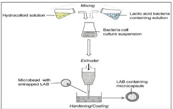
I.3.3.1. Extrusion
Extrusion is a simple and low cost procedure that makes minimal injuries where viability of probiotics is efficiently maintained (Mortazavian et al., 2007). It is an easy technique where a mixture of probiotic cells and encapsulating matrix was injected into a hardening solution through an extruder (syringe) as shown in figure I.10. The crosslinking occurred with calcium ions (Krasaekoopt et al., 2004). Mostly, alginate and calcium chloride concentration ranges from 0.5% to 4% and from 0.05 to 1.5 M, respectively, and the beads size ranges from 2 to 3 mm in diameter which mostly depends upon the distance between syringe and hardening solution, polymer type, viscosity, concentration and mainly diameter of the extruder orifice (Solanki et al., 2013).
Literature review
17
Figure 1.10. The encapsulation process of probiotics by extrusion technique (Feucht and Kwak, 2013).
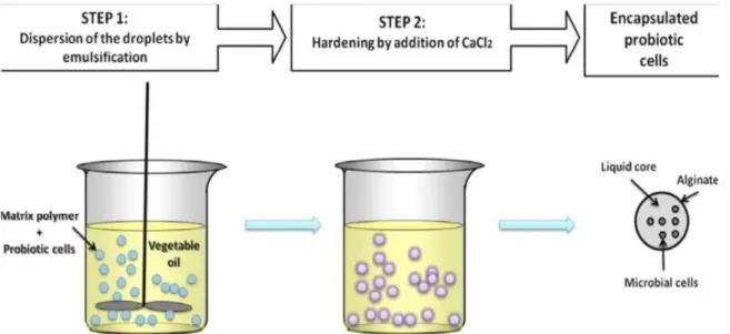
I.3.3.2. Emulsion
It is a chemical interaction between two phases, continuous (soybean, sunflower, canola, olive oil..) and discontinuous (cell polymer), calcium chloride is also needed as a hardening agent, beads sizes varied between small and large, they are encapsulating material, concentration and viscosity and agitation rate dependent (figure I.11) (Chávarri et al., 2012).
While emulsion technique is characterized with many advantages: high viability of cells and the ease of the scale-up; it has also some disadvantages: the large size range and shape in addition to the cost (expensive method). Alginate, carrageenan, and pectin are widely used in emulsion (Burgain et al., 2011).
Literature review
18
Figure I.11. The encapsulation process of probiotics by emulsion technique (Burgain et al., 2011).
I.3.3.3. Spray drying
It is so called because the final obtained capsules appeared as a dry powder, the principle consists of the addition of a mixture of bacterial cells and encapsulating material to a drying gas where atomization occurred (figure I.12) (Chávarri et al., 2012).
Comparing to the other techniques, small capsules are obtained, it is considerably cheap, rapid, widely scale-up and so adequate for industrial use, however, cell viability is reduced as consequence of the use of high temperature which is the limiting factor for this technique. The common matrices used are: polysaccharides, proteins, skim milk, gelatin, starch, gum Arabic (Burgain et al., 2011).
Literature review
19
I.3.3.4. Coacervation
This microencapsulation technique utilizes phase separation of one or more incompatible polymers from the initial coating polymer solution under specific temperature, pH or composition of the solution. The incompatible polymer is added to the coating polymer solution and the dispersion is stirred. Changes in the physical parameters lead to the separation of incompatible polymer and deposition of dense coacervate phase surrounding the core material resulting in the production of microspheres (John et al., 2011). It is a promising technique since it has a good encapsulation capacity and controlled liberation of the core material from the microspheres, conferring with a remarked resistance against harsh conditions (Oliveira et al., 2007).
I.3.3.5. Spray chilling/cooling/congealing
It is a technique with the same principle as spray drying, instead of injection of hot air, the cold air is injected, and the beads formed (Champagne and Fustier, 2007).
I.3.3.6. Freeze-drying
Another encapsulating technique where cells are first frozen and then dried by sublimation of the solvent, normally water under high vacuum. The disadvantage of this method is the formation of crystals which causes the cell destruction and the stress condition of high osmolarity. To solve the problem of osmolarity, several cryoprotectants have been used in order to keep viability of microbial cells during dehydration, such as glucose, trehalose, maltodextrine, skim milk powder and whey protein. They permit also the adaptation of microbial cells to the environment (Mokarram et al., 2009; Basholli-Salihu et al., 2014). These cryoprotectants accumulate within cells, decreasing the osmotic difference between the internal and external cell environments (Lopez-Rubio et al., 2009; Martın et al., 2015).
I.3.4. Factors affecting microencapsulation effectiveness
Sornplang and Piyadeatsoo (2016) summarized in their review the factors to which the microencapsulation is dependent: conditions of processing factors, bacterial cell suspension concentration, type of encapsulating material, interactions between bacterial cell and capsule, surrounding factors, capsule material coating and processes, encapsulating material concentration and diameter of the obtained capsules.
Many authors underline the industrial feasibility of probiotic microencapsulation (Burgain et
Literature review
20
spray drying and coacervation are considered by Chavarri et al. (2012) as the cheapest technique, even though the former is rarely applied because of thermal cell inactivation and the scale up of the latter is quite arduous. Instead, the same authors report that the scale up of vibrational extrusion is relatively simple. Similar opinion was expressed by Burgain et al, (2011), who referred to extrusion as a simple and low cost technique. On the other hand, microencapsulation can theoretically reduce the cost linked to the production of biomass for probiotication of food. In fact, the main goal in applying microencapsulation is the higher cell recovery at the end of food process and consumption leading to use lower amount of encapsulated cells to achieve the same probiotic effect of free cells (Deprisco and Mauriello, 2016).
I.3.5. Applications of microencapsulation
The encapsulation technique has a large spectrum of applications, and this is related to their advantages: production of quality food products, achieving new methods in food manufacture, increasing probiotics viability against harsh conditions, maintain food safety during fermentation, and improving functional properties of product (Sornplang and Piyadeatsoo, 2016).
I.3.6. Probiotics in food products
The concept of food having medicinal value is called functional foods. Designer foods, medicinal foods, nutraceuticals, therapeutic foods, superfoods, foodiceuticals, medifoods are different names of functional foods, foods that have been modified in some ways to become functional (Shah, 2007). Selection of food systems to deliver probiotics and microencapsulation use are important in developing functional probiotic foods since food matrices are vehicles of transport to gastrointestinal tract and since microencapsulation can also improve the viability of probiotic in some food matrices (Ranadheera et al., 2010; Ruiz and Segura-Campos, 2017). This is convinced by the study of Godward and Kailasapathy (2003) which tested the impact of addition of encapsulated cells to defined food matrices and concluded that the addition enhanced some foods whereas it is not necessary for others proving the importance of selection of food as matrix carrier.
I.3.6.1. Dairy products
Literature highlighted that dairy products are the preferred vehicles for delivering probiotic bacteria to the human gastrointestinal tract. The most frequently used matrices are cheese, yoghurt, ice cream and other dairy products (Kent and Doherty, 2014).
Literature review
21
I.3.6.1.1. Yoghurts
In spite the presence of considerable amount of organic acids and the low pH, yoghurt and other fermented milk beverages are the principal food carrier of probiotics, this is due to their nutritional value and their compliance with worldwide dietary habits acceptance by consumers (Sanders, 2008b). The incorporation of probiotic living cells in yoghurt enhances its therapeutic value (Chen and Chen, 2007). However, there is poor level of probiotic viability in yoghurt because of the low pH (from 4.2 to 4.6). Studies showed that the use of encapsulated probiotic bacteria was better for their survival. Furthermore, the incorporation of probiotic cells into yoghurts could be carried out without making many modifications from the traditional process (Kailasapathy, 2006).
I.3.6.1.2. Cheese
Because having specific criteria; low moisture content, presence of salt, starter cultures competing for nutrients and developing acid and flavor during the maturation, cheese is used as a vehicle for probiotic delivery. For example, Cheddar cheese presents the advantage of being a good carrier of probiotic microorganisms. In addition, its good buffering capacity and its relatively high fat content may offer a protection to probiotic bacteria against enzymatic degradation and acidic environment of the GI tract (Dinakar and Mistry, 1994).
I.3.6.1.3. Ice cream and frozen dairy desserts
These products have the advantage to be stored at low temperatures, which makes them less exposed to abusive temperatures and so they have higher viability at the time of consumption. It is not easy to incorporate probiotic microorganisms into frozen desserts because of high acidity in the product, high osmotic pressure, freeze injury and exposure to the incorporated air during freezing (Chen and Chen, 2007). The introduction of probiotic bacteria in an encapsulated form into frozen desserts may overcome these difficulties and could produce useful markets and health benefits (Chen and Chen, 2007).
I.3.6.2. Non-dairy products
Fruits, vegetables, legumes and cereals are examples of non dairy products. Because they are rich on minerals, vitamins, dietary fibers, and antioxidants, fruits and vegetables are considered as good matrices (Yoon et al., 2006; Antunes et al., 2013). Moreover, the increasing demand on products with low cholesterol content and free from animal derivatives and milk allergens makes non dairy products an interesting alternative food carriers (Espedes
et al., 2013). Fruit juices are considered acidic media for probiotics, however, for their
Literature review
22
category with easy and steady assumption, in addition, they would be the next food category where the healthy probiotic bacteria will make their mark (Prado et al., 2008).
I.3.6.3. Other food carriers
Other foods can serve as probiotic carriers; it is the case of mayonnaise and vinegar, where, encapsulated bifidobacteria were incorporated and exhibited good viability. The potential use of microencapsulation to protect the cells in meat products were also investigated (Muthukumarasamy and Holley, 2007). Moreover, dry fermented sausages enriched with encapsulated probiotics did not affect sensory properties of the product. The enrichment of meat products with probiotic bacteria concerns also their use as protective culture in order to achieve an antimicrobial effect against spoilage and pathogenic population (Gao et al., 2014; Sidira et al., 2014). The addition of probiotic cultures in bakery goods (bread, biscuits, cakes and pastries) is still little investigated (Malmo et al., 2013; Deprisco and Mauriello, 2016).
Materials and
methods
Materials and methods
23
The work took place in the Laboratory of Molecular Toxicology, Department of Applied Microbiology and Food Sciences at the University Mohamed Seddik Benyahia of Jijel (Algeria) between the years 2013 and 2018. A part of this work was conducted at the Department of Agriculture, University of Naples Federico II, Portici, Napoli, Italy. All experiments were conducted in duplicate.
II.1. Bacterial isolates
Lactic acid bacteria (LAB) used in this study were isolated from the Algerian traditional fermented cheese ‘Klila’ from Ouergla City. Samples (dried Klila) were transported to the laboratory for bacterial isolation.
Pathogenic bacteria used for antagonism activity were Escherichia coli ATCC 25922 (UHC, Constantine, Algeria), Bacillus subtilis, Salmonella sp., Staphylococcus aureus and Listeria
monocytogenes (Stock cultures: Laboratory of Biotechnology, Environment and Health,
University of Jijel, Algeria).
II.2. Media and chemicals
- MRS broth and agar (CONDA, pronadisa, Madrid, Spain).
- Gibson & Abd-El-Malek medium at pH 6.5 (2.5 g yeast extract, 50g glucose, 100 ml tomato juice, 50 ml skimmed milk, 200 ml nutrient agar). Sterilization was achieved by tyndalisation 3 times 20 mn at 100oC.
- Phosphate Buffer Saline (PBS) at pH 7.4: two solutions were prepared, A and B, then they were mixed together (Solution A: 13.8g of NaH2PO4 in 200ml of distilled water. Solution B :
63.6g of Na2 HPO4 in 800ml of distilled water).
- Mueller Hinton agar (Pasteur Institute, Algeria).
- Commercial pineapple beverage (N’Gaous, Algeria). The composition is (water, sugar, concentrate of pineapple, concentrate of apple, stabilizers “pectin, carob gum” (1.14g/l), colorant, aroma, carotene, acidity regulator, citric acid, antioxidant, ascorbic acid “vitamin C”).
- Commercial strawberry beverage (TOUDJA, Bejaia, Algeria). The composition is (water, sugar, concentrate of fruit minimum 20 “juice and pulp of strawberry, carrot concentrate”, natural aroma, food additives: citric acid, ascorbic acid, carboxymethyl cellulose, glycosides).
Materials and methods
24
II.3. Isolation and purification of lactic acid bacteria
One g of Klila was weighed and added to 9 ml of normal saline solution (0.85% NaCl w/v), then serial dilutions were made eight (08) folds. A volume of 0.1 ml of the appropriate dilutions was homogenously distributed on MRS agar surface and the plates were incubated at 37°C for 48h under anaerobic conditions. To prevent yeasts growth, MRS agar was supplemented with 0.14% sorbic acid. Well separated colonies were chosen randomly and carefully and inoculated in MRS broth and incubated for 24h, again these selected colonies were streaked on MRS agar and incubated for 24h at 37°C. This step was repeated till obtained colonies appeared with the same color, the same shape and the same size. These colonies were subjected to microscopic examination to confirm their purity.
II.4. Phenotypic characteristics II.4.1. Gram stain and catalase test
The purified isolates were subjected to the Gram stain where only Gram positive bacilli were chosen, subsequently, these bacteria were subjected to catalase test by the addition of H2O2
(10%) to the selected colonies, and only catalase negative (29 isolates) were used for further tests of identification (Xanthopoulos et al., 2000).
II.4.2. Production of gas (CO2)
To test the ability to produce gas from glucose and hence to determine the profile of fermentation homo or heterofermentavie, the method described by Guiraud (1998) was followed, the medium Gibson & Abd-El-Malek was inoculated with the isolates and the top of the medium was covered with paraffin than tubes were incubated at 37°C for 7days, any movement of this layer was due to gas production and hence bacteria are heterofermentative.
II.4.3. Fermentation of carbohydrates
10 μl of each isolate was inoculated in 1 ml of the bromocresol purple medium to which was added the selected sterile sugar (carbohydrate) then, some drops of paraffin were added to the top of each medium to form a protecting upper layer and thus anaerobic conditions were created, incubation was carried out for 24h at 37°C. The following sugars were used: glucose, D-xylose, cellobiose, levulose, sorbose, trehalose, mannose, inositol, galactose, sucrose and raffinose. Results were positive if yellow color appeared (Giuraud, 1998).
Materials and methods
25
II.4.4. ADH test
Following the method of Hariri et al. (2009), bacterial isolates were inoculated in arginine Moeller medium then incubated for 48h at 37°C. Apparition of purple color means positive results and so the isolate used arginine while the yellow color means negative results and the isolate was not able to use arginine.
II.4.5. Growth at different temperatures
Growth of the isolated bacteria at different temperature was also carried out. Two temperature values were used; 15°C and 45°C. Inoculated bacteria in MRS broth were incubated for 5 days. Any turbidity of the medium means the presence of the growth (Leveau et al., 1991).
II.4.6. Growth in presence of NaCl
MRS broth was supplemented with NaCl at concentrations of 4% and 6.5% then inoculated with two successive cultures of bacterial isolates. Tubes were incubated at 37oC for 48h. Any turbidity in the medium was considered as growth (Guessas et al., 2012).
II.5. Molecular characterization II.5.1. DNA extraction
Genomic DNA was extracted using the Insta-Gene matrix (Bio- Rad, Milan, Italy) according to the manufacturer’s protocol. Briefly, colonies of each microorganism were suspended in 0.05 M phosphate buffered saline (PBS) pH 7.0 and centrifuged for 1 min at 14,000 rpm. Pellet was washed with PBS then centrifuged again. Supernatant was removed while pellet was resuspended in 200 μl of Insta-Gene matrix and incubated for 30 min at 56°C. The mixture was vortexed for 10s before its transfer to a water bath for 10 min at 100°C. Then, it was centrifuged at 14,000 rpm for 3 min and the resulting supernatant, containing the bacterial DNA, was used for PCR.
II.5.2. Sequencing of 16S rDNA of bacteria
Amplification of the 16S rRNA gene of the isolates was conducted with the following primers: Universal primers (Invitrogen) fD1 AGAGTTTGATCCTGGCTCAG-3’) and rD1 (5’-AAGGAGGTGATCCAGCC-3’). PCR reaction mixture (final volume 50 μl) contained 50 ng of DNA template, 5 μl of 10×buffer (200 mM Tris-HCl pH 8.4, 500 mM KCl), 25 mM MgCl2,