Democratic and people’s Republic of Algeria
Academic year 2017-2018
Order number (library): ……….
يحي نب قيدصلا دمحم ةعماج
-
لجيج
University Mohammed Seddik Ben
yahia-Jijel-ةايحلاو ةعيبطلا مولع ةيلك مسق ايجولويبوركملا ةيقيبطتلا مولعو ةيذغتلا
Faculty of Nature and Life Sciences
Department Applied Microbiology
and Food Sciences
Thesis submitted to obtain Academic master in Biology
Option: Applied Microbiology
Theme
Ministry of higher Education and Scientific Research
Effect of encapsulation in sodium alginate, k-carrageenan and chitosan on the viability of some Lactic Acid Bacteria during storage in juice and
gastrointestinal conditions. Examiners: Chairman: Pr.M. Sifour Examiner: Pr.T.Idoui Supervisor: M.S.Amira Realesed by: Naima Adouani Zoulikha Bouridane
we would like to express our sincerest appreciation to our supervisor madam. Samia
Amira who have always been supportive, understanding, patient and generously offering
assistance to me, so that it was possible for me to work on a topic that was of my interest.
It’s honored and a pleasure to be their student.
we would like to express our gratefulness to our examiners,Pr.Mohamed Sifour and
Pr.Tayeb Idoui for their acceptance to be members of jury.
Our thanks also go to Mr.Tarek Khanouf for assisting and him advices in the laboratory
We are highly grateful to Najla,Rym and Badra technicians laboratory of microbiology for
their assistance .
A word of thanks is given to ALLAH, the source of all Knowledge, by whose abundant
grace this work came to fruition.
Special and sincere thanks are to my parents Ammar Adouani and Rabiha Idoui , whose
love and guidance are with me in whatever i pursue. They are the Ultimate role models.
Most importantly, I wish to thank my beloved mother for her love, pray and tears. You
are excellent mother.
I would like to thank my friends, Zoulaikha ,Sarra ,Aziza,Om keltoum ,Khadidja ,Meriem
B,Fahima,Semia, Meriem CH,Fatima ,Nawel ,Hadjer and Manel.
From the bottom of my heart, I’d like to thank to my husband Salim Fozar, for his love,
patience and hard working.I would to express my gratitude to my sisters and brothers and
i wish for them all the best and happy life.
All my thanks for my good that let me live this success in my study.
My first thank is for my mother, that she was always encourage me to study and reach the
best level in my life.
Also, to my father who was always with me and give me all things that I need. I would like
to say that without them I am nothing.
I send my thanks for my brothers, sisters and for the smaller one in my family Imrane.
To my lovely friends, who were with me in all times and lets me live the best moment
beginning with Naima, khadidja, Oum kaltoum, Aziza, Sarra, Meryem, and Soumia.
Without, forget my friends who help me during my work in the laboratory Nawel, Fatima,
Hadjer and Manel.
Abbreviations list………..i
Figures list………....ii
Tables list……….iii
Introduction……….01
I- LITTERATURE REVIEW I-1- Lactic acid bacteria………..02
I-1-1- Definition………....02
I-1-2- Habitat……….02
I-1-3- Classification………..02
I-1-4- Probiotic bacteria………03
I-1-5- Application of lactic acid bacteria……….……..03
I-2- Microencapsulation technology……….…..04
I-2-1- Definition of encapsulation………...04
I-2-2- Structure and types of microcapsules……….….04
I-2-2-1- Structure of microcapsules………..04
I-2-2-2-Types of microcapsules………05
I-2-3- Methods of microencapsulation………..06
I-2-3-1- Extrusion method……….06
I-2-3-2- Emulsification method……….…07
I-2-3-3- Drying methods………...08
I-2-3-4- Coacervation method………...10
I-2-3-5- Liposome entrapment………..10
I-2-3-6- Molecular inclusion……….11
I-2-3-7- Adhesion to starch method………..11
I-2-3-8- Rennet-gelled protein encapsulation………11
I-2-4- Factors affecting microencapsulation process………12
I-3-2- Whey protein………...…15
I-3-3- Chitosan………...…15
I-3-4- k-carrageenan.……….15
I-3-5- Cellulose acetate phthalate………..…16
I-3-6- Starch………...…...16
I-3-7- Gellan gum and xanthan gum………..…...16
I-3-8- Gelatin………... 16
II- MATERIALS AND METHODS II-1- Bacterial strains……….17
II-2- Media and buffers………...………...17
II-3- Polymers………...…………17
II-4- Apparatus………...17
II-5- Preparation of bacterial culture………..…...18
II-6- Microencapsulation of bacterial strains in different biopolymers………..…..18
II-7- Double coating alginate microcapsule with chitosan………18
II-8- Effect of storage at different temperature on the viability of free and encapsulated cells………..…….19
II-9- Effect of the storage in the fruit juice on the viability of free and encapsulated cells………...19
10- Simulated gastrointestinal (GI) conditions………...19
11- Release of encapsulated cells………...19
III- RESULTS AND DISCUSSION III-1- Characterization of beads………...20
III-2- Aspect of encapsulated bacterial strains………...…..20
III-3- Effect of storage at different temperatures on the viability of free and encapsulated cells………...22
III-5- Effect of storage under simulated gastrointestinal conditions on the viability of free and
encapsulated cells………..34
Conclusion………38
References ………39
CAP Cellulose Acetate Phthalate
CDs Cyclodextrins
CFU Colony forming unit
E Encapsulated
F Free
GIC Gastro Intestinal Conditions LAB Lactic acid bacteria
ME Microencapsulation
Figure. I.1. Types of capsules………..5
Figure. I.2. Scheme of extrusion procedure……….…7
Figure. I.3. Scheme of emulsification procedure………...8
Figure.I.4. Scheme of spray drying procedure and image of a mini spray dryer………....9
Figure.I.5. Rennet gelation of milk proteins procedure……….12
Figure .I.6. Structure of alginate………...14
Figure .I.7. Structure of K-carragenan………..15
Figure.III.1. Aspect of Encapsulated Lb.casei………...21
Figure.III.2. Aspect of encapsulated Lb. brevis (B1) ………..21
Figure.III.3. Aspect of encapsulated Lb. plantarum (B1) ………..……..22
Figure.III.4. Survival of free and encapsulated Lb.casei in alginate during storage at different temperatures………....23
Figure.III.5. Survival of free and encapsulated Lb.casei in alginate coated chitosan during storage at different temperatures……….24
Figure.III.6. Survival of free and encapsulated Lb. casei in glycogen during storage at different temperatures………....24
Figure.III.7. Survival of free and encapsulated Lb.casei in K-carragenan during storage at different temperatures………...25
Figure.III.8. Survival of free and encapsulated Lb.brevis in alginate during storage at different temperatures……….…...26
Figure.III.9. Survival of free and encapsulated Lb. brevis in alginate coated chitosan during storage at different temperatures……….………....26
Figure.III.10. Survival of free and encapsulated Lb.brevis in glycogen during storage conditions at different temperatures………...27
Figure.III.11. Survival of free and encapsulated Lb. brevis in k-carrageenan during storage at different temperatures………....27
Figure.III.12. Survival of free and encapsulated Lb. plantarum in alginate during storage at different temperatures………....28
Figure.III.13. Survival of free and encapsulated Lb. plantarum in alginate coated chitosan during
storage at different temperatures………...28
Figure.III.14.Survival of free and encapsulated L.plantarum in glycogen during storage at different
temperatures………..……..29
Figure.III.15. Survival of free and encapsulated Lactobacillus plantarum k-carrageenan during
storage at different temperatures……….29
Figure.III.16. Number of cells of the encapsulated Lb.casei (KBM2) storred in pineappele
juice……….32
Figure.III.17. Number of cells of the encapsulated Lb.plantarum (B10) stored in pineapple
juice……….…32
Figure.III.18. Number of cells of the encapsulated Lb.brevis stored in pineapple juice………...33
Figure.III.19. Number of free and encapsulated cells of Lb.casei subjected to SGJ and
SIJ………....35
Figure.III.20. Number of free and encapsulated cells of Lb.plantarum subjected to SGJ
andSIJ………...36
Figure.III.21. Number of free and encapsulated cells of Lb brevis subjected to SGJ and
Table. I.1: Current taxonomy of lactic acid bacteria………..3 Table. I.2: The members of LAB and their applications………...4 Table. III.1. General characteristics of bacterial beads………...21
In recent years, consumers awareness towards the relationship between food and health has led to an explosion of interest in functional foods which are fortified with ingredients capable of producing health benefits. In this context, the addition of living probiotic microorganisms to food is a prominent way to create functional foods. LAB are the most important probiotic that have an important role in food, agricultural and clinical applications and beneficial effect in the ecosystem of the human intestinal tract (Heidebach et al., 2012; Harel and Tang, 2014; Perricone et al., 2015; Bintsis,
2018).
The use of probiotic bacteria is not limited to dairy product but is extended to other forms of functional foods or beverages (Dianawati et al., 2015). Recently, beverages based on fruits juices have been reported as a novel and appropriate medium for probiotic, for their content of essential nutrients and do not contain starter cultures that compete for nutrients with probiotics as they can combine the appearance of healthy and fresh foods (Perricone et al., 2015; Hruyia et al., 2018).
Probiotics should be metabolically stable and active in the product, survive passage through the stomach and reach the intestine in large amounts (Al-Furaih et al., 2016). However, the loss of living probiotic cell numbers during processing, storage, and gastro intestinal transit caused by various stress factors is an important issue and has to be avoided (Heidebach et al., 2012).
Microencapsulation of probiotics has been investigated for improving their viability by creating a physical barrier against harsh environmental conditions during manufacturing process of food and those encountered during gastric juice passage to reduce cell injury or cell lose, in a way that results in appropriate cell release in the gut (Krasaekoopt et al., 2004; Al-Furaih et al., 2016; Jantarathin et
al., 2017).
In this work, we aim to study the effect of encapsulating method using different polymers on the viability of some lactic acid bacteria during storage under different temperatures and in pineapple juice, and to test if they can resist the simulated gastrointestinal conditions.
I.1. Lactic acid bacteria
I.1.1. Definition
Lactic acid bacteria (LAB) constitutes a group of Gram positive bacteria characterized by certain morphological, metabolic, and physiological characteristics (Siezen et al., 2002). They are chemotrophic, aero-anaerobe facultative or microaerophiles, rod or cocci shaped, non-spore forming and non-motile bacteria. Also, they showed positive tests of indole, methyl-red and nitrate reduction, and negative tests of catalase and oxidase production and citrate-utilization (Dhamale et
al., 2015; Jagadeesh, 2015). They have complex exigences such as growth factors, amino acids,
peptides, puric and pyrimidic bases, vitamins B and fatty acids (Bekhouche, 2006).
LAB have the capacity to produce lactic acid only for the homofermentative type using the Embden-Meyerhof-Parnas pathway, some times more than lactic acid, other compounds like acetic acid, ethanol and carbonic gas are produced for the heterofermentative type via hexose monophosphate or pentose pathway (Bekhouche, 2006; Dhamale et al., 2015).
I.1.2. Habitat
LAB are widespread microorganisms which can be found in any environment rich mainly in carbohydrates, such as plants, fermented foods (fermented meat and fish …), dairy products, beverages, sewage, mucosal surfaces of humans and in cavities (mouth, genital, intestinal and respiratory tract) of human and animals (Florou-Paneri, 2013; Konig et al., 2017).
I.1.3. Classification
The classification of LAB into different genera is largely based on morphology, mode of glucose fermentation, growth at different temperatures, configuration of the lactic acid produced, ability to grow at high salt concentrations, and acid or alkaline tolerance as well as fatty acid composition and cell wall composition, lactic acid isomers from glucose, behavior against oxygen, arginine hydrolysis, bile tolerance, type of hemolysis, production of extracellular polysaccharides, and presence of certain enzymes (Rattanachaikunsopon and Phumkhachorn, 2010; Konig et al.,
2017).
More recently, genetic techniques such as 16S rDNA sequencing have been developed which allows a more consistent and accurate identification of individual strains (Khalisanni, 2011). LAB belong to the phylum Firmicutes, Class Bacilli, and order Lactobacillales whereas Bifidobacterium belongs to Actinomycetes (Siezen et al., 2002; Mokoena, 2017). And they are divided to six genera
Table I.1: Current taxonomy of lactic acid bacteria (Mozzi et al., 2016).
Family Genus
Aerococcaceae Abiotrophia, Aerococcus, Dolosicoccus, Eremococcus, Facklamia,
Globicatella, Ignavigranum.
Carnobacteriaceae Alkalibacterium, Allofustis, Alloiococcus, Atopobacter, Atopococcus, Atopostipes, Carnobacterium, Desemzia, Dolosigranulum, Granulicatella, Isobaculum, Marinilactibacillus, Trichococcus.
Enterococcaceae Enterococcus, Melissococcus, Tetragenococcus, Vagococcus.
Lactobacillaceae Lactobacillus, Paralactobacillus, Pediococcus.
Leuconostocaceae Leuconostoc, Oenococcus, Weissella.
Streptococcaceae Lactococcus, Lactovum, Streptococcus.
I.1.4. Probiotic bacteria
Due to their health benefits, some LAB are used as probiotics. The term probiotics is derived from the Greek “probios” which means “for life” (Florou-Paneri, 2013; Mokoena, 2017). Probiotics are defined by the World Health Organization as “live microorganisms which, when administered in adequate amounts, confer a health benefit on the host” (FAO/WHO, 2001). Some of these health benefits are : immune stimulation, cholesterol reduction, inhibition of pathogen growth, maintain of a healthy gut microflora, prevention of cancer, improvement in lactose utilization, prevention of diarrheal diseases or constipation, absorption of calcium and synthesis of vitamins and predigestion of proteins (Yan Li et al., 2009; Khosravi Zanjani et al., 2014).
Their efficacy depends on the dose level and their viability throughout storage, product shelf life and their survival in gut environment (Sathyabama et al., 2014). Unfortunately, most of the probiotics lack the ability to survive in high quantity because of low pH in gastric juice, exposure to oxygen and bile concentration that limited their effectiveness (Ding and Shah, 2007; Shori, 2017). In the past, species from LAB have been used as probiotic such as E. faecium, S. thermophilus, Le. mesenteroides, L. lactis subsp. lactis, E. faecalis and, P. acidilactici. Probiotics belong to the genera of Lactobacillus and Bifidobacterium used for human have been isolated from the human gastrointestinal tract (Dhamale et al., 2015).
I.1.5. Application of lactic acid bacteria
LAB are widely used as starter cultures in the food industry for the production of fermented foods, including dairy products (yogurt, cheese), meat (sausages), fish, cereals (bread and beverages such as beer), fruit (malolactic fermentation processes in wine production), and vegetables (sauerkraut, kimchi, silage) (Calo-Mata et al., 2008).
They are also used for maintaining the nutritive quality and improving the shelf life of foods by inhibiting the growth of pathogenic and deteriorating microorganisms producing antagonistic substances such as bacteriocins. They have also been used as flavor and texture producers (Parada
et al., 2007). Table I.2 bellow summarizes the field of application of the genera of LAB. Table I.2: The genera of LAB and their applications (Dhamale et al., 2015).
Genera Application
Lactobacillus, Leuconostoc Food industry
Carnobacterium, Pediococcus Bacteriocin production
Aerococcus, Vagococcus Biopharmaceutical industry
Weissella, Enterococcus Food and health industry
Streptococcus, lactococcus Diary industry
I.2. Microencapsulation technology
I.2.1. Definition of encapsulation
Encapsulation is a physicochemical or mechanical process in which tiny particles, droplets or gas compounds are surrounded by a coating or embedded in a homogeneous or heterogeneous matrix to give small capsules that release their contents at controlled rates under the influences of specific conditions over prolonged periods of time (Anal and Singh, 2007; Champagne and
Fustier, 2007; Burgain et al., 2011). Microencapsulation can provide a physical barrier between
the core compound and the other components of the product that inhibits chemical interactions; and thus, protects against the effects of environmental factors (e.g., temperature, pH, enzymes, and oxygen) (Poshadri and Kuna, 2010; Dias et al., 2017).
From microbiological point of view, microencapsulation can be defined as the process of entrapment/enclosure of microorganisms cells by means of coating them with proper hydrocolloid(s) in order to separate the cells from the surrounding environment to reduce cell injury or cell loss in a way that results in appropriate cell release in the intestinal medium (Krasaekoopt et
al., 2003; Mortazavian et al., 2007).
I.2.2. Structure and types of microcapsules I.2.2.1. Structure of microcapsules
The microcapsules are generally spherical, ovoid, and even irregular and might have even/smooth or rough surfaces which have a size ranging from 1µm to 1mm (Lakkis, 2016). It consists of a semipermeable, spherical, thin, and strong membrane surrounding a solid/liquid core
(Anal and Singh, 2007). In fact, both the size and shape of formed microparticles depend on the
materials and methods used to prepare them (Poshadri and Kuna, 2010). The encapsulated substance called the core material is dispersed in a matrix also named coating or shell (Burgain et
al., 2011). The core may be composed of just one or several ingredients and the wall may be single
or double-layered in order to increase microencapsulation efficiency. The retention of these cores is governed by their chemical functionality, solubility, polarity and volatility (Poshadri and Kuna, 2010).
I.2.2.2. Types of microcapsules
These microparticles are made up of polymeric materials that can entrap active particles at various concentrarions. The active particles are made up of drugs, perfums, flavors, vitamins, pigments or dyes, minerals, probiotics and other biological materials (Lakkis, 2016). Emulsification generates oily or aqueous droplets commonly named capsules, while the extrusion gives gelled droplets called beads. The core of the capsule is liquid while the core of the bead presents a porous network (Gbassi and Vandamme, 2012).
Two main types of capsules might be distinguished
• The reservoir type; has a shell around the core material and this is why it can also be called a capsule, this type is also called, single-core, mono-core or core-shell type.
• The matrix type; the active agent is dispersed over the carrier material, it can be in the form of relatively small droplets or more homogenously distributed over the encapsulate and also present at the surface.
A combination of these two types gives a third type of capsule: coated matrix where the active agent is recovered by coating (Figure I.1) (Zuidam and Nedovic, 2010; Burgain et al., 2011).
Figure I.1: Types of capsules (Zuidam and Nedovic, 2010). I.2.3. Methods of microencapsulation
Various techniques for microencapsulation of microbial cells have been investigated over the past few years for the protection and viability enhancement of microorganisms, (Rathore et al., 2013;
The choice of the most suitable encapsulation method essentially depends on the type of core material and the characteristics of the final product where the encapsulation will be applied, also the application of the microspheres in order to guarantee the survival of bacteria during the encapsulation process, in storage conditions and consumption, as well as the controlled release in the specific desired area of gut. It is essential that the encapsulation process is performed under relatively mild conditions to ensure high viability of the encapsulated cells (Chávarri et al., 2012;
Rathore et al., 2013; Dias et al., 2017).
The selected method should be able to produce microspheres with the necessary physical/chemical attributes while causing minimal damage to cell integrity and viability and be easy to scale up with acceptable processing costs (Rathore et al., 2013).
I.2.3.1. Extrusion method
Extrusion is the oldest and the most commun technique, it is a physical technique used for micrencapsulating bacteria in hydrocolloid gel matrices (alginate and carrageenan) due to its ease; simplicity; low cost; and gentle conditions, which ensure high retention of the microencapsulated bacteria (Lakkis, 2016; Burgain et al., 2011).
Technically, the process involves preparing a hydrocolloid solution, adding micro-organisms to it, and extruding the cell suspension through a syringe needle in the form of droplets to free-fall into a hardening solution or setting bath (Figure I.2). The size and shape of the beads depend on the diameter of the needle and the distance of free-fall, respectively, as well as surface tension of the hardening solution. This method has been used for producing beads with 2 to 5 mm diameters
(Krasaekoopt et al., 2003; Lakkis, 2016).
Extrusion technologies have many advantages for encapsulation of microbes. It is relatively gentle, does not involve deleterious solvents, does not need organic solvents and it is easy to control the size of beads by varying the applied potentiel, and can be done under both aerobic and anaerobic conditions, this latter is especially advantages when anaerobic microorganisms are being applied in food products. Extrusion technologies are also applied for flavors, enzymes, and proteins (DeVos et
al., 2010; Martín et al., 2014).
The most important disadvantage of this method is that it is difficult to use in large scale productions due to the slow formation of the microbeads (Burgain et al., 2011).
Figure I.2: Scheme of extrusion procedure (Feucht and Kwak, 2013). I.2.3.2 Emulsification method
Encapsulation by emulsion technique was developed in the early 1980 to immobilize sensitive living cells and it is hence certainly one of the most applied methods to generate LAB containing microcapsules until today, it is a chemical technique based on the interaction between the continuous and discontinuous phases (Feucht and Kwak, 2013; Cheng, 2015). In this technique, the discontinuous phase (cell polymer suspension) is added to a large volume of oil (continuous phase) such as soyabean, sunflower, canola or corn oil, some studies have used white light paraffin oil and mineral oil (Figure I.3). Emulsifiers are also added to form a better emulsion. Tween 80 at the concentration of 0.2% has been recommended as the best choice, because the emulsifiers lower the surface tension, resulting in smaller particle (Martín et al., 2014).
The mixture is homogenized to form a water in oil emulsion. Once the water in oil emulsion is formed, the water-soluble polymer is insolubilized (cross-linked) to form the particles within the oil phase which is then separated by filtration or centrifugation (Favaro-Trindade et al., 2011).
This method can easily be scaled up and generate small beads with a diameter of around 25 μm to 2 mm. It has been reported that concentration and viscosity of the encapsulation mix before gelation and its agitation rate and type of emulsifier are the main parameters that control the diameter of the final formed microbeads (Huq et al., 2013).
Emulsification is more expensive because it requires additional raw materials such as vegetable oil and emulsifiers to stabilize the emulsion. It also presents difficulties in implementation including emulsion instability, need for vigorous stirring which can be detrimental to cell survival, random incorporation of cells into the capsules, and inability to sterilize vegetable oil if you have to work under conditions of strict asepsis (Gbassi and Vandamme, 2012).
The combination of processes can lead to promising results. Picot and Lacroix, 2004 combined emulsification with spray drying to develop an encapsulation method on a large scale. In this work, the powder containing the probiotic bacteria was added to milk fat (dispersed and oil phase) which was emulsified in a solution of serum isolated protein (aqueous and continuing phase) to form w/o emulsion then the material was spray dried (Garti, 2008).
Figure I.3: Scheme of emulsification procedure (Burgain et al., 2011).
I.2.3.3. Drying methods I.2.3.3.1. Spray drying
Spray drying is one of the oldest encapsulation methods used since the 1930 to prepare the first encapsulated flavors using gum acacia as wall material. In food industry, spray drying is a commonly applied encapsulation method producing large amounts of microcapsules in one continuous process step. This method is very suitable when microencapsulated probiotics need to be dried in order to allow storage over a long period (Gharsallaoui et al., 2007; Feucht and Kwak,
2013).
The process involves the dispersion of the core material into a polymer solution, forming an emulsion or dispersion, followed by homogenisation of the liquid, then atomisation of the mixture into the drying chamber (Figure I.4). This leads to evaporation of the solvent (water) and hence the formation of matrix type microcapsules (Kailasapathy, 2002). The core material retention during
microencapsulation by spray-drying is affected by the composition and the properties of the emulsion and by the drying conditions (Gharsallaoui et al., 2007).
Figure.I.4: Scheme of spray drying procedure and image of a mini spray dryer (Chávarri et al; 2012).
I.2.3.3.2. Lyophilization method (freeze drying)
Lyophilsation is a process used for the dehydration of almost all heat sensitive materials and aromas. Freeze-drying has been used to manufacture probiotic powders for decades but the combination of freeze drying and encapsulation is relatively new concept (Desai and Park, 2005;
Martín et al., 2014).
It is performed by the sublimation of ice of the frozen sample under vacuumat absolute pressure between 0.05 to 0.1 mBar and temperature between -50 to -30°C (Cook et al., 2005; Cock and
Castillo, 2012). Once lyophilized, cryoprotectants are added to preserve and stabilize the probiotic
activity during storage, the most common cryoprotectans are lactose, trehalose, sorbitol, sucrose, milkprotein and skim milk, they are able to accumulate within the cells, reducing the osmotic difference between the internal and external environments (Cock and Castillo, 2012; Martín et al.,
2014).
Freeze drying is a very expensive technology 4 to 7 time than spray drying need a high energy and long processing time, in addition freezing causes damage to the cell membrane because of crystal formation and also imparts stress condition by high osmolarity (Chávarri et al., 2012;
Martín et al., 2014).
I.2.3.3.3 Spray chilling or cooling method
Spray-chilling or spray-cooling is another technology to produce lipid-coated active agents, it
consist of making a dispersion of a matrix and the bioactive product but instead of evaporating the dispersion is cooled by the injection of cold air (with a temperature below the melting point of the lipid) into a chamber and their atomization through a pneumatic nozzle into a vessel in order to
solidify the gel particles, the size of the particles depends on the core particles, melt viscosity, melt temperature, disk configuration and the rotational speed (Kailasapathy, 2009; De Vos et al., 2010;
Zuidam and Nedovic, 2010; Dias et al., 2017). Spray chilling is considered to be the least
expensive encapsulation technology and offers a few advantages over other encapsulation techniques. The disadvantage of spray chilling and spray cooling is that special handling and storage conditions are required (Kailasapathy, 2009).
I.2.3.4. Coacervation method
Coacervation is a modified emulsification technology, it consists of three steps comprising of
phase separation, deposition and solidification (DeVos et al., 2010; Nag, 2011). The first step includes the formation of three immiscible phases; liquid manufacturing vehicle, core material and coating material (Venkata Naga Jyothi et al., 2010).The second step includes deposition of liquid polymer upon the core material. Finally, the prepared microcapsules are stabilized by cross-linking, desolvation, thermal treatment or enzymatic methods. The formed microparticles are then collected by filtration or mild centrifugation followed by drying (Nag, 2011).
The size of the capsule and its characteristics can be varied by changing the pH, the ion concentration, the ratio of matrix molecule and the bioactive component, and the type of matrix
(DeVos et al., 2010). The process can be identified according to the number of polymer type(s).The
simple method is the evaporation of the solvent surrounding the molecules of a colloid with the addition of other non-electrolyte solvent (e.g. salts or alcohols) in which the colloid is insoluble. The complex coacervation is the combination of two oppositely charged hydrocolloid solutions, causing interaction and precipitation of complex polymers (Zuidam and Nedovic, 2010;
Favaro-Trindade et al., 2011).
Compared with others methods, Coacervation is a promising microencapsulation technology because of the very high payloads achievable (up to 99%) and because it doesn’t need high temperatures or organic solvents and allow the incorporation of a large number of microorganisms in relation to the encapsulant (Chávarri et al., 2012; Gouin, 2004; Favaro-Trindade et al., 2011).
I.2.3.5. Liposome entrapment
Liposomes are spherical bilayers which enclose bioative molecules, it is formed by dispersion of polar lipids (mostly phospholipid) in aqueous solution, due to their structure, they are able to encapsulate both water-soluble and lipid-soluble bioactives as well as amphiphilic compounds, which make them suitable for the encapsulation of wide array of bioactive compounds (De Vos et
I.2.3.6. Molecular inclusion
This method involves entrapment of smaller molecules inside the hollow cavity of a larger
molecule. Cyclodextrins are commonly used but restricted in the certain countries. Cyclodextrins (CD) are a group of natural oligosaccharides containing six, seven or eight glucose residues, inter linked byα (1→4) glycoside bonds in a structure shaped like cylinder. The most important advantage of CD is that it enhances the solubility of poorly water-soluble bioactives, in addition, it was proved that β – CDs are highly heat stable, can tolerate up to 200°C, and are highly resistant to chemical degradation (Solanki et al., 2013; Trifković et al., 2016). Some of the major limitations of this molecular inclusion technology are low payload and high cost of raw material (Solanki et
al., 2013).
I.2.3.7. Adhesion to starch method
Adhesion property to starch by bacteria has been applied for the microencapsulation of probiotics. Thus, starch granules can be used as vehicles for their administration into the food. For the production of starch microcapsules, the granules are inoculated with the bacterial culture, fermented, and dehydrated (Favaro-Trindade et al., 2011).
A variety of starches and modified starches have been tested like: a calcium induced alginate polymer containing Hi-Maize™ starch, Hylon VII maize starch granules and the use starch granules combined with amylose coating (Rokka and Rantamäki, 2010). The disadvantage of this method is that the obtained particles affecte food texture (Favaro-Trindade et al., 2011).
I.2.3.8. Rennet-gelled protein encapsulation
The process is based on enzymatic gel formation of the encapsulating material and the subsequent application of one of the conventional encapsulation technologies (Cock and Castillo,
2013). Microcapsules can be produced using a food approved enzyme (rennet) and an aqueous milk
protein solution (Figure I.5). Cleavage of k-caseine by rennet produces the aggregation of the casein micelles, non-covalent cross-links are then progressively formed between chains of flocculating micelles to form a final gel. These microcapsules are able to encapsulate probiotics, without significant loss of cells during the encapsulation process (Martín et al., 2014).
Survival of encapsulated cells can probably be explained by a higher local pH value within the protein matrix of the capsules caused by the protein buffering capacity. It can protect the cells during incubation under simulated gastric conditions at low pH. Furthermore, these proteins alleviate the feasibility to control the capsule size of microcapsules, which is of high importance regarding the sensory impact of the particles in final products food (Vidhyalakshmi et al., 2009).
Figure I.5: Rennet gelation of milk proteins procedure (Solanki et al., 2013).
I.2.4. Factors affecting microencapsulation process
Different factors affecting the microencapsulation are discussed below: ➢ Effect of various biomaterials on viability of probiotics
Many biomaterials were tested to check their effects on the process of microencapsulation and the viability of probiotic bacteria (Solanki et al., 2013).
➢ Capsule characteristics with respect to the surrounding environment
Selection of the material respecting the surrounding environment is very important. microcapsule formed by alginate and different combination contribute to calcium ions leakage from alginate capsule structure leading to its decomposition. All the capsules must be resistant to the acidic conditions of gastric juices (selection of capsule material take in account the time of decomposition after subjecting them to gastric conditions) (Solanki et al., 2013).
➢ Coating of the capsule
Coating of capsules is an efficient way to improve their physicochemical characteristics. For example, shell coating on the alginate capsules makes them resistant to the chelating agents of calcium ions (Mortazavian et al., 2007).
➢ Concentration of capsule making solution and bead diameter
Encapsulation efficiency has affected by concentration of capsule making solution and final bead diameter. Protective effects against the violent environmental factors increase whith the increase of beads diameters (Mortazavian et al., 2007; Solanki et al., 2013).
Physiology of the gastrointestinal (GI) tract is important during the probiotic encapsulation process where environmental factors could reduce encapsulation effectiveness (Solanki et al., 2013).
➢ Modification of capsule materials
Chemical modification of encapsulating material (by direct structural changes and/or addition of special additives) improves encapsulation effectiveness (Solanki et al., 2013).
➢ Initial concentration of microbial cells
Efficiency of encapsulation increases whith the increase of microbial cell concentration (Solanki et
al., 2013).
➢ Conditions of processing factors
Microencapsulation processes such as freeze drying, spray drying, micronization, and storage conditions are employed in order to avoid injuries to the beads and contained cells. Also process factors can influence beads diameter (Solanki et al., 2013; Mortazavian et al., 2007).
➢ Miscellaneous affecting factors
Other remaining factors such as mixing sequence/order of the constituents during microencapsulation, their mixing proportion and mechanical tensions which might make crackling or fracturing of the beads can also affect effectiveness of microencapsulation (Mortazavian et al.,
2007).
I.2.5. The advantages and disadvantages of encapsulation
Shahidi and Han (1993) proposed six reasons for applying microencapsulation in food industry:
• to reduce the core reactivity with environmental factors;
• to decrease the transfer rate of the core material to the outside environment;
• to promote easier handling;
• to control the release of the core material;
• to mask the core taste
• and finally to dilute the core material when it is required to be used in very minute amounts
• stabilizing the core material,
• controlling the oxidative reaction,
• masking colours or odours,
• extending the shelf life and protecting components against nutritional loss,
• avoids the formation of harmful and undesirable compounds as a result of chemical changes over time.
Moreover, the encapsulation of probiotics may increase microbial survival and fermentation efficiency (Anal and Singh, 2007; Poshadri and Kuna, 2010; Dias et al., 2017).
The disadvantages of encapsulation are associated with the stability of the capsule polymer matrix and the challenge of scaling up the encapsulation process and the possible negatives that can overcoming are:
• Additional costs.
• Increased complexity of production process and/or supply chain.
• Undesirable consumer notice (visual or touch) of the encapsulates in foodproducts.
• Stability challenges of encapsulates during processing and storage of thefood product
(Zuidam and Nedovic, 2010; Dias et al., 2017). I.3.Encapsulation matrices
Several biopolymer materials are available to encapsulate microbes in hydrogel matrices, depending on the desired physicochemical properties of the delivery vehicle. The most commonly used food-grade biopolymers including proteins (e.g., whey proteins and caseins and gelatin) and carbohydrates (e.g., starch and gums, carrageenan, xanthan). The choice of the capsule materials is a major element for successful microencapsulation (ME) of probiotics and the use of ME probiotics in functional foods. These microgels must be engineered to encapsulate high concentrations of probiotics and protect them from environmental stresses, such as acidic conditions pH, bile salts, and digestive enzymes (Etchepare et al., 2015; Yeung et al., 2016).
I.3.1. Alginate
Alginates are natural anionic polysaccharides made up of D-mannuronic and L-guluronic acid residues joined linearly by (1–4)-glycosidic linkages (Figure I.6) (Annan et al., 2008). It is linear heteropolysaccharide found in brown algae and is also produced as an extracellular matrix by certain bacteria (Rehm, 2009).
Alginate has been used as the encapsulating material due to its ability to absorb water, to be easily manipulated and innocuousness, having also other features such as gelling, stabilizing and thickening, reasons which have been of great interest to the food industry. It is the most used polysaccharide as encapsulating material of lactic acid bacteria, due to ease of handling, non-toxic nature and low cost, besides increasing the viability of these bacteria when exposed to different conditions when are compared with non-encapsulated bacteria ( Etchepare et al., 2015).
I.3.2. Whey proteins (WP)
Whey proteins are natural vehicles for probiotics cells (Livney, 2010). They are used widely as encapsulating agents in food applications due to their broad functionality including gelation and emulsification (Heidbach et al., 2009; Doherty et al., 2011). Moreover, for their structural and physicochemical properties they can be used as delivery system (Heidbach et al., 2009). Encapsulated probiotic cells in these proteins based on their specificity of possessing showed excellent gelation properties (Sarao and Arora, 2015). Whey proteins have been used singly or in combination of several polysaccharides in microencapsulation of probiotics (Abd El-Salam et al.,
2015). B-Lactoglobulin (BLG) is the main whey protein component and its principal gelling agent (Gunasekaran et al., 2007).
I.3.3. Chitosan
Chitosan is the natural cationic polysaccharide generated by alkaline deacetylation of chitin, which carries positive charges at pH values below 6.5 (Shu et al., 2017). Derived from various crustaceans and insects shells (Cook et al., 2011). Chitosan exhibited inhibitory effects on different types of lactic acid bacteria and for this reason is preferred as a coating material (Martin et al.,
2014).
I.3.4. K-Carrageenan
Carrageenan is a natural carbohydrate (polysaccharide) obtained from edible red seaweeds. It is a suitable support material for the immobilization of the whole cells and is commonly used as a food additive (Figure I.7) (Necas and Bartosikova, 2013; Chopde et al., 2014).
It combined easily with milk proteins to improve solubility and texture; serve as thickening Agent, emulsifier, and stabilizer (Necas and Bartosikova, 2013). Carrageenan requires temperatures comprised between 40° and 50° C for dissolution especially when applied at high concentrations such as 2-5%. Gelation of carrageenan is induced by temperature changes (Krasaekoopt et al.,
2003)
I.3.5. Cellulose acetate phthalate (CAP)
CAP is widely used as a coating agent. It is used for controlling drug release in the intestine due to its safety nature and because it is physically inert (Mortazavia et al., 2008). The advantage of this component is that it is not soluble at acidic pH (less than 5) but it is soluble at pH higher than 6 as a result of the presence of phthalate groups. The encapsulation of probiotic bacteria using CAP provides good protection for microorganisms in simulated GI conditions (Favaro-Trindade et al.,
2011; Chopde et al., 2014). I.3.6. Starch
Starches are polysaccharides, composed of a number of monosaccharides or sugar (glucose) molecules linked together with a-D-(1-4) and/or a-D-(1-6) linkage. The starch consists of two main structural components, the amylose, which is essentially a linear polymer in which glucose residues are (1-4) linked, and amylopectin, which is a larger branched molecule with (1-4) and a-D-(1-6) linkages (Sajilata et al., 2006). Because of its low price, relative ease of handling and broad application starch is used as matrix of capsules for targeted delivery of a broad panel of bioactive components (De vos et al., 2010). Starch can be used to ensure the viability of probiotic populations from the food to the large intestine. Resistant starch also offers an ideal surface for adherence of the probiotics to the starch granule (Anal and Singh, 2007).
I.3.7. Gellan gum and xanthan gum
Gellan gum is an anionic hetero polysaccharide, secreted by microbe Sphingomonas elodea. It consists of glucose, rhamnose, glucuronic acid and are linked together to give a tetrasaccharide unit
(Nikode et al., 2016). Xanthan gum is an anionic polyelectrolyte consisting of a cellulosic
backbone with side chains of two mannoses andone glucuronic acid on every second glucose residue (Shu et al., 2017).
I.3.8. Gelatin
Gelatin is a protein of animal origin, it is obtained by a partial hydrolysis of the fibrous insoluble collagen, which is a protein widely found in nature and is the major constituent of skin, bones and connective tissue. it is useful as reversible gelling agent for probiotic encapsulation. Because of its amphoteric nature, it also is an excellent candidate for cooperation with anionic polysaccharides like gellan gum. (Dong et al., 2006; Chopde et al., 2014).
Period of work
All the manipulations of this work were conducted in the microbiology laboratory at the univers it y Mohammed Seddik Ben yahia/Jijel during the period of April- June 2018.
II.1. Bacterial strains
Tree strains of lactic acid bacteria previously isolated and identified by Samia Amira at the laboratory of …. from traditional Algerian cheese Klila have been used: Lactobacillus plantarum (B10), Lactobacillus casei (KBM2) and Lactobacillus brevis (B1).
II.2. Media and buffers
- MRS medium (Man Rogosa Sharp) broth and agar.
- Buffer solution (PBS).
- Normal saline solution (0.9%).
II.3. Polymers
- Sodium alginate polymer (2%).
- K- carrageenan- alginate polymer (1%- 1%).
- Prebiotic polymer (alginate 2%- glycogen 1%- glycerol 2.5%).
- Alginate – chitosan polymer (2%- 0.8%).
II.4. Apparatus
- Balance (Scout Pro)
- pH meter (HANNA instrument)
- Spectrophotometer (Amersham Biosciences)
- Optical microscope (Paralux)
- Vortex (Minishaker IKA)
- Autoclave (Slli AVX electronic)
- Centrifuge (HETTICH ZENTRFUGEN)
- Water bath
II.5. Preparation of bacterial culture
Stored cultures of Lactobacillus plantarum (B10), Lactobacillus casei (KBM2) and Lactobacillus
brevis (B1) isolated from Klila cheese, previously purified and identified as mentioned above, have
been used.
A check for purity before the use has been conducted with macroscopic and microscopic tests of catalase assay, the observation of colonies with same color, shape and size and Gram staining. These pure strains were inoculated into 10 ml MRS broth and incubated at 37°C for 24h under aerobic conditions. The cells were harvested by centrifuging at 6000 rpm/ 15 min washed once by 0.9% NaCl (10 ml) and suspended in 10 ml of normal saline solution NaCl (0.9%). The resulting cell suspensio ns were used directly for assessing the survival of free cells or subjected to encapsulation as described in section “Microencapsulation “.
II.6. Microencapsulation of bacterial strains in different biopolymers
The extrusion method described by De prisco et al. (2015) with some modifications was adopted for encapsulation of Lb. plantarum, Lb. casei and Lb. brevis cells.
the microencapsulation was carried out using 2% sodium alginate, 1% alginate with 1% K-carrageenan, and 2% alginate with 1% glycogen solution previously autoclaved (121°C for 20min) the previously obtained cell suspension was aseptically microencapsulated by mixing it with polymer solution (40ml) to obtain a final volume of 50ml. The mixture containing bacterial cells were introduced into a sterile syringe (2,5ml) and dropped and hardened in 200 ml of a 0.5 mol/L calcium chloride (CaCl2) solution (in ratio of 4:1 with polymer cell suspension) previously autoclaved and cooled.
The resulting beads formed were then left for 45 min with rotation at room temperature, filtered and then washed with sterile distilled water. Finally, the beads were conserved in normal saline at 4°C for further utilization. Free cells were managed in the same way and used as control sample.
II.7. Double coating of alginate microcapsule with chitosan
The chitosan solution was prepared based on the method described by Jantarathin et al. (2017) with some modifications. Chitosan solution was prepared by dissolving 8 g of chitosan in 1000 mL of distilled water. The pH was adjusted to 3.2-3.4. The solution was autoclaved at 121°C for 20 min. The alginate beads were transferred to 100 ml of 0.8% chitosan solution. The microcapsules were stirred gently with a magnetic bar for 15 min to ensure the evenly coated of the surface of the algina te bead. Such microcapsules were then separated by filtration, then rinsed with distilled water.
II.8. Effect of storage at different temperature on the viability of free and encapsulated cells:
The evaluation of the viability of encapsulated and free cells was realized by the addition of 1g of beads into 9ml of physiological normal saline and their incubation under various temperatures 0°C, 4°C and 25°C during 28 days
II.9. Effect of the storage in the fruit juice on the viability of free and encapsulated cells:
Commercial pineapple juice N’gaous (the composition is in the appendices) purchased from local supermarket was used for the storage of encapsulated (E) cells. According to the method of
Nualkaekul et al., (2013) and Nualkaekul et al., (2011), 1g of beads of each polymer prepared as
described in section II-6 was added to 9 ml of juice in sterile tube for all strains. The juice was stored at 4°C for 04 weeks. Samples were collected weekly and analyzed for cell viability.
II.10. Simulated gastrointestinal (GI) conditions
Simulated gastric and enteric juices were prepared according to Brinques et al. (2011) and
Deprisco et al. (2014). Simulated gastric solution (SGS) was prepared by adding 3g/L pepsine
(1038 U ml-1) to the gastric solution at pH 2. Simulated intestinal solution (SIS) was prepared by suspending pancreatin in the intestinal solution to a final concentration of 1g/l, with 5g/l bile salts and adjusting the pH to 7 with sterile 0.1 mol/L Na OH. Both solutions were sterilized by filtration (0.22 µm).
Two series of tubes are used, 1g of encapsulated cells of each polymer was suspended in 9 ml of simulated gastric solution (SGS) for the both series than incubated at 37°C. after the period of incubation, beads of the first series used for the counting of the number of cells, beads of the second series of tubes submitted to the SIS then incubated at 37 °C with shaking by hand each 30 min. After this period bacterial cells enumerated using serial dilution and count by Mallassez cell. Free cells were managed in the same way and used as control sample.
II.11. Release of encapsulated cells:
To determine the viable counts, the entrapped bacteria were released from the beads according to the method of Sultana et al., (2000) and Brinques et al., (2011), One gram of beads was re-suspended in 9 ml of phosphate buffer (0.5 M, pH 7.4) or sodium citrate 5% followed by homogenizing in a vortex 10 min. The formed solution was then used to determine the number of viable cells using serial dilution and counted by MALLASSEZ cell each week.
III.1. Characterization of beads
The beads sizes were determined by measuring diameters with a calibrated micrometer scale and the average weight of four (4) beads of each polymer was taken and the results were showed in the tables Table III 1.
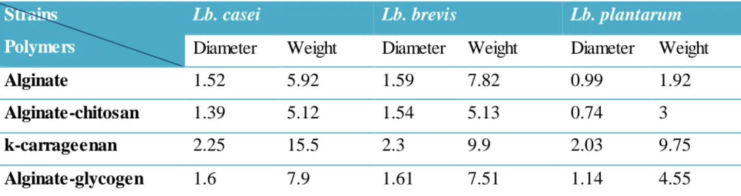
Table III.1: General characteristics of bacterial beads.
Strains Polymers
Lb. casei Lb. brevis Lb. plantarum
Diameter Weight Diameter Weight Diameter Weight
Alginate 1.52 5.92 1.59 7.82 0.99 1.92
Alginate-chitosan 1.39 5.12 1.54 5.13 0.74 3
k-carrageenan 2.25 15.5 2.3 9.9 2.03 9.75
Alginate-glycogen 1.6 7.9 1.61 7.51 1.14 4.55
The tables above demonstrate that beads diameters are different depending on the bacterial strain and the polymer of encapsulation. The average diameter of alginate beads is ranged between 0.99 mm and 1.59 mm, whereas the beads of alginate coated with chitosane showed the lowest diameter (0.74 mm). The k-carrageenan beads were the largest ones with diameter of 2.3 mm, while beads of alginate and alginate- glycogen showed near diameters.
III.2. Aspect of encapsulated bacterial strains
The shape of the beads obtained after the microencapsulation procedure was generally spherical; sometimes elliptical shaped beads were observed. The alginate beads and the alginate-glycogen beads had a rounded smoother surface for the Lb. casei and Lb. brevis strains of bacteria whereas
Lb. plantarum beads had irregular smaller shape. The k- carrageenan beads had an elliptical and
Figure III.1: Aspect of encapsulated Lb. casei (KBM2): (a) beads of alginate, (b) beads of alginate coated with chitosan, (c) beads of k-carrageenan, (d) beads of alginate-glycogen.
Figure III.2: Aspect of encapsulated Lb. brevis (B1): (e) beads of alginate, (f) beads of alginate coated with chitosan, (g) beads of K-carrageenan, (h) beads of alginate-glycogen.
b
c d
e f
g h
Figure III.3: Aspect of encapsulated Lb. plantarum (B1): (i) beads of alginate, (j) beads of alginate coated with chitosan, (k) beads of k-carrageenan, (l) beads of alginate-glycogen.
Compared to alginate beads, the chitosan-coated beads had jagged edges, according to (Yeung et
al., 2016) this observation suggests that the chitosan layer has been successfully deposited onto the
external surfaces of the alginate microgels. In addition, Fareez et al., (2015) found that the irregularity shape reflects higher polymer concentration on the surface of the beads.
In this work, in spite we used the same concentration of polymer (2%) according to (Chávarri et
al., 2010), there were different beads sizes among different kinds of beads because they had
different amount of probiotic inside.
III.3. Effect of storage at different temperatures on the viability of free and encapsulated cells
These experiments were performed in order to evaluate the efficiency of encapsulation to reduce the loss in number of the bacterial strains (free and encapsulated cells) under different temperatures and the results were represented in the figures below.
The initial cell count of all bacteria before encapsulation was about 10 (1og cells/ml) to 11 (log cells/ml).
i J
➢ Lactobacillus casei
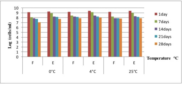
As shown in figure III.4, at 0°C the decrease in free cells is higher than the encapsulated cells, whereas E cells in alginate decrease from 9.22 (log cells /ml) to 7.75 (log cells/ml). At 4°C the E cells decreased from 9.40 (log cells/ml) to 8.04 (log cells/ml) and these results demonstrated a higher number than the free cells, also, at 25°C the number decreased from 9.38 (log cells/ml) to 7.86 (log cells /ml).
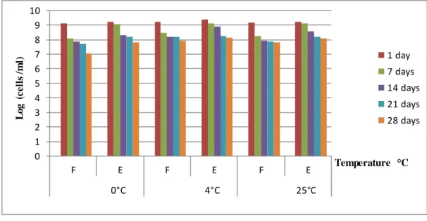
For the E cells in alginate coated with chitosan shown in figure III.5, the decrease was from 9.21 (log cells/ml) to 7.80 (log cells/ml) at 0°C, and it was from 9.39 (log cells/ml) to 8.12 (log cells /ml) at 4°C in the same storage time, whereas at 25°C, the E cells were declined from 9.23 (log cells /ml) to 8.07 (log cells/ml) in 28 days.
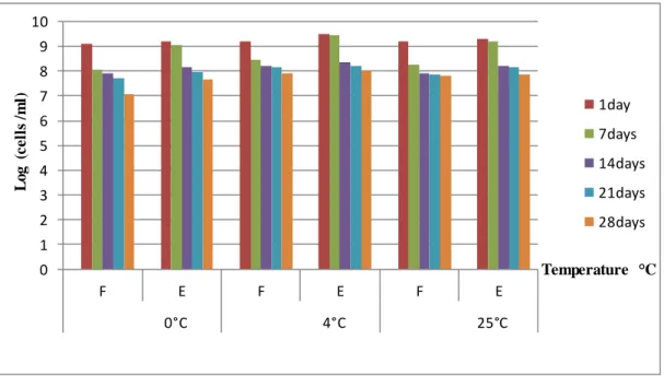
Figures III.6 and III.7 show that the number of cells released from alginate-glycogen and
k-carrageenan microcapsules respectively after storage. it can be seen that the number of cells released from k-carrageenan decreased during storage for 28 days, at 0°C 9.19 (log cells/ml) to 7.66 (log cells /ml), at 4°C, there was a loss of cells from 9.49 (log cells /ml) to 7.99 (log cells /ml) at the end of storage, whereas, at 25°C the number of cells decreased from 9.3 (log cells /ml) to 7.87 (log cells /ml). however, encapsulated cells in alginate-glycogen, showed a decreased from 9.1 to 7.20 (log cells /ml) at 0°C and from 9.29 to 7.98 (log cells /ml) at 4°C but at 25°C it reduced from 9.25 to 7.87 (log cells /ml).
Figure III.4: Survival of free and encapsulated Lb.casei in alginate during storage at different temperatures. 0 1 2 3 4 5 6 7 8 9 10 F E F E F E 0°C 4°C 25°C L o g ( c e ll s /m l) Temperature °C 1day 7days 14days 21days 28days
Figure III.5: Survival of free and encapsulated Lb. casei in alginate coated chitosan during storage at different temperatures.
Figure III.6: Survival of free and encapsulated Lb. casei in alginate-glycogen during storage at different temperatures. 0 1 2 3 4 5 6 7 8 9 10 F E F E F E 0°C 4°C 25°C L o g ( c e ll s / m l) Temperature °C 1 day 7 days 14 days 21 days 28 days 0 1 2 3 4 5 6 7 8 9 10 F E F E F E 0°C 4°C 25°C L o g ( c e ll s / m l) Temperature °C 1 day 7 days 14 days 21 days 28 days
Figure III.7: Survival of free and encapsulated Lb. casei in k-carrageenan during storage at different temperatures.
➢ Lactobacillus brevis
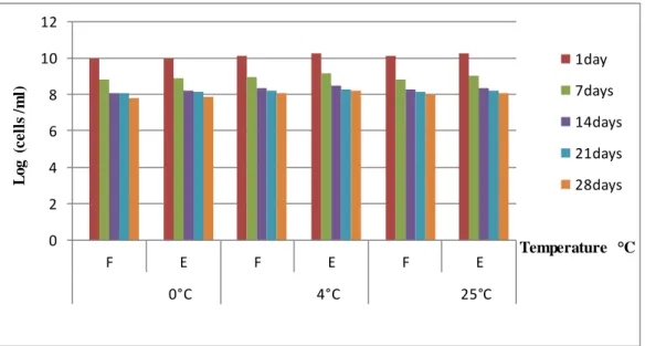
A decrease in number was observed for Lb. brevis encapsulated in all polymers of our experiment within 28 days in different temperatures of storage. Figure III.8 show that E cells in alginate were decreased from 10.24 (log cells /ml) to 8.19 (log cells /ml) at 4°C in which the highest number was observed compared to the other temperatures at 0°C from 10.02 to 7.89 (log cells / ml), at 25°C from 10.24 to 8.07 (log cells /ml).
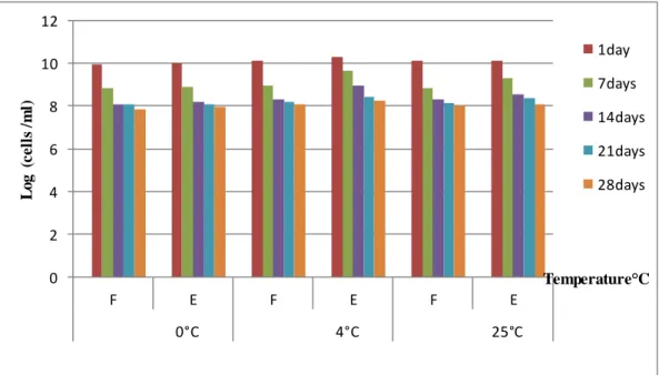
For alginate-chitosan encapsulation (figure III.9), we found a slight decrease over the 28 days, similarly to alginate, the number at 4°C was higher than others temperatures, from10.31 to 8.29 (log cells /ml) at 4°C; from 10.15 to 7.94(log cells /ml) at 0°C; and from 10.25 to 8.19 (log cells /ml) at 25°C. according to figures III.10 and III.11, the same results were observed with alginate-glycogen and K-carrageenan. However, the K-carrageenan presents better results compared to alginate-glycogen and alginate.
0 1 2 3 4 5 6 7 8 9 10 F E F E F E 0°C 4°C 25°C L o g ( c e ll s / m l) Temperature °C 1day 7days 14days 21days 28days
Figure III.8: Survival of free and encapsulated Lb. brevis in alginate during storage at different temperatures.
Figure III.9: Survival of free and encapsulated Lb. brevis in alginate coated chitosan during storage at different temperatures.
0 2 4 6 8 10 12 F E F E F E 0°C 4°C 25°C L o g ( c e ll s / m l) Temperature °C 1day 7days 14days 21days 28days 0 2 4 6 8 10 12 F E F E F E 0°C 4°C 25°C lo g ( c e ll s /m l) Temperature°C 1day 7days 14days 21days 28days
Figure III.10: Survival of free and encapsulated Lb. brevis in alginate-glycogen during storage at different temperatures.
Figure III.11: Survival of free and encapsulated Lb. brevis in k-carrageenan during storage at different temperatures.
➢ Lactobacillus plantarum
Lb. plantarum showed a decrease in number for both F and E cells during 28 days of storage at
refrigeration, freezing, and ambient temperature with all polymers (figure III.12, III.13, III.14 and 0 2 4 6 8 10 12 F E F E F E 0°C 4°C 25°C L o g ( c e ll s /m l) Temperature °C 1day 7days 14days 21days 28days 0 2 4 6 8 10 12 F E F E F E 0°C 4°C 25°C L o g ( c e ll s / m l) Temperature°C 1day 7days 14days 21days 28days
III.15), but the chitosan had the highest number of cells compared with alginate, glycogen and
K-carrageenan.
Compared to free cells, the encapsulation process gave a small decrease in the number of bacteria in all polymers and at different storage temperatures, because after 28 days, all encapsulated bacteria demonstrated high viability relative to free cells.
Figure III.12: Survival of free and encapsulated Lb. plantarum in alginate during storage at different temperatures.
Figure III.13: Survival of free and encapsulated Lb. plantarum in alginate coated chitosan during storage at different temperatures.
0 2 4 6 8 10 12 F E F E F E 0°C 4°C 25°C L o g c e ll s / m l Temperature °C 1day 7days 14days 21days 28days 0 2 4 6 8 10 12 F E F E F E 0°C 4°C 25°C L o g c e ll s / m l Temperature°C 1day 7days 14days 21days 28days