HAL Id: tel-01406276
https://tel.archives-ouvertes.fr/tel-01406276
Submitted on 30 Jun 2017
HAL is a multi-disciplinary open access archive for the deposit and dissemination of sci-entific research documents, whether they are pub-lished or not. The documents may come from teaching and research institutions in France or abroad, or from public or private research centers.
L’archive ouverte pluridisciplinaire HAL, est destinée au dépôt et à la diffusion de documents scientifiques de niveau recherche, publiés ou non, émanant des établissements d’enseignement et de recherche français ou étrangers, des laboratoires publics ou privés.
Migration and specification of CGE-derived GABAergic
cortical interneurons during mouse development
Audrey Touzot
To cite this version:
Audrey Touzot. Migration and specification of CGE-derived GABAergic cortical interneurons during mouse development. Agricultural sciences. Université Nice Sophia Antipolis, 2014. English. �NNT : 2014NICE4089�. �tel-01406276�
Ecole doctorale des Sciences de la Vie et de la Santé
THÈSE de Doctorat
pour obtenir le titre de
Docteur en Biologie
Spécialité: Interactions moléculaires et cellulaires
“Migration et spécification des interneurones
GABAergiques corticaux issus de la CGE au cours du
développement chez la souris”
“Migration and specification of CGE-derived GABAergic
cortical interneurons during mouse development
”
Audrey TOUZOT
Institut Biologie Valrose INSERM1091 CNRS7277 UNS 28 avenue Valrose 06108 Nice
Présidente:
Dr. BARDONI Barbara
Rapporteurs:
Prof. PRICE David
Dr. VITALIS Tania
Directrice de thèse: Dr. STUDER Michèle
Acknowledgments
“All truths are easy to understand once they are discovered, the point is to discover them.” - Galileo Galilei –
First and foremost, I would like to thank my advisor Michèle Studer for providing me with the opportunity to complete my Ph.D. thesis in her laboratory and encouraging me over the past years. It has been an honor to be her Ph.D. student. She has been actively interested in my work and has always been available to advise me. I am very grateful for your patience and I would like to thank you for encouraging my research and for allowing me to grow as a research scientist.
I would like to thank all the members of the Studer’s lab for sharing their knowledge and skills with me; in particular Christian and Maria Anna that are my primary resources for getting my science questions answered. A big thank you to those of you who were here when I started and now have moved on (Nadia, Lisa, Jessica, Salsa, Mariel), those who have been here throughout most of my Ph.D. (Kawssar, Elia, Joséphine) and all of you have joined the last few years (Eya, Michele). Thank you for your continued friendship, support, hugs, whatsApp chats, laughs and for all the conversations, philosophical debates and inspirational conversations over coffee and cake. I’ll never forget the many wonderful lunches and fun activities we’ve done all together. Thank you all for making the lab such an enjoyable and pleasant place to work.
I'm thankful for my years spent with my friends Yoann, Adrien, Marc, Jessica, Aude D, Lucile, Aude V, Antoine, Amélie, Nolwen, Julien for everything we shared, every chance we had to grow. I'll take the best of you and lead by your example where ever I go.
I especially thank my dad Denis, my siblings Laura and Michael. My hard-working parent has sacrificed his life for my sister, my brother and myself and provided unconditional love and care. I love him so much, and I would not have made it this far without him. My sister has been my best friend all my life and I love her dearly and thank her for all her advice and support. My brother has been my little boy and I thank him to be so strong and courageous. I know I always have my family to count on when times are rough. I also thank Audrey and her daughter Lou that are more than my cousins but my sister and my niece. I love you.
All these persons let me understand over the last years that I should look at everything as a lesson and shouldn’t want to walk around angry. And there are things we don't want to happen, but have to accept; things we don't want to know, but have to learn, and people we can't live without, but have to let go. Thank you.
Abstract
In the cerebral cortex, normal neuronal activity relies on the balance between excitation, provided by pyramidal glutamatergic neurons, and inhibition, executed by GABAergic cortical interneurons. Abnormal development of interneurons can alter cortical activity leading to various neurodevelopmental diseases, such as epilepsy, autism or other intellectual disabilities. Understanding the molecular mechanisms that control interneuron development and diversity has therefore received considerable attention by neuroscientists and clinicians. In rodents, cortical interneurons are highly heterogeneous and originate from the medial (MGE) and caudal ganglionic eminence (CGE) according to precise temporal schedules, express a defined combination of factors, and reach their final laminar position through tangential and radial cell migration. Although great progress have been made over the last decade in elucidating the diversity and fate-specification of MGE-derived interneuron subtypes, the molecular mechanisms controlling the migration and development of CGE-derived interneurons, which comprise around 30% of all cortical interneurons, are still very vague. In this study, I directly address this question, first by investigating the migratory paths of cortical interneurons using a reporter line (5HTGFP) specific to the CGE, and second by challenging the functional role of two transcription factors, COUP-TFI and COUP-TFII, highly expressed in the embryonic CGE during development. My data unraveled two major previously non-characterized migratory streams from the subpallium to the pallium; a dorsal stream (CLMS) in which CGE-derived cells migrate to the lateral GE (LGE), and a ventral one (CMMS) in which CGE-derived cells migrate to the MGE before reaching the pallium. Together with the already well-described caudal stream (CMS), I analyzed their mode of migration at different ages and identify a series of genes expressed in the migrating GFP+ cells. While Sp8 is mainly expressed in the CLMS, Prox1, COUP-TFI, COUP-TFII and Nrp2 are expressed in the CMMS. Beside COUP-TFII, I found that also COUP-TFI and Nrp1 are expressed in the CMS. By inactivating COUP-TFI and/or COUP-TFII in developing interneurons, these streams are perturbed and expression of these factors affected. As a consequence, adult mutant mice have an altered distribution of interneuron subpopulations, particularly the ones derived from the CGE. Taken together, my study identified and characterized two novel CGE-derived interneuron migratory routes to the cortex and showed that COUP-TF transcription factors directly contribute in modulating these paths by regulating the expression of distinct factors in the migrating cells.
Résumé
Dans le cortex cérébral, l’activité neuronale repose sur la maintenance d’une délicate balance entre l’excitation, assurée par les neurones pyramidaux glutamatergiques, et l’inhibition, réalisée par les interneurones corticaux GABAergique. L’altération du développement des interneurones peut modifier l’activité corticale et ainsi conduire à plusieurs troubles mentaux, tels que l'épilepsie et l'autisme. Les mécanismes qui contrôlent le développement et la diversité des interneurones sont donc une question de haute priorité non seulement pour les chercheurs en neurosciences, mais aussi pour les cliniciens. Chez les rongeurs, les interneurones corticaux sont issus de l’éminence ganglionnaire médiale et caudale (CGE et MGE respectivement) selon une séquence temporelle précise et expriment une combinaison de facteurs définis. Ils atteignent leur position laminaire définitive grâce à la migration cellulaire tangentielle et radiale. Bien que d’énormes progrès ont été faits pour élucider la diversité et la spécification des sous-types d’interneurones provenant de la MGE, les mécanismes moléculaires contrôlant la migration et la spécification des interneurones issus de la CGE, qui représentent environ 30% des interneurones corticaux, sont toujours incertains. Dans cette étude, je traite de cette question en examinant premièrement les voies de migration des interneurones grâce à une lignée de souris rapportrice des interneurones issus de la CGE (5HTGFP), et dans un second temps en comprenant le rôle fonctionnel de deux facteurs de transcription, COUP-TFI et COUP-TFII, qui sont hautement exprimés dans la CGE au cours du développement cortical. Mes données ont montré deux voies de migration précédemment non caractérisées qui vont du télencéphale ventrale à la plaque corticale ; une voie dorsale (CLMS) où les interneurones issus de la CGE migrent vers l’éminence ganglionnaire latérale (LGE) et une voie ventrale (CMMS) où les interneurones migrent vers la MGE avant de rejoindre le cortex. J’ai analysé le mode de migration de ces interneurones dans ces deux voies de migration ainsi que dans la voie de migration caudale (CMS) et j’ai identifié certains gènes exprimés dans les cellules utilisant ces voies de migration tels que Sp8 qui est principalement exprimé dans le CLMS, et Prox1, COUP-TFI, COUP-TFII et Nrp2 qui sont exprimés dans le CMMS. Dans le CMS, en plus de COUP-TFII, j’ai trouvé que COUP-TFI et Nrp1 s’y exprimés également. En inactivant conditionnellement COUP-TFI et/ou COUP-TFII dans les interneurones, les voies de migration sont altérées ainsi que l’expression des différents facteurs identifiés. Comme probable conséquence les souris mutantes adultes montrent une distribution altérées des sous-populations d’interneurones en particulier de celles issues de la CGE. De façon globale mon étude a donc permis d’identifier et de caractériser deux nouvelles voies de migration vers le cortex pour les interneurones provenant de la CGE et a montré que les facteurs de transcriptions COUP-TFs contribuent directement à la modulation de ces voies en régulant l’expression de facteurs dans les cellules en migration.
Table of Contents
Acknowledgments ... v
Abstract ... vii
Résumé ... viii
Abbreviations ... xi
Figures Index ... xii
Chapter I Introduction ... 1
1. The cerebral cortex: a delicate balance between excitation and inhibition ... 3
1.1. The excitatory projection neurons ... 4
1.2. The inhibitory cortical GABAergic interneurons ... 6
1.2.1. Parvalbumin-expressing interneurons ... 6
1.2.2. Somatostatin-expressing interneurons ... 7
1.2.3. Serotonin receptor 3a-expressing interneurons ... 7
2. Cortical interneuron neurogenesis: a tight spatio-temporal control ... 11
2.1. The anatomical origin of cortical interneurons ... 11
1.1. The time of generation of cortical interneurons ... 14
2. Molecular mechanisms of interneuron development ... 19
2.1. Transcription factors required in MGE- and LGE-derived interneuron specification ... 19
2.2. Molecular control of CGE-derived interneurons ... 22
2.2.1. Gsx, Mash1, Dlx and Prox1 ... 23
2.2.2. COUP-TFs ... 24
3. Interneuron migration: a long journey ... 31
3.1. The major dorsal routes taken by migrating interneurons ... 31
3.1.1. Tangential migration of cortical interneurons ... 31
3.1.2. Radial migration of cortical interneurons ... 33
3.2. Guidance factors involved in interneuron migration ... 35
3.2.1. Secreted molecules ... 35
3.2.2. Transcription factors... 38
4. Aim of the study ... 39
Materials and Methods ... 41
Chapter II Identification of two novel tangential paths of CGE-derived interneurons ... 45
1. CGE-derived interneurons migrate caudally but also rostrally along distinct dorsal and ventral routes ... 47
2. Semaphorins and Neuropilins might be involved in guiding CGE-derived migratory pathways ... 50
2.1. Neuropilin 1 (Nrp1) is expressed in caudally-migrating 5HTGFP+ interneurons (CMS) and might respond to local sources of Sema3A ... 50
2.2. Neuropilin 2 (Nrp2) is expressed in the CMMS and CMLS of 5HTGFP+ migrating interneurons and might respond to local sources of Sema3F ... 52
3. Expression of transcription factors in migrating CGE-derived interneurons ... 54
3.1. The MGE-marker Nkx2.1 is expressed in a subset of 5HTGFP+ cells in the MGE/POA region . 54 3.2. The transcription factor Sp8 labels two indpendent 5HTGFP+ sources of interneurons ... 56
3.3. The homeodomain gene Prox1 is expressed in a subset of CMMS 5HTGFP+ interneurons ... 58
3.4. COUP-TF genes are differentially expressed in 5HTGFP+ CGE-derived interneurons ... 58
4. Conclusion ... 64
Chapter III Role of COUP-TFI and COUP-TFII in CGE-derived interneuron migration and specification ... 67
1. Loss of COUP-TFI affects CGE proliferation and proper balance of CGE-derived interneuron migration during development ... 69
2. COUP-TFI controls the ratio between MGE- and CGE-derived interneurons in adult mice ... 77
3. Loss of COUP-TFII affects mainly CMS and CMMS migration ... 79
4. Opposed role of COUP-TFII in controlling the balance between MGE- and CGE-derived interneurons in adult mice ... 83
5. Partial additive effects on interneuron migration in COUP-TF compound mutants ... 85
Chapter IV Discussion and Perspectives ... 91
Discussion ... 93
Perspectives... 101
Supplementary ... 103
Loss of COUP-TFI Alters the Balance between Caudal Ganglionic Eminence- and Medial Ganglionic Eminence-Derived Cortical Interneurons and Results in Resistance to Epilepsy References ... 106
Abbreviations
5HT3aR 5-hydroxytryptamine (serotonin) receptor 3a
AEP Entopeduncular
BrdU Bromodeoxyuridine
CGE Caudal ganglionic eminence
CKO Conditional knock out
CMS Caudal migratory stream
COUP-TFI Chicken ovalbumin upstream promoter transcription factor I
COUP-TFII Chicken ovalbumin upstream promoter transcription factor II
CP Cortical plate
CR Calretinin
Ctrl Control
DAPI 4',6-diamidino-2-phenylindole
DNA Deoxyribonucleic acid
E0.5 Embryonic day 0.5
EDTA Ethylenediaminetetraacetic acid
GABA γ-aminobutyric acid
GE Ganglionic eminences
GFP Green fluorescent protein
ISH In situ hybridization
IZ Intermediate zone
LGE Lateral ganglionic eminence
LHX6 LIM homeodomain transcription factor 6
MGE Medial ganglionic eminence
MZ Marginal zone NKX2.1 NK2 homeobox 1 NPY Neuropeptide Y NRP1 Neuropilin-1 NRP2 Neuropilin-2 P21 Postnatal day 21
PBS Phosphate Buffered Saline
PFA Paraformaldehyde
POA Preoptic area
PROX1 Prospero homeobox protein 1
PV Parvalbumin
RNA Ribonucleic acid
SDS Sodium dodecyl sulfate
Shh Sonic hedgehog
SOX6 Sry-related HMG-box-containing transcription factor 6
SP Subplate
SP8 Specificity protein 8
SSC Sodium Chloride-Sodium Citrate
SST Somatostatin
SVZ Subventricular zone
VIP Vasoactive intestinal peptide
Figures Index
Figure 1 : Origin of cortical interneuron subpopulations.. ... 10
Figure 2: Schematic representation of the developing murine brain at E13.5.. ... 12
Figure 3: Temporal origin of cortical interneurons.. ... 15
Figure 4: Specification of GABAergic cortical interneurons. ... 18
Figure 5: Cortical interneuron migration in the developing telencephalon. ... 30
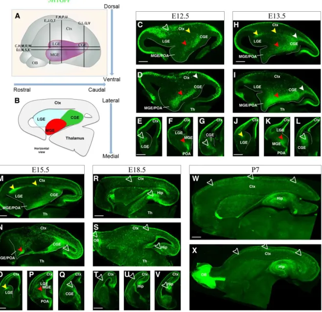
Figure 6: 5HTGFP expression pattern during development.. ... 46
Figure 7: Migrating pathways of CGE-derived interneurons. ... 49
Figure 8: Cell tracking along the CLMS and the CMMS.. ... 49
Figure 9: Guidance of 5HTGFP cells through the CMS by Nrp1/Sema3A. ... 51
Figure 10: Guidance of 5HTGFP cells through the CMMS by Nrp2/Sema3F. ... 53
Figure 11: Some 5HTGFP+ cells express Nkx2.1 in the MGE/POA.. ... 55
Figure 12: Sp8 labels specific subpopulations of interneneurons.. ... 57
Figure 13: A subpopulation of CGE-derived interneurons express Prox1 in the CMMS.. .... 59
Figure 14: COUP-TFI is expressed highly in the CMS and in a subpopulation of CMMS migrating cells. ... 61
Figure 15: COUP-TFII-expressing cells migrate through the CMMS and is present in cells exiting CGE to migrate caudally. ... 62
Figure 16: High co-expression of COUP-TFI and COUP-TFII. ... 63
Figure 17: Transcription factors are expressed in different migratory paths. ... 65
Figure 18: Altered migration in absence of COUP-TFI function. ... 68
Figure 19: Increased proliferation in absence of COUP-TFI.. ... 70
Figure 20: Downregulation of Prox1 in the CMMS in absence of COUP-TFI. ... 72
Figure 21: COUP-TFII subpopulation might compensate the decrease of Prox1 subpopulation in CMMS of TFICKO brain. ... 74
Figure 22: Some CGE-derived interneurons are redirected through the CLMS in absence of COUP-TFI.. ... 75
Figure 23: Nrp2 guides the CGE-derived interneurons along the CMMS whereas Nrp1 is required in the caudal cortex. ... 76
Figure 24: Altered balance between PV- and VIP- and CR-expressing cortical interneurons in the absence of COUP-TFI function.. ... 78
Figure 25: Altered migration in absence of COUP-TFII function. ... 80
Figure 26: Regulation of COUP-TFI by COUP-TFII. ... 80
Figure 27: Downregulation of Prox1 in the CMMS in absence of COUP-TFII. ... 82
Figure 28: Some CGE-derived interneurons are redirected through the CLMS in absence of COUP-TFII.. ... 82
Figure 29: Altered balance between PV- and VIP- and CR-expressing cortical interneurons in the absence of COUP-TFII function.. ... 84
Figure 30: Additive effects of COUP-TFI and COUP-TFII on the migration. ... 86
Figure 31: Increased number of Sp8+/5HTGFP+ in the CLMS at earlier stage.. ... 88
Figure 32: Upregulation of Prox1 in the CMMS at E15.5 in absence of COUP-TFI and COUP-TFII.. ... 89
Figure 33: Putative new migratory streams for CGE-derived cortical interneurons. ... 96
Figure 34: Putative regulatory model between the transcription factors in the CGE at E15.5 ... 100
Chapter I
Introduction
1. The cerebral cortex: a delicate balance between excitation and inhibition
The mammalian neocortex is a highly evolved organ and an extremely complex biological entity that is responsible for the higher brain functions of the central nervous system. Located in the roof of the dorsal telencephalon (pallium), the neocortex is the largest and most pivotal structure of the mammalian telencephalon and is organized into unique areas that serve distinct functions such as sensory perception, learning, memory, and motor outputs. The size of the primary cortical areas vary across species, and also within a specie (Leingärtner et al., 2007).
Arealization of the neocortex is controlled by a regulatory hierarchy beginning with morphogens secreted from patterning centres positioned at the perimeter of the dorsal telencephalon. These morphogens act in part in cortical progenitors to establish the differential expression of transcription factors that specify their area identity, which is inherited by their neuronal progeny, providing the genetic framework for area patterning (O'Leary and Sahara, 2008). The different areas are typically arranged in six layers (lamina), which differ in neuronal composition, density, and connectivity (Parnavelas, 2002). In the nervous system, the diversification of neuronal cell types is a common strategy to ensure functional complexity and flexibility of the neural networks. These circuits are functionally organized into radial units that span the cortical layers and consist of two major classes of neurons: glutamatergic excitatory cells (pyramidal and spiny stellate neurons) and GABA (γ-aminobutyric acid)-ergic inhibitory interneurons (Hensch, 2005).
Proper development and functioning of the neocortex critically depends on the coordinated production and migration of excitatory and inhibitory neurons. Pyramidal neurons, which make up approximately 75% of all neurons in the cortex, are the projection cells that send axons to other areas of the cortex and to distant parts of the brain. They utilize the excitatory amino acid glutamate as a neurotransmitter. Non-pyramidal cells are interneurons and their connections are all made locally. There are many varieties of interneurons based on differences in size and shape of their dendrites and patterns of axonal branching. They all contain the inhibitory neurotransmitter GABA and also one or more neuropeptides and/or calcium-binding proteins (Parnavelas, 2002).
For proper functioning, a delicate balance between excitatory and inhibitory inputs must be carefully maintained during assembly of cortical circuits. Disrupting this balance may very well contribute to the emergence of neuropsychiatric disorders, such as epilepsy, autism spectrum disorders, and intellectual disabilities. Recent studies in animal models demonstrate that the molecular basis of inhibitory circuit disruption is often linked to specific abnormalities in the development and function of interneurons (Marín, 2012) and that perturbations of regional and laminar identity may be important factors in neurodevelopmental diseases (Bedogni et al., 2010).
1.1. The excitatory projection neurons
Glutamatergic excitatory neurons are characterized by a typical pyramidal morphology. They comprise the majority of cells in the neocortex and extend their axons to distant intracortical, subcortical and subcerebral targets generating the output both within the cortex and to distant brain regions (Molyneaux et al., 2007).
During early development, there is a dramatic expansion of the neuroepithelium in the dorsolateral wall of the rostral neural tube that will give rise to neocortical projection neurons. The layer immediately adjacent to the ventricle is termed the ventricular zone (VZ). As neurogenesis proceeds, an additional proliferative layer known as the subventricular zone (SVZ) forms above the VZ at embryonic day (E) 13.5 in the mouse (Anderson et al., 2002; Gorski et al., 2002). Progenitors residing in the VZ and SVZ produce projection neurons of the different neocortical layers in a tightly controlled temporal order from E11.5 to E17.5 (Caviness and Takahashi, 1995; Rakic, 1974). There are at least three basic types of neurogenic progenitors within the developing neocortex: neuroepithelial cells, radial glia cells, outer radial glial cells and intermediate progenitors (Gotz and Huttner, 2005). The pseudostratified neuroepithelial cells undergo symmetric cell divisions to expand the pool of multipotent progenitors as well as a smaller percentage of asymmetric cell divisions to generate the earliest born neurons (Gotz and Huttner, 2005; Chenn and McConnell, 1995; Smart, 1973). These neurons form a layered structure termed the preplate, which is later split into the more superficial marginal zone and the deeply located subplate. The cortical plate (CP), which will give rise to the multilayered neocortex, begins to develop in between these two layers (Bayer and Altman, 1991). Then, neuroepithelial cells transform into radial glial
cells (Malatesta et al., 2003; Hartfuss et al., 2001). Radial glial cells possess long processes that span the entire neocortical wall and maintain contact both at the ventricular and pial surfaces throughout their mitotic division cycles. Radial glial cells undergo interkinetic nuclear migration, whereby the nucleus moves within the cytoplasm of elongated neuroepithelial progenitor cells in synchronization with the cell cycle phase (Gotz and Huttner, 2005; Noctor et al., 2004). The nucleus ascends to the upper region of the VZ during S phase, and later descends to the apical part of the VZ. The outer radial glial cells in mice were shown to be the progeny of ventricular radial glial cells (Shitamukai et al., 2011; Wang et al., 2011), and to undergo similar mitotic movements (Wang et al., 2011). In human timelapse imaging and fate analysis show that outer radial glial cells divide asymmetrically to self-renew, and give rise to an extended lineage of transit amplifying cells (Hansen et al., 2010). Unlike radial glial cells, outer radial glial cells are located far from the ventricle, with no apical contact to the luminal surface, but they possess a long basal fiber that often extends to the pial surface. Intermediate progenitor cells (also known as basal progenitors) reside within the VZ and often divide at ventricular surface at early stages of neurogenesis (Franco and Müller, 2013; Noctor et al., 2004). However, as neurogenesis proceeds, the intermediate progenitor cells migrate to the SVZ. Retroviral labelling and time-lapse imaging in embryonic rodent cortical slice cultures as well as staining for neuron markers was used to demonstrate that intermediate progenitor cells most often undergo one round of symmetric division to produce two neurons (Attardo et al., 2008; Haubensak et al., 2004; Kriegstein and Noctor, 2004; Noctor et al., 2004). The “two-step pattern” of neurogenesis, involving radial glial cells and intermediate progenitor cells, appears to be the predominant principle for cortical neurogenesis in rodents (Haubensak et al., 2004; Kriegstein and Noctor, 2004).
Upon induction of the telencephalon by gradients of extracellular signalling molecules, such as sonic hedgehog, fibroblast growth factors and bone morphogenetic proteins (Rallu M. et al., 2002), a number of genes that direct neocortical neurogenesis are expressed across the dorsolateral wall of the telencephalon. These include LIM homeobox 2 (Lhx2), forkhead box G1 (Foxg1), empty spiracles homologue 2 (Emx2) and paired box 6 (Pax6), each of which has crucial roles in specifying the progenitors that give rise to the projection neurons of the neocortex. Together, these four genes establish the neocortical progenitor domain by repressing dorsal midline (Lhx2 and Foxg1) (Vyas et al., 2003; Bulchand et al., 2001; Monuki et al., 2001) and ventral (Emx2 and Pax6) fates (Muzio et al., 2002).
1.2. The inhibitory cortical GABAergic interneurons
GABAergic interneurons are local circuit cells responsible for inhibitory transmission in the neocortex. While they only comprise approximately 20 % of all neocortical cells, interneurons are key regulators of cortical activity in all organisms, both invertebrates and vertebrates. They generate and maintain network oscillations, which provide the temporal structures that orchestrate the activities of neural assemblies (Isaacson and Scanziani, 2011). However interneurons that use GABA (γ-aminobutyric acid) as their neurotransmitter have far more functions than just the “inhibition” of other neurons. GABA transmission that typically acts through postsynaptic GABAA ionotropic receptors, regulates as well synaptic integration,
probability and timing of action potential, and plasticity in principal neurons (Huang et al., 2007). This variety of inhibitions is made possible by a myriad of GABAergic interneuron subtypes characterized by their neurochemical, morphological and physiological properties, the three main criteria recommended by The Petilla Interneuron Nomenclature Group (PING) (DeFelipe et al., 2013; Ascoli et al., 2008; Markram et al., 2004). Molecular features include transcription factors, neuropeptides, calcium-binding proteins, and receptors that these interneurons express, among many others. Morphologically speaking, cortical interneurons are classified in function of their soma, dendrites, axons, and the connections they make. Physiological characteristics include firing pattern, action potential measurements, passive or subthreshold parameters, and postsynaptic responses, just to name a few (Ascoli et al., 2008). Each of these properties influences the role of each interneuron within the cortical circuitry.
1.2.1. Parvalbumin-expressing interneurons
Current data suggest that around 40% of neocortical GABAergic interneurons express the calcium-binding protein parvalbumin (PV) in the somatosensory cortex (Lee et al., 2010; Xu et al., 2010; Fogarty et al., 2007). The majority of PV-expressing interneurons are basket cells, which can be further subdivided by size of their cell body (e.g., large basket cell, small basket cell, and nest basket cell), and chandelier cells (Helmstaedter et al., 2009; Uematsu et al., 2008; Markram et al., 2004). Physiologically, PV-expressing cells exhibit fast-spiking (FS) electrophysiological profiles, characterized by a high-frequency train of action potentials (APs) with little adaptation (Xu and Callaway, 2009; Gibson et al., 1999; Cauli et al., 1997; Kawaguchi, 1997; Kawaguchi et al., 1987). Additionally, these interneurons possess the lowest input resistance and the fastest membrane time constant of all interneurons (Ascoli et
al., 2008; Goldberg et al., 2008; Markram et al., 2004; Gibson et al., 1999; Cauli et al., 1997; Kawaguchi and Kubota, 1997; Connors and Gutnick, 1990). Far more is understood about basket cells, which are interneurons that make synapses at the soma and proximal dendrite of target neurons, and usually have multipolar morphology (Ascoli et al., 2008; Kawaguchi and Kubota, 1997). Fast-spiking basket neurons are the dominant inhibitory system in the neocortex, where they mediate the fast inhibition of target neurons, among many other functions (Cruikshank et al., 2007; Gabernet et al., 2005; Lawrence and McBain, 2003; Pinto et al., 2003; Miller et al., 2001; Pouille and Scanziani, 2001; Pinto et al., 2000) and play a key role in controlling the delicate balance between excitatory and inhibitory inputs in the cerebral cortex (Haider and McCormick, 2009; Hasenstaub et al., 2005). Less is known about the second subgroup of PV-expressing interneurons, the chandelier cells. Unlike basket neurons, chandelier cells target the axon initial segment of pyramidal neurons (Ascoli et al., 2008; Kawaguchi and Kubota, 1997).
1.2.2. Somatostatin-expressing interneurons
Interneurons expressing the neuropeptide somatostatin (SST) account for around 30% of the neocortical interneurons and represent the second-largest interneuron group in the mouse neocortex. SST-expressing interneurons have little overlap with PV-expressing interneurons (Lee et al., 2010; Xu et al., 2010; Fogarty et al., 2007). Representative cell types that belong to SST-expressing neurons include Martinotti cells in the neocortex, which possess ascending axons that arborize layer I and establish synapses onto the dendritic tufts of pyramidal neurons (Wang et al., 2004; Kawaguchi and Kubota, 1997). Martinotti cells are found throughout cortical layers II-VI, but are most abundant in layer V (Uematsu et al., 2008; Wang et al., 2004; Kawaguchi and Kubota, 1997). These GABAergic interneurons typically exhibit a regular adapting firing pattern, but also may initially fire bursts of two or more spikes on slow depolarizing humps when depolarized from hyperpolarized potentials (Xu et al., 2013; McGarry et al., 2010; Ma et al., 2006). In contrast to PV-positive interneurons, excitatory inputs onto Martinotti cells are strongly facilitating (Fanselow et al., 2008; Kapfer et al., 2007; Silberberg and Markram, 2007; Beierlein et al., 2003; Reyes et al., 1998).
1.2.3. Serotonin receptor 3a-expressing interneurons
The remaining 30% of neocortical interneurons largely express the 5-hydroxytryptamine (serotonin) receptor 3a (5-HT3aR) and are divided into two subclasses, vasoactive intestinal
peptide (VIP)-expressing cells and non-VIP-expressing cells. VIP is expressed in about 40% of the 5HT3aR population in the somatosensory cortex and does not overlap with SST and PV neurons (Rudy et al., 2011; Lee et al., 2010). VIP interneurons generally make synapses onto dendrites (Lee et al., 2010; Miyoshi et al., 2010; Cauli et al., 2000), and some have been observed to target other interneurons (Dávid et al., 2007; Acsády et al., 1996). Compared to all cortical interneurons, VIP interneurons possess a very high input resistance and are among the most excitable cells in the cortex (Lee et al., 2010; Miyoshi et al., 2010; Cauli et al., 2000). A significant number of VIP interneurons coexpress calretinin (CR) and display bitufted/bipolar morphology (Lee et al., 2010; Miyoshi et al., 2010; Caputi et al., 2009; Cauli et al., 2000). By physiological characterizations, these neurons are usually referred to as irregular-spiking cells (Miyoshi et al., 2010; Férézou et al., 2002; Cauli et al., 2000; Porter et al., 1998; Cauli et al., 1997). Irregular-spiking interneurons possess a vertically oriented, descending axon that extends to deeper cortical layers, and have an irregular firing pattern that is characterized by action potentials occurring irregularly during depolarization near threshold (Lee et al., 2010; Miyoshi et al., 2010; Galarreta et al., 2004; Férézou et al., 2002; Cauli et al., 2000; Porter et al., 1998; Cauli et al., 1997). The second major subtype of VIP neurons displays bitufted/multipolar morphology but does not express CR (Lee et al., 2010). These neurons are referred to as fast-adapting cells, which show rapidly adapting firing traits (Lee et al., 2010; Miyoshi et al., 2010).
In the 60% of non-VIP 5HT3aR neurons nearly 80% express the interneuronal marker Reelin. Neurogliaform cells are a type of cortical interneuron that belongs to this category. These cells have multiple dendrites radiating from a round soma and express neuropeptide Y (NPY) (Oláh et al., 2007; Kawaguchi and Kubota, 1997). They are unique relative to other GABAergic cortical interneurons because they are capable of forming synaptic connections with each other as well as with other interneuronal types (as opposed to other interneurons that can only make synapses onto homologous neurons), thus solidifying their important role in regulating neural circuitry (Price et al., 2005; Simon et al., 2005; Zsiros and Maccaferri, 2005). Furthermore, neurogliaform cells function by activating slow GABAA and GABAB
receptors in order to provoke long lasting inhibitory postsynaptic potentials onto pyramidal neurons and other interneurons (Ma et al., 2013; Armstrong et al., 2012; Rudy et al., 2011; Lee et al., 2010; Oláh et al., 2007; Tamás et al., 2003).
Figure 1 : Origin of cortical interneuron subpopulations. Schematic showing the
molecular expression profiles of GABAergic interneurons in the P21 somatosensory mouse barrel cortex. PV-, SST-, and 5HT3aR-immunopositive populations constitute mutually exclusive interneuron subtypes. PV- and SST-expressing populations are exclusively derived from the Lhx6-expressing MGE lineage (Fogarty et al., 2007). All CGE-derived interneurons express 5HT3aR. The MGE gives rise to a population of interneurons that coexpress Reelin and SST, while those Reelin expressing interneurons lacking SST-expression are CGE-derived. CR is coexpressed with all markers with the exception of PV. In addition, there appears to be a small population that is solely CR-positive within the CGE-lineage (adapted from Myioshi et al., 2010).
2. Cortical interneuron neurogenesis: a tight spatio-temporal control
The diversity of cortical interneurons appears to rely on differential developmental ontogeny that is becoming another criterion of their classification which helps in understanding their functional specificity. Previous genetic and transplantation studies have demonstrated that the distinct interneuron subtypes are produced in spatially and temporally distinct regions in the subpallium (Miyoshi et al., 2010; Wonders et al., 2008; Xu et al., 2008; Flames et al., 2007; Fogarty et al., 2007; Miyoshi et al., 2007; Butt et al., 2005; Xu et al., 2004).
2.1. The anatomical origin of cortical interneurons
In rodents, unlike in humans or primates (Letinic et al., 2002), telencephalic interneurons mainly derive from the ventral telencephalon (subpallium) (Corbin et al., 2001; Marín and Rubenstein, 2001). Similarly to the production of glutamatergic neurons in the dorsal telencephalon, neocortical interneuron neurogenesis occurs proximal to the ventricle of the developing neuroepithelium, with the majority of neocortical interneurons produced between E11 and E17. During embryonic development, the ventral telencephalon consists of the ganglionic eminences (GE) and preoptic area (POA)/ anterior entopeduncular (AEP) domains. The GE can be further subdivided into three anatomically distinct regions namely the medial (MGE), lateral (LGE), and caudal (CGE) ganglionic eminences (Fig. 1). The names of these different areas are based on their rostral-caudal location in the telencephalon. As embryonic development continues, the GEs grow and ultimately the morphological boundaries between these regions recede and are no longer present in the post-natal brain.
In vitro studies of cortical interneuron migration (Lavdas et al., 1999; Wichterle et al., 1999) and fate-mapping experiments (Xu et al., 2003) have shown that the ganglionic eminences (LGE, MGE, and CGE) are the principal sources of cortical interneurons (Xu et al., 2003; Nery et al., 2002). Within the subpallium, the MGE appears to be the primary source (around 70%) of cortical interneurons (Jiménez et al., 2002; Anderson et al., 2001; Wichterle et al., 2001; Marín et al., 2000). Transplantation experiments of MGE precursors have revealed that the majority of MGE-derived interneurons express either PV or SST (Xu et al., 2004; Valcanis and Tan, 2003; Wichterle et al., 2001; Wichterle et al., 1999) (Fig. 2). The MGE and POA express the homeobox transcription factor Nkx2.1, but the dorsal-most MGE additionally expresses Nkx6.2 and Gli1 and is partially Nkx2.1 negative
Figure 2: Schematic representation of the developing murine brain at E13.5. (A) Three
dimensional view of a developing brain indicating the ventral anatomical regions that give rise to interneurons. Both the MGE (red) and CGE (green) produce cortical interneurons. Whether the LGE (blue) produces cortical interneurons is still a matter of debate. (i-ii) Coronal views of the brain at two locations along the rostro-caudal axis. (iii) Horizontal view of the brain axis at one location in the middle of the dorso-ventral axis. CGE, caudal ganglionic eminence; LGE, lateral ganglionic eminence; MGE, medial ganglionic eminence; OB, olfactory bulb; Ctx, cortex. (adapted from Batista-Brito and Fishell, 2009).
(Sousa et al., 2009; Wonders et al., 2008; Fogarty et al., 2007; Rallu Murielle et al., 2002). In vitro transplantation studies of dorsal (d) and ventral (v) MGE-derived cells revealed that, while both regions produce a mixed population of interneurons, there is a strong bias for the production of SST+ and PV+ cells in the dMGE and vMGE, respectively (Wonders et al., 2008; Flames et al., 2007). In particular, some evidence suggest that the Nkx6.2-expressing progenitors in the dMGE preferentially generate SST-positive cells, whereas progenitors expressing both Nkx2.1 and Nkx6.2 are the sole contributors of the double SST/CR-positive Martinotti cells in the cortex (Sousa et al., 2009; Flames et al., 2007; Fogarty et al., 2007).
The next major source of inhibitory neurons is the CGE, which is responsible for producing about 30% of all cortical interneurons (Miyoshi et al., 2010; Nery et al., 2002; Anderson et al., 2001) (Fig. 2). The CGE generates diverse subtypes of interneurons that preferentially populate the superficial layers of the neocortex. Nery et al were the first to show, via in utero transplantation analyses, that a certain percentage of interneurons destined for the cortex indeed originates in the CGE (Nery et al., 2002). Other studies have utilized both in vitro and in vivo experiments to confirm this initial discovery, and have further added that CGE-derived interneurons are bipolar or double-bouquet in morphology. These interneurons express VIP and/or CR (not SST) (Butt et al., 2005; Pleasure et al., 2000). Recently, Miyoshi and colleagues used a genetic fate mapping approach to corroborate the finding that the CGE is indeed a source of cortical interneurons (Miyoshi et al., 2010). Findings from several studies indicate that 5HT3aR-expressing cortical interneurons are actually largely CGE-derived, as demonstrated by EGFP visualization in the 5HT3aR-BACEGFP mouse, a transgenic mouse generated by the GENESAT consortium (Vucurovic et al., 2010; Chameau et al., 2009; Inta et al., 2008). Lee et al compared cortices derived from the Nkx2.1-BACcre crossed to the Cre-dependent red R26RtdRFP reporter with the 5HT3aR-BACEGFP mouse to exclude the MGE as a possible source of this population of interneurons (Lee et al., 2010). No overlap of cells labelled in red and cells labelled in green was observed, and since the Nkx2.1-BACcre line labels MGE-derived cortical interneurons, the MGE was thus be discounted as a source of 5HT3aR-positive interneurons (Lee et al., 2010). In addition, the Mash1-BACCreER; R26RtdRFP mouse line, which is CGE-specific, displayed significant and near complete overlap with expressing cells, confirming the CGE as the major source of 5HT3aR-positive cortical interneurons (Lee et al., 2010).
While it is largely agreed that the MGE and CGE serve as primary sources of cortical interneurons in the developing rodent nervous system, the possibility of the LGE as a third
source has been heavily debated. Results from several studies have suggested that the LGE could as well be a contributor of cortical interneurons (Anderson et al., 2001; Wichterle et al., 2001; Wichterle et al., 1999). Effectively, Sussel and colleagues reported that Nkx2.1 mutants, in which the MGE fails to form, show a 50% reduction in cortical interneuron numbers relative to wild-type (Sussel et al., 1999). If the MGE were the origin of the majority of cortical interneurons, then a 50% reduction implies other regions of interneuron production beside the CGE that contribute only to 30%. More convincing evidence come from E15.5 Nkx2.1 mutant embryos in which the LGE-like region seem to heavily contribute to cells migration into the developing cortex (Nery et al., 2003; Anderson et al., 2001). BrdU labeling of neural progenitors also supports the notion of a cellular migratory route from the LGE to the cortex during embryogenesis, although only a portion of the BrdU-labeled cells were also GABA-positive (Anderson et al., 2001). Additionally, Jimenez and colleagues discovered that when the MGE is removed in explants taken from rat embryos, cellular migration from the LGE to the cortex continued to be observed, suggesting that the migrating cells are not MGE cells merely passing through the LGE (Jiménez et al., 2002).
Previous reports have also indicated the contribution of several other regions, such as the POA and AEP, to the pool of cortical interneurons. Indeed, a recent study by Gelman et al proposed the POA as an additional source of cortical GABAergic interneurons (Gelman et al., 2011). The POA is a region of the hypothalamus, and results from this study indicate that this area contributes approximately to 10% of all GABAergic interneurons of the murine cerebral cortex. On the contrary, the AEP which is located between the MGE and the hypothalamus would contribute to the generation of cortical and hippocampal oligodendrocytes (He et al., 2001).
1.1. The time of generation of cortical interneurons
Similarly to excitatory neurons, cortical interneurons are specified in an “inside-out” manner and this laminar distribution is produced in a temporal sequence (Miyoshi et al., 2007; Anderson et al., 2002; Cavanagh and Parnavelas, 1989). Furthermore, MGE and CGE derived cells are generated with different temporal profiles (Fig. 3). While MGE-derived interneurons
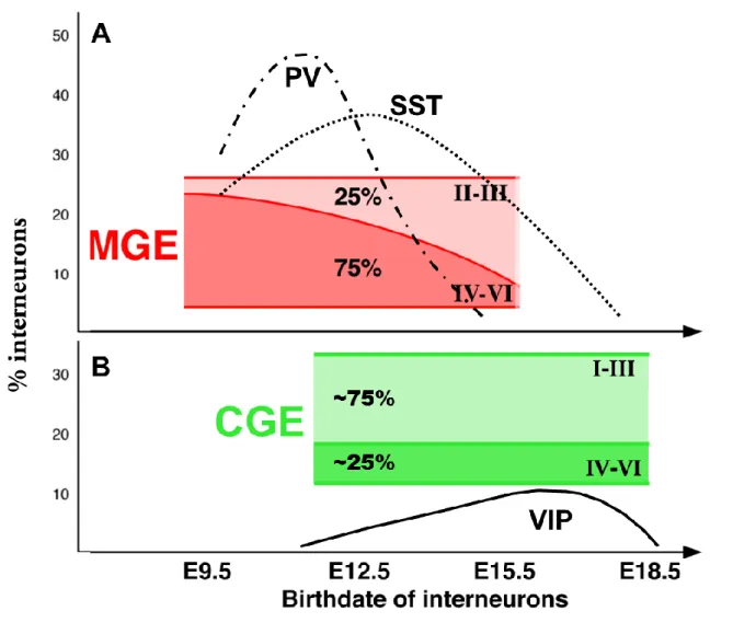
Figure 3: Temporal origin of cortical interneurons. (A) A schematic drawing showing the
contribution of MGE-derived interneurons with different cortical layers based on birthdate and diagram of temporal origin of two subtypes, the parvalbumin (PV-dash-dot line) and the somatostatin (SST-dot line). MGE-derived interneurons exhibit inside-out layering. (B) A schematic drawing showing the contribution of CGE-derived interneurons with different cortical layers based on birthdate and diagram of temporal origin of one subtype, the vasoactive intestinal peptide (VIP-continuous line). Interneurons derived from the CGE preferentially occupy superficial layers. (adapted from Miyoshi and Fishell, 2011 and Batista-Brito and Fishell, 2009).
are mostly born between E11 and E17.5 (with a peak at E13.5) (Fig. 3A), the majority of CGE-derived interneurons are produced at later developmental time points (E12.5–E18.5, with a peak at E16.5) (Fig. 3B), and generate distinct interneuron subtypes, suggesting that the time of origin plays an important role in interneuron specification (Taniguchi et al., 2013; Miyoshi et al., 2010; Miyoshi et al., 2007; Butt et al., 2005; Nery et al., 2002) (Fig. 3). Heterochronic transplants of progenitors indicated that the fate of transplanted cells is determined by the age of the donor and not by the age of the recipient (Butt et al., 2005). Moreover, both in vitro culture assays and fate mapping experiments of cohorts have revealed that the competence of MGE progenitors to produce different interneuron subtypes changes over the course of neurogenesis (Miyoshi et al., 2007; Xu et al., 2004). Specifically, a high proportion of SST+ cells are born at early developmental stages, but are almost absent in E15.5, while PV+ cells are generated at a consistent rate throughout MGE-derived interneuron production. Moreover, each cohort exhibits unique physiological properties characteristic of its birthdate. A particularly interesting example of this temporal diversity phenomenon is represented by the Chandelier cells. These neurons typically, though not always, express PV+, are fast-spiking and located both superficially and in deeper regions of the cortex. Recent studies have elegantly demonstrated that these cells are predominantly produced in the vMGE at later stages, around E15.5–E17.5, of interneuron production (Taniguchi et al., 2013; Inan et al., 2012). Contrary to the MGE, genetic fate-mapping analyses have shown that interneuron subtypes generated within the CGE appear to not significantly change over time. CGE-derived cells typically populate the superficial layers of the neocortex, but there is no correlation between their temporal origin and their specific layer destination (Miyoshi et al., 2010).
Figure 4: Specification of GABAergic cortical interneurons. With regard to the
specification of MGE-derived interneuronal progenitors, several transcription factors play a role. Shh signaling activates Nkx2.1, which is the key transcription factor in specifying PV- and SST-positive interneurons from this region. Lhx6 and Lhx8 are transcription factors that lie downstream of Nkx2.1; they also aid in the specification of PV and SST interneurons. Sox6 lies downstream of both Nkx2.1 and Lhx6/8. The Dlx homeobox family of genes play a key role in specification of CGE-derived cortical interneurons, although they also function to maintain the PV-expressing subset of MGE-derived interneurons (Dlx5 in particular). Gsx1 and Gsx2 are both required for the specification of cortical interneurons that originate in the CGE. Mash1 is a downstream transcription factor whose absence results in reduced cortical interneuron numbers; it is required for proper function of the Notch ligand Delta1, which, in the Notch signaling pathway, serves to repress neuronal differentiation. The Dlx genes lie further downstream and play a crucial role in CGE-derived interneuron specification. (adapted from Kelsom and Lu, 2013 and Gelman at al., 2012)
2. Molecular mechanisms of interneuron development
The first step toward the generation of GABAergic interneurons is the subdivision of the neuroepithelium into pallium (the dorsal telencephalon that generates cortical pyramidal neurons) and subpallium (the ventral telencephalon that generates cortical interneurons). Upon patterning of the neuroepithelium along the dorsal-ventral axis through the actions of morphogens, such as Shh (Butt et al., 2008; Fjose et al., 1993) and bone morphogenetic proteins (Tang et al., 2014; Matharu and Sweeney, 1992), different sets of transcription factors begin to be expressed by progenitor cells in distinct progenitor domains (Fig. 4). In the telencephalon, similarly to the spinal cord, a specific combination of transcription factors seems to specify the identity of each class of interneurons (Fig. 4) and to be sufficient to target one interneuron type to a specific prosencephalic area and more precisely to distinct cortical lamina (Tsai and Tsai, 1997b;a). Most transcription factors are turned off in part or entirely at postnatal stages (such as Nkx2.1 or Lhx6) when interneurons are mature and express neurochemical markers used for their classification (Leng et al., 1996). So far, only few factors have been identified as being continuously expressed, from the embryonic to adult ages, by a distinct subtype of interneurons,
2.1. Transcription factors required in MGE- and LGE-derived interneuron specification
Several studies report on the identification and characterization of a transcriptional network playing key roles in regulating proper development and specification of MGE-derived GABAergic cortical interneurons. While transcription factors, such as the Dlx homeobox genes, Lhx6 and Sox6 are crucial toward specification of the PV- and SST-positive subsets of interneurons derived from the MGE, the transcription factor that seems to play a major role in MGE interneuron specification is Nkx2.1 (Cai et al., 2013; Vucurovic et al., 2010; Xiaoren et al., 1995; Ladias and Karathanasis, 1991b; Tsai et al., 1987; Wang et al., 1987), whose expression is localized and confined within the MGE (Xu et al., 2008; Xianming and Janet, 2001) (Fig. 4).
Nkx2.1 plays a pivotal role in the establishment of MGE progenitors as well as the specification of MGE-derived interneuron subtypes located throughout the cortical lamina (Xiaoren et al., 1995; Tsai and O'Malley, 1994). In addition, maintenance of Nkx2.1
expression is regulated by Sonic hedgehog (Shh) signalling (Butt et al., 2008). More specifically, the levels of Shh signalling to the MGE interneuronal progenitors seem to be determinant for the specification of the interneuron sub-type, either PV or SST: Xu et al have shown that high levels of Shh signalling preferentially give rise to SST-expressing interneurons, which in turn results in reduced production of PV-expressing interneurons (Kanatani et al., 2008). A loss-of-function study revealed that in Nkx2.1 null mutant mice, the MGE is mis-specified to the fate of the LGE and as a consequence, more than half of the cortical interneurons, including SST-, NPY-, and calbindin-expressing neurons, are lost (Pereira et al., 2000). A subsequent study utilizing a conditional Nkx2.1 allele demonstrated that removal of Nkx2.1 gene function after establishment of the MGE identity alters the fate of MGE-derived progenitors to CGE ones, so that VIP/CR-expressing neurons are generated instead of MGE-derived PV- and SST-expressing neurons (Xiaoren et al., 1995). These results indicate that Nkx2.1 functions as a molecular switch that favours the fate of MGE progenitors rather than that of LGE and CGE.
A second transcription factor whose role is crucial to MGE-derived interneuronal specification, and whose expression is also confined to the MGE, is Lhx6 (Vucurovic et al., 2010; Xiaoren et al., 1995; Ladias and Karathanasis, 1991b) (Fig. 4). Lhx6 is a LIM homeodomain tarnscription factor and a direct downstream target of Nkx2.1 (Tran et al., 1992). It is expressed in intermediate progenitors in the SVZ of the MGE (Pereira et al., 2000; Qiu et al., 1994b; Tran et al., 1992) and continues to be expressed in developing and mature postmitotic cortical interneurons. In the mature cortex, the expression of Lhx6 is confined to PV- and SST-expressing neurons (Vucurovic et al., 2010; Xianming and Janet, 2001; Ladias and Karathanasis, 1991b). Manipulation of Lhx6 expression levels either through genetic engineering has confirmed its importance in determining the fate of both PV- and SST-positive interneurons. In the absence of Lhx6, null mutant mice have two major phenotypes: cell migration and cell type specification defects. First, Lhx6-deficient neurons show a delay in reaching the cortex and are unable to properly integrate into their respective cortical layers (Vucurovic et al., 2010; Ladias and Karathanasis, 1991b). Consistent with these findings, expression of several receptor molecules such as CXCR4, CXCR7, and ErbB4, which are involved in interneuron migration and positioning, is reduced (Vucurovic et al., 2010). This observation suggests that factors downstream of Lhx6 could contribute to the process of cortical integration. Second, MGE-derived neural progenitors are still able to migrate to the pallium in Lhx6 null mice, but most of these interneurons fail to express either PV or SST;
rather, they seem to express NPY, which turned out to be increased in the cortices of these mice (Vucurovic et al., 2010; Xiaoren et al., 1995; Ladias and Karathanasis, 1991b).
Transcription factors, other than Nkx2.1 and Lhx6, are important for the specification of MGE-derived, PV and SST interneurons and one such factor is Sox6 (Fig. 4). Sox6 is a Sry-related HMG-box-containing transcription factor that is expressed by immature and mature MGE-derived GABAergic cortical interneurons. It has been shown that Sox6 controls interneuron subtype differentiation by controlling the temporal segregation of transcriptional programs between progenitors and post-mitotic neurons (Tsai et al., 1987; Wang et al., 1987). Genetic removal of Sox6 in mice results in a dramatic reduction in the number of PV- and SST-expressing interneurons (93% and 70% respectively) and an alteration in their laminar position (Tsai et al., 1987; Wang et al., 1987). However, although interneurons fail to express PV, they are morphologically like basket cells and continue to exhibit fast-spiking, albeit immature, electrophysiological features (Tsai et al., 1987). Interestingly, Lhx6 function is required for the maintenance of Sox6 expression in neurons that are actively migrating toward the cortex, but not in MGE-derived interneuronal progenitors (Renaud et al., 1995; Tsai et al., 1987). These data suggest that Sox6 is necessary for proper positioning and maturation, but not specification of MGE-derived interneurons.
Another transcription factor that works in conjunction with Lhx6 is Lhx8, which is regulated by Nkx2.1 and is co-expressed with Lhx6 in MGE-derived neuronal progenitors (Tang et al., 2012; Pereira et al., 2000) (Fig. 4). While Lhx8-positive cells are not specified to become GABAergic cortical interneurons, genetic analysis of mice lacking both Lhx6 and Lhx8 provided some unexpected insights toward the Lhx8 role in cortical interneuron specification. Lhx6/8 double mutants exhibited a significantly reduced number of MGE-derived interneuron and defective migratory paths. However, this phenotype is not observed in mice mutant for only Lhx8 (Renaud et al., 1995). Genetic and molecular biological analyses have also revealed that the expression of Shh in the MZ is controlled by redundant activities of Lhx6 and Lhx8, which can bind to the Shh enhancer and regulate its expression (Renaud et al., 1995).
Differently from the previous genes, the Dlx family of homeobox genes, specifically Dlx1, 2, 5, and 6, play a role in the specification of all interneuronal progenitors (Fig. 4). They are expressed in most subpallial neural progenitor cells (Ladias and Karathanasis, 1991a; Wang et al., 1989) and continue to be expressed in most of their postmitotic progenies in embryonic, postnatal, and mature cortices (Cooney et al., 1993). Cortical interneurons show similar
tangential migration defects in Dlx1/2 and Dlx5/6 double mutants (Cai et al., 2013; Avram et al., 1999; Thummel, 1995). However, recent studies have shown that different members of the Dlx genes have unique gene expression dynamics and specific functions throughout development and maturation. For instance, Wang et al utilized transplantation experiments to demonstrate that loss of Dlx5 or both Dlx5 and Dlx6 in mice leads to a significant reduction of PV-expressing interneurons, alteration in dendritic morphology, and epilepsy (Cai et al., 2013). In the adult cortex, Dlx1 is preferentially expressed in SST- and CR-positive neurons (Cooney et al., 1993). Consistent with its expression pattern, Dlx1 knockout mice exhibit specific and progressive loss of SST- and CR-expressing cortical interneurons, mainly due to apoptotic cell death and immature dendritic arborization in these classes of interneurons (Cooney et al., 1993).
Unlike the MGE, the contribution of cortical interneurons by the LGE remains controversial but some evidences suggest that the LGE produces a small population of Sp8-expressing cortical interneurons (Tricoire et al., 2010; Li et al., 2003; Tsai and O'Malley, 1994). Sp8 is a member of the Sp1 zinc finger transcription factor family. It is widely expressed in the mouse embryonic telencephalon (Bell et al., 2003; Treichel et al., 2003). Its expression in pallial progenitors regulates patterning of the cerebral cortex (Sahara et al., 2007; Zembrzycki et al., 2007), and its subpallial expression regulates differentiation of olfactory bulb (OB) interneurons (Waclaw et al., 2006). Sp8 is strongly expressed in the dorsal LGE (dLGE), a domain that prenatally generates OB interneurons, and unlike other transcription factors such as Emx1, Gsh2, Nkx2.1, Dlx2, and Mash1, Sp8 appears to be continuously expressed in the majority of mature OB interneurons (Wei et al., 2011; Liu et al., 2009; Waclaw et al., 2006). Moreover, Sp8 is required for the production of OB calretinin-expressing (CR+) and PV+ interneurons (Li et al., 2011; Waclaw et al., 2006).
2.2. Molecular control of CGE-derived interneurons
As mentioned above, approximately 30% of GABAergic cortical interneurons originate from the CGE. While it was previously thought that the CGE was merely an extension of the LGE (Yozu et al., 2005; Qiu et al., 1994b; Ladias and Karathanasis, 1991a; Wang et al., 1989), recent work considers the CGE an independent source of cortical interneurons, separated from the LGE and MGE, and with a proper transcriptional network (Lin et al., 2011; van der Wees
et al., 1996; Ladias and Karathanasis, 1991a). However, the factors and molecular mechanisms controlling the fate determination of the CGE remain largely elusive.
2.2.1. Gsx, Mash1, Dlx and Prox1
Among the transcription factors expressed in LGE and CGE, the homeobox transcription factors Gsx1 and Gsx2 are both required for the specification of interneuronal progenitors in these regions (Fig. 4). Xu et al have shown through conditional loss and gain of Gsx2 function that Gsx2 is required in the generation of CR-expressing bipolar cortical interneurons (Kanatani et al., 2008). Investigation of genes downstream of Gsx1 and Gsx2 resulted in the discovery of Mash1 (also called Ascl1), a basic-helix-loop-helix transcription factor also involved in interneuron specification (Fig. 4). Mash1 mutants show a significant neuronal loss early in development, as well as reduced cortical interneuron numbers (Pastorcic et al., 1986). Mash1 has a cell-autonomous function in the development of a subset of early telencephalic progenitors and a non-cell autonomous function in mediating lateral inhibition through positively regulating Notch signalling (Miyoshi et al., 2010; Mlodzik et al., 1990; Pastorcic et al., 1986). Thus, loss of Mash1 results in the premature differentiation of cells within the SVZ expressing the Dlx genes (Miyoshi et al., 2010; Pastorcic et al., 1986). The Dlx genes are, as in the MGE, key players in the specification of CGE-derived interneurons and lie downstream of both Gsx2 and Mash1 (Alfano et al., 2014; Reinchisi et al., 2011; Tripodi et al., 2004; Chan et al., 1992). Onset of expression of the Dlx genes follows a Dlx2, Dlx1, Dlx5, and Dlx6 temporal progression (Kastner et al., 1995; Wang et al., 1990). Gsx2- and Mash1-positive neural progenitor cells simultaneously express Dlx1/2 (Miyoshi et al., 2010; Thummel, 1995; Wang et al., 1989) and loss of Dlx1/2 function results in the failure of Dlx5/6 expression (Miyoshi et al., 2010; Thummel, 1995; Wang et al., 1989). Importantly, Dlx1/2 double mutants exhibit incorrect specification of the LGE/CGE, with inappropriate expression of ventral cortical markers (Ladias and Karathanasis, 1991a; Wang et al., 1989). A detailed analysis of the transcriptional network in Dlx1/2 double mutants has resulted in the identification of almost 100 transcription factors that may play a role, both dependently and independently of the Dlx genes, in MGE, LGE and CGE-derived cortical interneuron specification (Ladias and Karathanasis, 1991a; Wang et al., 1989).
A recent study showed that Prox1, a homeobox encoding gene, is expressed in a subset of CGE/LGE- and POA-derived interneurons during embryonic development and maintained in
the mature cortex (Miyajima et al., 1988). Indeed, during early development Prox1 expression is observed in the SVZ of all three ganglionic eminences as well as the POA, which suggests that the gene may be actively down-regulated in MGE interneurons migrating to the cortex. In the striatum however, expression of Prox1 in interneurons is independent of origin and can be detected in nearly all subtypes (Miyajima et al., 1988). The function of Prox1 in cortical interneurons is unknown. However, in the dentate gyrus of the embryonic and adult hippocampus, Prox1 is activated downstream of Wnt signals and is required for maintenance of intermediate precursors (Karalay et al., 2011; Lavado et al., 2010). In these cells Prox1 also acts as a postmitotic cell fate determinant specifying granule cell identity over CA3 pyramidal cell fate (Iwano et al., 2012) and later on it is required for maturation and survival of immature granule cells (Lavado et al., 2010). In the lymphatic system Prox1 expression is maintained in lymphatic endothelial cells (LEC) and is required throughout life for maintenance of cell identity (Johnson et al., 2008).
2.2.2. COUP-TFs
The orphan nuclear receptors, COUP-TFI and COUP-TFII are the only transcription factors identified so far that become either gradually restricted to the CGE (Ritchie et al., 1990) or widely expressed within CGE (Kanatani et al., 2008; Tripodi et al., 2004). COUP-TFs stands for Chicken Ovalbumin Upstream Promoter Transcriptional Factors, since they were shown to be transcriptional regulators binding specifically to the promoter region of the chicken ovalbumin gene and regulating its expression in chicken oviducts (Wang et al., 1989; Tsai et al., 1987; Wang et al., 1987; Pastorcic et al., 1986). More generally, COUP-TFs are nuclear receptors belonging to the steroid/thyroid hormone receptor superfamily (Tsai and Tsai, 1997a). Members of this family are internalized to the nucleus both in a ligand-dependent or independent manner and act as strong transcriptional regulators by binding to the DNA of their target genes. COUP-TFs are defined as orphan receptors, since ligands regulating their activity have not been identified so far. They are required in many vital processes, such as organogenesis, angiogenesis, and metabolic homeostasis, as well as in a variety of developmental programs. These developmental processes involve COUP-TFs in different cellular functions, such as cell fate determination, cell differentiation, cell survival, and cell migration (reviewed in (Alfano et al., 2014; Lin et al., 2011; Park et al., 2003)).