HAL Id: hal-01093687
https://hal.archives-ouvertes.fr/hal-01093687
Submitted on 20 Dec 2014
HAL is a multi-disciplinary open access
archive for the deposit and dissemination of
sci-entific research documents, whether they are
pub-lished or not. The documents may come from
teaching and research institutions in France or
abroad, or from public or private research centers.
L’archive ouverte pluridisciplinaire HAL, est
destinée au dépôt et à la diffusion de documents
scientifiques de niveau recherche, publiés ou non,
émanant des établissements d’enseignement et de
recherche français ou étrangers, des laboratoires
publics ou privés.
relax: the analysis of biomolecular kinetics and
thermodynamics using NMR relaxation dispersion data
Sébastien Morin, Troels Linnet, Mathilde Lescanne, Paul Schanda, Gary
Thompson, Martin Tollinger, Kaare Teilum, Stephane Gagné, Dominique
Marion, Christian Griesinger, et al.
To cite this version:
Sébastien Morin, Troels Linnet, Mathilde Lescanne, Paul Schanda, Gary Thompson, et al.. relax: the
analysis of biomolecular kinetics and thermodynamics using NMR relaxation dispersion data. Applied
Bioinformatics, Adis, 2014, 30 (15), pp.2219 - 2220. �10.1093/bioinformatics/btu166�. �hal-01093687�
relax: the analysis of biomolecular kinetics and
thermodynamics using NMR relaxation dispersion data
S ´ebastien Morin
1,2, Troels E. Linnet
3, Mathilde Lescanne
4, Paul Schanda
4,
Gary S. Thompson
5, Martin Tollinger
6, Kaare Teilum
3, St ´ephane Gagn ´e
1,
Dominique Marion
4, Christian Griesinger
7, Martin Blackledge
4, and Edward
J. d’Auvergne
4,7∗1
PROTEO, Universit ´e Laval, Qu ´ebec G1V 0A6, Canada.
2International AIDS Society HQ, CH-1202
Geneva, Switzerland.
3Dept. of Biology, University of Copenhagen, DK-2200, Denmark.
4Institut de
Biologie Structurale, Grenoble F-38027, France.
5Astbury Centre for Structural Molecular Biology,
University of Leeds, Leeds LS2 9JT, UK.
6Institute of Organic Chemistry & CMBI, University of
Innsbruck, A-6020, Austria.
7NMR-based Structural Biology, Max Planck Institute for Biophysical
Chemistry, D-37077 G ¨ottingen, Germany.
ABSTRACT
Nuclear Magnetic Resonance (NMR) is a powerful tool for observing the motion of biomolecules at the atomic level. One technique, the analysis of relaxation dispersion phenomenon, is highly suited for studying the kinetics and thermodynamics of biological processes. Built on top of the relax computational environment for NMR dynamics is a new dispersion analysis designed to be comprehensive, accurate and easy to use. The software supports more models, both numeric and analytic, than current solutions. An automated protocol, available for scripting and driving the GUI, is designed to simplify the analysis of dispersion data for NMR spectroscopists. Decreases in optimisation time are granted by parallelisation for running on computer clusters and by skipping an initial grid search by using parameters from one solution as the starting point for another – using analytic model results for the numeric models, taking advantage of model nesting, and using averaged non-clustered results for the clustered analysis.
Availability: The software relax is written in Python with C modules and is released under the GPLv3+ licence. Source code and precompiled binaries for all major operating systems are available from http://www.nmr-relax.com.
Contact: edward@nmr-relax.com
Biological macromolecules are intricate machines and their functions are closely related to their motions. These motions can be studied experimentally at the atomic level by NMR spectroscopy. Many important biological processes occur on the µs to ms timescale and, for atoms exchanging between different states, NMR relaxation dispersion can be observed. By studying this exchange process, kinetic and thermodynamic information can be obtained.
For exchanging atoms, their nuclear spin magnetisation is described by the Bloch-McConnell equations (McConnell, 1958). ∗to whom correspondence should be addressed
Using experimental data the solution to these equations reveals both populations of the molecular states (thermodynamics) and rates of exchange between them (kinetics). Though the general solution valid for all motions remains intractable, analytic solutions with restricted motions are available and are frequently used. The equations can also be solved numerically.
Two NMR dispersion methods are used for analysing motions: single, zero, double, or multiple quantum (SQ, ZQ, DQ, MQ)
CPMG (Carr and Purcell, 1954; Meiboom and Gill, 1958); or R1ρ
(Deverell et al., 1970). Combined SQ, ZQ, DQ, and MQ data will be labelled as multiple-MQ or MMQ data. Various models are used to analyse different data and motions. The simplest is that of no motion (No Rex). For SQ CPMG-type experiments, analytic models include the original Luz and Meiboom (1963) multiple-site fast exchange models (LM63), the Carver and Richards (1972) and population-skewed Ishima and Torchia (1999) 2-site models for most time scales (CR72, IT99), and the Tollinger et al. (2001) 2-site very slow exchange model (TSMFK01). The CR72 model has been extended
by Korzhnev et al. (2004) for MMQ data. For R1ρ-type data analytic
equations include the Meiboom (1961) 2-site fast exchange model for on-resonance data (M61), extended by Davis et al. (1994) to off-resonance data (DPL94), and the Trott and Palmer (2002) and Miloushev and Palmer (2005) 2-site models for non-fast and all time scales (TP02, MP05). Different numeric solutions (NS) can be designed for SQ or MMQ data.
Diverse software solutions exist for analysing relaxation dispersion data including CPMGFit (http://www.palmer.hs.columbia .edu/software/cpmgfit.html), cpmg fit (available upon request from Dmitry Korzhnev), CATIA (Hansen et al., 2008), NESSY (Bieri and Gooley, 2011), GUARDD (Kleckner and Foster, 2012), ShereKhan (Mazur et al., 2013), and GLOVE (Sugase et al., 2013). The software relax (d’Auvergne and Gooley, 2008) is a platform for studying molecular dynamics using experimental NMR data and can be used as a numerical computing environment. Herein support for relaxation dispersion within relax is presented. Distributed as part of
1
© The Author(s) 2014. Published by Oxford University Press.
This is an Open Access article distributed under the terms of the Creative Commons Attribution License
(http://creativecommons.org/licenses/by/3.0/), which permits unrestricted reuse, distribution, and
reproduction in any medium, provided the original work is properly cited.
Associate Editor: Prof. Anna Tramontano
Bioinformatics Advance Access published April 9, 2014
by guest on May 4, 2014
http://bioinformatics.oxfordjournals.org/
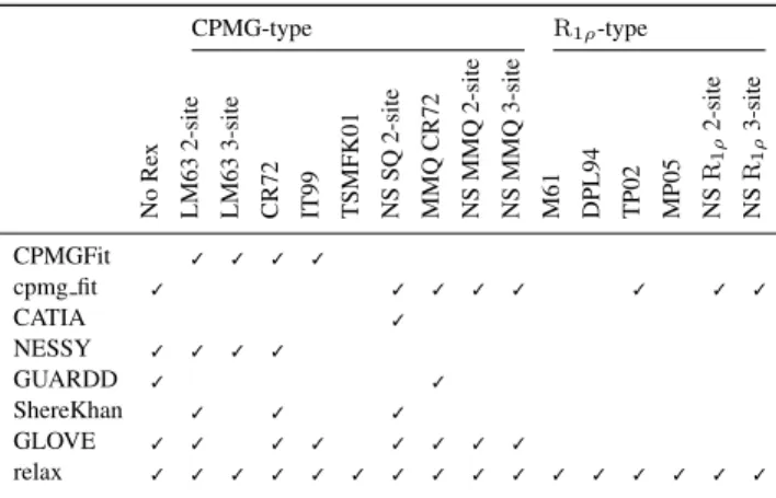
Table 1. Comparison of model support for different dispersion software CPMG-type R1ρ-type No Re x LM63 2-site LM63 3-site CR72 IT99 TSMFK01 NS SQ 2-site MMQ CR72 NS MMQ 2-site NS MMQ 3-site M61 DPL94 TP02 MP05 NS R1 ρ 2-site NS R1 ρ 3-site CPMGFit ✓ ✓ ✓ ✓ cpmg fit ✓ ✓ ✓ ✓ ✓ ✓ ✓ ✓ CATIA ✓ NESSY ✓ ✓ ✓ ✓ GUARDD ✓ ✓ ShereKhan ✓ ✓ ✓ GLOVE ✓ ✓ ✓ ✓ ✓ ✓ ✓ ✓ relax ✓ ✓ ✓ ✓ ✓ ✓ ✓ ✓ ✓ ✓ ✓ ✓ ✓ ✓ ✓ ✓
relax, this is the most comprehensive dispersion package supporting the greatest number of dispersion models and NMR data types.
The number of dispersion models supported by relax is extensive (Table 1). This allows for detailed comparisons between modern numeric and traditional analytic approaches. Different user interfaces (UIs) can be employed to analyse dispersion data including the prompt, scripting, and graphical user interface (GUI). The scripting UI enables the greatest flexibility and allows for most analysis protocols to be replicated. By implementing a novel automated analysis and providing an easy to use GUI based on this auto-analysis, the study of dispersion data is much simplified.
The setup of the auto-analysis includes defining the molecular system, loading the dispersion data directly from peak lists, clustering atoms with the same kinetics, modifying the list of dispersion models, and setting up Monte Carlo (MC) simulations for error propagation. Execution involves sequential optimisation of the models, fixed model elimination rules to remove failed models and failed MC simulations increasing both parameter reliability and accuracy (d’Auvergne and Gooley, 2006), and a final run whereby AIC model selection is used to judge statistical significance (Akaike, 1973; d’Auvergne and Gooley, 2003). The optimisation is designed for absolute accuracy and robustness but, as this can take time, it has been parallelised at the spin cluster and MC simulation level to run on computer clusters using OpenMPI. Three additional methods are used to speed up calculations, all designed to skip the computationally expensive grid search. The first is model nesting – the more complex model starts with the optimised parameters of the simpler. The second is model equivalence – when two models have the same parameters. For example the CR72 model parameters are used as the starting point for the CPMG numeric models resulting in a huge computational win. The third is for spin clustering – the analysis starts with the averaged parameter values from a completed non-clustered analysis.
The dispersion analysis in relax is implemented in Python using NumPy and the GUI using wxPython. Optimisation using the Nelder-Mead simplex and log-barrier constraint algorithms from the minfx library (https://gna.org/projects/minfx/) removes the need
for numerical gradient approximations which add a second numeric layer to the NS models. Data visualisation is via the software Grace.
ACKNOWLEDGEMENT
Nikolai Skrynnikov is thanked for his generous feedback and code contributions for implementing many of the numeric models and Flemming Hansen and Dmitry Korzhnev for kindly providing their software and published dispersion data.
REFERENCES
Akaike, H. (1973). Information theory and an extension of the maximum likelihood principle. In B. N. Petrov and F. Csaki, editors, Proceedings of the 2nd International Symposium on Information Theory, pages 267–281, Budapest. Akademia Kiado. Bieri, M. and Gooley, P. R. (2011). Automated NMR relaxation dispersion data analysis
using NESSY. BMC Bioinformatics, 12, 421.
Carr, H. Y. and Purcell, E. M. (1954). Effects of diffusion on free precession in nuclear magnetic resonance experiments. Phys. Rev., 94(3), 630–638.
Carver, J. and Richards, R. (1972). General 2-site solution for chemical exchange produced dependence of T2 upon Carr-Purcell pulse separation. J. Magn. Reson., 6(1), 89–105.
d’Auvergne, E. J. and Gooley, P. R. (2003). The use of model selection in the model-free analysis of protein dynamics. J. Biomol. NMR, 25(1), 25–39.
d’Auvergne, E. J. and Gooley, P. R. (2006). Model-free model elimination: A new step in the model-free dynamic analysis of NMR relaxation data. J. Biomol. NMR, 35(2), 117–135.
d’Auvergne, E. J. and Gooley, P. R. (2008). Optimisation of NMR dynamic models. J. Biomol. NMR, 40(2), 107–133.
Davis, D., Perlman, M., and London, R. (1994). Direct measurements of the dissociation-rate constant for inhibitor-enzyme complexes via the T1rho and T2 (CPMG) methods. J. Magn. Reson., 104(3), 266–275.
Deverell, C., Morgan, R. E., and Strange, J. H. (1970). Studies of chemical exchange by nuclear magnetic relaxation in rotating frame. Mol. Phys., 18(4), 553–559. Hansen, D. F., Vallurupalli, P., Lundstrom, P., Neudecker, P., and Kay, L. E. (2008).
Probing chemical shifts of invisible states of proteins with relaxation dispersion NMR spectroscopy: how well can we do? J. Am. Chem. Soc., 130(8), 2667–2675. Ishima, R. and Torchia, D. (1999). Estimating the time scale of chemical exchange of
proteins from measurements of transverse relaxation rates in solution. J. Biomol. NMR, 14(4), 369–372.
Kleckner, I. R. and Foster, M. P. (2012). GUARDD: user-friendly MATLAB software for rigorous analysis of CPMG RD NMR data. J. Biomol. NMR, 52(1), 11–22. Korzhnev, D. M., Kloiber, K., Kanelis, V., Tugarinov, V., and Kay, L. E. (2004).
Probing slow dynamics in high molecular weight proteins by methyl-TROSY NMR spectroscopy: application to a 723-residue enzyme. J. Am. Chem. Soc., 126(12), 3964–3973.
Luz, Z. and Meiboom, S. (1963). Nuclear magnetic resonance study of protolysis of trimethylammonium ion in aqueous solution - order of reaction with respect to solvent. J. Chem. Phys., 39(2), 366–370.
Mazur, A., Hammesfahr, B., Griesinger, C., Lee, D., and Kollmar, M. (2013). ShereKhan–calculating exchange parameters in relaxation dispersion data from CPMG experiments. Bioinformatics, 29(14), 1819–1820.
McConnell, H. (1958). Reaction rates by nuclear magnetic resonance. J. Chem. Phys., 28(3), 430–431.
Meiboom, S. (1961). Nuclear magnetic resonance study of proton transfer in water. J. Chem. Phys., 34(2), 375–388.
Meiboom, S. and Gill, D. (1958). Modified spin-echo method for measuring nuclear relaxation times. Rev. Sci. Instrum., 29(8), 688–691.
Miloushev, V. Z. and Palmer, 3rd, A. G. (2005). R(1rho) relaxation for two-site chemical exchange: general approximations and some exact solutions. J. Magn. Reson., 177(2), 221–227.
Sugase, K., Konuma, T., Lansing, J. C., and Wright, P. E. (2013). Fast and accurate fitting of relaxation dispersion data using the flexible software package GLOVE. J. Biomol. NMR, 56(3), 275–283.
Tollinger, M., Skrynnikov, N. R., Mulder, F. A., Forman-Kay, J. D., and Kay, L. E. (2001). Slow dynamics in folded and unfolded states of an SH3 domain. J. Am. Chem. Soc., 123(46), 11341–11352.
Trott, O. and Palmer, 3rd, A. G. (2002). R1rho relaxation outside of the fast-exchange limit. J. Magn. Reson., 154(1), 157–160.
2
by guest on May 4, 2014
http://bioinformatics.oxfordjournals.org/