A Bacterial Chromosome Structuring Protein
Binds Overtwisted DNA to Stimulate Type II
Topoisomerases and Enable DNA Replication
The MIT Faculty has made this article openly available.
Please share
how this access benefits you. Your story matters.
Citation
Guo, Monica S. et al. "A bacterial chromosome structuring protein
binds overtwisted DNA to stimulate type II topoisomerases and
enable DNA replication" Cell 175, 2 (October 2018): P583-597.e23 ©
2018 Elsevier Inc.
As Published
http://dx.doi.org/10.1016/j.cell.2018.08.029
Publisher
Elsevier BV
Version
Author's final manuscript
Citable link
https://hdl.handle.net/1721.1/125266
Terms of Use
Creative Commons Attribution-NonCommercial-NoDerivs License
A bacterial chromosome structuring protein binds overtwisted
DNA to stimulate type II topoisomerases and enable DNA
replication
Monica S. Guo1,4, Diane L. Haakonsen1,4,‡, Wenjie Zeng3, Maria A. Schumacher3,*, and Michael T. Laub1,2,5,*
1Department of Biology
2Howard Hughes Medical Institute,Massachusetts Institute of Technology, Cambridge, MA 02139 3Department of Biochemistry, Duke University School of Medicine, Durham, NC 27710
4Contributed equally, listed alphabetically 5Lead Contact
Summary
When DNA is unwound during replication, it becomes overtwisted and forms positive supercoils in front of the translocating DNA polymerase. Unless removed or dissipated, this superhelical tension can impede replication elongation. Topoisomerases, including gyrase and topoisomerase IV in bacteria, are required to relax positive supercoils ahead of DNA polymerase, but may not be sufficient for replication. Here, we find that GapR, a chromosome structuring protein in
Caulobacter crescentus, is required to complete DNA replication. GapR associates in vivo with positively supercoiled chromosomal DNA, and our biochemical and structural studies demonstrate that GapR forms a dimer-of-dimers that fully encircles overtwisted DNA. Further, we show that GapR stimulates gyrase and topo IV to relax positive supercoils, thereby enabling DNA replication. Analogous chromosome structuring proteins that locate to the overtwisted DNA in front of replication forks may be present in other organisms, similarly helping to recruit and stimulate topoisomerases during DNA replication.
Graphical abstract
*correspondence: laub@mit.edu (Lead Contact), maria.schumacher@duke.edu.
‡present address: Department of Molecular and Cell Biology, University of California, Berkeley, Berkeley, CA 94720 Author Contributions
D.L.H. performed the cell fractionation, RNA-sequencing, and ChIP-sequencing. M.S.G. performed the flow cytometry, DNA sequencing, and biochemical experiments. W.Z. purified proteins for structural studies. M.A.S. performed the structural studies. D.L.H., M.S.G., M.A.S., and M.T.L. conceived experiments, analyzed data, and wrote the manuscript.
Declaration of Interests
The authors declare no competing interests.
Publisher's Disclaimer: This is a PDF file of an unedited manuscript that has been accepted for publication. As a service to our
customers we are providing this early version of the manuscript. The manuscript will undergo copyediting, typesetting, and review of
HHS Public Access
Author manuscript
Cell
. Author manuscript; available in PMC 2019 October 04.Published in final edited form as:
Cell. 2018 October 04; 175(2): 583–597.e23. doi:10.1016/j.cell.2018.08.029.
A
uthor Man
uscr
ipt
A
uthor Man
uscr
ipt
A
uthor Man
uscr
ipt
A
uthor Man
uscr
ipt
eTOC blurb
A bacterial protein that specifically recognizes and encircles overtwisted DNA is required to stimulate the activity of type II topoisomerases and enable DNA replication.
Introduction
In all organisms, chromosomes must be compacted ~1000-fold to fit within the confines of a cell. However, DNA cannot be haphazardly packed, and instead must be organized to facilitate a range of cellular processes, including DNA replication, transcription, and chromosome segregation. Chromosome compaction is achieved in large part by the supercoiling of DNA, forming a series of plectonemic loops in bacteria (Higgins, 2016). DNA can be either positively (+) or negatively (−) supercoiled, depending on whether the DNA duplex winds around a superhelical axis in a left- or right-handed fashion, respectively. The supercoiling of DNA is tightly controlled by topoisomerases, enzymes that can break and rejoin DNA segments to introduce or relieve superhelical strain (Vos et al., 2011). Despite major advances in our understanding of topoisomerases, the molecular mechanisms that regulate when, where, and how these enzymes act in vivo remain poorly understood. Supercoils are also introduced by cellular processes that unwind the double helix, such as transcription and DNA replication. The ‘twin supercoiled domain’ model posits that, as RNA or DNA polymerase translocates along and unwinds the DNA, it produces overtwisted and (+) supercoiled DNA ahead of it, with underwound, (−) supercoiled DNA arising in its wake (Liu and Wang, 1978; Wu et al., 1988). These supercoils must be dissipated by topoisomerases to prevent buildup of torsional stress that would inhibit unwinding of the duplex and prevent translocation of RNA or DNA polymerase along the DNA (Postow et al., 2001). The action of topoisomerases is especially critical during DNA replication as ~50– 100 (+) supercoils per second are generated ahead of the translocating replisome (Postow et
A
uthor Man
uscr
ipt
A
uthor Man
uscr
ipt
A
uthor Man
uscr
ipt
A
uthor Man
uscr
ipt
al., 2001). Despite the importance of topoisomerases to replication, how the activities of these enzymes are controlled to promote replication elongation remains largely unknown. Topoisomerases fall into two classes, type I and type II, which respectively cleave either one or two strands of DNA (Vos et al., 2011). Bacteria, which maintain their chromosomes with net-negative supercoil, typically have a type I topoisomerase, topo I, that can relieve excess (−) supercoils, and two type II topoisomerases, DNA gyrase and topo IV, that can relax (+) supercoils. Gyrase also introduces (−) supercoils into relaxed DNA, while topo IV helps decatenate replicated chromosomes. Because gyrase, and to a lesser extent topo IV, are found in nearly all bacteria but are absent from eukaryotes, these enzymes are targeted by a number of clinically-relevant antibiotics, such as the coumerins and quinolones (Vos et al., 2011). Bacteria treated with such drugs cannot replicate their DNA, underscoring the importance of topoisomerases to DNA replication (Khodursky et al., 2000).
Although gyrase, perhaps with topo IV, is often assumed to be sufficient to manage the (+) supercoils that arise during replication (Khodursky et al., 2000), a role for other factors cannot be ruled out. In particular, bacteria, which do not encode histones, harbor a bevy of so-called nucleoid-associated proteins (NAPs) that may influence topoisomerases. NAPs are a functionally heterogeneous set of proteins that are not conserved at the sequence level, but share many similarities such as small size, high basic amino-acid content, and DNA-binding ability (Badrinarayanan et al., 2015). NAPs typically bind DNA sequence non-specifically by recognizing DNA shape, e.g., narrow minor grooves, and many structure the chromosome by bending, wrapping, or bridging DNA, activities that can introduce or constrain supercoils. As NAPs are often highly abundant, their loss can lead to global changes in supercoiling (Lal et al., 2016; Weitao et al., 2000). Several NAPs in Escherichia coli, including SeqA and the larger chromosome structuring protein MukB, can bind to and stimulate topo IV to relax supercoils or decatenate interlinked circular DNA (Hayama and Marians, 2010; Kang et al., 2003; Li et al., 2010). However, to the best of our knowledge, no NAPs have been shown to stimulate gyrase to relax (+) supercoils and a direct role for NAPs in replication elongation has not been established.
Here, we demonstrate that a highly-conserved NAP from the α-proteobacterium Caulobacter crescentus called GapR is essential for DNA replication in fast growth conditions by promoting the ability of gyrase and topo IV to relieve the (+) supercoils that accumulate ahead of the replication fork. GapR was identified as an essential DNA-binding protein that binds AT-rich DNA and has pleiotropic effects when depleted (Arias-Cartin et al., 2017; Ricci et al., 2016; Taylor et al., 2017), but its molecular function and precise roles in the bacterial cell cycle have not been elucidated. Our studies show that a loss of GapR leads to global changes in superhelicity and an inability to complete DNA replication. Intriguingly, we find that, in addition to AT-rich DNA, GapR globally associates with the 3’ ends of highly-expressed genes, but only when they are actively transcribed. These results suggest that GapR recognizes and helps dissipate the (+) supercoils that arise during unwinding of the DNA duplex. Consistent with this model, we demonstrate that GapR preferentially binds overtwisted DNA and can stimulate gyrase and topo IV to remove (+) supercoils. Crystal structures of GapR reveal a unique fold with dimeric GapR that transitions, upon DNA binding, to form a dimer-ofdimers that completely encircles DNA. Strikingly, the structure
A
uthor Man
uscr
ipt
A
uthor Man
uscr
ipt
A
uthor Man
uscr
ipt
A
uthor Man
uscr
ipt
shows that the GapR clamp specifically recognizes the shape of overtwisted DNA, and cannot accommodate B-form DNA, supporting a model in which GapR specifically localizes to the overtwisted DNA ahead of replication forks where it can stimulate type II
topoisomerases.
Results
Identification of the nucleoid-associated protein, GapR
Despite their importance in chromosome organization, most NAPs are not broadly
conserved. To identify new NAPs in Caulobacter, we isolated intact nucleoids using sucrose gradient centrifugation and performed mass spectrometry on the proteins that remained tightly associated (Fig. S1A). After sedimentation, fractions were DAPI stained to identify the nucleoid fraction and the slower migrating cytoplasmic fraction, which contains RNA and highly sheared DNA (Fig. S1A). We verified by immunoblotting that a known NAP, integration host factor (IHF), was stably associated with the nucleoid, while DNA binding proteins with higher dissociation rates, such as the transcription factor CtrA, were found in the cytoplasmic fraction (Fig. 1A). Mass spectrometry identified multiple, independent peptides of known NAPs, including HU and IHF, and other tight DNA-binding proteins, including subunits of RNA polymerase (RNAP) and single-stranded DNA binding protein; no known canonical transcription factors were recovered (Table S1).
To identify NAPs, we examined our mass spectrometry data for proteins with no annotated function but with properties common to many NAPs: (i) small (< 25 kDa), (ii) a basic region to promote tight DNA binding, (iii) high expression during exponential growth and (iv) conservation across α-proteobacteria. One candidate was an 89 amino-acid protein encoded by the highly-conserved gene gapR (CCNA_03428, Fig. 1B, S1B). We confirmed that GapR was nucleoid-associated by repeating the nucleoid purification on cells expressing
GapR-3×FLAG from its native locus. Epitope-tagged GapR was highly enriched in the nucleoid fraction and not detectable in the cytoplasmic fraction, in contrast to α-subunit of RNAP which was present in both fractions (Fig. 1A), suggesting that GapR binds the nucleoid stably and with high affinity.
GapR was also identified in searches for essential Caulobacter DNA-binding proteins (Arias-Cartin et al., 2017; Ricci et al., 2016) and origin-binding proteins (Taylor et al., 2017). These studies suggested that GapR is an essential and highly expressed protein that has pleiotropic effects when depleted, but the molecular function of GapR was not determined.
Depleting GapR leads to cellular filamentation and DNA replication defects
To understand the essential function of GapR, we constructed a depletion strain in which gapR is driven by a glucose-repressible, xylose-inducible promoter on a low-copy plasmid (ΔgapR Pxyl-gapR). Even in the presence of xylose, this strain has reduced levels (~15-fold)
of GapR relative to the wild type. The strain remains viable in xylose, but has a significant growth defect (Fig. 1C-D, S1C-D). Further inhibiting gapR expression by shifting cells to a medium with glucose, led to undetectable levels of GapR protein and a five-log defect in
A
uthor Man
uscr
ipt
A
uthor Man
uscr
ipt
A
uthor Man
uscr
ipt
A
uthor Man
uscr
ipt
plating viability. The GapR depletion strain was not able to grow in glucose and exhibited cell-division defects, becoming highly filamentous, often with multiple, incomplete
constriction sites after a shift to a rich medium containing glucose (Fig. 1E). Flow cytometry analysis of chromosome content indicated that most GapR-depleted cells did not accumulate chromosomes despite becoming filamentous (Fig. 1E). Instead, the culture was enriched for G1 cells suggesting that DNA replication is disrupted at the level of initiation and/or elongation.
We used RNA-seq to analyze changes in gene expression after 6 hr of GapR depletion in glucose relative to wild-type cells grown in glucose. We identified 1245 genes that changed expression at least 2-fold (~32% of all genes; 996 genes upregulated and 249 genes downregulated; Fig. 1F) indicating that GapR has broad, pervasive effects on gene
expression (Arias-Cartin et al., 2017; Ricci et al., 2016), including a general stress response, indicated by a correlation with the expression changes seen in cells treated with ethanol (Pearson’s r=0.72, p=1.2×10−67; Fig. S1E). We observed similar, but more modest, changes when GapR levels were reduced in xylose (Fig. S1E, Table S2). The GapR-depletion strain in glucose also exhibited a DNA damage response (Arias-Cartin et al., 2017), with
expression changes similar to cells treated with DNA damaging agents or expressing the toxin SocB, which leads to replication fork collapse (Pearson’s r=0.57, p=1.6×10⁻⁶, Fig. 1G). A DNA damage response was not observed when cells were challenged by ethanol or subjected to cell-cycle disruption by depleting SciP.
GapR binding is not correlated with GapR-dependent changes in gene expression
To gain more insight into GapR function, we analyzed its global DNA binding profile by ChIP-seq using a strain expressing GapR-3×FLAG as the sole copy of GapR from its native locus (Fig. S1F). This profile revealed widespread GapR-3×FLAG binding across the genome (Fig. 2A). Using the 34 highest peaks, we identified two highly enriched motifs, each consisting almost exclusively of A and T, whereas the Caulobacter genome overall is 68% GC, suggesting that GapR preferentially binds AT-rich DNA (Fig. 2B-D). These data agree with previous studies indicating that AT-content of the DNA is one determinant of GapR binding (Arias-Cartin et al., 2017; Ricci et al., 2016). However, there are important exceptions to this trend not previously noted that are discussed below.
To assess whether GapR directly affects gene expression, we computed the average GapR enrichment within each open reading frame (Fig. 2E) or at each promoter (Fig. S2A) in the genome and plotted these values against the expression changes in GapR-depleted cells. There was no correlation, suggesting that GapR does not directly affect gene expression.
GapR may associate with (+) supercoils at the 3’ end of highly-expressed genes and operons
How, then, does GapR affect global patterns of transcription? Because some NAPs influence the supercoiling state of cells and consequently alter the expression of supercoiling-sensitive genes, we compared the expression profile of cells depleted of GapR in glucose to that of cells treated with novobiocin, an inhibitor of gyrase and, to a reduced extent, topo IV (Table S2). For the 289 genes that changed at least 4-fold in novobiocin-treated cells, there was a
A
uthor Man
uscr
ipt
A
uthor Man
uscr
ipt
A
uthor Man
uscr
ipt
A
uthor Man
uscr
ipt
strong correlation with the changes observed in GapR-depleted cells (Pearson’s r=0.61, p=5.0×10⁻³¹), although the magnitudes of the changes due to GapR-depletion were typically smaller (Fig. 1H). The expression profiles of cells expressing SocB toxin, treated with ethanol, or cell-cycle arrested by SciP depletion did not similarly correlate with the novobiocin response, suggesting that the changes in supercoiling-sensitive genes following GapR depletion are not just a result of general stress or a disruption of the cell cycle (Fig. 1H). The change in supercoiling-sensitive genes suggested to us that GapR-depleted cells may have reduced (−) supercoiling. Consistent with this conclusion, we found that the net (−) superhelicity of chromosomes from GapR-depleted cells was reduced relative to that of wild-type cells, but higher than that of novobiocin-treated cells (Fig. S1G).
Interestingly, we noticed that GapR accumulated in genomic regions that were not AT-rich, but instead corresponded to the 3’ ends of highly-expressed transcription units such as tRNAs (Fig. 2F; S2B). At these locations, the binding of GapR was broad (spanning several kb) and lower in magnitude compared to the AT-rich locations noted above (Fig. 2B-D). GapR binding in these regions was dependent on active transcription, as it was reduced by the addition of the transcriptional inhibitor rifampicin (Fig. 2F; S2B). This transcription-dependent accumulation of GapR was strongly transcription-dependent on the expression level of the transcription unit (two-sided t-test: p=1.8×10⁻³³) and was specific to the 3’ ends of transcription units (Fig. 2G, S2D-E). In contrast, the binding of GapR at ATrich genomic regions was not affected by rifampicin (Fig. 2D, S2C). Although it is possible that GapR helps terminate transcription, like E. coli H-NS (Ray-Soni et al., 2016), our RNA-seq analysis of GapR-depleted cells did not reveal any transcriptional read-through (Fig. S2B). Why would GapR associate with the 3’ ends of highly-expressed operons? Unwinding of the DNA duplex during transcription leads to the overtwisting of DNA and the accumulation of (+) supercoils ahead of RNAP. Thus, GapR may associate preferentially with the DNA structures that arise at the ends of highly-expressed transcription units. Further, we hypothesized that GapR may help eliminate or dissipate (+) supercoils as GapR-depleted cells exhibited decreased (−) superhelicity (Fig. 1H, S1G). If GapR indeed helps relieve (+) supercoil stress, then depleting GapR should disrupt DNA replication, as the unwinding of DNA during replication leads to a significant accumulation of (+) supercoils ahead of replication forks.
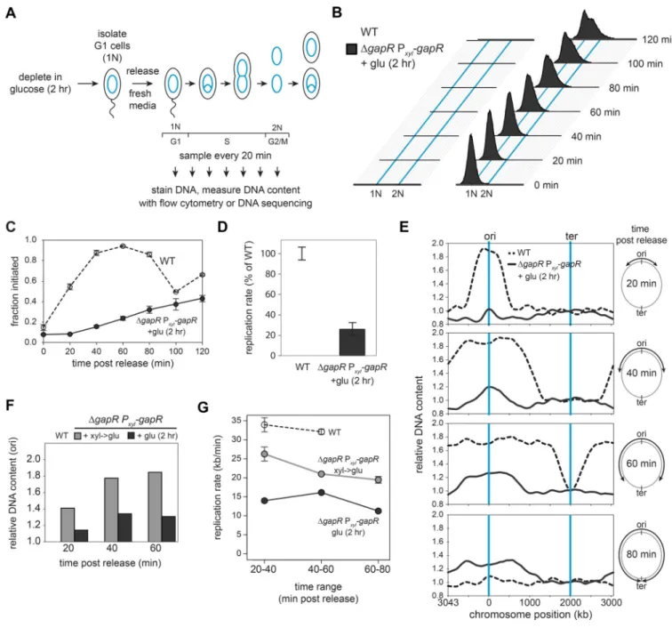
GapR is required for proper DNA replication initiation and elongation
To assess the role of GapR in DNA replication, we used flow cytometry to measure DNA content in synchronized, GapR-depleted cells over time (Fig. 3A). The GapR depletion strain was grown in glucose for 2 hr to deplete GapR, synchronized to isolate cells in G1, and released into fresh media with glucose to continue repressing gapR expression. After release, DNA content was monitored by flow cytometry (Fig. 3B). For the wild type, nearly all cells initiated replication by ~40 min postsynchronization, with cell division occurring within 100 min, indicating that replication of the entire chromosome took < 60 min (Fig. 3B-C). In contrast, GapR-depleted cells showed significant defects in both initiation and elongation. Only 20% of cells had initiated within 40 min post-synchronization and only ~60% had initiated after 120 min (Fig. 3C). The rate of replication elongation was also
A
uthor Man
uscr
ipt
A
uthor Man
uscr
ipt
A
uthor Man
uscr
ipt
A
uthor Man
uscr
ipt
slowed significantly (Fig. 3D), with few cells achieving 2N content (Fig. 3B). We also examined the GapR depletion strain under xylose-inducing conditions, which showed more modest but significant defects in replication initiation and elongation rate (Fig. S3A-D). We then used DNA sequencing to more precisely assess the replication defects of
synchronized wildtype and GapR-depleted cells. Replicated regions of the chromosome are easily detected and tracked over time as they have ~2-fold more reads than unreplicated regions (Fig. 3E). Using this assay, we found that nearly all wild-type cells (~90%) initiated replication ~20 min post-synchronization (Fig. 3F), with replication progressing steadily toward the terminus at a rate of ~32–34 kb/min (Fig. 3G), consistent with prior estimates (Jensen, 2006). In contrast, for GapR-depleted cells, <20% of cells had initiated after 20 min, and only ~35% had initiated by 60 min post-release (Fig. 3F). Additionally, the replication forks of GapR-depleted cells showed a progressive slowdown over time, with a rate of only 12 kb/min between 60 and 80 min, and with no cells having finished replication by the 80 min time point when all wild-type cells had completed. We also performed this experiment on the GapR depletion strain shifted to glucose only upon synchronization rather than for 2 hr before synchronization, again observing delays in replication initiation and a progressive decrease in elongation rate (Fig. 3F-G, S3E).
Together, our flow cytometry and DNA-sequencing data suggested that GapR-depleted cells are not competent to finish replication. We directly tested this model by: (i) Monitoring replication by flow cytometry over an extended (4 hr) period. Whereas wild-type cells replicated twice during this period, GapR-depleted cells accumulated intermediate (<2N) chromosome content (Fig. S4A). (ii) If any GapR-depleted cells finish replication, they should constrict and divide; while wild-type cells divided by 120 min, GapR-depleted cells did not divide and became progressively more filamentous over time (Fig. S4B). (iii) The origin-terminus ratio in GapR-depleted cells remained constant after ~60 min (Fig. S4C), indicating that replication initiated but did not complete. Taken together, our results demonstrate that GapR is required for the timely and successful completion of DNA replication.
GapR depleted cells are sensitized to inhibitors of type II topoisomerases
Because our ChIP experiments suggested that GapR associates with (+) supercoiled DNA, we hypothesized that GapR promotes DNA replication by helping to alleviate the (+) supercoils that accumulate ahead of replication forks. This model predicts that cells depleted of GapR should be sensitive to sub-lethal doses of antibiotics that inhibit gyrase and topo IV, the enzymes that remove (+) supercoils. To test this, we measured the plating viability of the GapR depletion strain grown in xylose to reduce GapR levels (Fig. 1C) after treatment with sub-lethal levels of novobiocin, an antibiotic that inhibits gyrase and topo IV. Notably, the GapR depletion strain exhibited nearly 5-logs lower plating viability than wild type in novobiocin (Fig. 4A). The GapR depletion strain was also sensitized to rifampicin and hydroxyurea (Fig. S4D), which also inhibit DNA replication. However, the GapR depletion strain was not more sensitive than the wild type to low doses of mitomycin C, which directly damages DNA, or chloramphenicol, which inhibits translation (Fig. 4A). The antibiotic sensitivities of the GapR depletion strain contrast with that of ΔrecA cells, which had nearly
A
uthor Man
uscr
ipt
A
uthor Man
uscr
ipt
A
uthor Man
uscr
ipt
A
uthor Man
uscr
ipt
identical plating viability as the wild type on novobiocin and chloramphenicol, but was highly sensitized to mitomycin C. Thus, GapR is likely not required to repair DNA damage or for a general stress response. Instead, these results support a model in which GapR promotes DNA replication by relieving (+) supercoils in front of the elongating replisome. To test whether the synergistic effects of reduced GapR and novobiocin treatment on viability result from defects in DNA replication elongation, we used flow cytometry to monitor DNA content in synchronous populations of wild-type and GapR-depleted cells in the presence of novobiocin. Whereas wild-type cells continued to replicate at a slightly reduced rate in novobiocin, cells with reduced levels of GapR showed very little
accumulation of DNA and never reached a 2N state when treated with novobiocin (Fig. 4B-C), having a rate of replication elongation ~20% that of untreated wild-type cells (Fig. 4D). These data confirm that without sufficient GapR, the replication fork becomes highly sensitized to topoisomerase inhibition, suggesting that GapR may affect topoisomerase activity.
GapR binds DNA and can alter DNA topology
Collectively, our in vivo results suggested that GapR promotes DNA replication by attenuating (+) supercoils, thereby relieving the topological stress associated with fork progression. To directly test whether GapR binds and affects DNA topology, we expressed and purified untagged GapR. Gel filtration of GapR produced a single peak that eluted faster than expected for a ~10 kDa protein (Fig. S5A). Size-exclusion chromatography coupled to multi-angle light scattering (SEC-MALS) revealed a single, monodisperse (Mw/Mn = 1.000)
peak, with an observed molecular weight of 26.4 kDa (Fig. S5A).
To investigate how GapR binds DNA, we performed electrophoretic mobility shift assays (EMSAs) in the presence of 300 bp, linear double-stranded DNA derived from a GapR ChIP peak (Fig. 5A). We observed the formation of GapR-DNA complexes, with multiple species observed at subsaturating levels of GapR, with an apparent KD of ~220 nM (Fig. S5C). As
we progressively reduced the probe length, the number of shifted species also decreased, with two species for a 20 bp probe and a single shift for a 16 bp probe (Fig. 5B, S5B), suggesting that GapR binds to an ~10 bp stretch of DNA. For probes >16 bp, the multiple bands seen could reflect multiple binding events or GapRmediated bridging of different probes. To test the latter possibility, we performed an EMSA on a mixture of probes of two different lengths (Fig. 5C). The resulting EMSA pattern was a union of the individual EMSAs, indicating that GapR does not bridge DNA in vitro. The apparent KD of GapR on
DNA was only slightly (~3-fold) lower on a 20 bp probe compared to a 300 bp probe or a ~3 kb plasmid (Fig. S5C), suggesting that GapR does not spread along DNA.
To test whether GapR affects DNA topology, we incubated GapR with relaxed plasmid DNA. If GapR introduces or stabilizes any (+) supercoils that arise, then compensatory (−) supercoils will accumulate elsewhere in the plasmid to maintain a net relaxed state. Addition of calf-thymus topoisomerase I (topo I) can then be added to remove the (−) supercoils; subsequent Proteinase K treatment will remove topo I and GapR, yielding plasmid DNA harboring any supercoils introduced or stabilized by GapR. Changes in supercoiling were assessed by agarose electrophoresis, with each band representing a unique topoisomer.
A
uthor Man
uscr
ipt
A
uthor Man
uscr
ipt
A
uthor Man
uscr
ipt
A
uthor Man
uscr
ipt
Notably, when both GapR and topo I were added, we observed conversion of relaxed plasmids into supercoiled forms (Fig. 5D), but only when the concentration of GapR was >2.5 μM. At the highest concentration of GapR (20 μM), we saw reduced changes in DNA topology as GapR may coat the plasmid, preventing topo I from accessing the DNA (Fig. S5D). Indeed, we found that such concentrations of GapR also protected plasmid DNA from DNase digestion (Fig. S5F).
We also tested whether GapR could affect DNA topology using a similar assay in which a circular, nicked plasmid was first incubated with GapR and then treated with T4 DNA ligase to trap any supercoils introduced or constrained by GapR; any compensatory supercoils induced by GapR binding would dissipate because of the nick present before ligase is added (Fig. 5E). As above, bound proteins were then degraded and plasmid topology examined by agarose electrophoresis. Ligating nicked plasmid in the absence of GapR yielded relaxed plasmid, as expected. However, in the presence of GapR, we observed a clear increase in plasmid migration rates, again at concentrations of GapR >2.5 μM. To determine if these GapR-constrained topoisomers were (+) or (−) supercoils, we performed two-dimensional chloroquine gel electrophoresis. In the presence of >2.5 μM GapR, the ligated DNA migrated as expected for a (+) supercoiled plasmid, indicating that GapR can constrain or stabilize (+) supercoils (Fig. 5F).
GapR-mediated changes in (+) supercoiling could arise from a propensity for DNA with (+) writhe or for overtwisted DNA, which can convert into (+) writhe. We favored the latter since the increases in (+) writhe measured by gel electrophoresis (Fig. 5D-F) required GapR concentrations >2.5 μM. As GapR binds DNA with a KD of ~200–300 nM, many GapR
binding events are likely required to generate the abrupt change in plasmid writhe observed (Fig. 5D-F, S5C). The simplest explanation is that each GapR binding event introduces or stabilizes a small amount of twist in the DNA, with twist from multiple binding events inducing a change in writhe. Importantly however, GapR probably does not directly introduce substantial numbers of supercoils in vivo as immunoblots estimated only ~3,000 GapR molecules per cell (Fig. S1F). Instead, our findings collectively suggest that GapR preferentially recognizes and associates with overtwisted, (+) supercoiled DNA, as occurs in front of active replication forks and at the 3’ ends of highly expressed genes.
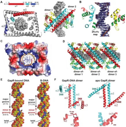
GapR forms a dimer-of-dimer clamp that encircles overly twisted DNA
GapR shows no homology to any structurally characterized DNA binding protein and, in fact, contains no known DNA binding motif. To determine how GapR binds DNA and recognizes overtwisted DNA, we solved a crystal structure of C. crescentus GapR in complex with DNA. We obtained this structure by single wavelength anomalous diffraction (SAD) using crystals obtained with selenomethionine substituted GapR in complex with an 11-mer, AT-rich site and refined the structure to final Rwork/Rfree values of 24.1%/26.8% to
2.3 Å resolution (Fig. 6A-B, Table S4). Strikingly, the structure shows that GapR
oligomerizes to form a dimer-of-dimers, which completely encircles the 11-mer DNA inside a large, central cavity (Fig. 6A; Movie S1), consistent with our EMSA indicating a binding site of ~10 bp (Fig. 5B). Each GapR subunit consists of three helices. An N-terminal helix (residues 14–51), α1, forms an antiparallel coiled coil dimer with α1 of a second GapR
A
uthor Man
uscr
ipt
A
uthor Man
uscr
ipt
A
uthor Man
uscr
ipt
A
uthor Man
uscr
ipt
subunit. The α1-α1´ dimer (where ´ indicates the other subunit in a dimer) buries 1340 Å2 of protein surface from solvent. α2 (residues 55–66) and α3 (residues 69–86) are positioned at opposite ends of the α1-α1´ antiparallel coiled coil dimer. The α2-α3 helical pairs from each GapR subunit interdigitate with a α2-α3 helical pair from a second GapR dimer to form the dimer-of-dimers. These cross α2-α3 zipper interactions seal the ends of the dimer-of-dimer, creating the large central DNA binding cavity (Fig. 6A). An electrostatic surface representation of the GapR dimer-of-dimers shows that it forms a DNA-binding clamp with a remarkably electropositive inner DNA binding surface (Fig. 6C; Movie S1). DALI searches revealed no structures with similarity to the GapR monomer, dimer, or dimer-of-dimers.
The electron density for the GapR-bound DNA is clearly resolved (Fig. 6B) and forms a pseudocontinous ladder in the crystal, resulting in multiple dimer-of-dimer molecules along the DNA (Fig. 6D). There are no significant contacts between any of the dimers-of-dimers, supporting the notion that GapR does not cooperatively coat or bridge DNA. Consistent with the idea that GapR does not bind DNA sequence specifically, GapR makes no base contacts and only interacts with the DNA phosphate backbone. Residues from both α1 and α2 helices make key contacts with the DNA. Specifically, Lys34, Lys42 and Lys49 from each
α1 helix and Lys56, Lys59, Arg63 and Lys66 from the basic regions of the four α2 helices
provide a total of 28 phosphate contacts to DNA (Fig. S6A; Movie S1). The α1 helices (Fig. 6A) and these basic residues track along the phosphate backbone suggesting that they “read” a specific DNA conformation. Indeed, analysis of GapR-bound DNA revealed a significantly narrowed minor groove of 9.8 Å (compared to 12 Å for B-DNA) and widened major groove of 21 Å (compared to 17.2 Å for B-DNA) (Fig. 6E). However, most strikingly, GapR-bound DNA is significantly overtwisted in the structure, with the helical DNA twist being 44.5º compared to 36.0º for B-DNA (Fig. 6E). Importantly, attempts to fit B-DNA, which has a greater diameter than overtwisted DNA, within the GapR DNA binding cavity resulted in clash. Thus, the GapR DNA binding clamp appears to recognize an overtwisted DNA conformation that features narrow minor grooves and widened major grooves, not a specific DNA sequence. Notably, the oligonucleotide used in the crystal structure had high AT-content, which is known to more readily adopt an overtwisted state with narrow minor grooves. GapR does not bind as well to oligonucleotides with reduced AT-content (Fig. S6D).
To investigate whether apo GapR adopts a different conformation, we sought the apo GapR structure. Although apo C. crescentus GapR failed to produce data quality crystals, we obtained the structure of apo Bosea sp. Root381 GapR (herein termed Bosea GapR), which shares 60% sequence identity with the C. crescentus protein (Fig. S6B). We confirmed that the Bosea GapR ortholog also adopts a dimer-of-dimer conformation with DNA in the central cavity (see Methods, Table S4, and Fig. S6C). The apo Bosea GapR structure was solved by SAD phasing and refined to final Rwork/Rfree values of 22.2%/25.9% to 2.4 Å
resolution (Table S4). Interestingly, the apo Bosea GapR forms a dimer rather than a dimer-of-dimers (Fig. 6F). The apo structure harbors the same α1-α1´ antiparallel dimer
interaction observed in the DNA-bound Caulobacter GapR structure, but the α2 and α3 helices form a continuous helix (Fig. 6F). Although we did not obtain the apo Caulobacter GapR structure, our SEC-MALS analysis was consistent with Caulobacter GapR forming an
A
uthor Man
uscr
ipt
A
uthor Man
uscr
ipt
A
uthor Man
uscr
ipt
A
uthor Man
uscr
ipt
elongated dimer in solution (Fig. S5A). The finding that GapR is dimeric in the absence of DNA indicates that DNA is not threaded through the dimer-of-dimer clamp, which is implausible, but rather that it adopts the dimerof-dimer state upon binding target DNA.
GapR stimulates the activities of topo IV and gyrase
Collectively, our results indicate that GapR recognizes overtwisted DNA such as that arising in front of replication forks. At these locations GapR may stimulate gyrase and/or topo IV to relieve superhelical stress and promote replication elongation. To test whether GapR promotes the activity of either gyrase or topo IV, we purified the two subunits of both Caulobacter gyrase and topo IV, and reconstituted each enzyme.
We first tested whether GapR can stimulate topo IV to remove (+) supercoils, deliberately selecting reaction conditions that yield relatively low topoisomerase activity to maximize our ability to observe any stimulation by GapR. We found that GapR at 0.4 μM stimulated the rate of (+) supercoil relaxation by topo IV ~4-fold (Fig. 7A). After 60 min, a reaction lacking GapR still had ~60% of the initial supercoiled substrate remaining, whereas the reaction containing GapR had almost no starting substrate left. Notably, the concentrations of GapR that stimulated topo IV activity (Fig. S7A) are insufficient to introduce (+) supercoils alone (Fig. 5D-E, S5E). We also tested whether GapR stimulates topo IV to relax (−) supercoils, and observed a modest, but reproducible ~1.3-fold increase in the rate of this reaction (Fig. S7B). The stimulatory effects of GapR on topo IV were specific as other proteins, including glucose-6-phosphate dehydrogenase (G6PD) and the DNA-binding protein GcrA, did not stimulate topo IV activity (Fig. S7C). We also tested whether GapR can stimulate other bacterial type II topoisomerases, by repeating relaxation assays with E. coli topo IV. We found that 0.4 μM GapR was unable to stimulate (+) supercoil relaxation by E. coli topo IV, but produced a modest increase in (−) supercoil relaxation (Fig. S7D). We then tested whether GapR stimulates Caulobacter gyrase. As with topo IV, we observed a clear and reproducible, GapR-dependent increase of ~2-fold for the rates at which gyrase relaxes (+) supercoiled DNA and introduces (−) supercoils into relaxed DNA (Fig. 7B, S7E-F). The stimulatory effects on gyrase were specific as neither G6PD nor GcrA stimulated gyrase (Fig. S7G). GapR had only a modest, but reproducible, stimulatory effect on (+) supercoil relaxation and (−) supercoil introduction by E. coli gyrase (Fig. S7H). These results demonstrate that GapR promotes the activities of both type II topoisomerases from Caulobacter, gyrase and topo IV, including their removal of (+) supercoils. Taken together with our in vivo studies, we conclude that the association of GapR with overtwisted DNA ahead of replication forks helps stimulate the type II topoisomerases, promoting replication fork progression, the timely removal of (+) supercoils ahead of replisomes, and,
consequently, the completion of DNA replication in Caulobacter.
Discussion
The successful completion of DNA replication in any organism requires the resolution of complex topological structures that build up around the fork. Although the importance of topoisomerases in this process is well established, how topoisomerases are recruited to, and how their activity is controlled at, the replication forks is still poorly understood. Here, we
A
uthor Man
uscr
ipt
A
uthor Man
uscr
ipt
A
uthor Man
uscr
ipt
A
uthor Man
uscr
ipt
identified GapR as a crucial regulator of topoisomerases and replisome progression in α-proteobacteria. Loss of GapR sensitizes cells to the inhibition of type II topoisomerases and leads to chromosomal relaxation, significantly slowed replication, and eventual cell death. We propose that the essential function of GapR is to promote the activities of type II
topoisomerases, both to maintain global supercoiling levels and, importantly, to attenuate (+) supercoils that would otherwise impede replication forks (Fig. 7C).
Type II topoisomerases have long been known to enable DNA replication by alleviating the torsional stress that accumulates near replication forks. Indeed, the efficacy of many front-line antibiotics, e.g. coumerins and quinolones, is based on their ability to inhibit gyrase, and thereby block bacterial DNA replication (Vos et al., 2011). However, although type II topoisomerases such as gyrase or topo IV are required for DNA replication in vivo, whether these enzymes are sufficient has not been established. Our results indicate that they may not be, and instead may require activators or co-factors, such as GapR.
Recognition of highly twisted DNA by GapR
GapR specifically binds to overtwisted DNA (Fig. 5E-F, 6E), which is precisely the form of DNA that initially arises in front of replication forks. Overtwisted DNA also arises in front of translocating RNA polymerase and our ChIP-seq results demonstrated that GapR binds such regions in a sequenceindependent, but transcription-dependent manner. Consistent with these results, the structure of GapR revealed that the protein makes no base-specific contacts with DNA, but instead recognizes the shape of the DNA. When bound to DNA, GapR adopts a structure with an internal cavity lined with basic residues that contact the negatively charged backbone of the encircled DNA. Importantly, our results indicate that GapR is only able to recognize overtwisted DNA, as the larger diameter of B-DNA cannot be
accommodated within the DNA-binding cavity of GapR. Thus, the structure of GapR in complex with DNA reveals a simple, but compelling mechanism for its localization to regions of the genome that are more highly twisted by the action of translocating DNA or RNA polymerase.
Our ChIP-seq analyses and prior studies of GapR revealed that GapR shows a preference for binding AT-rich DNA (Arias-Cartin et al., 2017; Ricci et al., 2016). Notably, such DNA often contain Atracts that can adopt intrinsically bent structures, with narrow minor grooves and increased superhelicity (Haran and Mohanty, 2009). Thus, AT-rich regions and the DNA ahead of replisomes or RNA polymerase may be the primary binding sites for GapR by virtue of a common, overtwisted shape. Our biochemical studies showed that GapR can also associate with relaxed or (−) supercoiled DNA, likely via local regions of overtwisted DNA within these substrates, arising from sequences with intrinsically narrow minor grooves or from thermodynamic fluctuations in twist.
GapR forms a snug clamp around overtwisted DNA raising the question of how it is loaded. The apo structure revealed that GapR is a dimer in the absence of DNA, with α2 and α3 forming a continuous helix (Fig. 6F), suggesting that the region between α2 and α3 is kinked upon DNA binding. A specific residue, Asp68, appears to function as the pivot point between α2 and α3. Among GapR homologs, an aspartic acid is highly conserved at this position, although glutamic acid, glutamine, and aspargine are also found. Notably, these
A
uthor Man
uscr
ipt
A
uthor Man
uscr
ipt
A
uthor Man
uscr
ipt
A
uthor Man
uscr
ipt
residues are all known to function as N-cap residues and helix breakers. Indeed, while Asp68 points into the solvent in the apo GapR structure as part of the continuous α2 helix, it forms stabilizing N-cap contacts with α3 in the DNA-bound structures (Fig. 6F). Combined, these data suggest a unique mechanism for GapR clamp formation and loading in which the protein uses its α1-α1´ helices to track along the DNA as a caliper to search for sites with narrow minor and expanded major grooves, which then results in stable dimer-of-dimer formation through kinking to form α2-α3 and subsequent interactions between α2-α3 pairs around the DNA. Thus, unlike other DNA binding clamps, GapR likely does not require a clamp loader to dock onto its target DNA.
The preferential binding of GapR to highly twisted DNA explains why many GapR binding events were required to generate increases in plasmid writhe (Fig. 5D-E). Because
supercoiling is a combination of twist and writhe, each GapR binding event introduces a small amount of twist into DNA, such that multiple binding events are necessary before inducing a change in writhe. Assuming GapR binds a ~10 bp site with half the available sites bound, the change of +3 writhe we observed implies an overtwisting of the DNA by ~8° per GapR bound, similar to the 8.5° change measured in the structure. However, GapR likely does not directly introduce large numbers of supercoils in vivo as there are only ~3,000 GapR molecules present per cell (Fig. S1F), significantly less than NAPs such as HU, which is present at ~60,000 copies per cell (Rouviere-Yaniv et al., 1979). Additionally, we found that GapR stimulates gyrase and topo IV at lower concentrations than is required to introduce topological changes. These observations, along with the structure of GapR bound to DNA, indicate that the primary role of GapR is not to directly alter DNA topology, but rather to associate with regions of (+) supercoiling, promoting the activity of type II topoisomerases precisely where their activities are needed the most.
Role of GapR in modulating topoisomerase activity
How, mechanistically, might GapR promote gyrase and topo IV activity? The simplest model is that GapR directly binds gyrase and topo IV to promote their binding to DNA or catalytic activity (Fig. S7I, left). By recognizing overtwisted DNA that arises in front of replication forks, GapR may recruit or stimulate gyrase and topo IV to act on the (+) supercoiled DNA that arises in front of forks and that must be dissipated for replication to progress. Notably, the structure of GapR revealed two acidic patches that are solvent-exposed in the DNA-bound, dimer-of-dimers configuration. Most of these acidic residues are highly conserved (Fig. S6B) and they may mediate a direct interaction with the topoisomerases. Alternatively, GapR could stimulate gyrase and topo IV without directly binding these enzymes, instead stabilizing DNA in a way that promotes topoisomerase binding and/or processivity (Fig. S7I, right). For instance, (+) writhed DNA that arises in front of replication forks may normally diffuse away from the fork. GapR binding to overtwisted DNA could ‘pin’ a plectonemic region of DNA, effectively trapping DNA for relaxation by topoisomerases. Further biochemical and enzymatic studies are needed to fully dissect the detailed mechanisms by which GapR stimulates gyrase and topo IV.
A
uthor Man
uscr
ipt
A
uthor Man
uscr
ipt
A
uthor Man
uscr
ipt
A
uthor Man
uscr
ipt
Regulation and control of DNA replication elongation
Whether the loss of viability in GapR-depleted cells arises from the defects in replication or the broad changes in transcription, or both, is unclear. The transcriptional changes are, however, likely an indirect effect of GapR depletion as the GapR ChIP profile did not correlate with the global changes in gene expression. We favor the notion, supported by our biochemical and structural studies, that the primary function of GapR is to promote type II topoisomerase activity during DNA replication. Consistent with this model, GapR-venus was suggested to localize to the DNA ahead of the replisome (Arias-Cartin et al., 2017). Other NAPs have been implicated in controlling bacterial DNA replication. For instance, E. coli IHF and Fis are both critical for DNA replication initiation (Magnan and Bates, 2015). There are also several proteins in E. coli, including MukB and SeqA, that interact with topo IV to stimulate (−) supercoil removal and chromosome decatenation, although neither is required for replication elongation (Hayama and Marians, 2010; Kang et al., 2003; Li et al., 2010). E. coli HU was suggested to stimulate gyrase’s decatenation activity in vitro, but whether this function of HU promotes DNA replication in vivo is unknown (Marians, 1987). E. coli YejK stimulates topo IV to relax (+) supercoils in vitro, but YejK inhibits gyrase and ΔyejK strains have only modest, possibly indirect, effects on DNA replication and exhibit no growth defect (Lee and Marians, 2013). Thus, to our knowledge, GapR is the first NAP required for replication elongation and the first characterized activator of gyrase. In sum, we suggest that GapR plays a critical role in resolving the superhelical stress associated with DNA replication. GapR may also be required to alleviate the torsional stress that arises in regions of the genome where (+) supercoils accumulate and cannot diffuse, e.g. when the replication forks converge near the terminus or, more frequently, when the
transcription and replication machineries converge. At such sites, (+) supercoils must be resolved for replication to continue.
GapR is highly conserved throughout the α-proteobacteria where it likely plays a similar role in activating type II topoisomerases. GapR homologs are not found in other classes of bacteria, but a role for NAPs in replication elongation has not been well explored. There also may be as yet unidentified NAPs that share no homology to GapR, but recognize highly twisted DNA to similarly promote replication. In eukaryotes, there may also be analogous proteins. For example, high mobility group (HMG) proteins bind DNA relatively non-specifically, contributing to chromosome architecture in poorly defined ways. Intriguingly, yeast Hmo1 and the type II topoisomerase Top2 were recently shown to bind together near genes transcribed during S phase to somehow suppress chromosome fragility (Bermejo et al., 2009), and human HMGB1 interacts with and stimulates Top2α decatenation activity (Stros et al., 2007). We anticipate that interactions between stablybound, chromosome architecture proteins and topoisomerases, like that described here for GapR, will ultimately prove essential to the maintenance of genome integrity and the successful execution of DNA replication in most organisms.
A
uthor Man
uscr
ipt
A
uthor Man
uscr
ipt
A
uthor Man
uscr
ipt
A
uthor Man
uscr
ipt
STAR Methods
Contact for Reagent and Resource Sharing
Questions about or requests for methods, strains, and resources generated in this study can be directed to the Lead Contact, Michael T. Laub (laub@mit.edu).
Experimental Model and Subject Details
Growth conditions and chemical treatments—Caulobacter strains were grown in PYE (2 g/L bactopeptone, 1g/L yeast extract, 0.3 g/L MgSO4, 0.5 mM 0.5M CaCl2) at 30 °C
unless noted. The Pxyl promoter was induced by supplementing media with xylose (0.3%)
and repressed with glucose (0.2%). Antibiotics were used at the following concentrations unless noted (liquid/plates): oxytretracycline (1 μg mL−1 / 2 μg mL−1), spectinomycin (25 μg mL−1 / 200 μg mL−1), kanamycin (5 μg mL−1 / 25 μg mL−1), gentamycin (NA / 5 μg mL−1). E. coli strains were grown in LB (10 g/L NaCl, 10 g/L tryptone, 5 g/L yeast extract) and supplemented with antibiotics at the following concentrations (liquid/plates): carbenicillin (50 μg mL−1 / 100 μg mL−1), oxytretracycline (12 μg mL−1 / 12 μg mL−1), spectinomycin (50 μg mL−1 / 50 μg mL−1), kanamycin (30 μg mL−1 / 50 μg mL−1), gentamycin (15 μg mL
−1 / 20 μg mL−1). When required, 400 mM IPTG was used to induce gene expression unless
noted. Optical density was measured at 600 nm using a Genesys 10 Bio Spectrophotometer.
Strain construction—All Caulobacter strains were derivatives of ML76, an isolate of strain CB15N/NA1000. The insertion of a 3×FLAG at the C-terminus of GapR was constructed via a two-step recombination method using sacB as a counterselection marker. Strain ML2794 was constructed by electroporating plasmid pNPTS138-UP-gapR-3×FLAG-DW into ML76 and first integrants were selected on kanamycin plates. A second
recombination step was performed by growing first integrants in PYE overnight and then plating on sucrose. Sucrose resistant clones were restreaked to verify they were kan sensitive and colonies were verified by PCR with primer pair CCNA_3428–3×flag-2-R and
CCNA_03428-UP-F-less-hindiii.
The GapR depletion strain, ML2795, was constructed by electroporating plasmid pMT4427-CCNA_03428 into ML76 and selecting on kanamycin plates. As a second step, plasmid pNTPSSPEC-CCNA_03428-TET was electroporated and cells were selected on kanamycin, spectinomycin and tetracycline. First integrants were then grown overnight with kanamycin, tetracycline, and xylose and then plated on kanamycin, tetracycline, xylose + sucrose plates for sacB counterselection. Sucrose resistant plates were restreaked to verify sensitivity to spectinomycin. ML2954 was constructed by electroporating plasmid pMT4260-PgapR-gapR into ML76 and selecting on kanamycin plates. GapR was subsequently deleted by
integrating pNTPS-SPEC-CCNA_03428-TET as described previously.
Plasmid construction
Integration plasmids: pNPTS138-UP-gapR-3×FLAG-DW was constructed by amplifying
a fragment containing the upstream region of gapR with primers CCNA_03428-UP-F-less-hindiii and CCNA_03428–3×flag-1-R to which the coding region for 3×-FLAG was added by a second round of PCR with primers CCNA_3428–3×flag-2-R and
CCNA_03428-UP-F-A
uthor Man
uscr
ipt
A
uthor Man
uscr
ipt
A
uthor Man
uscr
ipt
A
uthor Man
uscr
ipt
less-hindiii. This upstream fragment was fused by splice-overlap-extension (SOE) PCR to a fragment containing the region downstream of gapR amplified with primers 3428dw-fuse-3×flag-F and CCNA_03428-DW-R. The resulting PCR product was digested with NheI and HindIII restriction enzymes and ligated into pNPTS-138 digested with the same
enzymes.
The pNTPS-SPEC-CCNA_03428-TET plasmid was constructed by multiple SOE PCR reactions. First, a fragment containing 703 bp upstream of CCNA_03428 was amplified with the primer pair CC3319TETDELUP-R and FOR_UP3319PNTPSPSTI, the tetracycline (tet) resistance cassette was amplified from template pKO3 using primer pair OL7_tet_F and OL8_tet_R, and a fragment containing 703 bp downstream of CCNA_03428 was amplified using primers CC3319TETDELDWF and CC3319TETDELDW_SPEI-R. Subsequent SOE PCRs were performed to fuse the three fragments together. The resulting product was digested with PstI and SpeI and ligated into pNTPSSPEC digested with the same restriction enzymes.
The pMT4260-PgapR-gapR plasmid was constructed by amplifying a fragment containing
the promoter and coding region of GapR with GapR_up_KpnI and GapR_down_NheI, digesting with KpnI and NheI, and ligated pMT4260 digested with the same enzymes.
Replicative plasmids: pMT4427-CCNA_03428 was constructed by ligation of a PCR
fragment amplified with primers CCNA_03428-F-NdeI and CCNA_03428-R-SacI and digested with NdeI and SacI.
Expression plasmids: pET28-CCNA_03428 was constructed by ligation of a PCR
fragment amplified with primers CCNA_3428-F-NdeI and CCNA_3428_R_SacI and digested with NdeI and SacI. pKS22b-hSUMO-GapR was constructed by ligation of a PCR fragment amplified with primers GapR-F-BamHI and GapR-R-NotI and digested with BamHI and NotI.
pKS22b-hSUMO-GyrA was constructed by Gibson assembly cloning with a PCR fragment amplified with primers GyraseA-F-BamHI-homology and GyraseA-R-NotI-homology and the pKS22b vector digested with BamHI and NotI. pKS22b-hSUMO-GyrB was constructed by Gibson assembly cloning with a PCR fragment amplified with primers GyrB-F-BamHI-homology and GyrB-R-NotI-GyrB-F-BamHI-homology and the pKS22b vector digested with BamHI and NotI. pKS22b-hSUMO-ParC was constructed by Gibson assembly cloning with a PCR fragment amplified with primers ParC-F-BamHI-homology and ParC-R-NotI-homology and the pKS22b vector digested with BamHI and NotI. pKS22b-hSUMOParE was constructed by Gibson assembly cloning with a PCR fragment amplified with primers ParE-F-BamHI-homology and ParE-R-NotI-ParE-F-BamHI-homology and the pKS22b vector digested with BamHI and NotI. For pET15b-GapR(Cres) and pET15b-(Bosea) and their variants, the full length Bosea sp. Root 381 GapR protein and C. crescentus GapR residues 11–89 were purchased as E. coli codon optimized genes from Genscript with NdeI and BamHI sites and subcloned into pET15b using restriction cloning. This resulted in the placement of a cleavable hexa-histidine tag at the N-terminus of the expressed proteins. C. crescentus GapR(11–89) was used for structural studies as the N-terminal 10 residues of the C. crescentus protein are not
A
uthor Man
uscr
ipt
A
uthor Man
uscr
ipt
A
uthor Man
uscr
ipt
A
uthor Man
uscr
ipt
conserved among GapR homologs (and not present in several homologs including the Bosea sp. Root 381 GapR protein) and are predicted to be disordered.
Method Details
Nucleoid purification and mass spectrometry analysis
The nucleoid purification protocol was performed as follows. A 25 mL culture of
exponentially growing ML1241 was centrifuged at 8,500 rpm at 4 °C for 5 min. Cell pellets were resuspended in 0.5 mL of ice-cold buffer A (10 mM Tris-HCl [pH 8.2], 100 mM NaCl, and 20% sucrose) followed by the addition of 0.1 mL of ice-cold buffer B (100 mM Tris-HCl [pH 8.2], 50 mM EDTA, 0.6 mg/mL lysozyme) and incubated for 1 minute on ice. Then, 0.5 mL of Buffer C (10 mM Tris-HCl [pH 8.2], 10 mM EDTA, 10 mM spermidine, 1% Brij-58, and 0.4% deoxycholate) was added and the mixture incubated for 4 min at 5 °C. The lysed cells were loaded onto linear sucrose density gradients (15–50%) containing 10 mM Tris-HCl (pH 8.2) and 100 mM NaCl, and centrifuged for 20 minutes at 10,000 rpm in a Beckmann SW 41 Ti rotor. Fractions (~1 mL each) were collected by piercing a hole at the bottom of the ultracentrifugation tube and DNA content quantified by DAPI (Invitrogen) fluorescence using a plate reader (Spectramax). The fractions were then incubated with 20 U of benzonase at room temperature for 30 min. Fractions were run on a 15% Tris-HCl gel (BioRad) and submitted for tandem mass spectrometry analysis at the Koch Institute Biopolymers and Proteomics core facility. The region of the gel corresponding to proteins in the range of 5–25 kDa was isolated and cut into two fragments for mass spectrometry analysis. Mass spectra were analyzed using Mascot (Matrix Science, London, UK), searching against the Sprot_extra_022412 database assuming digestion with trypsin. To compare chromosomal supercoiling, nucleoids were isolated as above and loaded on to 10–30% linear sucrose gradients containing varying amounts of ethidium bromide. For novobiocin treated nucleoids, cells were treated with 25 μg/mL novobiocin (Sigma) for 15 min before cell harvest. Intercalation of ethidium bromide into DNA first causes relaxation of the chromosome and decreased sedimentation and then at higher concentrations of ethidium increased sedimentation due to introduction of (+) supercoils. The amount of ethidium bromide at the point of slowest migration is reflective of the concentration of negative supercoils in the chromosome (Worcel and Burgi, 1972). Gradients were
centrifuged at 10,000 rpm in a Beckman SW41 for 15 min. The distance sedimented by the nucleoid from the top of the gradient was measured. Sedimentation data from multiple runs with different ethidium bromide concentrations were pooled for analysis. The concentration of ethidium required at the point of slowest migration was estimated by fitting the data with a LOWESS fit using Prism 7 software.
Expression profiling with microarrays and RNA-sequencing
RNA was extracted using hot trizol lysis and the Direct-zol RNA MiniPrep (Zymo). rRNAs were removed using the Ribo-Zero Kit for Gram-negative bacteria (Illumina). The rRNA-depleted RNA was then fragmented using the RNA fragmentation reagents (Ambion) at 70 °C for 8 min. Fragmented RNA was recovered by ethanol precipitation and resuspended in 6 μL water. RNA-seq libraries were prepared based on the previously described dUTP
A
uthor Man
uscr
ipt
A
uthor Man
uscr
ipt
A
uthor Man
uscr
ipt
A
uthor Man
uscr
ipt
protocol as follows. First-strand cDNA synthesis was performed by addition of 1 μL of 3 μg/ μL random primers (Invitrogen) followed by incubation at 5 min at 65 °C. Samples were then placed on ice for 1 min and the following reagents were added: 4 μL first-strand synthesis buffer, 2 μL 100 mM DTT, 1 μL 10 mM dNTPs, 1 μL Superase-in (ThermoFisher) and 4 μL water. Reactions were incubated at room temperature for 2 min and 1 μL
Superscript III (ThermoFisher) was added and the following thermocycler program was used: 10 min at 25 °C, 1hr at 50 °C and 15 min at 70 °C. The reaction volumes were brought to 200 μL and a phenol:chloroform:isoamyl alcohol (25:24:1) (Sigma) extraction was performed. cDNA was resuspended in 104 μL water and second-strand synthesis reactions were set up with 30 μL of 5X second-strand synthesis buffer (ThermoFisher), 4 μL of 100 mM dNTPs with dUTP (Promega) instead of dTTP, 4 μL of 5X first-strand synthesis buffer and 2 μL 100 mM DTT. Reactions were incubated on ice for 5 min and 1 μL RNase H (NEB), 1 μL E.coli DNA ligase (NEB) and 4 μL DNA Pol I (NEB) were added, followed by an incubation at 16 °C for 2.5 hrs. cDNA was recovered by Ampure XP (Beckman Coulter) bead purification with 100 μL beads in 450 μL 20% PEG/NaCl solution.
Sequencing libraries were built by first end repairing cDNA with 5 μL T4 DNA polymerase (NEB), 5 μL T4 PNK (NEB), and 1 μL Klenow large fragment (NEB) in 100 μL T4 DNA ligase buffer with 0.25 mM dNTPs for 30 min at room temperature. Repaired DNA was recovered by Ampure XP (Beckman Coulter) bead purification, using 100 μL beads in 300 μL 20% PEG/NaCl solution. Beads were washed twice with 80% ethanol, dried, and resuspended in 32 μL EB. The bead slurry was directly treated with 3 μL Klenow (3’→5’ exo-) (NEB) in 50 μL NEB Buffer #2 with 0.2 mM ATP at 37 °C for 30 min to add 3’ overhangs to DNA. The reaction was cleaned up by addition of 150 μL 20% PEG/NaCl and capture of the Ampure XP beads. After two washes with 80% ethanol, the beads were resuspended in 23 μL EB and the supernatant was transferred to a clean tube. Y-shaped Illumina adapters were ligated onto the DNA in 50 μL total volume in Quick Ligase buffer with 3 μM Y-shaped adaptors and 1 μL Quick Ligase (NEB) for 15 min at room temperature. Y-shaped adaptors were prepared by annealing Illumina PE adapter 1 and Illumina PE adapter 2. 75 μL 20% PEG/NaCl was added, and the DNA then recovered by addition of Ampure beads followed by two ethanol washes. After resuspension in 23 μL water, the Ampure beads were captured and the DNA in the supernatant was transferred to a clean tube. Digestion of the second strand was performed by addition of 6 μL of 5× HF Phusion buffer and 1 μL USER (NEB) and incubation at 37 °C for 15 min, followed by 5 min at 95 °C. DNA libraries were amplified in 80 μL final volume with Phusion DNA polymerase (NEB) in Phusion High GC buffer supplemented with Betaine (0.4 M final) (Sigma). The total number of cycles was optimized for each sample such that 10–14 cycles of PCR was used for each sequencing library. Libraries were purified by addition of 240 μL 20% PEG/ NaCl and capture of the Ampure XP beads. After two washes with 80% ethanol, the beads were resuspended in 20 μL EB. Elutions were then run on a 8% TBE polyacrylamide gel (ThermoFisher) for 30 min at 180 V and a 230–500 bps region was gel extracted using a Spin-X 0.22 μm cellulose acetate column (Costar). Single-end sequencing was performed on an Illumina Hi-Seq 2000 at the MIT Bio Micro Center.
For DNA microarray experiments, 10 mL cultures of ML76 were grown in PYE + 0.3% xylose, M2X or M2G to mid-exponential phase and treated with 25 μg/mL novobiocin
A
uthor Man
uscr
ipt
A
uthor Man
uscr
ipt
A
uthor Man
uscr
ipt
A
uthor Man
uscr
ipt
(Sigma) for 15 min or left untreated to use as a reference for the microarray. RNA was extracted using the RNeasy Mini Kit (Qiagen). The generation of labeled cDNA was performed using 20 μg of RNA in 14 μL RNase-free water to which 1 μL of random primers (Invitrogen) was added. The samples were incubated at 65 °C for 10 min, followed by 2 min incubation on ice. 9.6 μL of trimix (6 μL 5X first-strand buffer, 3 μL 0.1 M DTT, and 0.6 μL dNTP mix [25 mM A,G,T, 10 mM C]), 1 μL of Superscript II (ThermoFisher), and 3 μL of labeled dCTP (Cy5 dye for experimental and Cy3 dye for reference) (Sigma) were added and incubated covered with foil for 10 min at room temperature. Samples were then incubated at 42 °C for 1 hr, after which an additional 1 μL of Superscript II was added for another hour. RNA was degraded by addition of 1.5 μL of 0.5 N NaOH and cDNA purified by addition of 1.5 μL of 0.5 N HCl followed by purification with a PCR Purification Kit (Qiagen). After a wash with PE buffer, cDNA was eluted in 50 μL EB. 1 μL of 20 μg/μL yeast tRNA (Sigma) was added and the total volume was adjusted to 55 μL. 55 μL of 2X-HiRPM hybridization buffer (Agilent) as added to the samples and incubated for 3 min at 95 °C followed by 1 min at 42 °C. 100 μL was loaded onto a custom Agilent array placed in a hybridization chamber and incubated overnight rotating at 65 °C. Arrays were washed for 5 min in Oligo aCGH/Chip-on-chip wash buffer 1 (Agilent), washed for 5 min in Oligo aCGH/Chip-on-chip wash buffer 2 (Agilent) at 30 °C, and washed with acetonitrile for 2 min at room temperature. Arrays were dried Stabilization and Drying Solution (Agilent) and imaged using an Agilent scanner at the MIT Bio Micro Center.
Chromatin Immunoprecipitation Sequencing (ChIP-seq)
ChIP was performed using strain ML2794 (gapR::gapR-3×FLAG) or wild-type CB15N grown in PYE. The ChIP procedure was as follws. Briefly, cell cultures (20 mL) were grown to OD600 ~0.3 and fixed by the addition of 10 mM sodium phosphate [pH 7.6] and 1%
formaldehyde (final concentrations) (Sigma). When appropriate, 25 μg/mL of rifampicin (Sigma) was added to cells for 20 minutes prior to fixation. Fixed cells were incubated at room temperature for 10 minutes and then quenched with 0.1 M glycine (Sigma) for 5 min at room temperature followed by 15 min on ice. Cells were washed three times with 1X PBS [pH 7.4] and resuspended in 500 μL of TES buffer (10 mM Tris-HCl [pH 7.5], 1 mM EDTA, 100 mM NaCl) to which 35,000 U of Ready-Lyse (Epicentre) was added. Following 15 min incubation at RT, 500 μL of ChIP buffer (16.7 mM Tris-HCl [pH 8.1], 167 mM NaCl, 1.1% Triton X-100, 1.2 mM EDTA) containing protease inhibitors (Roche cOmplete EDTA-free tablets) was added. After 10 min at 37 °C, the lysates were sonicated on ice and cell debris cleared by centrifugation. Supernatant protein concentration was measured by Bradford assay (Thermo Scientific) and 500 μg of proteins were diluted into 1 mL of ChIP buffer + 0.01% SDS. The diluted supernatants were pre-cleared for 1 hr at 4 °C on a rotator with 50 μL of Protein-A Dynabeads (Life Technologies) pre-blocked overnight in ChIP buffer + 0.01% SDS and 100 μg ultrapure BSA (Ambion). Beads were pelleted and 90 μL of the supernatant was removed as input DNA and stored at −80 °C, the remaining pre-cleared supernatant was incubated rotating at 4 °C overnight with 1 μL of FLAG antibody (Sigma). The immune complexes were captured for 2 hr at 4 °C with 50 μL of pre-blocked Protein-A Dynabeads. Beads were then washed consecutively at 4 °C for 15 min with 1 mL of the following buffers: low salt wash buffer (0.1% SDS, 1% Triton X-100, 2 mM EDTA, 20 mM Tris-HCl [pH 8.1], 150 mM NaCl), high salt wash buffer (0.1% SDS, 1% Triton X-100, 2
A
uthor Man
uscr
ipt
A
uthor Man
uscr
ipt
A
uthor Man
uscr
ipt
A
uthor Man
uscr
ipt
mM EDTA, 20 mM Tris-HCl [pH 8.1], 500 mM NaCl), LiCl wash buffer (0.25 M LiCl, 1% NP-40, 1% deoxycholate, 1 mM EDTA, 10 mM Tris-HCl [pH 8.1] and twice with TE buffer (10 mM Tris-HCl [pH 8.1], 1 mM EDTA). Complexes were then eluted twice from the beads with 250 μL of freshly prepared elution buffer (1% SDS, 0.1 M NaHCO3). To reverse
crosslinking, 300 mM of NaCl and 2 μL of RNase A (0.5 mg / mL) (Qiagen) were added to the collective eluates which were incubated at 65 °C overnight. Samples were then incubated at 45 °C for 2 hr with 5 μL of Proteinase K (NEB) in the presence of 40 mM EDTA (pH 8.0) and 40 mM Tris-HCl [pH 6.8]. DNA from the samples was then extracted twice with phenol:chloroform:isoamyl alcohol (25:24:1) (Sigma) and subsequently precipitated by adding sodium acetate (pH 5.2), 100 μg glycogen (ThermoFisher) and 1 volume of ice cold isopropanol, and stored at −20 °C overnight. DNA was pelleted and washed with 75% ethanol and resuspended in TE buffer [pH 8.0].
Sequencing libraries were built by first end repairing cDNA with 5 μL T4 DNA polymerase (NEB), 5 μL T4 PNK (NEB), and 1 μL Klenow large fragment (NEB) in 100 μL T4 DNA ligase buffer with 0.25 mM dNTPs for 30 min at room temperature. Repaired DNA was recovered by Ampure XP (Beckman Coulter) bead purification, using 100 μL beads in 300 μL 20% PEG/NaCl solution. Beads were washed twice with 80% ethanol, dried, and resuspended in 32 μL EB. The bead slurry was directly treated with 3 μL Klenow (3’→5’ exo-) (NEB) in 50 μL NEB Buffer #2 with 0.2 mM ATP at 37 °C for 30 min to add 3’ overhangs to DNA. The reaction was cleaned up by addition of 150 μL 20% PEG/NaCl and capture of the Ampure XP beads. After two washes with 80% ethanol, the beads were resuspended in 23 μL EB and the supernatant was transferred to a clean tube. Y-shaped Illumina adapters were ligated onto the DNA in 50 μL total volume in Quick Ligase buffer with 3 μM Y-shaped adaptors and 1 μL Quick Ligase (NEB) for 15 min at room temperature. Y-shaped adaptors were prepared by annealing Illumina PE adapter 1 and Illumina PE adapter 2. 75 μL 20% PEG/NaCl was added, and the DNA then recovered by addition of Ampure beads followed by two ethanol washes. After resuspension in 30 μL water, the Ampure beads were captured and the DNA in the supernatant was transferred to a clean tube. DNA libraries were amplified in 80 μL final volume with Phusion DNA polymerase (NEB) in Phusion High GC buffer supplemented with Betaine (0.4 M final) (Sigma). The total number of cycles was optimized for each sample such that 10–14 cycles of PCR was used for each sequencing library. Libraries were purified by addition of 240 μL 20% PEG/ NaCl and capture of the Ampure XP beads. After two washes with 80% ethanol, the beads were resuspended in 20 μL EB. Elutions were then run on a 8% TBE polyacrylamide gel (ThermoFisher) for 30 min at 180 V and the 250–500 bps region was gel extracted using a Spin-X 0.22 μm cellulose acetate column (Costar). Single-end sequencing was performed on an Illumina Hi-Seq 2000 at the MIT Bio Micro Center. RpoC-3×FLAG ChIP profiles were from (GSE73925).
Caulobacter synchronizations
For synchronizations, Caulobacter strains were grown to mid-exponential phase and G1/ swarmer cells were isolated using Percoll (GE Healthcare) density gradient centrifugation. Cells were pelleted at 8000 × g for 5 min and resuspended in M2 salts (0.87 g/L Na2HPO4,
0.53 g/L KH2PO4, 0.5 g/L NH4Cl) with an equal volume of Percoll. The suspension was