LA BORRELIOSE
DE LYME
Solène ANSQUER CHU de Poitiers
JOURNEES DE DES de NEUROLOGIE
14 et 15 mars 2014
Borréliose de Lyme
☐ Infection causée par un groupe de Spirochètes (Famille : Spirochaetaceae, Genre : Borrelia) ☐ 5 espèces en Europe : Borrelia garinii > B. burgdorferi > B. afzelii > spielmanii et bavariensis
GENERALITES
Borrelia Burgdorferi
↳ Variabilité de sa présentation clinique • Borrelia afzelii : tropisme cutanée • Borrelia burgdorferi : tropisme articulaire
Transmission par les tiques Ixodes
☐ Développement de la tique en 4 stades dans des régions tempérées, hygrométrie > 70%
GENERALITES
Tick Extraction
1 cm
Hypostom
☐ Infestation de la tique lors de repas sanguin sur un vertébré contaminé par Borrelia
☐ Cycle développemental impliquant 3 hôtes différents avec une gâme d’hôtes très variée • petits mammifères
• rongeurs et écureuil gris (Sciurius carolensis) et écureuil roux (Sciurius vulgaris) • les lagomorphes
• les oiseaux
Seminar
www.thelancet.com Vol 379 February 4, 2012 463
days and explains why transmission occurs only after a delay. Expression of OspC plays an essential part in the establishment of infection in a mammalian host, although the mechanism by which OspC promotes borrelial infectivity is unknown.26,27
When feeding, an infected tick deposits spirochaetes into the skin of a host animal. Later, Lyme borrelia disseminate from that site through blood or perhaps tissue planes to other locations. Evidence indicates that the risk of haematogenous dissemination by B burgdorferi is strain dependent.28
Infection of human beings or animals elicits innate and adaptive immune responses, resulting in both macrophage-mediated and antibody-macrophage-mediated killing of spirochaetes. Despite a robust humoral and cellular immunological response, however, infection with Lyme borrelia can persist. Virulence factors that cause persistence include the spirochaete’s ability to downregulate expression of specifi c immunogenic surface-exposed proteins, including OspC, and to alter rapidly and continually by recombination of the antigenic properties of a surface lipoprotein known as variable major protein-like sequence expressed (VlsE). The ability of spirochaetes to bind to various components of the extracellular matrix might also contribute to persistence.29–31
Lyme borrelia are not known to produce toxins. Most tissue damage seems to result from host infl ammatory reactions. The intensity of the infl ammatory response varies according to the Borrelia genospecies that causes an infection.32 Although host genetic factors have an
important role in the expression and severity of infection in animals, the only role established in man is in the development of antibiotic refractory Lyme arthritis, which is seen most often in patients with specifi c HLA-DR alleles.30
Clinical manifestations and epidemiological aspects
Localised infection is typically manifested by a erythema migrans skin lesion. Early disseminated disease is usually characterised by two or more erythema migrans skin lesions or as an objective manifestation of Lyme neuroborreliosis or Lyme carditis. Late Lyme borreliosis usually manifests as arthritis or the skin disorder known as acrodermatitis chronica atrophicans, but can also include specifi c rare neurological manifestations. The often used division of the disease into stages is somewhat theoretical and sometimes not in agreement with clinical fi ndings.33 For example, in some studies, most patients
who present with Lyme arthritis have no recollection of having had an earlier clinical manifestation of Lyme borreliosis.9
Of the various objective clinical presentations of Lyme borreliosis in Europe, erythema migrans is the most common.34–36 In one case series of patients with
Lyme borreliosis,35 89% had erythema migrans by
itself, 5% had arthritis, 3% had early neurological
mani festations, 2% had borrelial lymphocytoma, 1% had acrodermatitis chronica atrophicans, and less than 1% had cardiac manifestations. None of the patients had late neurological Lyme borreliosis. A similar distribution of cases has been seen in a case series in the USA,37–39 but
no patients had borrelial lymphocytoma or acrodermatitis chronica atrophicans. Yearly incidence rates in Europe seem to increase from northern Europe to the southern parts of central Europe, and range from 69 cases per 100 000 population in Sweden to 111 cases per 100 000 in
5 mm
Figure 2: Developmental stages of Ixodes ricinus
From left to right: larva, nymph, adult female, adult male. Reproduced with permission from the European Concerted Action on Lyme Borreliosis.
Eggs hatch to larva Larvae seek new host Eggs laid by female Fully fed female drops from host to ground
Female attaches to and feeds on third host Nymph attaches to and feeds on second host Larva moults to nymph Host two Host three Host one
Larva feeds on first host
Nymph moults to adult Borrelia afzelii
Borrelia garinii Borrelia burgdorferi
Fully fed larva drops to gound ?
Fully fed female drop from host to ground from host Female attaches to and feeds on Host three Larv Lar to n Host two
feeds on first host
Figure 3: Infectious cycle of the European Borrelia burgdorferi sensu lato genospecies
The size of the coloured closed circles indicates the relative involvement of the the diff erent vertebrate reservoirs for the diff erent genospecies. B burgdorferi sensu stricto is the only pathogenic genospecies present in the USA and, as in Europe, both rodents and birds are reservoirs. Reproduced with permission from the European Concerted Action on Lyme Borreliosis. A red cross indicates a non-reservoir host.
Seminar
www.thelancet.com Vol 379 February 4, 2012 463
days and explains why transmission occurs only after a delay. Expression of OspC plays an essential part in the establishment of infection in a mammalian host, although the mechanism by which OspC promotes borrelial infectivity is unknown.26,27
When feeding, an infected tick deposits spirochaetes into the skin of a host animal. Later, Lyme borrelia disseminate from that site through blood or perhaps tissue planes to other locations. Evidence indicates that the risk of haematogenous dissemination by B burgdorferi is strain dependent.28
Infection of human beings or animals elicits innate and adaptive immune responses, resulting in both macrophage-mediated and antibody-macrophage-mediated killing of spirochaetes. Despite a robust humoral and cellular immunological response, however, infection with Lyme borrelia can persist. Virulence factors that cause persistence include the spirochaete’s ability to downregulate expression of specifi c immunogenic surface-exposed proteins, including OspC, and to alter rapidly and continually by recombination of the antigenic properties of a surface lipoprotein known as variable major protein-like sequence expressed (VlsE). The ability of spirochaetes to bind to various components of the extracellular matrix might also contribute to persistence.29–31
Lyme borrelia are not known to produce toxins. Most tissue damage seems to result from host infl ammatory reactions. The intensity of the infl ammatory response varies according to the Borrelia genospecies that causes an infection.32 Although host genetic factors have an
important role in the expression and severity of infection in animals, the only role established in man is in the development of antibiotic refractory Lyme arthritis, which is seen most often in patients with specifi c
HLA-DR alleles.30
Clinical manifestations and epidemiological
aspects
Localised infection is typically manifested by a erythema migrans skin lesion. Early disseminated disease is usually characterised by two or more erythema migrans skin lesions or as an objective manifestation of Lyme neuroborreliosis or Lyme carditis. Late Lyme borreliosis usually manifests as arthritis or the skin disorder known as acrodermatitis chronica atrophicans, but can also include specifi c rare neurological manifestations. The often used division of the disease into stages is somewhat theoretical and sometimes not in agreement with clinical fi ndings.33 For example, in some studies, most patients
who present with Lyme arthritis have no recollection of having had an earlier clinical manifestation of Lyme borreliosis.9
Of the various objective clinical presentations of Lyme borreliosis in Europe, erythema migrans is the most common.34–36 In one case series of patients with
Lyme borreliosis,35 89% had erythema migrans by
itself, 5% had arthritis, 3% had early neurological
mani festations, 2% had borrelial lymphocytoma, 1% had acrodermatitis chronica atrophicans, and less than 1% had cardiac manifestations. None of the patients had late neurological Lyme borreliosis. A similar distribution of cases has been seen in a case series in the USA,37–39 but
no patients had borrelial lymphocytoma or acrodermatitis chronica atrophicans. Yearly incidence rates in Europe seem to increase from northern Europe to the southern parts of central Europe, and range from 69 cases per 100 000 population in Sweden to 111 cases per 100 000 in
5 mm
Figure 2: Developmental stages of Ixodes ricinus
From left to right: larva, nymph, adult female, adult male. Reproduced with permission from the European Concerted Action on Lyme Borreliosis.
Eggs hatch to larva Larvae seek new host Eggs laid by female Fully fed female drops from host to ground
Female attaches to and feeds on third host Nymph attaches to and feeds on second host Larva moults to nymph Host two Host three Host one
Larva feeds on first host
Nymph moults to adult
Borrelia afzelii Borrelia garinii Borrelia burgdorferi
Fully fed larva drops to gound ?
Fully fed female drop from host to ground from host Female attaches to and feeds on Host three Larv Lar to n Host two
feeds on first host
Figure 3: Infectious cycle of the European Borrelia burgdorferi sensu lato genospecies
The size of the coloured closed circles indicates the relative involvement of the the diff erent vertebrate reservoirs for the diff erent genospecies. B burgdorferi sensu stricto is the only pathogenic genospecies present in the USA and, as in Europe, both rodents and birds are reservoirs. Reproduced with permission from the European Concerted Action on Lyme Borreliosis. A red cross indicates a non-reservoir host.
Homme : un hôte accidentel dans le cycle de la tique Ixodes Ricinus
GENERALITES
- Travailleurs sylvicoles - Randonneurs
- Chasseurs
Epidémiologie
☐ Foyers endémiques
• Incidence moyenne en France : 43 cas / 100 000 habitants (27000 cas / an)
• France : large distribution, absent > 1500m altitude et autour bassin Méditerranéen
☐ Présentation saisonnière (1er pic au Printemps et 2ème pic fin de été/début de automne)
GENERALITES
Stade I : phase précoce, localisée
☐ Erythème migrant (40% des patients en Europe)
• Début des signes entre 3 et 30 jours après morsure de tique
• Disparait spontanément en 3 à 4 semaines en l’absence de traitement
☐ Signes généraux et adénopathies régionales
MANIFESTATIONS CLINIQUES
• Macule ou papule rougeâtre au site de morsure • Extension centrifuge progressive
• Variations possible de érythème (dyscoloration bleuâtre, surélévation par rapport à la peau saine, démangeaisons ou douleurs)
• localisation : membres inférieurs, creux axillaire et plan inguinal
BMJ | 23 JUNE 2012 | VOLUME 344 45
PRACTICE
1Brownlee Centre for Infection,
Tropical Medicine and Counselling, Gartnaval Hospital, Glasgow G12 0YN, UK
2Sir William Dunn School of
Pathology, Oxford OX1 3RE, UK
3Kyles Medical Centre,
Tighnabruaich, Argyll PA21 2BE, UK Correspondence to: C J A Duncan [email protected] Cite this as: BMJ 2012;344:e3124 doi: 10.1136/bmj.e3124
This is part of a series of occasional articles on common problems in primary care. The BMJ welcomes contributions from GPs
• Asymptomatic individuals with tick bite should not be tested for Lyme borreliosis (false positives occur because of past resolved infection and cross reactive antibodies), nor should they receive prophylactic treatment (see below).2 Clinical features should be
explained with advice to return if symptoms develop.2
• Treat erythema migrans for 14 days (range 14–21 days)4 with oral doxycycline (100 mg twice daily) or
amoxicillin (500 mg three times daily). For pregnant or breastfeeding women or children aged <12 years, the British Infection Association lists alternative treatments.2 (See the British National Formulary for
paediatric dosing.2)
• Explore the patient’s concerns. Early Lyme borreliosis has a good clinical outcome.2-4 Cure was observed
in 95% of those treated for erythema migrans in a prospective study.5 Non-specific symptoms such
as fatigue or headache are common in the general population6 and are no more likely in people treated for
Lyme borreliosis at 6-12 months.5 7
• Give advice on prevention:
– Cover skin with long sleeved clothing in forested areas. Insect repellents such as DEET applied to skin and permethrin treatment of clothing are also effective.
– Check carefully at least daily for ticks, and remove them gently (without twisting) by grasping as close to the skin as possible with tweezers or a commercial tick removal device. Using nail polish, match ends, etc, to remove ticks can increase the risk of transmission by irritating or rupturing the tick, causing injection or release of infected material.2
– Prophylactic treatment of tick bites is rarely
indicated in the UK. It may be indicated in special circumstances such as immunocompromise2 or
after exposure to tick bites in specific regions such as parts of New England, USA, where tick infection prevalence is >20% and exposure occurred <72 hours previously.4 Discuss chemoprophylaxis with an
infection specialist.
A 48 year old man removed two ticks attached to his leg while walking in the Scottish
Highlands. A week later he develops a rash and, worried about Lyme disease, consults you
What you should cover
Epidemiology—Lyme borreliosis is the commonest
tick-borne infection in the northern hemisphere. It is relatively uncommon in the UK overall (about 1200 cases in 2009), but marked geographical variation is observed. Risk is highest in rural forested areas and heathland such as the Highlands (incidence 56.35/100 000 in 2009-101), Lake
District, and New Forest. Some 15–20% of infections are acquired in Europe or the US. There has been a steady rise in cases diagnosed in the UK over the past decade.2 3
Risk assessment—Ask about duration of hard
bod-ied (Ixodes) tick attachment, with or without engorge-ment (fig 1). Transmission of pathogenic Borrelia species is unlikely if ticks are attached for <24 hours and unengorged.2 4
Clinical features—Erythema migrans (fig 2) occurs in
90% of symptomatic Lyme borreliosis 2–40 days after exposure.2-4 Classic erythema migrans is annular with
central clearing (differential diagnosis includes ringworm and erythema multiforme), but in the early stages it can be homogeneous and easily confused with cellulitis or insect bite hypersensitivity, and multiple lesions can fol-low haematogenous spread.4 Non-specific febrile illness
without rash occurs in 7% of early Lyme borreliosis in the US.3 Neuroborreliosis can occur in early infection and
usually presents with meningitis or cranial nerve palsies (such as facial nerve).2-4 Cardiac involvement (heart block)
is extremely rare.2-4
What you should do
• Examine the rash.
• Discuss testing. Erythema migrans is a clinical diagnosis and does not require serological
confirmation. Serology is indicated only for diagnostic uncertainty or neurological involvement. Such
patients (and any with immunocompromise) should be discussed with an infection specialist. Paired blood samples taken at a four week interval may be required since seroconversion can take several weeks.2
10-MINUTE CONSULTATION
Tick bite and early Lyme borreliosis
Christopher J A Duncan,1, 2 George Carle,3 R Andrew Seaton1
Fig 1 | Fully engorged female Ixodes ricinus (courtesy of Dr Alan S Bowman, University of Aberdeen)
Fig 2 | Classic erythema migrans. Note the central clearing
PATIENT INFORMATION
• Health Protection Scotland. What do I need to know about ticks and tick borne diseases? www.documents.hps.scot.nhs.uk/giz/general/tick-factsheet-2009-04.pdf
• The Royal Parks, Health Protection Agency. Tick bites and Lyme disease. www.hpa.org.uk/web/HPAwebFile/HPAweb_C/1271256716650
• Wolters Kluwer Health. Patient information: what to do after a tick bite to prevent Lyme disease (beyond the basics). www.uptodate.com/contents/patient-information-what-to-do-after-a-tick-bite-to-prevent-lyme-disease
Stade II : phase disséminée précoce
☐ Signes généraux (asthénie) et adénopathies multiples, splénomégalie ☐ Atteinte cutanée
• Erythème migrant multiple
MANIFESTATIONS CLINIQUES
Seminar
www.thelancet.com Vol 379 February 4, 2012
465
are summarised in table 1 (photographic examples of
clinical manifest ations are given in fi gure 4 and fi gure 5).
Diff erential diagnoses are summarised in table 2.
Uncommon skin manifestations such as localised
scleroderma (morphea) and lichen sclerosus et
atrophicus might be caused by borrelia infection, but
this association is controversial.
46–48Sclerotic lesions that
are clinically and histologically indistinguishable from
localised sclero derma or lichen sclerosus et atrophicus
develop in about 10% of patients with typical
acro-dermatitis chronica atrophicans.
49,50Another
manifes-tation suspected to be associated with Lyme borrelia
infection is cutaneous B-cell lymphoma because of
positive serological and PCR results and isolation of
Lyme borrelia from skin lesions in European patients.
51–53However, this association has not been seen in cases
from Asia or the USA.
54,55Prospective clinical studies are
necessary to ascertain whether, or how often, these
dermatological disorders are caused by infection with
Lyme borrelia.
Late Lyme neuroborreliosis
Late Lyme neuroborreliosis is uncommon.
33,45,56–61Mono-phasic, slowly progressive encephalomyelitis is the
most severe neurological manifestation—it mainly
involves white matter and is more common in Europe
than in the USA.
60,61Examination of cerebrospinal fl uid
typically shows a lymphocytic pleocytosis, a slightly
raised protein concentration, and a normal glucose
concen tration, with evidence of intrathecal production
of antibodies to Lyme borrelia. MRI of the aff ected part
of the neuraxis can show areas of infl ammation,
typically with increased signal on T2 and FLAIR
imaging and enhancement after addition of contrast. A
mild axonal neuropathy and an imprecisely defi ned
subtle enceph alopathy have been reported, mostly by
researchers from the USA.
62Peripheral neuropathy of
the involved limb occurs in more than half of patients
with a long-lasting acrodermatitis chronica atrophicans
skin lesion.
60Laboratory testing in Lyme borreliosis
White blood cell count, packed cell volume and
haemoglobin concentrations, and platelet counts of
patients with Lyme borreliosis are usually no diff erent
from those of healthy individuals, unless co-infected
with Anaplasma phagocytophilum or Babesia microti, or
tick-borne encephalitis virus is present. In early localised
and early disseminated Lyme borreliosis, especially in
patients with erythema migrans, slightly raised liver
function test results (particularly aspartate and alanine
amino transferase concentrations) can be seen in about
35% of patients in the USA and in up to 20% of patients
in Europe. Erythrocyte sedimentation rates can be
slightly raised in all stages of Lyme borreliosis, but values
greater than 80 mm/h are very uncommon.
Cerebro-spinal fl uid examination in Lyme neuro borreliosis
typically shows a pleocytosis with more than
90% lymphocytes, a slightly raised protein concen
tra-tion, and a normal glucose concentration. Synovial
fl uid examination in Lyme arthritis typically shows
about 25 000 white cells/mm³, ranging from 500 white
cells per mm³ to 110 000 white cells per mm³, with a
poly morphonuclear predominance.
33,63Laboratory diagnosis by serological testing
Typical erythema migrans is usually suffi ciently
distinctive to allow a clinical diagnosis in the absence of a
supporting laboratory test. Serological assays for
antibodies to Lyme borrelia are positive infrequently at
this stage, and thus should be obtained only in atypical
cases, and then in conjunction with convalescent phase
serological testing 2–6 weeks after obtaining the acute
sample (table 1).
For non-erythema migrans presentations of
Lyme borreliosis, the mainstay of laboratory diagnosis is
two-tier serological testing in which the fi rst tier is usually
a sensitive enzyme linked immunosorbent assay
(EIA).
33,64–68If the EIA is positive or equivocal, then
separate IgM and IgG immunoblots are done on the
same serum sample. If symptoms have persisted for at
least 4 weeks, then the IgG immunoblot should be
A
B
C
D
Figure 4: Examples of erythema migrans
(A) Erythema migrans on a patient’s right thigh; time from tick bite to onset of erythema migrans is 9 days—
duration of erythema migrans is 5 days. (B) Widely expanded erythema migrans with central clearing on a patient’s
back. (C) Erythema migrans on a patient’s left thigh. (D) Borrelial lymphocytoma: reddish-blue nodule on a
patient’s left ear lobe.
Seminar
www.thelancet.com Vol 379 February 4, 2012 465
are summarised in table 1 (photographic examples of clinical manifest ations are given in fi gure 4 and fi gure 5). Diff erential diagnoses are summarised in table 2.
Uncommon skin manifestations such as localised scleroderma (morphea) and lichen sclerosus et atrophicus might be caused by borrelia infection, but this association is controversial.46–48 Sclerotic lesions that
are clinically and histologically indistinguishable from localised sclero derma or lichen sclerosus et atrophicus develop in about 10% of patients with typical acro-dermatitis chronica atrophicans.49,50 Another
manifes-tation suspected to be associated with Lyme borrelia infection is cutaneous B-cell lymphoma because of positive serological and PCR results and isolation of Lyme borrelia from skin lesions in European patients.51–53
However, this association has not been seen in cases from Asia or the USA.54,55 Prospective clinical studies are
necessary to ascertain whether, or how often, these dermatological disorders are caused by infection with Lyme borrelia.
Late Lyme neuroborreliosis
Late Lyme neuroborreliosis is uncommon.33,45,56–61
Mono-phasic, slowly progressive encephalomyelitis is the most severe neurological manifestation—it mainly involves white matter and is more common in Europe than in the USA.60,61 Examination of cerebrospinal fl uid
typically shows a lymphocytic pleocytosis, a slightly raised protein concentration, and a normal glucose concen tration, with evidence of intrathecal production of antibodies to Lyme borrelia. MRI of the aff ected part of the neuraxis can show areas of infl ammation, typically with increased signal on T2 and FLAIR imaging and enhancement after addition of contrast. A mild axonal neuropathy and an imprecisely defi ned subtle enceph alopathy have been reported, mostly by researchers from the USA.62 Peripheral neuropathy of
the involved limb occurs in more than half of patients with a long-lasting acrodermatitis chronica atrophicans skin lesion.60
Laboratory testing in Lyme borreliosis
White blood cell count, packed cell volume and haemoglobin concentrations, and platelet counts of patients with Lyme borreliosis are usually no diff erent from those of healthy individuals, unless co-infected with Anaplasma phagocytophilum or Babesia microti, or tick-borne encephalitis virus is present. In early localised and early disseminated Lyme borreliosis, especially in patients with erythema migrans, slightly raised liver function test results (particularly aspartate and alanine amino transferase concentrations) can be seen in about 35% of patients in the USA and in up to 20% of patients in Europe. Erythrocyte sedimentation rates can be slightly raised in all stages of Lyme borreliosis, but values greater than 80 mm/h are very uncommon. Cerebro-spinal fl uid examination in Lyme neuro borreliosis
typically shows a pleocytosis with more than 90% lymphocytes, a slightly raised protein concen tra-tion, and a normal glucose concentration. Synovial fl uid examination in Lyme arthritis typically shows about 25 000 white cells/mm³, ranging from 500 white cells per mm³ to 110 000 white cells per mm³, with a poly morphonuclear predominance.33,63
Laboratory diagnosis by serological testing
Typical erythema migrans is usually suffi ciently distinctive to allow a clinical diagnosis in the absence of a supporting laboratory test. Serological assays for antibodies to Lyme borrelia are positive infrequently at this stage, and thus should be obtained only in atypical cases, and then in conjunction with convalescent phase serological testing 2–6 weeks after obtaining the acute sample (table 1).
For non-erythema migrans presentations of Lyme borreliosis, the mainstay of laboratory diagnosis is two-tier serological testing in which the fi rst tier is usually a sensitive enzyme linked immunosorbent assay (EIA).33,64–68 If the EIA is positive or equivocal, then
separate IgM and IgG immunoblots are done on the same serum sample. If symptoms have persisted for at least 4 weeks, then the IgG immunoblot should be
A B
C D
Figure 4: Examples of erythema migrans
(A) Erythema migrans on a patient’s right thigh; time from tick bite to onset of erythema migrans is 9 days— duration of erythema migrans is 5 days. (B) Widely expanded erythema migrans with central clearing on a patient’s back. (C) Erythema migrans on a patient’s left thigh. (D) Borrelial lymphocytoma: reddish-blue nodule on a
Stade II : phase disséminée précoce
☐ Signes généraux (asthénie) et adénopathies multiples, splénomégalie ☐ Atteinte cutanée
• Lymphocytome borrélien (lymphocytome cutané bénin) (0,3 à 3% en Europe)
MANIFESTATIONS CLINIQUES
Seminar
www.thelancet.com Vol 379 February 4, 2012 465
are summarised in table 1 (photographic examples of clinical manifest ations are given in fi gure 4 and fi gure 5). Diff erential diagnoses are summarised in table 2.
Uncommon skin manifestations such as localised scleroderma (morphea) and lichen sclerosus et atrophicus might be caused by borrelia infection, but this association is controversial.46–48 Sclerotic lesions that
are clinically and histologically indistinguishable from localised sclero derma or lichen sclerosus et atrophicus develop in about 10% of patients with typical acro-dermatitis chronica atrophicans.49,50 Another
manifes-tation suspected to be associated with Lyme borrelia infection is cutaneous B-cell lymphoma because of positive serological and PCR results and isolation of Lyme borrelia from skin lesions in European patients.51–53
However, this association has not been seen in cases from Asia or the USA.54,55 Prospective clinical studies are
necessary to ascertain whether, or how often, these dermatological disorders are caused by infection with Lyme borrelia.
Late Lyme neuroborreliosis
Late Lyme neuroborreliosis is uncommon.33,45,56–61
Mono-phasic, slowly progressive encephalomyelitis is the most severe neurological manifestation—it mainly involves white matter and is more common in Europe than in the USA.60,61 Examination of cerebrospinal fl uid
typically shows a lymphocytic pleocytosis, a slightly raised protein concentration, and a normal glucose concen tration, with evidence of intrathecal production of antibodies to Lyme borrelia. MRI of the aff ected part of the neuraxis can show areas of infl ammation, typically with increased signal on T2 and FLAIR imaging and enhancement after addition of contrast. A mild axonal neuropathy and an imprecisely defi ned subtle enceph alopathy have been reported, mostly by researchers from the USA.62 Peripheral neuropathy of
the involved limb occurs in more than half of patients with a long-lasting acrodermatitis chronica atrophicans skin lesion.60
Laboratory testing in Lyme borreliosis
White blood cell count, packed cell volume and haemoglobin concentrations, and platelet counts of patients with Lyme borreliosis are usually no diff erent from those of healthy individuals, unless co-infected with Anaplasma phagocytophilum or Babesia microti, or tick-borne encephalitis virus is present. In early localised and early disseminated Lyme borreliosis, especially in patients with erythema migrans, slightly raised liver function test results (particularly aspartate and alanine amino transferase concentrations) can be seen in about 35% of patients in the USA and in up to 20% of patients in Europe. Erythrocyte sedimentation rates can be slightly raised in all stages of Lyme borreliosis, but values greater than 80 mm/h are very uncommon. Cerebro-spinal fl uid examination in Lyme neuro borreliosis
typically shows a pleocytosis with more than 90% lymphocytes, a slightly raised protein concen tra-tion, and a normal glucose concentration. Synovial fl uid examination in Lyme arthritis typically shows about 25 000 white cells/mm³, ranging from 500 white cells per mm³ to 110 000 white cells per mm³, with a poly morphonuclear predominance.33,63
Laboratory diagnosis by serological testing
Typical erythema migrans is usually suffi ciently distinctive to allow a clinical diagnosis in the absence of a supporting laboratory test. Serological assays for antibodies to Lyme borrelia are positive infrequently at this stage, and thus should be obtained only in atypical cases, and then in conjunction with convalescent phase serological testing 2–6 weeks after obtaining the acute sample (table 1).
For non-erythema migrans presentations of Lyme borreliosis, the mainstay of laboratory diagnosis is two-tier serological testing in which the fi rst tier is usually a sensitive enzyme linked immunosorbent assay (EIA).33,64–68 If the EIA is positive or equivocal, then
separate IgM and IgG immunoblots are done on the same serum sample. If symptoms have persisted for at least 4 weeks, then the IgG immunoblot should be
A B
C D
Figure 4: Examples of erythema migrans
(A) Erythema migrans on a patient’s right thigh; time from tick bite to onset of erythema migrans is 9 days—
duration of erythema migrans is 5 days. (B) Widely expanded erythema migrans with central clearing on a patient’s back. (C) Erythema migrans on a patient’s left thigh. (D) Borrelial lymphocytoma: reddish-blue nodule on a
Stade II : phase disséminée précoce
☐ Atteinte cardiaque (1% des cas en Europe)
• Myocardite
- début 4 à 8 semaines après EM
- rarement symptômatique (syncopes, malaises, dyspnée d’effort) - BAV de sévérité variable
MANIFESTATIONS CLINIQUES
Stade II : Neuroborréliose du stade disséminé précoce
☐ Méningoradiculite (20% des cas avec EM)
• Radiculalgies avec ou sans atteinte motrice et +/- abolition des ROTs - Territoire thoracique et lombaire > cervical
- Douleurs (à type de serrement, striction, brûlures, intensité +++, prédominance nocturne) - Début aigu, entre 2 et 4 semaines après érythème migrant (EM)
- Persistance plusieurs mois avec une amélioration progressive en l’absence de traitement
MANIFESTATIONS CLINIQUES
• EMG :
- atteinte axonale prédominante • Ponction lombaire :
- pléiocytose modérée avec prédominance de lymphocytes - hyperprotéinorachie modérée sans hypoglycorachie
- élévation des taux d’IgG, IgM, IgA, bandes oligoclonales d’IgG - Ac spécifiques anti-Borrelia
Stade II : Neuroborréliose du stade disséminé précoce
☐ Méningoradiculite avec atteinte des nerfs craniens
• Paralysie faciale périphérique (90% des cas d’atteinte NC)
- début subaigu en 1 à 2 jours
- Atteinte bilatérale asymétrique, décalée dans le temps le plus souvent - Absence de dysgueusie +/- douleur mastoidienne
• Autres :
• Atteinte des nerfs oculomoteurs (diplopie) • Neuropathie optique
• Atteinte du nerf trijumeau
• Atteinte du VIII avec surdité brusque, syndrome vestibulaire • PL :
- pléiocytose modérée avec prédominance de lymphocytes - hyperprotéinorachie modérée sans hypoglycorachie
- élévation des taux d’IgG, IgM, IgA, bandes oligoclonales d’IgG - Ac spécifiques anti-Borrelia
Stade II : Neuroborréliose du stade disséminé précoce
☐ Méningoradiculite avec atteinte des nerfs craniens
• IRM cérébrale
MANIFESTATIONS CLINIQUES
d’après Campbell et al. Neurology 2014
RESIDENT & FELLOW SECTION Section Editor Mitchell S.V. Elkind, MD, MS
Jamie Campbell, MB
John McNamee, MB
Peter Flynn, MB
Gavin McDonnell, MD
Correspondence to Dr. Campbell: [email protected]Download teaching slides: www.neurology.org
Teaching NeuroImages:
Facial diplegia due to neuroborreliosis
A 39-year-old man presented with progressive facial
diplegia and mild headache. He reported a self-limited
migratory rash 3 weeks previously following a walk in
Castlewellan Forest Park in Northern Ireland.
Brain MRI revealed contrast enhancement of
cra-nial nerves III, V, and VII (figures 1 and 2). CSF
revealed 356 lymphocytes/mL, protein 2.64 g/L,
and normal glucose. Serology was positive for Borrelia
antibodies. Clinical manifestations of
neuroborrelio-sis may include meningitis, cranial neuropathies, and
radiculoneuritis. MRI brain can show enhancement
of multiple cranial nerves.
1This patient was
sympto-matic only of facial nerve involvement. Treatment is
with oral doxycycline or IV cephalosporin. Our
Figure 1 MRI brain shows multiple cranial nerve involvement
(A) Axial T1-weighted image postcontrast shows enhancement of the cisternal segments of the oculomotor nerves bilater-ally (white arrows). (B) Axial T1-weighted image postcontrast demonstrates enhancement of the interpeduncular portions of the oculomotor nerves bilaterally (white arrows). (C) Axial T1-weighted image postcontrast demonstrates enhancement of the cisternal portion of the right trigeminal nerve (white arrow).
Figure 2 MRI brain shows multiple cranial nerve involvement
(A) Axial T1-weighted image postcontrast demonstrates enhancement of the intracanilicular, labyrinthine, and proximal tympanic segments of the left facial nerve. The first genu is clearly visible (white arrow). (B) Axial T1-weighted image post-contrast demonstrates enhancement of the cisternal and intracanilicular portions of the right facial nerve (white arrow). (C) Axial diffusion-weighted imaging (B1000) demonstrates restricted diffusion within the cisternal segment of the right tri-geminal nerve.
From the Department of Neurology, Belfast Health and Social Care Trust (J.C., G.M.), and the Department of Neuroradiology (J.M., P.F.), Royal Victoria Hospital, Belfast, UK.
Go to Neurology.org for full disclosures. Funding information and disclosures deemed relevant by the authors, if any, are provided at the end of the article.
e16 © 2014 American Academy of Neurology
Stade II : Neuroborréliose du stade disséminé précoce
☐ Myélite aigüe transverse (4 à 5% des formes de neuroborréliose)
• Tableau bruyant associant signes atteinte médullaire : - déficit sensitif (niveau métamérique)
- paraparésie ou tétraparésie - troubles vésicosphinctériens • IRM médullaire
• Diagnostic sérologique éliminant les autres causes
MANIFESTATIONS CLINIQUES
1.
Introduction
La maladie de Lyme est une anthropozoonose bacte´rienne syste´mique cause´e par une spiroche`te appartenant au groupe Borrelia burgdorferi sensu lato, dont le vecteur est une tique de la famille des Ixodes. L’atteinte neurologique, ou neuro-borre´liose, est une des formes possibles de la maladie,et dont la mye´lite aigue¨ constitue une manifestation clinique rare mais non exceptionnelle. Nous rapportons le cas d’un ado-lescent ayant pre´sente´ une mye´lite transverse aigue¨ dans le cadre d’une neuroborre´liose de Lyme, dont la symptomato-logie digestive bruyante constituait un pie`ge diagnostique.
2.
Observation
Cet adolescent de 16 ans, sans ante´ce´dent notable en dehors d’une oste´ochondrite des deux talons, avait pre´sente´ des vomissements ite´ratifs post-prandiaux et matinaux, associe´s a` une dysphagie aux solides. Une alte´ration de l’e´tat ge´ne´ral avec un amaigrissement de 15 kg en 6 semai-nes avait e´galement e´te´ note´e. Ces symptoˆmes avaient fait suite a` une visite chez un e´tiopathe qui avait manipule´ le rachis cervical, devant des cervicalgies d’installation re´cente. Ces douleurs e´taient d’horaire me´canique, sans irradiation radiculaire. Il n’y avait pas d’autre signe fonc-tionnel ou ge´ne´ral. L’interrogatoire n’avait pas releve´ la notion d’e´rythe`me migrant.
A` l’examen, un aspect cachectique (poids a` 48,9 kg pour une taille a` 172 cm, soit un indice de masse corporelle a` 16,5 kg/m2) et un de´ficit moteur du membre supe´rieur droit minimeet isole´ (les re´flexes oste´otendineux e´tantconserve´s) e´taient note´s. Le reste de l’examen e´tait normal. Biologique-ment, l’he´mogramme e´tait normal, il n’y avait pas de syndrome inflammatoire (prote´ine C re´active (CRP) plasma-tique < 5 mg/L), l’ionogramme sanguin et le bilan he´patique e´taient e´galement normaux. La thyre´ostimuline (TSH) e´tait dans les limites hautes de la normale a` 4,97 mU/L (normale entre0,27et4,20).L’intradermore´actiona` la tuberculinee´tait ne´gative, de meˆme que le test QuantiFERONW, les tubages
gastriques et la re´action en chaıˆne par polyme´risation (PCR) pour Mycobacterium tuberculosis dans le liquide ce´phalora-chidien (LCR). Le scanner ce´re´bral sans injection e´tait normal. L’imagerie par re´sonance magne´tique (IRM) me´dullaire mon-trait un e´largissement du cordon me´dullaire de C2 a` C5, associe´ a` un hypersignal sur les se´quences ponde´re´es en T2 dans la meˆme re´gion (fig. 1). Les se´quences ponde´re´es en T1 avec injection de gadolinium mettaient en e´vidence un rehaussement me´ninge´ et des racines en regard (fig. 2). L’examen du LCR apre`s la ponction lombaire montrait une ple´iocytose avec 500 e´le´ments/mm3, a` pre´dominance lym-phocytaire (94 %), associe´e a` une hyperprote´inorachie (a` 2,07 g/L) et une glycorachie normale. Dans le sang, la se´rologie de Lyme e´tait fortement positive a` 2 reprises
en immunoglobulines (Ig) M (6,5 U/mL puis 5,5 U/mL) et en IgG (181,6 U/mL et 198,4 U/mL). La se´rologie e´tait e´gale-ment positive dans le LCR. Sous une antibiothe´rapie intra-veineuse par ceftriaxone a` la dose de 2 g/j pendant 28 j, l’e´volution e´tait favorable avec disparition de la symptoma-tologie douloureuse et de´ficitaire et une reprise ponde´rale satisfaisante (un mois apre`s la fin du traitement, le poids de l’adolescent e´tait de 58,5 kg).
Mye´lite aigue¨ et maladie de Lyme
Figure 1. Imagerie par re´sonance magne´tique me´dullaire, coupe sagittale T2. E´largissement du cordon me´dullaire de C2 a` C5, le´sion hyperintense compatible avec un œde`me.
Figure 2. Imagerie par re´sonance magne´tique me´dullaire, coupe sagittale T1 apre`s injection de gadolinium. Rehaussement me´ninge´ (1) et
radiculaire (2) a` l’injection de gadolinium.
647
1. Introduction
La maladie de Lyme est une anthropozoonose bacte´rienne
syste´miquecause´eparunespiroche`teappartenantaugroupe Borrelia burgdorferi sensu lato, dont le vecteur est une tique de la famille des Ixodes. L’atteinte neurologique, ou neuro-borre´liose,estunedesformespossiblesdelamaladie,etdont la mye´lite aigue¨ constitue une manifestation clinique rare mais non exceptionnelle. Nous rapportons le cas d’un ado-lescent ayant pre´sente´ une mye´lite transverse aigue¨ dans le cadre d’une neuroborre´liose de Lyme, dont la symptomato-logie digestive bruyante constituait un pie`ge diagnostique.
2. Observation
Cet adolescent de 16 ans, sans ante´ce´dent notable en
dehorsd’uneoste´ochondritedesdeuxtalons,avaitpre´sente´
des vomissements ite´ratifs post-prandiaux et matinaux,
associe´s a` une dysphagie aux solides. Une alte´ration de l’e´tatge´ne´ral avec un amaigrissement de15 kg en 6 semai-nesavait e´galement e´te´ note´e. Ces symptoˆmes avaient fait suite a` une visite chez un e´tiopathe qui avait manipule´ le
rachis cervical, devant des cervicalgies d’installation
re´cente. Ces douleurs e´taient d’horaire me´canique, sans irradiation radiculaire. Il n’y avait pas d’autre signe fonc-tionnel ou ge´ne´ral. L’interrogatoire n’avait pas releve´ la notion d’e´rythe`me migrant.
A` l’examen, un aspect cachectique (poids a` 48,9kg pour une taille a` 172 cm, soit un indice de masse corporelle a` 16,5kg/m2) et un de´ficitmoteur du membresupe´rieur droit
minimeetisole´ (lesre´flexesoste´otendineuxe´tantconserve´s) e´taientnote´s.Lerestedel’examene´taitnormal.
Biologique-ment, l’he´mogramme e´tait normal, il n’y avait pas de
syndrome inflammatoire (prote´ineC re´active (CRP) plasma-tique< 5mg/L),l’ionogrammesanguinetlebilanhe´patique e´taient e´galement normaux. La thyre´ostimuline (TSH) e´tait dansles limiteshautesde lanormale a` 4,97 mU/L (normale entre0,27et4,20).L’intradermore´actiona` latuberculinee´tait ne´gative, de meˆme que le test QuantiFERONW, les tubages
gastriqueset lare´action enchaıˆnepar polyme´risation(PCR) pour Mycobacterium tuberculosis dans le liquide ce´phalora-chidien(LCR).Lescannerce´re´bralsansinjectione´taitnormal. L’imagerieparre´sonancemagne´tique(IRM)me´dullaire mon-trait un e´largissement du cordon me´dullaire de C2 a` C5, associe´ a` un hypersignal sur les se´quences ponde´re´es en T2 dans la meˆme re´gion (fig. 1). Les se´quences ponde´re´es enT1avecinjectiondegadoliniummettaientene´videnceun rehaussement me´ninge´ et des racines en regard (fig. 2). L’examen du LCR apre`s la ponction lombaire montrait une ple´iocytose avec 500 e´le´ments/mm3, a` pre´dominance
lym-phocytaire (94 %), associe´e a` une hyperprote´inorachie (a` 2,07g/L) et une glycorachie normale. Dans le sang, la se´rologie de Lyme e´tait fortement positive a` 2 reprises
en immunoglobulines (Ig) M (6,5 U/mL puis 5,5 U/mL) et
en IgG (181,6 U/mL et 198,4 U/mL). La se´rologie e´tait e´gale-ment positive dans le LCR. Sous une antibiothe´rapie intra-veineuse par ceftriaxone a` la dose de 2g/j pendant 28j, l’e´volution e´taitfavorable avecdisparitionde la symptoma-tologie douloureuse et de´ficitaire et une reprise ponde´rale satisfaisante(un moisapre`slafindutraitement,lepoidsde l’adolescente´tait de 58,5kg).
Mye´lite aigue¨ et maladie deLyme
Figure1. Imagerieparre´sonancemagne´tiqueme´dullaire,coupesagittale T2.E´largissementducordonme´dullairede C2a` C5,le´sion hyperintense compatible avecun œde`me.
Figure2. Imagerieparre´sonancemagne´tiqueme´dullaire,coupesagittale T1 apre`sinjection de gadolinium. Rehaussementme´ninge´ (1) et
radiculaire (2)a` l’injection de gadolinium.
Stade II : Neuroborréliose du stade disséminé précoce
☐ Autres neuroborrélioses précoces (plus rares)
• Méningite isolée • Encéphalopathie
• Méningo-encéphalo-myélite • Syndrome de Guillain Barré
Stade III : phase tardive, chronique
☐ Atteinte rhumatologique (rare en Europe)
• Monoarthrite
- d’apparition brutale (2 semaines à 2 ans après début de infection)
- évolution par brêves poussées (de quelques jours à semaines) périodes de rémissions - localisation : genou ++
• Oligoarthrite asymétrique prédominant aux membres inférieurs
Seminar
466 www.thelancet.com Vol 379 February 4, 2012
positive.33,65,68–70 Untreated patients who remain
sero-negative despite symptoms persisting for more than 6 weeks are unlikely to have Lyme borreliosis and other potential diagnoses should be actively pursued.
Omission of the fi rst tier EIA, or interpretation of the immunoblot with criteria that are not evidence-based, will potentially decrease the specifi city of testing and are contributing factors to misdiagnosis. A particular concern with the IgM immunoblot in clinical practice has been the many false positive results caused by the over-reading of non-specifi c weak bands.66
Background rates of seropositivity, which can exceed 4% in highly endemic areas of the USA,70 with even
higher rates in Europe, can also confound the interpretation of seroreactivity. Indeed, seropositivity rates of more than 50% have been reported for Austrian hunters older than 50 years.71 In such populations,
additional testing, such as tests for intrathecal anti-body production in patients with suspected Lyme neuro borreliosis, PCR testing of joint fl uid for sus-pected Lyme arthritis, or skin biopsies for sussus-pected
acro dermatitis chronica atrophicans or borrelial lympho-cytoma, might increase diagnostic accuracy. Clearly, a positive serological test does not mean that a patient necessarily has active Lyme borreliosis. The positive predictive value is usually most informative when the pretest probability based on the clinical features is at least 20%. Serological testing is not indicated in routine follow-up of patients after treatment, because either IgM or IgG borrelial antibodies can persist for many years in successfully treated patients.33,45,61
Testing for borrelial antibodies that are produced locally in the CNS (ie, intrathecal synthesis of specifi c antibodies) is a mainstay of the diagnosis of Lyme neuroborreliosis in Europe, and detection of antibody in cerebrospinal fl uid has been reported to precede that of serum antibody in some European patients.44,69,72,73 However, intrathecal synthesis of
anti-bodies can persist for several months to several years after successful antibiotic treat ment.44,69,72,73
Other diagnostic modalities
Culture for Lyme borrelia is not routinely done or available to diagnose Lyme borreliosis because it is unnecessary for patients with erythema migrans and too insensitive for patients with extracutaneous mani-fest ations of Lyme borreliosis. However, PCR for detection of borrelial DNA in synovial fl uid specimens is positive in up to about 80% of untreated patients with Lyme arthritis, and a positive result lends support to this diagnosis in a patient with a positive IgG immunoblot.33,69,74 The positivity rate of PCR in
cerebrospinal fl uid tends to be much lower than it is in synovial fl uid,68 however, and was only about 5% in a
study of children from the USA with early neurological Lyme borreliosis.75 A negative PCR result on either
cerebrospinal or synovial fl uid does not exclude Lyme borreliosis. PCR on blood or urine samples, tests for urine antigen detection, tests for T-lymphocyte recognition of borrelial antigens (as a measure of a cellular immune response), measurement of the number of CD57 natural killer cells, and use of live microscopy on blood to search for spirochaetes, have not been shown to be reliable and are not recommended for clinical use.33,69,76
Treatment
In-vitro studies have shown that Lyme borrelia are susceptible to tetracyclines, most penicillins, many second-generation and third-generation cephalosporins, and macrolides.33,77,78 Lyme borrelia are resistant to specifi c
fl uoroquinolones, rifampicin, and fi rst-generation cephalosporins.33,77,78
Although erythema migrans will eventually resolve without antibiotic treatment, oral antibiotic treatment is recommended to prevent dissemination and development of later sequelae (table 3). Doxycycline, amoxicillin, phenoxymethylpenicillin, and cefuroxime axetil are
A
B C
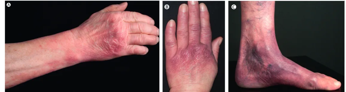
Figure 5: Examples of acrodermatitis chronic atrophicans
Acrodermatitis chronic atrophicans (ACA) is typically located on the extensor sites of extremities: (A) ulnar and hand lesions, (B) bluish-red lesion on the back of a patient’s hand and waxy appearance of the skin of fi ngers, (C) lesions on a patient’s left foot and lower leg.
Seminar
466 www.thelancet.com Vol 379 February 4, 2012
positive.33,65,68–70 Untreated patients who remain
sero-negative despite symptoms persisting for more than 6 weeks are unlikely to have Lyme borreliosis and other potential diagnoses should be actively pursued.
Omission of the fi rst tier EIA, or interpretation of the immunoblot with criteria that are not evidence-based, will potentially decrease the specifi city of testing and are contributing factors to misdiagnosis. A particular concern with the IgM immunoblot in clinical practice has been the many false positive results caused by the over-reading of non-specifi c weak bands.66
Background rates of seropositivity, which can exceed 4% in highly endemic areas of the USA,70 with even
higher rates in Europe, can also confound the interpretation of seroreactivity. Indeed, seropositivity rates of more than 50% have been reported for Austrian hunters older than 50 years.71 In such populations,
additional testing, such as tests for intrathecal anti-body production in patients with suspected Lyme neuro borreliosis, PCR testing of joint fl uid for sus-pected Lyme arthritis, or skin biopsies for sussus-pected
acro dermatitis chronica atrophicans or borrelial lympho-cytoma, might increase diagnostic accuracy. Clearly, a positive serological test does not mean that a patient necessarily has active Lyme borreliosis. The positive predictive value is usually most informative when the pretest probability based on the clinical features is at least 20%. Serological testing is not indicated in routine follow-up of patients after treatment, because either IgM or IgG borrelial antibodies can persist for many years in successfully treated patients.33,45,61
Testing for borrelial antibodies that are produced locally in the CNS (ie, intrathecal synthesis of specifi c antibodies) is a mainstay of the diagnosis of Lyme neuroborreliosis in Europe, and detection of antibody in cerebrospinal fl uid has been reported to precede that of serum antibody in some European patients.44,69,72,73 However, intrathecal synthesis of
anti-bodies can persist for several months to several years after successful antibiotic treat ment.44,69,72,73
Other diagnostic modalities
Culture for Lyme borrelia is not routinely done or available to diagnose Lyme borreliosis because it is unnecessary for patients with erythema migrans and too insensitive for patients with extracutaneous mani-fest ations of Lyme borreliosis. However, PCR for detection of borrelial DNA in synovial fl uid specimens is positive in up to about 80% of untreated patients with Lyme arthritis, and a positive result lends support to this diagnosis in a patient with a positive IgG immunoblot.33,69,74 The positivity rate of PCR in
cerebrospinal fl uid tends to be much lower than it is in synovial fl uid,68 however, and was only about 5% in a
study of children from the USA with early neurological Lyme borreliosis.75 A negative PCR result on either
cerebrospinal or synovial fl uid does not exclude Lyme borreliosis. PCR on blood or urine samples, tests for urine antigen detection, tests for T-lymphocyte recognition of borrelial antigens (as a measure of a cellular immune response), measurement of the number of CD57 natural killer cells, and use of live microscopy on blood to search for spirochaetes, have not been shown to be reliable and are not recommended for clinical use.33,69,76
Treatment
In-vitro studies have shown that Lyme borrelia are susceptible to tetracyclines, most penicillins, many second-generation and third-generation cephalosporins, and macrolides.33,77,78 Lyme borrelia are resistant to specifi c
fl uoroquinolones, rifampicin, and fi rst-generation cephalosporins.33,77,78
Although erythema migrans will eventually resolve without antibiotic treatment, oral antibiotic treatment is recommended to prevent dissemination and development of later sequelae (table 3). Doxycycline, amoxicillin, phenoxymethylpenicillin, and cefuroxime axetil are
A
B C
Figure 5: Examples of acrodermatitis chronic atrophicans
Acrodermatitis chronic atrophicans (ACA) is typically located on the extensor sites of extremities: (A) ulnar and hand lesions, (B) bluish-red lesion on the back of a patient’s hand and waxy appearance of the skin of fi ngers, (C) lesions on a patient’s left foot and lower leg.
MANIFESTATIONS CLINIQUES
☐ Atteinte cutanée
Stade III : Neuroborréliose du stade tardif
☐ Polyneuropathies sensitives axonales
• Troubles sensitifs :
- douleurs, dysesthésies distales - hypopallesthésie
- hypoesthésie thermo-algique • abolition des ROTs inconstante
• EMG : polyneuropathie de forme axonale • PL : rarement réaction cellulaire lymphocytaire • Biopsie nerf / muscle :
- myosite (phase initiale) : infiltrat monocytaire périmysial et périvasculaire, rares fibres musculaires en nécrose
- nerf : dégénérescence axonale avec perte des grosses fibres myélinisées, infiltrat inflammatoire monocytaire du péri et endonévre et périvasculaire
Stade III : Neuroborréliose du stade tardif
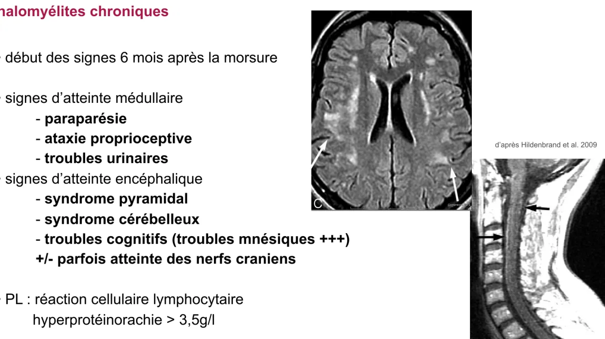
☐ Encéphalomyélites chroniques
• début des signes 6 mois après la morsure • signes d’atteinte médullaire
- paraparésie
- ataxie proprioceptive - troubles urinaires
• signes d’atteinte encéphalique - syndrome pyramidal - syndrome cérébelleux
- troubles cognitifs (troubles mnésiques +++)
+/- parfois atteinte des nerfs craniens
• PL : réaction cellulaire lymphocytaire hyperprotéinorachie > 3,5g/l
• IRM cérébrale : hypersignaux T2 aspécifiques de la substance blanche • IRM médullaire : hypersignal T2 du cordon médullaire
MANIFESTATIONS CLINIQUES
Pediatric LNB
Thirty percent of Lyme disease cases occur in children.3 The presence of erythema migrans in 89% of children significantly aids the clinical diagnosis.81 It is speculated that the CNS le-sions in children represent a spirochete-triggered auto-immune process.82 Headache is the most frequent neurologic symptom; the most common neurologic signs of pediatric
LNB include facial nerve palsy (3%–5%) and meningitis (1%). Less common manifestations are sleep disturbance and pap-illedema associated with increased intracranial pressure.83,84 Ataxia, chorea, myelitis, pseudotumor cerebri, meningitis, and encephalopathy are very uncommon.84-90Peripheral neu-ropathies, radiculopathies, and Bannwarth syndrome are other rare pediatric LNB manifestations.
Fig 8. A 56-year-old woman with neck, bilateral shoulder,
and bilateral arm pain. In 2 weeks, she subsequently devel-oped left facial palsy and positive serum EIA and Western blot (IgM and IgG) and CSF Lyme IgG and IgM antibodies. Complete resolution of symptoms occurred with oral doxycy-cline. Postcontrast sagittal and axial T1-weighted MR images show diffuse thin uniform cervical spinal cord leptomeningeal enhancement without apparent root or ganglion enhancement.
Fig 9. A 17-year-old boy with right papilledema and orbital
pain and rule out pseudotumor. The patient had positive serum EIA and Western blot (IgM and IgG) and CSF Lyme IgM and IgG antibodies. Lyme PCR in the CSF was negative. Complete resolution of symptoms occurred after intravenous ceftriaxone therapy. Right optic nerve edema on fat-saturated T2-weighted fast spin-echo images (A ) and right-greater-than-left optic nerve enhancement on coronal fat-saturated contrast-enhanced T1-weighted images (B ). C, Bilateral third cranial nerve enhancement (arrowheads) and bilateral retro-bulbar compartment congestion (arrow). Note the generalized extraocular muscle enlargement and enhancement, including the insertions. Additional imaging findings included enhance-ment of right fifth cranial nerve, optic chiasm, and intracanal-icular right seventh nerve, all of which were occult to neurologic examination.
AJNR Am J Neuroradiol 30:1079–87 ! Jun-Jul 2009 ! www.ajnr.org 1085 rect manifestations of LNB antibiotic-reversible frontal
hypoperfusion.65,76,77 Spinal Cord LNB
Spinal cord involvement by B burgdorferi is very rare. As a function of geography, LNB would be a rare differential con-sideration in the evaluation of transverse myelitis. MR imag-ing findimag-ings with LNB myelopathy are characterized by diffuse or multifocal T2-weighted cord lesions. In contrast to the clas-sic cervical spinal cord MR imaging abnormalities seen in MS, most patients with LNB do not have macroscopic lesions or magnetization-transfer ratio changes.58 Nerve root involve-ment is best seen on postcontrast T1-weighted sequences.40 Spinal involvement has demonstrated diffuse or multifocal T2-weighted cord lesions and nerve root enhancement on postcontrast T1-weighted sequences (Fig 8).40
Orbital and Ocular Lyme Disease
Rare ocular LNB may occur at all 3 stages of the disease. Uveitis and optic neuritis are the most common ocular
complica-tions.78 Conjunctivitis and episcleritis are the most frequent manifestations of the early stage. Neuro-ophthalmic disorders and uveitis occur in the second stage, whereas keratitis, chronic intraocular inflammation, and orbital myositis are seen in the third stage of Lyme disease.79A nonspecific follic-ular conjunctivitis occurs in approximately 10% of patients with Lyme disease.39 Direct ocular infection and a delayed
hypersensitivity mechanism may be involved at different dis-ease stages.
Borrelial orbital myositis is most probably an immunolog-ically mediated response subsequent to hematogenous dis-semination of Borrelia species.80 The clinical and imaging
manifestations of orbital myositis Lyme disease closely mimic those of orbital pseudotumor (Fig 9). The differential diagno-sis includes lymphoma or possibly thyroid dysorbitopathy. Criteria for orbital and ocular Lyme disease include the lack of evidence of other diseases, occurrence in patients living in an endemic area, positive serology, and, in most cases, response to treatment.39
Fig 6. A 50-year-old woman with a history of tick bite and erythema migrans rash treated with doxycycline, who had recurrent erythema migrans rash with headache, fever, nausea, and
nuchal rigidity. The patient had CSF pleocytosis with positive Lyme serum EIA and IgM Western blot and negative Lyme antibodies in the CSF. Gradual symptomatic improvement occurred following intravenous ceftriaxone therapy. There has been stable MR imaging for 5 years. Sagittal (A and B ) and axial (C ) fluid-attenuated inversion recovery images show arcuate and confluent subcortical white matter involvement and callososeptal interface involvement remarkably similar to that in MS, but without involvement of the periventricular white matter.
Fig 7. A 74-year-old man with 2-year cognitive decline and memory loss. The patient had Lyme-positive serum EIA and Western blot (IgG and IgM) and CSF pleocytosis with CSF positive
Lyme IgM and IgG antibodies. The patient improved with intravenous ceftriaxone therapy. The “dot-dash” callososeptal interface (A ) and periventricular distribution of involvement (B ) would be routinely ascribed to a demyelinating process.
1084 Hildenbrand ! AJNR 30 ! Jun-Jul 2009 ! www.ajnr.org
EXAMENS BIOLOGIQUES
Techniques directes
☐ Examen direct
• Non recommandé en routine (faible quantité d’organismes présents) • Examen sur fonds noir, coloration argentique : Se et Sp faibles
☐ Mise en culture
• Non recommandée en routine (faible rendement) • Culture longue, délai de positivité : 7 jours à 4 mois
☐ PCR
• Sur biopsie de peau, liquide synovial, biopsie d’organes divers (Se = 60-80 %)
EXAMENS BIOLOGIQUES
Techniques indirectes
☐ ELISA
• Stade d’EM : taux d’anticorps faibles voir nuls • Stade II : taux d’anticorps faibles
• Stade III : taux d’anticorps d’autant plus élevés que l’évolution est longue
• Neuroborréliose : Synthèse intrathécale d’anticorps spécifiques si :
(titres anticorps LCR/sérum) / (concentration albumine LCR/sérum) > 2
☐ Confirmation par Western Blot
DIAGNOSTIC SEROLOGIQUE
Techniques indirectes
☐ ELISA
• Stade d’EM : taux d’anticorps faibles voir nuls • Stade II : taux d’anticorps faibles
• Stade III : taux d’anticorps d’autant plus élevés que l’évolution est longue
• Neuroborréliose : Synthèse intrathécale d’anticorps spécifiques si :
(titres anticorps LCR/sérum) / (concentration albumine LCR/sérum) > 2
☐ Confirmation par Western Blot 90 kD72 kD
60 kD 41 kD 34 kD 31 kD 22 kD 20 kD
EXAMENS BIOLOGIQUES
Mesures préventives
☐ Interventions sur l’environnement (diminuer la densité du vecteur) ☐ Mesures de protection individuelles
• Protection mécanique (vêtements couvrants) • Répulsifs (< 3 applications / jour)
Mesures préventives
☐ Auto examen après exposition (caractère indolore de la piqure) ☐ Extraction de la tique
• présence de la tique > 8h-24h (B. afzelii) ou > 48-72h (B. burgdorferi) : risque maximal • technique d’extraction par pince à tique, rotation sens anti-horaire
• antiseptique après retrait de la tique (sinon risque de regurgitation de la tique)
PRISE EN CHARGE
Tick Extraction
1 cm HypostomTick Extraction
1 cm HypostomTick Extraction
1 cm
Hypostom
Traitement antibiotique de érythème migrant (stade 1)
☐ Adulte
• Doxycycline P.O. 100mg x2/j pendant 14 à 21 jours
• ou Amoxicilline P.O. 1g x3/j pendant 14 à 21 jours
• ou Céfuroxime-axétil P.O. 500mg x2/j pendant 14 à 21 jours • ou azithromycine P.O. 500mg / jour pendant 10 jours
TRAITEMENTS
☐ Enfant
• < 8 ans : Amoxicilline P.O. 50 mg/kg/j en 3 prises pendant 14 à 21 jours
• > 8 ans : Doxycycline 4 mg/kg/j en 2 prises (maxi 100mg/prise) pendant 14 à 21 jours
ou Amoxicilline P.O. 50mg/kg/j pendant 14 à 21j
• ou Céfuroxime-axétil P.O. 30mg/kg/j en 2 prises pendant 14 à 21 jours • ou azithromycine P.O. 20mg/kg/j en 1 prise pendant 10 jours
☐ Femme enceinte ou allaitante
• Amoxicilline P.O. 1g x3/j pendant 14 à 21 jours
• ou Céfuroxime-axétil P.O. 500mg x2/j pendant 14 à 21 jours • ou azithromycine P.O. 500mg / jour pendant 10 jours
Traitement antibiotique de la neuroborréliose (stade 2 et 3)
☐ Paralysie faciale isolée sans méningite
• Amoxicilline P.O. 1g x 3/jour pendant 14 à 21 jours • ou Ceftriaxone I.V. 2 g/j pendant 14 à 21 jours
• ou Doxyxycline P.O. 200mg/j pendant 14 à 21 jours
☐ Neuroborréliose de adulte dont la paralysie faciale avec méningite
• Ceftriaxone I.V. 2g/j pendant 21 à 28 jours
• ou Penicilline G I.V. 18 à 24 MUI/j réparties en 6 doses pendant 21 à 28 jours • ou Doxycycline P.O. 200mg/j pendant 21 à 28 jours
TRAITEMENTS
☐ Femme enceinte
• Idem sauf contre-indication des cyclines ☐ Enfant
• Contre indication des cyclines avant âge de 8 ans
Traitement de la neuroborréliose (stade 2 et 3)
☐ Suivi
• Evaluation clinique 2 mois après la fin du traitement antibiotique
• Si réponse partielle : antibiothérapie complémentaire (autre famille que le traitement initial) • Suivi sérologique inutile
Démarche diagnostique de la borréliose de Lyme
CONCLUSION
Démarche diagnostique de la borréliose de Lyme
CONCLUSION
Interrogatoire / Examen clinique
Stade I Stade II et III
EM typique EM atypique Sérologie
Pas de sérologie
Antibioprophylaxie inutile la plupart du temps si morsure de tique asymptomatique sauf :
- enfant et/ou femme enceinte avec : - morsures multiples
- exposition > 48h
Traitement antibiotique Sérologie de contrôle
3 semaines plus tard Western blot
négative positive ou douteuse
négative positive négatif positif
Traitement antibiotique
Borréliose de Lyme
WB Absence de
Borréliose de Lyme Traitement
selon évolution clinique