HAL Id: hal-01233095
https://hal.sorbonne-universite.fr/hal-01233095
Submitted on 24 Nov 2015
HAL is a multi-disciplinary open access
archive for the deposit and dissemination of sci-entific research documents, whether they are pub-lished or not. The documents may come from teaching and research institutions in France or abroad, or from public or private research centers.
L’archive ouverte pluridisciplinaire HAL, est destinée au dépôt et à la diffusion de documents scientifiques de niveau recherche, publiés ou non, émanant des établissements d’enseignement et de recherche français ou étrangers, des laboratoires publics ou privés.
IL-1β-Induced Group IIA Secretory Phospholipase A2
Expression in VSMCs
Khadija El Hadri, Chantal Denoyelle, Lucas Ravaux, Benoit Viollet, Marc
Foretz, Bertrand Friguet, Mustapha Rouis, Michel Raymondjean
To cite this version:
Khadija El Hadri, Chantal Denoyelle, Lucas Ravaux, Benoit Viollet, Marc Foretz, et al.. AMPK Sig-naling Involvement for the Repression of the IL-1β-Induced Group IIA Secretory Phospholipase A2 Ex-pression in VSMCs. PLoS ONE, Public Library of Science, 2015, 10 (7), pp.e0132498. �10.1371/jour-nal.pone.0132498�. �hal-01233095�
AMPK Signaling Involvement for the
Repression of the IL-1
β-Induced Group IIA
Secretory Phospholipase A2 Expression in
VSMCs
Khadija El Hadri1*, Chantal Denoyelle2, Lucas Ravaux1, Benoit Viollet3,4,5, Marc Foretz3,4,5, Bertrand Friguet1, Mustapha Rouis1, Michel Raymondjean1
1 Sorbonne Universités, Université Pierre et Marie Curie, Biological Adaptation and Ageing (B2A) CNRS UMR8256/INSERM ERL-U1064, F-75005 Paris, France, 2 Université Paris Diderot, Sorbonne Paris Cité, BFA CNRS UMR8251, 4 Rue MA Lagroua Weill Hallé, 75013 Paris, France, 3 Institut Cochin, Inserm U1016, Paris, France, 4 CNRS, UMR 8104, Paris, France, 5 Université Paris Descartes, Sorbonne Paris Cité, Paris, France
Abstract
Secretory Phospholipase A2 of type IIA (sPLA2 IIA) plays a crucial role in the production of lipid mediators by amplifying the neointimal inflammatory context of the vascular smooth muscle cells (VSMCs), especially during atherogenesis. Phenformin, a biguanide family member, by its anti-inflammatory properties presents potential for promoting beneficial effects upon vascular cells, however its impact upon the IL-1β-induced sPLA2 gene expres-sion has not been deeply investigated so far. The present study was designed to determine the relationship between phenformin coupling AMP-activated protein kinase (AMPK) func-tion and the molecular mechanism by which the sPLA2 IIA expression was modulated in VSMCs. Here we find that 5-aminoimidazole-4-carboxamide-1-β-D-ribonucleotide (AICAR) treatment strongly repressed IL-1β-induced sPLA2 expression at least at the transcriptional level. Our study reveals that phenformin elicited a dose-dependent inhibition of the sPLA2 IIA expression and transient overexpression experiments of constitutively active AMPK demonstrate clearly that AMPK signaling is involved in the transcriptional inhibition of sPLA2-IIA gene expression. Furthermore, although the expression of the transcriptional repressor B-cell lymphoma-6 protein (BCL-6) was markedly enhanced by phenformin and AICAR, the repression of sPLA2 gene occurs through a mechanism independent of BCL-6 DNA binding site. In addition we show that activation of AMPK limits IL-1β-induced NF-κB pathway activation. Our results indicate that BCL-6, once activated by AMPK, functions as a competitor of the IL-1β induced NF-κB transcription complex. Our findings provide insights on a new anti-inflammatory pathway linking phenformin, AMPK and molecular control of sPLA2 IIA gene expression in VSMCs.
OPEN ACCESS
Citation: El Hadri K, Denoyelle C, Ravaux L, Viollet B, Foretz M, Friguet B, et al. (2015) AMPK Signaling Involvement for the Repression of the IL-1β-Induced Group IIA Secretory Phospholipase A2 Expression in VSMCs. PLoS ONE 10(7): e0132498. doi:10.1371/ journal.pone.0132498
Editor: Ping Song, Georgia State University, UNITED STATES
Received: December 11, 2014 Accepted: June 15, 2015 Published: July 10, 2015
Copyright: © 2015 El Hadri et al. This is an open access article distributed under the terms of the
Creative Commons Attribution License, which permits unrestricted use, distribution, and reproduction in any medium, provided the original author and source are credited.
Data Availability Statement: All relevant data are within the paper.
Funding: This work was supported by the Université Pierre et Marie Curie (University Paris 6) and by grants from the“Leg Poix-2007” and "Leg Poix-2009" of the Chancellerie des Universités de Paris. Dr. Lucas Ravaux was supported by doctoral fellowships from the Ministère de l’Education Nationale de la Recherche et de la Technologie.
Competing Interests: The authors have declared that no competing interests exist.
Introduction
Phenotypic modulation of vascular smooth muscle cells (VSMCs) observed in the pathogenesis of vascular diseases such as hypertension, restenosis, aneurysm and atherosclerosis is charac-terized by the downregulation of the expression of VSMC-specific marker genes concomitant with the upregulation of the expression of genes regulating proliferation, migration and secre-tion of inflammatory mediators [1,2,3]. Atherosclerosis is a complex pathological process and accumulating evidences indicate that cardiovascular disorders are closely associated with chronic inflammation linked most often to metabolic syndrome which includes abdominal obesity, atherogenic dyslipidemia, elevated blood pressure and insulin resistance [4,5]. In vivo correlative data indicate that VSMCs, beside macrophages, play also an important role in the initiation of atherosclerosis [6]. After migration from the media to the intima, VSMCs present atherogenic properties. Hence, they are the major producers of extracellular matrix that in turn provoke cell adhesion and oxidized low density lipoproteins accumulation [7]. The initiation of atherosclerosis results from complex interactions of circulating inflammatory mediators. VSMCs, endothelial cells, and macrophages produce a myriad of proatheogenic proinflamma-tory and prothrombogenic molecules including lipid mediators such as various forms of eicosa-noides, mainly prostaglandin E2 (PGE2) which in turn amplify the effects of cytokines [8,9]. Several observations argue for a major implication of type IIA secretory PLA2 (sPLA2-IIA) [10]. Phospholipase A2 enzymes (EC 3.1.1.4) hydrolyze ester bonds at the sn-2 position of gly-ceroacylphospholipids to produce lysophospholipids and nonesterified fatty acids such as ara-chidonic acid (C20:4 n-6), the precursor of these proinflammatory mediators encompassing PGE2 [11,12]. The isoform sPLA2 IIA gene (sPla2g2a) is largely expressed in VSMCs in response to cytokines, interleukin-1β (IL-1β), IL-6 and tumor necrosis factor alpha (TNF–α) [13]. We have shown that oxysterols, PGE2, high mobility group B1 (HMGB1) and serum amyloid A (SAA) also induce sPLA2 IIA gene transcription in VSMCs [14,9,15–16]. The inter-play of various transcription factors (NF-κB, C/EBPβ, Ets, LXR, AP1) that stimulate the rat sPLA2 promoter activity by these inducers has been demonstrated [14,17]. More recently, we demonstrated that peroxisome proliferator-activated receptor (PPAR)-α,-γ,-β significantly inhibited cytokine-stimulated sPLA2 expression and secretion in VSMCs [18]. However, unlike to PPARα and γ, the inhibitory effect of PPARβ ligands was not affected by PPRE muta-tion. In concordance with primary results of Lee et al [19], we demonstrated that PPARβ ligand promotes B cell lymphoma 6 (BCL-6) binding to its specific sequence element in proximal position of the sPLA2IIA gene promoter. Repression mechanisms triggered by BCL-6 have been investigated in multiple cell systems. Interaction with histone-deacetylases, association with various corepressors SMRT/NCoR, SIN3A [20] and proximate binding to NF-κB response elements have been demonstrated [21]. Importantly, promoter structure as well densation of binding sites driven induction of this sPla2g2a gene illustrate the pathological con-sequences of losing BCL-6 repression in time course of chronic inflammation.
Interestingly, anti-hyperglycemic agents currently prescribed to treat type 2 diabetes, i.e. biguanides such as metformin and phenformin, induce gene expression changes in the liver including Bcl6 [22] and activate the highly conserved AMP-activated protein kinase (AMPK) [23–24]. AMPK traditionally activated by increase in cellular AMP/ATP ratio, has been pro-posed to function as a metabolic master switch to coordinate cellular adaptation to nutritional and environmental variations[25–26]. AMPK, considered as an energy-sensing system, occurs as heterotrimeric complexes which is only active after phosphorylation of a critical threonine (Thr 172) in the activation loop of theα catalytic subunit. Upstream kinases have been identi-fied as the tumor suppressor liver kinase B1 (LKB1) associated to STRAD and MO25 accessory subunits [27–29] and the calmodulin-dependent protein kinase kinase-β (CaMKKβ) [30–31].
The binding of AMP to theγ subunit of AMPK hinder the dephosphorylation of Thr 172 by protein phosphatase. Recently, AMPK pathway appeared to be a critical downstream mediator for vascular adiponectin signalling in endothelial cells and macrophages [32–33]. In addition, the AMPK activator, AICAR (5-aminoimidazole-4-carboxamide-1-β-D-riboside), suppressed serum-induced proliferation of human aortic SMCs [34] and mediated the modulation of leu-cocyte adhesion observed onto aortic endothelial cells. Therefore, AMPK activation makes up a potential anti-atherogenic mechanism [35].
The study was performed to determine the impact of the AMPK signaling pathway in the regulation by biguanides of the sPLA2 IIA gene expression in VSMCs. As shown in the liver with phenformin, we hypothesize that BCL-6 may serve as a target for the control of the sPLA2 gene expression. We demonstrate here that AMPK activation significantly inhibited cytokine-stimulated sPLA2 promoter. Thus, the anti-inflammatory effect of AMPK could represent a promising therapeutic target to limit the sPLA2-dependent production of proinflammatory lipid mediators, namely PGE2, in the vascular wall.
Materials and Methods
Isolation and culture of VSMCs from rat aortas
VSMCs were isolated from thoracic aortas of adult male Wistar rats by enzymatic digestion as described previously [36,17]. Cells were seeded on dishes coated with type I collagen from calf skin (Sigma) and cultured in DMEM, 4mM glutamine, 100U/ml penicillin and 100μg/ml strep-tomycin, supplemented with 10% (vol/vol) fetal calf serum (Gibco BRL). Experiments were performed with confluent cells made quiescent by incubating them for 24h in serum-free medium containing 0.2% SVF before treatment with appropriate agents for 24h. The culture medium was then removed for measurements of sPLA2 activity and cells were lysed for total RNA preparation and western blotting. Experiments with animals and cell culture were con-ducted in accordance with guidelines of the "Haut Conseil des Biotechnologies" committee and according to the accepted project "OGM" with the agreement from October 11–2011. Agree-ment number: 5876. All procedures involving animal handling and their care were in accor-dance with the University Pierre and Marie Curie Guidelines for Husbandry of Laboratory Mice.
PLA2 activity
sPLA2 activity was measured using the fluorescent substrate 1-hexadecanoyl-2-(1-pyrenylde-canoyl)-sn-glycero-3-phosphoglycerol (Interchim, France) as described previously [36] Total hydrolysis of the substrate obtained with 0.1 unit of PLA2 from bee venom (Sigma) was used as reference to calculate the sPLA2 activity in the samples. The basal level fluctuates fol-lowing the preparation of VSMCs albeit the response to cytokine was of the same order until the 5th passage where the induction becomes less intense reflecting a progressive loss of differentiation.
RNA extraction and qRT-PCR analysis
Total RNA was extracted by using RNAeasy kit (Qiagen) according to the supplier’s instruc-tions. Total RNA (1μg) was used as template to synthesize for 1h at 37°C the first strand cDNA with 200 U of mouse mammary lentivirus-reverse transcriptase (RT) (Invitrogen), 100μM ran-dom primers and the buffer supplied by the manufacturer in a total volume of 20μl. The reac-tion was terminated by heating to 95°C for 5 min. Quantitative PCR was performed using the LightCycler LC480 (Roche Diagnostics). The PCR mix included 5μl of each complementary
DNA (cDNA) (diluted 1:25) and 300 nM of each primer in 1× LightCycler DNA SYBR Green 1 Master Mix. The forward and reverse primer sequences for cDNA were designed with the Primer Express software according to European Molecular Biology Laboratory accession num-bers: the rat sPLA2-IIA, 5’-ATGGCCTTTGGCTCAAT-3’ (Ex1-2) and 5’-GCAACCGTA GAAGCCATA-3’ (Ex2); the rat cyclophilin A, 5’-TGCTGGACCAAACACAAATG-3’ (Ex4) and 5’-CTTCCCAAAGACCACATGCT-3’ (Ex5). The q-PCRs were performed using the fol-lowing thermal settings: denaturation and enzyme activation at 95°C for 5 minutes, followed by 40 cycles of 95°C (10s), 60°C (15s), and 72°C (15s). Post-amplification dissociation curves were performed to verify the presence of a single amplification product and the absence of primer dimers. Controls and water blanks were included in each run; they were negative in all cases. Real-time quantitative PCR data represent the amount of each target messenger RNA (mRNA) relative to the amount of cyclophilin gene mRNA, estimated in the logarithmic phase of the PCR. Serial dilutions were used to determine the fit coefficients of the relative standard curve.
Western blotting
VSMCs were harvested, homogenized in a lysis buffer at 4°C and centrifuged as described [18]. Proteins (20μg/lane), evaluated using Quick Start Bradford protein assay from Biorad, were separated on SDS-PAGE gel electrophoresis (8% gel). After electroblotting onto nitrocellulose Amersham Hybond-ECL (GE Heathcare) we determined the efficiency of the protein transfer with Ponceau S (Sigma-Aldrich). After blocking with 5% non-fat dry milk in Tris Buffered Saline (TBS) the membrane was incubated overnight with specific antibodies, P-AMPKα (Thr172), AMPKα, P-acetyl-CoA carboxylase (Ser79) and acetyl-CoA carboxylase, BCL-6, phospho-IκBα, β-actin, phospho-IκBα, I-κBα, phospho-p65 and p-65 NF-κB (Cell Signaling Technology) in TBS containing 0.1% tween 20. After three successive 10 min washes the blot was incubated 1 h with HRP conjugated (Sigma) secondary antibody (1:2000) in TBS contain-ing 0.1% tween 20. Detection of immune complexes was visualized uscontain-ing enhanced chemilumi-nescence reagents onto an Image captur Las 3000 (Fujifilm).
Plasmid transfection
VSMCs were seeded 48 h before transfection in 24 well plates at a concentration of 2x10⁴cells per plate in DMEM (2% glutamine, 1% penicillin-streptomycin, 10% SVF). At 70% confluence cells were washed 20 min before transfection. The transfection mixture was as follow: 400ng of luciferase reporter DNA [-1153; +46]sPLA2-Luc or multimeric-NF-κB[(Ig-κB]-Luc already described, 100ng of CMVβ-galactosidase, 1.6μl of Lipofectamine plus and Lipofectamine (Invi-trogen) in 200μl of OPTIMEM (Sigma) and for transactivation studies 10ng of pCMV-AMPKα2 catalytic and negative dominant expression vector were added to the mix. The cells were refed 3h later with 200μl of DMEM containing 5% SVF for 1h and then the culture medium was replaced by DMEM containing 0.2% SVF. Twenty hours later after the start of the transfection, the medium was changed and the cells were pretreated with indicated concentration of AICAR (5-aminoimidazole-4-carboxamide-1-β–D-riboside), phenformin (phenylbiguanide) for 4h, then stimulated with IL-1β (10ng/ml) and the incubation was continued for a further 24h. Lucif-erase activity was measured using lucifLucif-erase reporter assay kit (PROMEGA), with signal detec-tion for 12s by a luminometer (Berthold, Pforzheim, Germany) and normalized by dividing the relative light units byβ-galactosidase activity. The degree of induction was calculated relative to the control (minus IL-1β).
Statistical analysis
All quantitative data are presented as mean of at least 3 independent experiments ± SEM. Sta-tistical analyses were performed using an unpaired Student’s t-test and comparisons involving repeated measurements were analyzed by repeated-measures ANOVA followed by Bonferroni posttest. Results are expressed as the means +/- SE. Differences were considered significant at P<0.05.
Results
AICAR and phenformin activate AMPK in rat VSMCs
Experiments were conducted with primary cultures of VSMCs isolated from rat aorta. These cells undergo phenotypic changes in response to proinflammatory conditions. Indeed, in response to proinflammatory cytokines, these cells express several biomarkers of inflammation such as VCAM-1, MCP1, extracellular metalloproteinases and acute phase enzymes such as secreted sPLA2 and COX-2 [7,37–39]. Moreover, we have previously observed that prostaglan-dins E2 combined with IL-1β progressively synergizes the secretion of sPLA2 and causes a complete disorganization of the cytoskeletal framework [9]. To explore the effect of AMPK activation on IL-1β-induced sPLA2IIA gene expression and activity, cultured VSMCs were pretreated or not with the AMPK activators AICAR or phenformin prior IL-1β treatment. We chose to use phenformin which is more lipophilic than metformin and more efficiently inter-nalized in cell culture in absence of cationic transporters. We performed immunoblotting to show whether treatment with AICAR or phenformin led to the activation of AMPK, character-ized by the phosphorylation of Thr172 within the AMPKα subunit and of Ser79 in acetyl-coA carboxylase (ACC), a well-established target of AMPK [26]. As shown inFig 1, treatment of VSMCs with 2mM AICAR or 1mM phenformin led to a significant increase in Thr172 AMPK and Ser79 ACC phosphorylation. In the presence of IL-1β, AICAR and phenformin induced phosphorylation of both Thr172 AMPK and Ser79 ACC. These results confirm that AICAR and phenformin stimulate AMPK signaling pathway in primary cultured rat VSMCs, consis-tent with previous studies showing AMPK activation by the biguanide metformin in BAECs or HUVECs [40].
AICAR and phenformin inhibit IL-1β-induced sPLA2-IIA gene expression
and activity
Next, in order to investigate whether AMPK might hinder the IL-1β-induced production of sPLA2, cultured rat VSMCs were pretreated with AICAR or phenformin prior to be incubated with IL-1β. Clearly, AICAR dose-dependently caused a significant inhibition of IL-1β-induced sPLA2 activity secreted by VSMCs, whereas it has no effect on basal sPLA2 activity in the absence of IL-1β (Fig 2A). We observed that AICAR strongly inhibited IL-1β-induced sPLA2 IIA gene expression as soon as 3 hours after IL-1β treatment (Fig 2B). This result indicates that activation of AMPK pathway for 4 hours was optimal for complete inhibition of the IL-1β induced sPLA2 gene expression. Similarly, incubation with phenformin resulted in a progres-sive dose-dependent decrease in IL1β-induced sPLA2 activity secreted in the culture medium (Fig 3A) and significantly lowered IL-1β-induced expression of sPLA2 IIA to the basal level (Fig 3B). Our results showed that AICAR and phenformin are both potent anti-inflammatory agents that suppress sPLA2 IIA production in rat VSMCs incubated with IL-1β.
AMPK activation represses sPLA2 IIA promoter activity in VSMCs
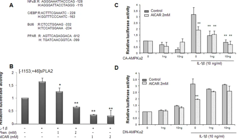
Since, phenformin and AICAR modulate IL1β-induced sPLA2 IIA gene expression in rat VSMCs, these results prompted us to examine the ability of AMPK to regulate the sPLA2 pro-moter activity. We have previously characterized the interplay of various transcription factors involved in the modulation of sPLA2 promoter activity [17–18]. Interestingly, all the binding sites were conserved and perfectly aligned between human and rat promoters (Fig 4A). A reporter construct encompassing the rat sPLA2 promoter ([-1153; +46]sPLA2-Luc) was tran-siently transfected into VSMCs preincubated with phenformin or AICAR for 4 hours prior to the treatment with IL-1β (10ng/ml) for 18 hours. We observed that the activity of the sPLA2 promoter was significantly decreased in a dose dependent manner with phenformin and drasti-cally with AICAR (Fig 4B). In an attempt to determine the role of AMPK activation on the sPLA2 promoter activity, we expressed constitutively active (CA-AMPK) or dominant negative (DN-AMPK) AMPK mutants in VSMCs [41]. Expression of CA-AMPK strongly decreased the [-1153; +46]sPLA2 promoter induction by IL-1β (Fig 4C). Addition of 2mM AICAR in the cell culture medium did not show additive inhibitory effect with expression of
CA-AMPK, indicating that AICAR signals through the AMPK signaling pathway to inhibit sPLA2 promoter activity. In contrast, expression of DN-AMPKα2 has no effect on basal and IL-1β-induced sPLA2 promoter activity but severely blunted the inhibitory action of AICAR on IL-1β-induced sPLA2 promoter activity, excluding off-target effect of AICAR (Fig 4D). Altogether, the experiments indicate that activation of AMPK exerts a repressive effect on
Fig 1. AICAR and phenformin treatments induce the phosphorylation of AMPK and ACC in isolated rat VSMCs in primary culture. After 1 hour pretreatment with AICAR (2mM) or phenformin (1mM), the cells were incubated or not with IL-1β (10ng/ml) for 30 minutes. Total proteins (20μg/lane) were separated by SDS PAGE and western blotted with specific antibodies (part A) against phospho-AMPK, total AMPK (65 KDa), and (part B) phospho-ACC, total ACC.β–actin was used as loading control. Representative blots of 3 independent experiments are shown. Data of the quantification are expressed as mean +/- SEM.*, p < 0,05; **, p < 0,01, AICAR or Phenformin treated vs control cells.
IL-1β-stimulated expression of the type IIA sPLA2 gene in VSMCs. As a consequence, AMPK stimulation may reduce the extended production of proinflammatory mediators as lysopho-spholipids and arachidonic acid.
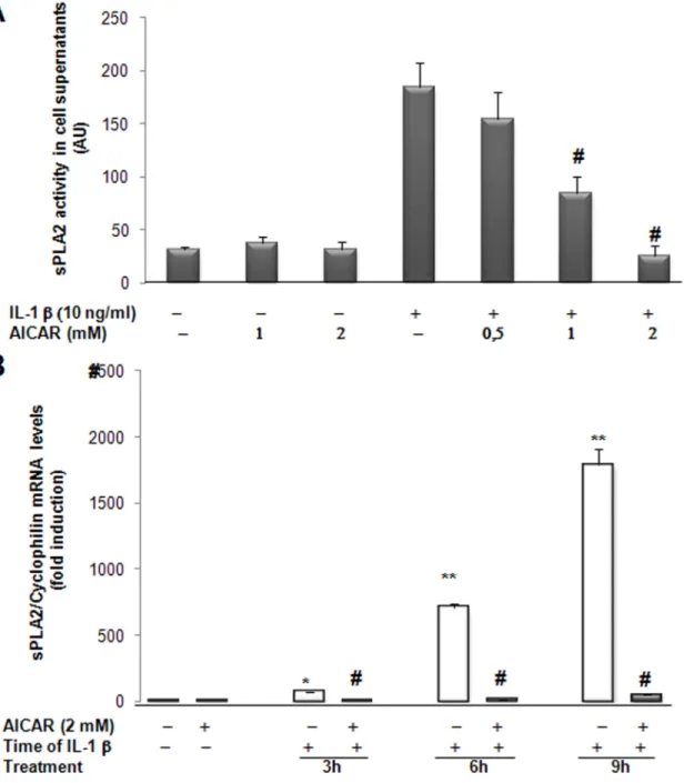
Fig 2. AICAR modulates IL-1β-induced sPLA2 activity and sPLA2 IIA mRNA expression in rat VSMCs. (A) VSMCs were preincubated for 4 hours with various concentrations of AICAR and then cultured in absence or presence of IL-1β (10ng/ml) for 18 hours. The sPLA2 activity was measured
spectrofluorimetrically from the supernatant of the cells. (B) Cells were preincubated for 4 hours with 2mM AICAR and then treated with IL-1β for 3, 6 or 9 hours. Total RNA was extracted and real time PCR was performed to evaluate sPLA2 mRNA levels as described under Experimental procedures. Data are shown as mean +/- SEM from 3 separate experiments.*, p<0.01;**, p <0.001, IL-1β–treated vs control cells, IL-1β-treated vs control cells; #, p< 0.001 AICAR treatedvs IL-1β treated VSMCs.
Role of BCL6 as repressor stimulated by AICAR
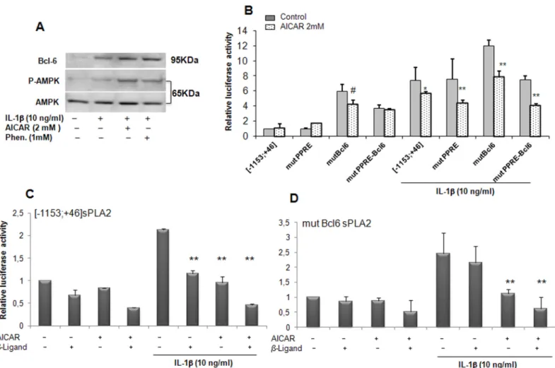
We previously showed that VSMCs expressed the proto-oncogene BCL-6 and demonstrated its ability to repress the expression of sPLA2 IIA [18], in concordance with results reported with another proinflammatory gene, MCP1 in macrophages [19]. We first determined the expres-sion of BCL-6 in rat VSMCs by Western blot analysis. As already observed its expresexpres-sion was slightly augmented in the presence of IL-1β. BCL-6 expression was further increased in the presence of AICAR and phenformin concomitantly with AMPK signalling activation (Fig 5A). This finding led us to specifically consider the repressor, BCL-6, as a potential target of the AMPK pathway. We previously demonstrated that PPARβ agonist (L165041) elicited the bind-ing of BCL-6 to the sPLA2 promoter and led to its inhibition under inflammatory conditions [18]. In an attempt to examine the role of BCL-6 binding site in the regulation of sPLA2IIA promoter transcription, VSMCs were transiently transfected with the [-1153; +46]sPLA2 pro-moter in which BCL-6 binding site was mutated in combination or not with mutation in the PPRE binding site. Mutation of BCL6 binding site in the [-1153; +46]sPLA2 promoter strongly increased sPLA2IIA promoter activity in basal condition, confirming the repressive role of BCL6 on sPLA2 gene expression. Interestingly, incubation of VSMCs with AICAR yielded to the same levels of inhibition observed with mutPPRE or the mutBCL-6 constructs as the wild type construct, [-1153; +46]sPLA2 in basal and IL-1β-stimulated conditions, suggesting that BCL-6 and PPRE binding sites are not necessary for the repression of sPLA2IIA promoter by AMPK (Fig 5B). As we previously demonstrated that PPARβ agonist (L165041) induced a BCL-6 binding to the sPLA2 promoter under inflammatory conditions, we performed transient transfection in the presence of either PPARβ ligand or AICAR or both. We observed that the IL-1β-induction of the [-1153; +46]sPLA2 promoter was attenuated to similar levels by PPARβ ligand and AICAR (Fig 5C). Importantly, this inhibition was further enhanced by co-treatment with PPARβ ligand and AICAR (Fig 5C). When activity of the mutBCL-6[-1153; +46]sPLA2 promoter was examined in the presence of IL1β, the inhibitory effect of PPARβ ligand was abolished but in contrast the inhibitory effect of AICAR alone or in combination with PPARβ ligand was preserved (Fig 5D). Altogether, these results suggest that the ability of AMPK to inhibit the sPLA2IIA promoter is independent on the BCL-6 binding site.
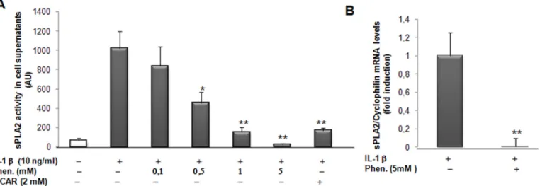
Fig 3. Effect of phenformin and AICAR treatment on sPLA2 activity and gene expression in rat VSMCs. VSMCs were preincubated for 4 hours with phenformin (0.1, 0.5, 1, 5mM) and AICAR (2mM) and cultured in absence or presence of IL-1β (10ng/ml) for 18 hours. (A) sPLA2 activity was measured in the supernatant of cells. (B) sPLA2 mRNA levels were determined by real time PCR analysis. Data are shown as mean +/- SEM from 3 separate experiments.*, p<0,01; **, p<0,001, IL-1β and Phenformin or AICAR treated vs IL-1β-treated cells.
Inhibition of IL-1
β-induced NF-κB dependent transactivation by AMPK in
VSMCs
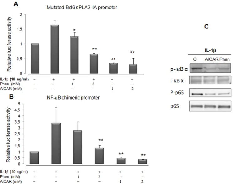
The absence of AICAR and phenformin action through the BCL-6 binding site on the sPLA2 promoter (Fig 6A) prompted us to further investigate whether AMPK acts on another regula-tory site. We demonstrated previously that the sPLA2 gene promoter contains a crucial NF-κB binding site at position -131bp which is crucial for the stimulation of sPLA2 promoter by IL-1β [17]. It was recently shown that AICAR and a constitutively active AMPK reduced the expres-sion of VCAM-1 in TNFα-activated aortic endothelial cells by attenuating NF-κB acetylation [41–42]. In order to assess the impact of phenformin and AICAR on the activity of the tran-scriptional factor NF-κB in VSMCs, we transiently transfected VSMCs with a chimeric con-struct, [(IgKB)3-cona]-Luc, where 3 IgK enhancerκB sites were fused to the conalbumin promoter [43,17]. The activity of the chimeric NF-κB-Luc promoter was also strongly dimin-ished with AICAR and phenformin treatment (Fig 6B). This result reveals that the treatment of VSMCs by either AICAR or phenformin abolished the IL-1β-induced activity of a promoter which transcription was strickly dependent upon the NF-κB pathway. To further confirm the impact of AICAR and phenformin on the activation of NF-κB transcription factor by IL-1β,
Fig 4. Phenformin, AICAR and overexpression of the AMPKα2 subunit inhibit the sPLA2 IIA gene promoter activity in VSMCs. (A) Alignment of rat (R) and human (H) NFκB, C/EBP, BCL-6, PPAR binding sequences of the sPLA2 IIA gene promoters. (B) VSMCs were transitory transfected with Lipofectamine plus with the sPLA2-Luc reporter plasmid: rat [-1153; +46]sPLA2 IIA-Luc construct. Cells were pretreated with phenformin (1 and 2 mM) or AICAR (1 and 2 mM) for 4 hours before prolonged incubation for 18 hours with or without IL-1β (10 μg/ml). (C and D) VSMCs were cotransfected with 1 and 10 ng of pCMV AMPKα2 constitutively active or dominant negative expression vector. As a control cells were transfected with empty pcDNA3. After transfection, cells were treated or not with IL-1β and AICAR (2mM) for 18 hours. The relative luciferase activities were calculated after normalization to β-galactosidase activity. Results (mean +/- SEM) were from 3 independent experiments.*, P<0.05; **, P<0.01 compared with IL-1β treated cells. doi:10.1371/journal.pone.0132498.g004
we analyze degradation of NF-κB inhibitor IκBα and NF-κB translocation (Fig 6C). The blot probed with antiphospho-IκB antibody shows a phosphorylated IκBα specific band detected in IL-1β treated cells. When VSMCs where pretreated with AICAR and phenformin the impact of IL-1β upon the phosphorylation of IκBα was diminished and the IL-1β induced phosphoryla-tion of p65 protein was clearly attenuated. These results demonstrate that activaphosphoryla-tion of AMPK pathway interfere with IL-1β activation of the NF-κB signaling cascade. In consequence, the activation of proinflammatory genes, encompassing a NF-κB binding site, like sPLA2 IIA gene was strongly hindered in vascular SMCs.
Discussion
In the present study we investigated the anti-inflammatory properties of the AMPK activators, AICAR and the antidiabetic drug phenformin, by focusing our attention on the modulation of sPLA2 IIA gene expression by inflammatory signals in VSMCs. The above findings have war-ranted a revisiting of the transcriptional regulation of the IL-1β-induced sPLA2 IIA gene
Fig 5. sPLA2 gene promoter inhibition is independent of the BCL-6 binding site in VSMCs. (A) protein expression was assessed by Western blot analysis, BCL-6 (95 KDa), phospho-AMPK-Thr172and AMPK (65 KDa). VSMCs were treated with IL-1β (10ng/ml) alone or with AICAR (2mM) or phenformin
(1mM) for 18 hours. (B) VSMCs were transfected with the sPLA2-Luc reporter plasmids: [-1153;+46]sPLA2Luc, mutated PPREsPLA2 (mutPPRE), mutated BCL-6 site (mutBcl6), double-mutated version of the sPLA2 promoter (mutPPRE-Bcl6) and VSMCs were then treated as inFig 4. (C) VSMCs were transfected with the [-1153;+46]sPLA2Luc plasmid, treated with PPARβ ligand (L165041, 10mM) or 2mM AICAR or both 4 hours before addition or not of IL-1β for an additional 18 hours. (D) VSMCs were transfected with BCL-6-mutated sPLA2 version, (mBCL-6[-1153;+46])sPLA2Luc construct and incubated as in part C. Data are expressed as mean +/-SEM of 4 experiments. #, P<0.05 (compared with control) and *, P<0.05; **, P<0.01 compared with IL-1β treated. doi:10.1371/journal.pone.0132498.g005
promoter activity. Our results highlight new integrated molecular mechanisms involving AMPK signaling pathway in the inhibition of sPLA2 IIA gene expression and provide new insights through the interplay between the protooncogene BCL-6 and NF-κB transcription factors.
Inflammation in the vascular intima has emerged as a crucial factor in the pathogenesis of atherosclerosis induced by hypercholesterolemia, shear stresses of hypertension or disordered blood flow and during restenosis after coronary intervention. Circumscribed chronic inflam-matory states originate from a partial destruction of cells followed by a partial healing of the tis-sue. The VSMCs proliferation and progressive dedifferentiation, is a critical event of the progression of vascular neointimal lesion formation. Beside chemokines, several mediators as biologically active oxidized lipids, named eicosanoids (mainly PGE2) are involved. These lipid
Fig 6. Phenformin and the activator of AMPK repress the sPLA2 gene promoter activity through NF-κB binding sites and inhibit IL-1β-induced NF-κB activation. VSMCs were transfected with either mutated-BCL-6 sPL2 IIA-Luc reporter (part A) or a NFNF-κB-mediated-Luc-reporter (part B) construct made up a multimerized NF-κB binding site chimeric promoter and then incubated with phenformin (1 or 2 mM) and with AICAR (1 or 2 mM) in the same conditions asFig 5. Data are expressed as mean +/-SEM of 3 experiments.*, ** P<0.05 and P<0.01 respectively compared with IL-1β treated VSMs. In part C, IkBα, phospho-IκBα, p65NF-κB and phospho-p65NF-κB expression was assessed by western blots. VSMCs were preincubated with or without AICAR (2mM) or phenformin (2mM) for 30 minutes before addition of IL-1β (10ng/ml) for 15 minutes. Then the cells were harvested and lysed as described and subjected to western blotting to detect phospho-ⅠBα, IκBα, phospho-p65NF-κB. Total. p65NF-κB is provided as a control.
mediators outcome from arachidonic acid (AA) and released by induced phospholipase A2 activity generate an amplification loop of the inflammation leading to a non-controlled response. The involvement of sPLA2 IIA as a key regulator in chronic and acute inflammatory diseases is well documented [44,12,45], likewise its major contribution through different path-ways is clearly demonstrated in the arterial wall and VSMCs during atherosclerosis develop-ment[46–47,39]. We have taken advantage of our expertise in the field of the transcriptional regulation of the sPLA2 IIA gene expression in VSMCs to explore the action of AMPK signal-ing pathway. AMPK originally thought to be a major survival factor in a variety of metabolic stresses, appeared to be a critical downstream mediator of vascular adiponectin signaling [48]. In endothelial cells, it was shown that adiponectin inhibits CRP production through its ability to modulate AMPK signaling pathways that in turn suppress NF-κB activity [49]. Furthermore, AICAR reduces fatty acid-induced NF-κB activation [33] and diminishes endothelial cell pro-liferation under conditions of ischemia [50]. Particularly interesting, metformin (the parent phenformin biguanide) treatment of insulin resistant type 2 diabetic patients was proved to be very effective for lessening macrovascular morbidity. Pre-treatment of HUVECs with metfor-min was shown to have anti-inflammatory effects by suppressing TNF-α-induced IκBα degra-dation through the AMPK pathway [51–52]. Metformin was first reported as an AMPK agonist in hepatocytes [23], H-2K cells [51] and skeletal muscle [52] not acting through changes in energy status. However, it was recently demonstrated that metformin-induced changes in cellular AMP/ATP ratio is a pre-requisite for the activation of AMPK [30]. Particu-larly interesting, it was shown that activation of AMPK by AICAR suppresses the proliferation of human aortic SMCs by inducing S-15 phosphorylation and expression of p53. Induction of CDKI p21 by AICAR suggested an AMPK-dependent G1 arrest through inactivation of E2F transcription factor [34]. In the present study, we report that the expression of IL-1β-induced sPLA2IIA mRNA in VSMCs was dose-dependently inhibited by phenformin and interestingly also by resveratrol (trans-3,4’,5-trihydroxystilbene) (data not shown), a natural compound known for its beneficial effect on cardiovascular system and activation of AMPK [53–54]. This result confirms the protective activity of resveratrol in VSMCs likely by inhibiting transcription factors such as NF-κB or AP-1 following the increase of AMPK activity [55–57]. In fine the repressive effect that occurs through the AMPK signaling pathway affects a large coordinated network of genes involved in the inflammatory amplification loop in VSMCs. In endothelial cells, the mechanism by which metformin activates AMPK has been reported [58]
The inter-gene network of the genes affected by phenformin includes many transcription factors and specifically the transcriptional repressor BCL-6 which targets many inflammatory genes [22]. Here, we addressed the question about the action of AMPK on the transcriptional repressor BCL-6 reported as a key regulator of sPLA2 IIa gene expression. AMPK activity could in fine suppress inflammation through various mechanisms involving the stabilization of repressive complexes on several inflammatory gene promoters. Recently, Gongol et al demon-strated, in endothelial cells, that AMPKα2 facilitated the phosphorylation of PARP-1 and its dissociation from the intron 1 of the repressor POZ/zinc-finger BCL-6 and hence led to the extinction of inflammatory genes VCAM-1 and MCP-1 [59]
Nevertheless, to the best of our knowledge this is the first time that IL-1β-induced sPLA2 IIA activity is shown to be inhibited by AMPK activation mainly at a transcriptional level. Since AMPK regulates the inhibition of a network of proinflammatory genes, we focused on the molecular candidate repressor BCL-6 [60]. We previously demonstrated that sPLA2 IIA gene is a direct target of BCL-6 acting through a specific binding site in the proximal promoter. In accordance with the results reported by Lee et al [19] on another proinflammatory gene, MCP1, we demonstrated that PPARβ activation by PPAR ligands led to the translocation of the protooncogene BCL-6 and its binding to a specific site located between position -342 and
-351bp relative to the transcription initiation start. BCL-6 could contribute to the prevention of atherosclerosis by recruiting, with a high degree of overlap, SMRT and NCoR-corepressor complexes to control the expression of inflammatory and atherogenic genes [61–62]. Precisely, these studies revealed that BCL-6 SMRT/NCoR subcistromes are still enriched for proximally bound NF-κB driven inflammatory and tissue remodelling genes in bone marrow of ldlr-/-mice. Effectively, the sPLA2 IIA promoter contains NF-κB and C/EBP binding sites highly conserved between rat and human (-131bp and 178bp respectively for rat and -138bp and -235bp for human, unpublished results) and a typical BCL-6 binding site located at -340bp. The organization of the sPLA2 IIA promoter is reminiscent of many promoters of inflamma-tory genes where NF-κB and BCL-6 sites are co-localized within a 200bp space [21]. We also observed in the present study that AMPK treatment stimulated BCL-6 expression and interest-ingly, we confirm, as already observed by RT-PCR and western blotting analysis [18], that BCL-6 is potentially an IL-1β-inducible transcriptional repressor, probably via the NF-κB pathway. It was also shown that DNA binding activity or stability of the BCL-6 protein is mod-ulated by several mechanisms [63–64,8] and that BCL-6 mediated a powerful repression through a large set of interactions with tissue-specific repressors [65].
Interestingly, in contrast, to its action in the presence of the PPARβ ligand L165041, our experiments with either phenformin or AICAR did not reveal a direct role for BCL-6. We found that the mutation of the BCL-6 binding site on the sPLA2 promoter did not abolish the repression, although the inhibition of the promoter following PPARβ activation by a synthetic ligand was clearly dependent on BCL-6 DNA binding. A plausible mechanism could be that AMPK activation targets more preferentially the IL-1β-induced transcription factors such as NF-κB, C/EBPβ and AP1. According to this hypothesize transfection experiments revealed that a multimeric NF-κB synthetic promoter was perfectly repressed by phenformin and AICAR. In addition, we have shown that AMPK pathway also inhibits IL-1β phosphorylation of IκBα. In accordance, it was recently demonstrated that AMPK could also play a key role in endothelial cells as a pleiotropic inhibitor of immune response by acting through the nuclear binding activity of the NF-κB transcription factor [41]. Indeed metformin was shown to inhibit NF-κB activation by TNF-α and IL-1β via a possible AMPK pathway in endothelial cells and VSMCs [66–67].In the present study we show that AMPK activation by AICAR or Phenformin suppresses cytokine-induced NF-κB-dependent gene transcription and therefore modulates inflammatory responses in VSMCs and contributes to the partial resolution of the inflamma-tion. Thus the findings may have significance for treatment of atherosclerotic vascular disease.
At overall, our two studies on the transcriptional repression mechanisms of the IL-1β-induced sPLA2 IIA promoter reveal at least two distinct pathways, one through the action of the PPARβ ligand, as probably PGI2 eicosanoids, in liberating sequestrated BCL-6 which binds to the BCL-6 consensus sequence and the second when AMPK is activated in blocking NF-B activation complex. AMPK activation that occurs after phenformin treatment in VSMCs could attenuate the recruitment of coactivators and inhibits IKK activity. Barroso et al. showed that the PPARβ agonist, GW501516 prevented TNF–α-induced expression of NF-κB target genes through AMPK activation by reducing the p300 and p65 interaction and by stimulating SIRT1 expression [63]. However, the existence of another non-exclusive inhibitory mechanisms medi-ated by BCL-6 may occur in VSMCs. It was shown that the C-terminal domain of BCL-6 physi-cally interacts with the Rel-homology domain of NF-κB in vitro as with AP1 family members and that BCL-6 behaves as a mutual negative regulator of NF-κB target genes in diffuse large B-cell lymphomas [68–69]. The present study did not demonstrate a repressible effect stimu-lated by AMPK through a direct DNA binding of BCL-6, but may be consistent with a protein-protein interaction with NF-κB complexes. This leads us to hypothesize a plausible mechanism for the inhibition of the transcriptional activity of the sPLA2 IIA gene activity. In VSMCs,
AMPK activation by phenformin could phosphorylate the DNA binding domain of BCL-6 which could hinder its binding to the sPLA2 IIA promoter located at -340 bp of the initiation site without affecting its protein-protein interaction with the NF-κB transcriptional factor located downstream at -131 bp. We postulate that, once phosphorylated, BCL-6 could stabilize a SMRT/NCoR repressor complex that blocks IL-1β-induced NF-κB activity and then poten-tially diminishes sPLA2 IIA gene transcription. In fact, our close examination of the BCL-6 sequence reveals a putative phosphorylation site by AMPK located between amino acids 11 and 16 in the N-terminal domain of BCL-6 which are conserved in human, rat, mouse and chicken: FTRHASDVLL. This putative sequence matches well with the consensus one: FxRxxSxxxL[69–70].
In addition, we cannot exclude the role of miRNA, such as miR-155, that in macrophages was shown to repress the expression of BCL-6 in attenuating NF-κB signalling in advanced atherosclerosis [71]. Interestingly, a cascade of mRNA targeted by miR-155 would be involved in the regulation of vascular inflammation as described with the use of polyphenolic compound as resveratrol [72].
The knowledge gained by this study about the sPLAIIA gene promoter will improve the overall understanding of how cytokine-induced genes are regulated. On account of the closed disposition of the regulatory elements, the study of the transcriptional activity of the promoter will allow to identify new signalling pathways. A novel repression mechanism of the cytokine-mediated induction of sPLA2 IIA in hepatocytes was recently deciphered [73]. The gene activ-ity was blocked by the recruitment of corepressors SMRT and NCoR to the T3-liganded TRβ bound to a non canonic site located between -102bp and -82bp, on the proximal region of the rat sPLA2 IIA gene promoter. In fact, DNA binding interactions were precisely characterized in the same mapped region (from -101 to -77bp) by DNA footprinting and EMSA assays with VSMCs crude extracts (Antonio V. and Raymondjean M., unpublished results). This new report and our present study show evidence about a network of positive and negative mecha-nisms mediating the sPLA2 IIA promoter activity. The complexity and the overlapping of the transcription factors highlight the crucial role played by the sPLA2 IIA in the control of cell fate, i.e., proliferation, dedifferentiation and secretory status of VSMCs.
Interestingly, recently AMPK was shown to be the central target for the metabolic effects of resveratrol in vivo by increasing the NAD to NADH ratio, thus contributing indirectly to the stimulation of SIRT1 [74–75]. These evidences illustrate perfectly the central role played by the fuel-sensing kinase activated by various metabolic and stress conditions. More recent studies investigating the vascular consequences of AMPK deletion in vivo have shown that knockout of AMPKα2 contributes to neointima formation after vascular injury and moreover, upregula-tion of proinflammatory markers was observed in arteries ofα1AMPK-knockout mice after ATII infusion [76–77].
In summary, our study highlights the mutual exclusive regulation mechanism plays by BCL-6 when therapeutic interventions by PPARβ ligands and antidiabetic drugs are adminis-trated to patients Indisputably, more studies are needed to further understand how biguanide molecules acting through the AMPK signaling pathway may have various beneficial metabolic effects by reducing associated atherogenesis [78]. These results point to the sPLA2 IIA as a potential therapeutic target for numerous inflammatory diseases.
Acknowledgments
This work was supported by the Université Pierre et Marie Curie (University Paris 6) and by grants from the“Leg Poix” of the Chancellerie des Universités de Paris. We thank Ms Adelaide Deschamps, Laura Bouchareychas and Fanny Canesi for help in performing experiments
during their laboratory masterships and Claire Monné for technical assistance. Dr Lucas Ravaux was supported by doctoral fellowships from the « Ministère de l’Education Nationale de la Recherche et de la Technologie ».
Author Contributions
Conceived and designed the experiments: KEH CD LR BV MF BF M. Rouis M. Raymondjean. Performed the experiments: KEH CD LR M. Raymondjean. Analyzed the data: KEH CD LR BV MF BF M. Rouis M. Raymondjean. Contributed reagents/materials/analysis tools: KEH CD LR BV MF BF M. Rouis M. Raymondjean. Wrote the paper: KEH BV BF M. Rouis M.
Raymondjean.
References
1. Orr AW, Hastings NE, Blackman BR, Wamhoff BR (2010) Complex regulation and function of the inflammatory smooth muscle cell phenotype in atherosclerosis. J Vasc Res 47: 168–180. doi:10.1159/ 000250095PMID:19851078
2. Moore KJ, Tabas I (2011) Macrophages in the pathogenesis of atherosclerosis. Cell 145: 341–355. doi:10.1016/j.cell.2011.04.005PMID:21529710
3. Glass CK, Witztum JL (2001) Atherosclerosis. the road ahead. Cell 104: 503–516. PMID:11239408
4. Lusis AJ (2000) Atherosclerosis. Nature 407: 233–241. PMID:11001066
5. Ross R (1999) Atherosclerosis is an inflammatory disease. Am Heart J 138: S419–420. PMID:
10539839
6. Doran AC, Meller N, McNamara CA (2008) Role of smooth muscle cells in the initiation and early pro-gression of atherosclerosis. Arterioscler Thromb Vasc Biol 28: 812–819. doi:10.1161/ATVBAHA.107. 159327PMID:18276911
7. Owens GK, Kumar MS, Wamhoff BR (2004) Molecular regulation of vascular smooth muscle cell differ-entiation in development and disease. Physiol Rev 84: 767–801. PMID:15269336
8. Hansson GK (2001) Immune mechanisms in atherosclerosis. Arterioscler Thromb Vasc Biol 21: 1876– 1890. PMID:11742859
9. Clement N, Glorian M, Raymondjean M, Andreani M, Limon I (2006) PGE2 amplifies the effects of IL-1beta on vascular smooth muscle cell de-differentiation: a consequence of the versatility of PGE2 receptors 3 due to the emerging expression of adenylyl cyclase 8. J Cell Physiol 208: 495–505. PMID:
16741924
10. Hurt-Camejo E, Camejo G (1997) Potential involvement of type II phospholipase A2 in atherosclerosis. Atherosclerosis 132: 1–8. PMID:9247353
11. Tischfield JA (1997) A reassessment of the low molecular weight phospholipase A2 gene family in mammals. J Biol Chem 272: 17247–17250. PMID:9211858
12. Murakami M, Taketomi Y, Girard C, Yamamoto K, Lambeau G (2010) Emerging roles of secreted phos-pholipase A2 enzymes: Lessons from transgenic and knockout mice. Biochimie 92: 561–582. doi:10. 1016/j.biochi.2010.03.015PMID:20347923
13. Andreani M, Olivier JL, Berenbaum F, Raymondjean M, Bereziat G (2000) Transcriptional regulation of inflammatory secreted phospholipases A(2). Biochim Biophys Acta 1488: 149–158. PMID:11080684
14. Antonio V, Janvier B, Brouillet A, Andreani M, Raymondjean M (2003) Oxysterol and 9-cis-retinoic acid stimulate the group IIA secretory phospholipase A2 gene in rat smooth-muscle cells. Biochem J 376: 351–360. PMID:12882648
15. Jaulmes A, Thierry S, Janvier B, Raymondjean M, Marechal V (2006) Activation of sPLA2-IIA and PGE2 production by high mobility group protein B1 in vascular smooth muscle cells sensitized by IL-1beta. Faseb J 20: 1727–1729. PMID:16807371
16. Sullivan CP, Seidl SE, Rich CB, Raymondjean M, Schreiber BM (2010) Secretory phospholipase A2, group IIA is a novel serum amyloid A target gene: activation of smooth muscle cell expression by an interleukin-1 receptor-independent mechanism. J Biol Chem 285: 565–575. doi:10.1074/jbc.M109. 070565PMID:19850938
17. Antonio V, Brouillet A, Janvier B, Monne C, Bereziat G, Andreani M, et al. (2002) Transcriptional regula-tion of the rat type IIA phospholipase A2 gene by cAMP and interleukin-1beta in vascular smooth mus-cle cells: interplay of the CCAAT/enhancer binding protein (C/EBP), numus-clear factor-kappaB and Ets transcription factors. Biochem J 368: 415–424. PMID:12188923
18. Ravaux L, Denoyelle C, Monne C, Limon I, Raymondjean M, El Hadri K (2007) Inhibition of interleukin-1beta-induced group IIA secretory phospholipase A2 expression by peroxisome proliferator-activated receptors (PPARs) in rat vascular smooth muscle cells: cooperation between PPARbeta and the proto-oncogene BCL-6. Mol Cell Biol 27: 8374–8387. PMID:17908795
19. Lee CH, Chawla A, Urbiztondo N, Liao D, Boisvert WA, Evans R, et al. (2003) Transcriptional repres-sion of atherogenic inflammation: modulation by PPARdelta. Science 302: 453–457. PMID:12970571
20. Basso K, Dalla-Favera R (2010) BCL6: master regulator of the germinal center reaction and key onco-gene in B cell lymphomaonco-genesis. Adv Immunol 105: 193–210. doi:10.1016/S0065-2776(10)05007-8
PMID:20510734
21. Barish GD, Yu RT, Karunasiri M, Ocampo CB, Dixon J, Benner C, et al. (2010) Bcl-6 and NF-kappaB cistromes mediate opposing regulation of the innate immune response. Genes Dev 24: 2760–2765. doi:10.1101/gad.1998010PMID:21106671
22. Heishi M, Hayashi K, Ichihara J, Ishikawa H, Kawamura T, Kanaoka M, et al. (2008) Comparison of gene expression changes induced by biguanides in db/db mice liver. J Toxicol Sci 33: 339–347. PMID:
18670165
23. Zhou G, Myers R, Li Y, Chen Y, Shen X, Fenyk-Melody J, et al. (2001) Role of AMP-activated protein kinase in mechanism of metformin action. J Clin Invest 108: 1167–1174. PMID:11602624
24. Foretz M, Guigas B, Bertrand L, Pollak M, Viollet B (2014) Metformin: from mechanisms of action to therapies. Cell Metab 20: 953–966. doi:10.1016/j.cmet.2014.09.018PMID:25456737
25. Hardie DG (2007) AMP-activated/SNF1 protein kinases: conserved guardians of cellular energy. Nat Rev Mol Cell Biol 8: 774–785. PMID:17712357
26. Kahn BB, Alquier T, Carling D, Hardie DG (2005) AMP-activated protein kinase: ancient energy gauge provides clues to modern understanding of metabolism. Cell Metab 1: 15–25. PMID:16054041
27. Hawley SA, Boudeau J, Reid JL, Mustard KJ, Udd L, Makela T, et al. (2003) Complexes between the LKB1 tumor suppressor, STRAD alpha/beta and MO25 alpha/beta are upstream kinases in the AMP-activated protein kinase cascade. J Biol 2: 28. PMID:14511394
28. Boudeau J, Scott JW, Resta N, Deak M, Kieloch A, Komander D, et al. (2004) Analysis of the LKB1-STRAD-MO25 complex. J Cell Sci 117: 6365–6375. PMID:15561763
29. Neumann D, Suter M, Tuerk R, Riek U, Wallimann T (2007) Co-expression of LKB1, MO25alpha and STRADalpha in bacteria yield the functional and active heterotrimeric complex. Mol Biotechnol 36: 220–231. PMID:17873408
30. Hawley SA, Pan DA, Mustard KJ, Ross L, Bain J, Edelman A, et al. (2005) Calmodulin-dependent pro-tein kinase kinase-beta is an alternative upstream kinase for AMP-activated propro-tein kinase. Cell Metab 2: 9–19. PMID:16054095
31. Woods A, Dickerson K, Heath R, Hong SP, Momcilovic M, Johnstone SR, et al. (2005) Ca2+/calmodu-lin-dependent protein kinase kinase-beta acts upstream of AMP-activated protein kinase in mammalian cells. Cell Metab 2: 21–33. PMID:16054096
32. Ouchi N, Walsh K (2008) A novel role for adiponectin in the regulation of inflammation. Arterioscler Thromb Vasc Biol 28: 1219–1221. doi:10.1161/ATVBAHA.108.165068PMID:18565846
33. Devaraj S, Torok N, Dasu MR, Samols D, Jialal I (2008) Adiponectin decreases C-reactive protein syn-thesis and secretion from endothelial cells: evidence for an adipose tissue-vascular loop. Arterioscler Thromb Vasc Biol 28: 1368–1374. doi:10.1161/ATVBAHA.108.163303PMID:18451326
34. Igata M, Motoshima H, Tsuruzoe K, Kojima K, Matsumura T, Kondo T, et al. (2005) Adenosine mono-phosphate-activated protein kinase suppresses vascular smooth muscle cell proliferation through the inhibition of cell cycle progression. Circ Res 97: 837–844. PMID:16151020
35. Ewart MA, Kohlhaas CF, Salt IP (2008) Inhibition of tumor necrosis factor alpha-stimulated monocyte adhesion to human aortic endothelial cells by AMP-activated protein kinase. Arterioscler Thromb Vasc Biol 28: 2255–2257. doi:10.1161/ATVBAHA.108.175919PMID:18802013
36. Couturier C, Brouillet A, Couriaud C, Koumanov K, Bereziat G, Andreani M (1999) Interleukin 1beta induces type II-secreted phospholipase A(2) gene in vascular smooth muscle cells by a nuclear factor kappaB and peroxisome proliferator-activated receptor-mediated process. J Biol Chem 274: 23085– 23093. PMID:10438477
37. Bousserouel S, Brouillet A, Bereziat G, Raymondjean M, Andreani M (2003) Different effects of n-6 and n-3 polyunsaturated fatty acids on the activation of rat smooth muscle cells by interleukin-1 beta. J Lipid Res 44: 601–611. PMID:12562859
38. Bousserouel S, Raymondjean M, Brouillet A, Bereziat G, Andreani M (2004) Modulation of cyclin D1 and early growth response factor-1 gene expression in interleukin-1beta-treated rat smooth muscle cells by n-6 and n-3 polyunsaturated fatty acids. Eur J Biochem 271: 4462–4473. PMID:15560787
39. Jaulmes A, Janvier B, Andreani M, Raymondjean M (2005) Autocrine and paracrine transcriptional reg-ulation of type IIA secretory phospholipase A2 gene in vascular smooth muscle cells. Arterioscler Thromb Vasc Biol 25: 1161–1167. PMID:15802623
40. Zou MH, Kirkpatrick SS, Davis BJ, Nelson JS, Wiles WGt, Schlattner U, et al. (2004) Activation of the AMP-activated protein kinase by the anti-diabetic drug metformin in vivo. Role of mitochondrial reactive nitrogen species. J Biol Chem 279: 43940–43951. PMID:15265871
41. Zhang Y, Qiu J, Wang X, Zhang Y, Xia M (2011) AMP-activated protein kinase suppresses endothelial cell inflammation through phosphorylation of transcriptional coactivator p300. Arterioscler Thromb Vasc Biol 31: 2897–2908. doi:10.1161/ATVBAHA.111.237453PMID:21940946
42. Hattori Y, Akimoto K, Nishikimi T, Matsuoka H, Kasai K (2006) Activation of AMP-activated protein kinase enhances angiotensin ii-induced proliferation in cardiac fibroblasts. Hypertension 47: 265–270. PMID:16380538
43. Le Bail O, Schmidt-Ullrich R, Israel A (1993) Promoter analysis of the gene encoding the I kappa B-alpha/MAD3 inhibitor of NF-kappa B: positive regulation by members of the rel/NF-kappa B family. Embo J 12: 5043–5049. PMID:8262046
44. Hamaguchi K, Kuwata H, Yoshihara K, Masuda S, Shimbara S, Oh-ishi S, et al. (2003) Induction of dis-tinct sets of secretory phospholipase A(2) in rodents during inflammation. Biochim Biophys Acta 1635: 37–47. PMID:14642775
45. Wu Y, Raymond B, Goossens PL, Njamkepo E, Guiso N, Paya M, et al. (2010) Type-IIA secreted phos-pholipase A2 is an endogenous antibiotic-like protein of the host. Biochimie 92: 583–587. doi:10.1016/ j.biochi.2010.01.024PMID:20144678
46. Rosengren B, Jonsson-Rylander AC, Peilot H, Camejo G, Hurt-Camejo E (2006) Distinctiveness of secretory phospholipase A2 group IIA and V suggesting unique roles in atherosclerosis. Biochim Bio-phys Acta 1761: 1301–1308. PMID:17070102
47. Hurt-Camejo E, Gautier T, Rosengren B, Dikkers A, Behrendt M, Grass DS, et al. (2013) Expression of type IIA secretory phospholipase A2 inhibits cholesteryl ester transfer protein activity in transgenic mice. Arterioscler Thromb Vasc Biol 33: 2707–2714. doi:10.1161/ATVBAHA.113.301410PMID:
24115030
48. Cai XJ, Chen L, Li L, Feng M, Li X, Zhang K, et al. (2010) Adiponectin inhibits lipopolysaccharide-induced adventitial fibroblast migration and transition to myofibroblasts via AdipoR1-AMPK-iNOS path-way. Mol Endocrinol 24: 218–228. doi:10.1210/me.2009-0128PMID:19889816
49. Cacicedo JM, Yagihashi N, Keaney JF Jr., Ruderman NB, Ido Y (2004) AMPK inhibits fatty acid-induced increases in NF-kappaB transactivation in cultured human umbilical vein endothelial cells. Bio-chem Biophys Res Commun 324: 1204–1209. PMID:15504342
50. Motoshima H, Wu X, Mahadev K, Goldstein BJ (2004) Adiponectin suppresses proliferation and super-oxide generation and enhances eNOS activity in endothelial cells treated with oxidized LDL. Biochem Biophys Res Commun 315: 264–271. PMID:14766203
51. Fryer LG, Parbu-Patel A, Carling D (2002) The Anti-diabetic drugs rosiglitazone and metformin stimu-late AMP-activated protein kinase through distinct signaling pathways. J Biol Chem 277: 25226– 25232. PMID:11994296
52. Musi N, Hirshman MF, Nygren J, Svanfeldt M, Bavenholm P, Rooyackers O, et al. (2002) Metformin increases AMP-activated protein kinase activity in skeletal muscle of subjects with type 2 diabetes. Dia-betes 51: 2074–2081. PMID:12086935
53. Csiszar A (2011) Anti-inflammatory effects of resveratrol: possible role in prevention of age-related car-diovascular disease. Ann N Y Acad Sci 1215: 117–122. doi:10.1111/j.1749-6632.2010.05848.xPMID:
21261649
54. Thompson AM, Martin KA, Rzucidlo EM Resveratrol induces vascular smooth muscle cell differentia-tion through stimuladifferentia-tion of SirT1 and AMPK. PLoS One 9: e85495. doi:10.1371/journal.pone.0085495
PMID:24416418
55. Zang M, Xu S, Maitland-Toolan KA, Zuccollo A, Hou X, Jiang B, et al. (2006) Polyphenols stimulate AMP-activated protein kinase, lower lipids, and inhibit accelerated atherosclerosis in diabetic LDL receptor-deficient mice. Diabetes 55: 2180–2191. PMID:16873680
56. Tili E, Michaille JJ (2011) Resveratrol, MicroRNAs, Inflammation, and Cancer. J Nucleic Acids 2011: 102431. doi:10.4061/2011/102431PMID:21845215
57. Kundu JK, Shin YK, Kim SH, Surh YJ (2006) Resveratrol inhibits phorbol ester-induced expression of COX-2 and activation of NF-kappaB in mouse skin by blocking IkappaB kinase activity. Carcinogenesis 27: 1465–1474. PMID:16474181
58. Xie Z, Dong Y, Scholz R, Neumann D, Zou MH (2008) Phosphorylation of LKB1 at serine 428 by protein kinase C-zeta is required for metformin-enhanced activation of the AMP-activated protein kinase in
endothelial cells. Circulation 117: 952–962. doi:10.1161/CIRCULATIONAHA.107.744490PMID:
18250273
59. Gongol B, Marin T, Peng IC, Woo B, Martin M, King S, et al. (2013) AMPKalpha2 exerts its anti-inflam-matory effects through PARP-1 and Bcl-6. Proc Natl Acad Sci U S A 110: 3161–3166. doi:10.1073/ pnas.1222051110PMID:23382195
60. Chang CC, Ye BH, Chaganti RS, Dalla-Favera R (1996) BCL-6, a POZ/zinc-finger protein, is a sequence-specific transcriptional repressor. Proc Natl Acad Sci U S A 93: 6947–6952. PMID:8692924
61. Dhordain P, Albagli O, Lin RJ, Ansieau S, Quief S, Leutz A, et al. (1997) Corepressor SMRT binds the BTB/POZ repressing domain of the LAZ3/BCL6 oncoprotein. Proc Natl Acad Sci U S A 94: 10762– 10767. PMID:9380707
62. Barish GD, Yu RT, Karunasiri MS, Becerra D, Kim J, Tseng T, et al. (2012) The Bcl6-SMRT/NCoR cis-trome represses inflammation to attenuate atherosclerosis. Cell Metab 15: 554–562. doi:10.1016/j. cmet.2012.02.012PMID:22465074
63. Barroso E, Eyre E, Palomer X, Vazquez-Carrera M (2011) The peroxisome proliferator-activated recep-tor beta/delta (PPARbeta/delta) agonist GW501516 prevents TNF-alpha-induced NF-kappaB activa-tion in human HaCaT cells by reducing p65 acetylaactiva-tion through AMPK and SIRT1. Biochem Pharmacol 81: 534–543. doi:10.1016/j.bcp.2010.12.004PMID:21146504
64. Bereshchenko OR, Gu W, Dalla-Favera R (2002) Acetylation inactivates the transcriptional repressor BCL6. Nat Genet 32: 606–613. PMID:12402037
65. Parekh S, Polo JM, Shaknovich R, Juszczynski P, Lev P, Ranuncolo SM, et al. (2007) BCL6 programs lymphoma cells for survival and differentiation through distinct biochemical mechanisms. Blood 110: 2067–2074. PMID:17545502
66. Hattori Y, Suzuki K, Hattori S, Kasai K (2006) Metformin inhibits cytokine-induced nuclear factor kap-paB activation via AMP-activated protein kinase activation in vascular endothelial cells. Hypertension 47: 1183–1188. PMID:16636195
67. Isoda K, Young JL, Zirlik A, MacFarlane LA, Tsuboi N, Gerdes N, et al. (2006) Metformin inhibits proin-flammatory responses and nuclear factor-kappaB in human vascular wall cells. Arterioscler Thromb Vasc Biol 26: 611–617. PMID:16385087
68. Perez-Rosado A, Artiga M, Vargiu P, Sanchez-Aguilera A, Alvarez-Barrientos A, Piris M (2008) BCL6 represses NFkappaB activity in diffuse large B-cell lymphomas. J Pathol 214: 498–507. doi:10.1002/ path.2279PMID:18189332
69. Hardie DG (2011) AMP-activated protein kinase: an energy sensor that regulates all aspects of cell function. Genes Dev 25: 1895–1908. doi:10.1101/gad.17420111PMID:21937710
70. Gwinn DM, Shackelford DB, Egan DF, Mihaylova MM, Mery A, Vasquez DS, et al. (2008) AMPK phos-phorylation of raptor mediates a metabolic checkpoint. Mol Cell 30: 214–226. doi:10.1016/j.molcel. 2008.03.003PMID:18439900
71. Nazari-Jahantigh M, Wei Y, Noels H, Akhtar S, Zhou Z, Koenen RR, et al. (2012) MicroRNA-155 pro-motes atherosclerosis by repressing Bcl6 in macrophages. J Clin Invest 122: 4190–4202. doi:10. 1172/JCI61716PMID:23041630
72. Tili E, Michaille JJ, Adair B, Alder H, Limagne E, Taccioli C, et al. (2010) Resveratrol decreases the lev-els of miR-155 by upregulating miR-663, a microRNA targeting JunB and JunD. Carcinogenesis 31: 1561–1566. doi:10.1093/carcin/bgq143PMID:20622002
73. Sharma P, Thakran S, Deng X, Elam MB, Park EA (2013) Nuclear corepressors mediate the repression of phospholipase A2 group IIa gene transcription by thyroid hormone. J Biol Chem 288: 16321–16333. doi:10.1074/jbc.M112.445569PMID:23629656
74. Um JH, Park SJ, Kang H, Yang S, Foretz M, McBurney M, et al. (2010) AMP-activated protein kinase-deficient mice are resistant to the metabolic effects of resveratrol. Diabetes 59: 554–563. doi:10.2337/ db09-0482PMID:19934007
75. Mihaylova MM, Shaw RJ (2011) The AMPK signalling pathway coordinates cell growth, autophagy and metabolism. Nat Cell Biol 13: 1016–1023. doi:10.1038/ncb2329PMID:21892142
76. Song P, Wang S, He C, Wang S, Liang B, Viollet B, et al. (2011) AMPKalpha2 deletion exacerbates neointima formation by upregulating Skp2 in vascular smooth muscle cells. Circ Res 109: 1230–1239. doi:10.1161/CIRCRESAHA.111.250423PMID:21980125
77. Schuhmacher S, Foretz M, Knorr M, Jansen T, Hortmann M, Wenzel P, et al. (2011) alpha1AMP-acti-vated protein kinase preserves endothelial function during chronic angiotensin II treatment by limiting Nox2 upregulation. Arterioscler Thromb Vasc Biol 31: 560–566. doi:10.1161/ATVBAHA.110.219543
PMID:21205985
78. O'Neill LA, Hardie DG (2013) Metabolism of inflammation limited by AMPK and pseudo-starvation. Nature 493: 346–355. doi:10.1038/nature11862PMID:23325217