fMRI of Simultaneous Interpretation Reveals the Neural Basis of Extreme Language Control
Alexis Hervais-Adelman
1, Barbara Moser-Mercer
2, Christoph M. Michel
3and Narly Golestani
11
Brain and Language Lab, Department of Clinical Neuroscience,
2Faculty of Translation and Interpreting and
3Functional Brain
Mapping Lab, Department of Fundamental Neuroscience, University of Geneva, Geneva, Switzerland
Address correspondence to Dr Alexis Hervais-Adelman, Brain and Language Lab, Campus Biotech, Neuroscience Department, 9 Chemin des Mines, Case Postale 60, Geneva 20, Switzerland. Email: [email protected]
We used functional magnetic resonance imaging (fMRI) to examine
the neural basis of extreme multilingual language control in a group
of 50 multilingual participants. Comparing brain responses arising
during simultaneous interpretation (SI) with those arising during
sim-ultaneous repetition revealed activation of regions known to be
involved in speech perception and production, alongside a network
incorporating the caudate nucleus that is known to be implicated in
domain-general cognitive control. The similarity between the
net-works underlying bilingual language control and general executive
control supports the notion that the frequently reported bilingual
advantage on executive tasks stems from the day-to-day demands of
language control in the multilingual brain. We examined neural
corre-lates of the management of simultaneity by correlating brain activity
during interpretation with the duration of simultaneous speaking and
hearing. This analysis showed signi
ficant modulation of the putamen
by the duration of simultaneity. Our
findings suggest that, during SI,
the caudate nucleus is implicated in the overarching selection and
control of the lexico-semantic system, while the putamen is
impli-cated in ongoing control of language output. These
findings provide
the
first clear dissociation of specific dorsal striatum structures in
polyglot language control, roles that are consistent with previously
described involvement of these regions in nonlinguistic executive
control.
Keywords: caudate, cognitive control, language control, multilingualism,
putamen
Introduction
Bilingualism confers a variety of cognitive advantages (
Abuta-lebi et al. 2009
;
Diamond 2010
), including improved
non-linguistic executive skills (
Bialystok et al. 2012
), and delaying
the appearance of symptoms of Alzheimer
’s disease (
Schwei-zer et al. 2012
). It has been suggested that the observed
bene-fits of bilingualism stem from enhanced inhibitory control,
required for continual selection between multiple languages in
bilinguals (
Green 1998
). More recently, it has been proposed
that the bilingual advantage arises from increased use of a
more general
“conflict monitoring” system in bilinguals
com-pared with monolinguals (
Costa et al. 2009
;
Hilchey and Klein
2011
;
Abutalebi et al. 2012b
). Consistent with these
sugges-tions, brain-imaging evidence indicates that bilingual language
control depends upon a cortico-subcortical network
incorpor-ating the dorsal striatum, the anterior cingulate cortex (ACC)
and the supplementary motor area (SMA) (
Abutalebi and
Green 2008
;
Kroll et al. 2008
;
Hervais-Adelman et al. 2011
;
Abutalebi et al. 2012a
). These brain regions are known to
par-ticipate in nonlinguistic executive-control processes such as
response selection (
Grahn et al. 2008
), response inhibition
(
Aron 2008
), and con
flict monitoring (
Botvinick et al. 2004
;
Abutalebi et al. 2012b
).
Electroencephalographic
(EEG)
research
on
language
control has principally focused on the time course of
event-related potentials, and has provided some results that are
complementary to those of the functional magnetic resonance
imaging (fMRI) and positron emission tomography (PET)
literature (
Hervais-Adelman et al. 2011
). The EEG literature is
somewhat heterogeneous; it includes studies of perception
and production with focuses on several different evoked
com-ponents related to both language and executive function. This
literature has been thoroughly reviewed and synthesized by
Moreno et al. (2008
). Among the domain-general components
studied is the N200, which has been observed in several
bilin-gual tasks (
Rodriguez-Fornells et al. 2006
); it is related to
re-sponse suppression and has been localized to the ACC
(
Nieuwenhuis et al. 2003
;
Huster et al. 2010
).
Moreno et al.
(2008)
conclude that the event-related potential (ERP)
evi-dence indicates that language switching requires active
inhib-ition, and that the EEG signatures of language switching bear
some similarity to those underlying withholding of responses
in nonlinguistic paradigms.
Other ERP studies have allowed researchers to draw
conclu-sions about the time course of language selection during
speech production (for reviews see
Costa 2005
;
Kroll et al.
2008
). The weight of the evidence points toward simultaneous
activation of a bilingual
’s two languages (e.g.,
Hoshino and
Thierry 2011
), in support of bilingual language models such as
the bilingual interactive activation + model (
Dijkstra and van
Heuven 2002
) that posit an integrated bilingual lexicon which
is accessed in a language nonselective manner (
van Heuven
and Dijkstra 2010
).
While great strides have been made in our understanding of
polyglot language control, much of the existing evidence
re-garding the neural basis of bilingual language control comes
from the study of tasks requiring only punctate applications of
language control, such as language switching (
Khateb et al.
2007
;
Wang et al. 2009
;
Garbin et al. 2011
) and translation
(
Klein et al. 1995
;
Price et al. 1999
;
Quaresima et al. 2002
;
Leh-tonen et al. 2005
). Distinct neural bases for general, executive
task set maintenance, and for moment-to-moment cognitive
control have previously been established (
Dosenbach et al.
2008
), and this distinction likely applies to polyglot language
control. Given the mounting interest in the role of language
control networks and the bene
ficial nonlinguistic
conse-quences of their daily use, it is timely to explore the neural
bases of extreme multilingual language control, using a task
that places exceptional demands on both continuous and
moment-to-moment control. We therefore examined the neural
basis of SI in a group of 50 multilingual participants having
had no previous SI experience. Successful execution of SI
© The Author 2014. Published by Oxford University Press. All rights reserved. Cerebral Cortex December 2015;25:4727–4739
doi:10.1093/cercor/bhu158
depends heavily upon verbal working memory, simultaneous
speech perception and articulation, and switching and
divid-ing of attention between languages and between input and
output modalities (
Moser-Mercer et al. 2000
). The existing
neu-roimaging literature on SI is very sparse; only 1 PET study on 8
professional interpreters revealed activation in the left inferior
frontal gyrus and the SMA during interpretation (
Rinne et al.
2000
;
Tommola et al. 2000
).
Fabbro (1999)
proposes a
“neurofunctional” account of SI,
which attempts to break down the task of SI into its cognitive
subcomponents, and which incorporates a broad network of
relatively left-lateralized brain areas. Fabbro proposes that
acoustic analyses and motor output are handled bilaterally by
the auditory and sensorimotor cortices, respectively. He
sug-gests that phonological decoding takes place in left
hemi-sphere subcortical and temporo-parietal structures, that
semantic associations required to convert the message from
one language to another occur in the left anterior inferior
frontal cortex, and that articulatory processing for production
is effected in left-lateralized subcortical structures, SMA and
premotor regions, as well as in Broca
’s area. The right
hemi-sphere is characterized as being implicated in prosodic,
emo-tional, and pragmatic decoding as well as in attentional
control, for both production and perception. This account,
however, focuses primarily on the mechanistic aspects of
lan-guage perception and production implicated in SI, and does
not directly concern itself with the question of the basis of
lan-guage control or the executive components (such as working
memory, planning, error monitoring, etc.) of the task. As
out-lined above, in addition to producing and comprehending
speech, successful SI depends heavily on verbal working
memory, simultaneous speech perception and production, and
switching of attention between languages and between input
and output modalities (
Moser-Mercer 2000
;
Moser-Mercer et al.
2000
).
Neuroimaging of SI provides a unique insight into the
me-chanisms of language control as it allows us both to examine
the global network implicated by the task (task set
mainten-ance), and to speci
fically test for brain regions whose activity is
modulated by the duration of simultaneous speaking and
lis-tening (moment-to-moment control).
Materials and Methods
Participants
A group of 50 multilingual individuals (26 females, 7 left-handed, mean age: 25 years, range: 18–33 years) took part in the study. We present analyses of data from the 43 right-handed participants in the main text. For completeness, we additionally present analyses of data from the whole group, including the left-handed participants, in the Supplementary Materials. All participants had a high level of language proficiency in at least 3 languages. (Three languages are usually the minimum number required for admission to the Masters in Conference Interpretation offered by the Faculty of Translation and Interpretation at the University of Geneva. Early, balanced bilingual individuals are also admitted, if their proficiency in both their native languages is suffi-ciently high. Admission to the MA program is granted only upon suc-cessful passing of both written and oral proficiency examinations.) Twenty-three of the participants (of which 3 were left-handed) were enrolled in the Master’s degree in conference interpretation offered by the Faculty of Translation and Interpretation of the University of Geneva, and were scanned at the beginning of their training. (Thefirst semester of the program focuses on consecutive interpretation. These participants were all scanned for thefirst time within a few weeks of
beginning their training, at least 3 months prior to beginning training in simultaneous interpretation.) The remaining participants were re-cruited from the student body of the University of Geneva. (Prelimin-ary analysis of the data was performed in order to check for the presence of a main effect of group, or for a group by condition inter-action. As expected, none was found, and the groups were combined for subsequent analyses.) Participants were asked to report the lan-guages they speak or have studied, the age of acquisition of these, and to provide their own assessment of theirfluency in each. A continuous language experience score was calculated for each individual, as follows: Knowledge of each language was weighted according to the age at which it had been learnt (higher weight for languages learnt earlier in life) and according to thefluency with which it was spoken (higher weight for morefluently spoken languages). The following weights were used: 1) proficiency: not fluent = 1, somewhat fluent = 2, moderately fluent = 3, quite fluent = 4, very fluent = 5, native = 6; 2) age of acquisition: ages ≥21 = 1, ages 13–20 = 2, ages 7–12 = 3, ages 1–6 = 4, at birth = 5 (this schema has previously been used byGolestani et al. 2011). All participants spoke English or Frenchfluently, if not natively (see Table1).
The experimental procedure was approved by the local research ethics committee (Geneva University Hospital, reference number: 09-161). Participants gave informed consent, and the experiment was conducted in accordance with the Declaration of Helsinki.
Design and Materials
Participants were scanned using a Siemens 3-T Trio MRI scanner with a 12-channel head coil. Stimuli were presented through MRI-compatible MR ConFon electrodynamic headphones. A sparse imaging procedure (Hall et al. 1999) was employed, enabling us to both sample the peak of the hemodynamic response function, and to present stimuli and record responses during silent intervals between image acquisitions.
In order to distinguish the neural basis of SI from that of simultan-eous listening and speaking within one language (i.e., monolingual language control), we compared the brain activity elicited by simultan-eous interpretation (SI) with that elicited by simultansimultan-eous repetition of speech (known as shadowing, SH). We also included a more basic, passive listening (PL) control condition.
In the“Listen” condition, participants were asked simply to listen at-tentively to the sentences being presented, and to make no response. In the“Shadow” condition, they were asked to overtly repeat the sen-tences they heard, beginning their response as soon as possible after the onset of each sentence (i.e., to speak while simultaneously hearing each sentence). In the“Interpret” condition, they were instructed to overtly simultaneously interpret the content of the sentences they heard into their mostfluent language, which was almost always their native tongue. Again, participants were instructed to begin their re-sponses as quickly as possible, and ideally before the input stream had stopped. Verbal responses were recorded for offline analyses aimed at ensuring that participants complied and that they accurately performed the interpretation task.
Professional simultaneous interpreters usually, though not invari-ably, work from a highly proficient language into their native tongue, which is considered to be substantially the easier direction (Fabbro 1999). We wished to ensure that the task was achievable, even for par-ticipants with no experience of SI. We therefore asked parpar-ticipants to interpret from the source language (i.e., that in which the stimuli were presented) into their native language (the target language). The source language was always either English or French, according to the partici-pants’ preference. All conditions were carried out with the same source language for any given participant. This source language was always very well mastered (i.e., it was a veryfluent non-native language), and for 2 early bilingual participants it was one of their 2 native languages [L1]). The following 9 L1s were represented: English, French, German, Spanish, Italian, Russian, Romanian, Portuguese, and Lithuanian (see Table2for a list of the source-target language combinations).
Stimuli consisted of a set of 156 French or English sentences. These were grammatically simple, having no more than one embedded clause, and grouped into quartets composing a four-sentence scenario, allowing participants to generate context-based expectation about the
content of subsequent phrases. The thematic content of the French and English sets was highly overlapping, though nonidentical, due to matching constraints. (An example of an English quartet:“The cargo
ship has been found several weeks after disappearing at sea,” “The crew are all alive and well, and are currently answering questions,” “The few details that have emerged have left more questions than answers,” “There are rumors of secret cargoes and ransom demands”; and an example of a French quartet:“Il y a beaucoup de spéculation sur les circonstances de l’incident,” “Nous soupçonnons que le requin a pu confondre le surfeur avec un poisson blessé,” “Le jeune homme a été grièvement blessé dans l’attaque,” “Les médecins devront l’opérer toute la nuit pour sauver ses jambes” [in English: “There is a lot of speculation about the circumstances of the incident,” “We suspect that the shark mistook the surfer for an injuredfish,” “The young man was seriously injured in the attack,” “Doctors will have to operate all night to save his legs”].) Sentences were recorded by a bilingual male speaker of French and of Southern British English. Participants heard sentences in all conditions exclusively in one of the 2 available lan-guages (see Table2). Sentence quartets were presented in random order, and were randomly allocated to conditions.
Immediately prior to the onset of each quartet, participants were presented with an on-screen cue for the duration of the preceding MRI acquisition (2.1 s), consisting of an instruction:“Listen,” “Shadow,” or “Interpret,” which they carried out for the subsequent 4 sentences. Conditions were pseudorandomly ordered such that the same instruc-tion occurred for no more than 3 quartets in succession. The design is illustrated in Figure1.
Prior to the experiment, participants underwent a brief practice session inside the scanner during which they were familiarized with the tasks and with the speaker’s voice and accent. The experimenters moni-tored performance during practice to ensure that participants were capable of carrying out the interpretation task in a simultaneous fashion. We specifically verified that participants began their responses while the stimuli were ongoing, and that they were capable of produ-cing relativelyfluent output in the target language during interpretation. In order to provide an adequate control for brain activations asso-ciated with non-native language perception and comprehension, as well as within-language output monitoring, and division of attention between speaking and listening, we elected to use SH in the non-native language (which would be the source language during the SI trials). This is potentially somewhat different to L1 SH, it may, for example, induce co-activation of L1 (as would be suggested by the BIA+ model ofDijkstra and van Heuven 2002). SH in the non-native language (the source language for SI) thus allows us to control for any associated automatic co-activation of L1 induced when hearing the non-native lan-guage, since this likely also occurs during interpretation. It additionally provides the best available control for the perception, comprehension, monitoring, and switching components of a SI task from the same source language, enabling us to separate those from the additional cerebral activations elicited by the multilingual nature of the interpret-ation task.
fMRI Data Acquisition, Processing, and Analysis
A total of 195 functional volumes were acquired; divided over 3 runs that lasted ∼10 min each (T2*-weighted echo-planar imaging (EPI), Table 2
Distribution of language combinations for the task
Source language Target language Number of participants Of which LH
English French 11 1 English Spanish 6 1 English Italian 5 1 English German 5 27 from English French Italian 6 French Spanish 5 2 French Russian 3 French German 3 1 French Romanian 2 1 French Lithuanian 1 French Portuguese 2 French English 1
(Total: 9 target languages) 23 from French Table 1
Language proficiency scores, list of languages, and number of languages
Subject L1 L2 L3 N lang L score Source language AoA of source H
S1 RU EN FR 5 33 fr 7 R S2 ES EN IT 7 49 en 7 R S3 DE IT FR 6 54 fr 15 R S4c NLa FRa EN 6 46 en 12 R S5 EN FR ES 4 28 fr 14 R S6 FR ES EN 5 39 en 11 R S7 DE EN ES 5 38 en 8 R S8 ES FR EN 6 38 fr 11 R S9 IT EN DE 5 39 en 7 L S10 RU FR EN 4 31 fr 12 R S11 FR DE EN 7 51 en 9 R S12 IT EN FR 5 37 en 6 R S13 FRa DEa EN 4 34 fr 0 R S14 IT FR EN 3 27 fr 6 R S15 PTa FRa EN 3 30 fr 0 R S16 ES FR EN 4 34 fr 10 L S17 ITa FRa EN 3 30 fr 0 R S18 ES EN SV 4 28 en 15 R S19 ITa FRa PT 4 37 fr 0 R S20 ES FR EN 4 33 fr 12 L S21 IT FR EN 4 33 fr 7 R S22 DE FR EN 9 59 fr 10 R S23 ES FR EN 3 24 fr 16 R S24 FR DE EN 7 48 en 11 R S25 ES EN DE 5 35 en 10 R S26 PT FR EN 4 37 fr 15 R S27 FRa ENa ES 3 29 en 0 R S28 IT EN DE 4 27 en 12 L S29 DE FR EN 3 27 fr 10 R S30 ESa FRa EN 3 28 fr 0 R S31 LT EN DE 5 39 en 10 R S32 RO FR IT 6 51 fr 4 L S33 FR IT DE 4 34 en 16 R S34 RU EN FR 4 33 fr 20 R S35 RO ES FR 5 39 fr 8 R S36 FRa DEa ES 4 37 fr 0 L S37 ES EN FR 3 28 fr 16 R S38 DE FR ES 4 31 fr 12 R S39 ES FR DE 4 32 fr 16 R S40 DEa RMa FR 6 55 fr 11 R S41 IT FR EN 4 34 fr 8 R S42 FR ES EN 7 51 en 11 R S43 CATa ESa EN 4 38 en 10 L S44 FR EN IT 4 32 en 12 R S45 IT DE FR 4 38 fr 12 R S46 IT FR EN 4 33 fr 7 R S47b HR FR RU 5 46 en 8 L S48 IT EN FR 3 28 fr 11 R S49 IT FR EN 4 31 en 11 R S50 FR EN ES 3 27 en 12 R
Notes: Languages are designated using ISO-639 codes: EN, English, FR, French, IT, Italian, ES, Spanish, RU, Russian, DE, German, RO, Romanian, RM, Romansch, HR, Croatian, LT, Lithuanian, SV, Swedish, CAT, Catalan.
L1, L2, and L3 designatefirst, second, and third most fluent language, respectively. N lang is the total number of languages reported as somewhat proficient by the participants. L score is a language proficiency score, calculated on the basis of the participants’ self-report as follows: Knowledge of each language was weighted according to the age at which it had been learnt (higher weight for languages learnt earlier in life) and according to thefluency with which it was spoken (higher weight for morefluently spoken languages). The following weights were used: 1) proficiency: not fluent = 1, somewhat fluent = 2, moderately fluent = 3, quite fluent = 4, very fluent = 5, native = 6; 2) age of acquisition: ages ≥21 = 1, ages 13–20 = 2, ages 7–12 = 3, ages 1–6 = 4, at birth = 5. Source language is the language in which the task was carried out. AoA of source is the age of acquisition of the source language, in years. H is handedness. a
Dual L1 for early (raised in a bilingual environment prior to schooling) bilinguals. The language into which these participants interpreted is marked in bold.
b
This participant was the only one not to carry out simultaneous interpretation into their mother tongue, but rather into their L2. However, it must be noted that the proficiency of this individual in the L2 was sufficiently high for them to be granted a place to study simultaneous interpretation into this L2 at the Faculty of Translation and Interpretation in Geneva. This indicates a high-level mastery of the language.
c
36 × 3.2 mm slices, 20% interslice gap, 3.2 × 3.2 mm in-plane resolution, angled away from the eyes to prevent ghost artifacts from aliasing of eye movements, TA: 2.1 s, TR: 9 s, TE: 80 ms). A total of 13 quartets were presented per condition over the course of the experiment, resulting in 52 trials per condition. Sentences were presented between scans, and every quartet was succeeded by a single null event in which no stimuli were presented, yielding 65 scans per run. Within each run, 13 different quartets of sentences were presented. Each run contained 4 occurrences of each of 2 conditions and 5 of the third. The allocation of these was constrained such that each of the conditions could occur 5 times in only 1, pseudorandomly assigned run, counterbalanced across partici-pants. High-resolution (1 mm × 1 mm × 1.2 mm voxels) T1-weighted anatomical images were also acquired for each participant.
Preprocessing and analysis of the data were carried out in SPM8 (http://www.fil.ion.ucl.ac.uk/spm/). Data preprocessing for each subject included: 1) rigid realignment of each EPI volume to thefirst in the session, 2) co-registration of the structural volume to the mean EPI image, 3) normalization of the structural scan to a standard template (SPM8’s single-subject T1 template) using the SPM8 unified segmenta-tion routine, 4) normalizasegmenta-tion of all EPI volumes by applicasegmenta-tion of the deformation parameters estimated in the normalization step, and 5) spatial smoothing using a Gaussian kernel of 8 mm.
First-level analysis was carried out using a general linear model for each participant in which every scan was coded for condition (Listen, Shadow, or Interpret), and null events were left unmodeled. For the 2 conditions in which participants made responses (shadow and inter-pret) the duration of overlap between listening and speaking for each trial was included as a parametric modulator for the condition in ques-tion. The duration of sentences was included as a regressor in the design matrix. Each run was modeled separately and the effect of block was coded separately. Each event was modeled using a single finite impulse response function in SPM8. Six parameters were ap-pended in each run to code for the effects of movement (x, y, and z translations andx, y, and z rotations derived from the rigid realignment step of the preprocessing). A high-passfilter (cutoff 128 s) and AR1 correction for serial autocorrelation were applied. Contrast images for the pairwise comparisons of Shadow–Listen, Interpret–Shadow, and Interpret–Listen were produced for each participant, as were images showing the parametric modulation of activations during interpret and shadow trials by the duration of overlap.
Analysis of group data was achieved by entering contrasts of param-eter estimates from single-subject models into random-effects analyses (single-samplet-tests), comparing differences of parameter estimates over subjects to zero. To account for potential differences due to Figure 1. Illustration of fMRI paradigm. Main time-line represents sequence of events. N, null event, during which instructions were visually presented. PL, passive listening, SH, speech shadowing, SI, simultaneous interpretation. Panels illustrate composition of events. Every condition was presented in mini-blocks of 4 events (13 mini-blocks per condition, giving 52 scans per condition, plus 39 null events). The sentences presented within one mini-block were thematically linked and composed of a brief self-contained narrative. The order of conditions was pseudo-randomized such that the same condition never occurred for more than 3 mini-blocks in succession.
participants’ different language expertise, their language experience scores were included as a covariate. The anatomical location of peaks was determined with reference to the automatic anatomical labeling (Eickhoff et al. 2009) and Brodmann area (BA) templates provided with the MRIcron software package (http://www.mccauslandcenter.sc. edu/mricro/mricron/). All data reported reach a statistical significance threshold of whole-brain familywise error (FWE) correctedP < 0.05 at the voxel level.
Behavioral Responses
Participants’ verbal responses were recorded and checked to ensure compliance with the instructions. Audio recordings were preprocessed to achieve noise-reduction and speech enhancement using Audacity (http://audacity.sourceforge.net/). Onsets and offsets of responses were determined using an automated detection procedure, achieved using an in-house Praat (http://praat.org,Boersma and Weenink 2011) script to scan wavfiles for increases in amplitude in combination with increases in zero-crossings of the waveform relative to baseline (similar to that implemented byKello and Kawamoto (1998)). The duration of simultaneous speech listening and speaking (overlap) was calculated on the basis of the onsets and offsets of verbal responses and the known onsets and durations of the stimulusfiles.
Responses were scored by a panel of accredited professional simul-taneous interpreters trained to work in the language combinations used during the interpretation and SH tasks by the participants (one rater per language). Raters were asked to evaluate SH responses on a binary scale to indicate failure to comply with instructions (score of 0) and compliance (score of 1). They were asked to assess the interpret-ation trials on afive-point scale as follows: 0 = no output, 1 = 1 content word, 2 = 2 content words (minimally a subject and object), 3 = 3 content words or more to make a meaningful interpretation, 4 = complete interpretation.
Results
Results presented here are for the 43 right-handed participants.
For the sake of completeness, an analysis of all 50 participants,
incorporating the 7 left-handed individuals, is provided in the
Supplementary Materials.
Ratings of Performance
Due to equipment failure (microphone preampli
fier glitches),
only 46 complete sets of recordings out of 50 (including
left-handed participants) were assessed. For the 43
right-handed participants included here, 41 complete datasets were
evaluated. For these, interpretation and SH performance were
highly satisfactory. Participants on average responded
correct-ly and completecorrect-ly to over 90% of the SH trials (standard
devi-ation [SD] over participants: 13.8), and attempted responses in
over 95% of the interpretation trials (i.e., 5% missed trials or
nonresponses). On average, participants
’ interpretations were
rated at 3.16 (SD over participants: 0.49) on the scale described
in the methods, indicating that average performance lay
between completely accurate interpretations and highly
mean-ingful responses. These results demonstrate that participants
executed the task with a high degree of compliance and a
good level of accuracy.
Neural Basis of SH: Monolingual Language Control
Comparing the activation elicited by SH with that elicited by
PL allowed us to examine the neural systems that support
monolingual language control arising from speech production
and self-monitoring while simultaneously attending to an
ex-ogenous speech stream. The results of this contrast are shown
in Table
3
and Figure
2
, and are broadly concordant with
previously reported data on SH (
Peschke et al. 2009
) and on
speech production (
Peeva et al. 2010
). We note that despite the
SH having been in a language other than the participants
’ L1,
the activation pattern does not seem qualitatively different
from that previously reported for SH tasks. Further, we
observe no signi
ficantly elevated activity in the caudate nuclei
or in the putamen, subcortical structures that have previously
been associated with L2 repetition tasks (
Klein et al. 1994
,
2006
). For the sake of brevity, we will not further discuss the
comparison of SH and listening in this article.
Neural Basis of SI: Bilingual Language Control
An initial comparison of activation during SI with that during
PL revealed that SI recruits all the brain regions implicated in
SH. Brain areas recruited by SI over and above those recruited
by SH are involved in handling the additional demands of
in-terpretation, which can be super
ficially characterized as the
real-time conversion of the input stream from one language to
another. Achieving this successfully depends upon multiple
cognitive processes: deep but rapid semantic and syntactic
analysis of the input, retrieval of appropriate lexical, syntactic
and stylistic alternatives in the other language (i.e.,
transla-tion), planning of the corresponding linguistic and semantic
output, execution of speech output (speech production),
mon-itoring of the output, error correction, and,
finally, continuous
attention to the incoming speech stream (
Moser 1978
). A
com-parison of responses observed during SI with those observed
during SH revealed involvement of the left anterior SMA and
pre-SMA, the left anterior insula, the left premotor cortex, the
caudate nuclei, crus I of the right cerebellum and the dorsal
ACC (dACC) [Fig.
2
, Table
4
]. Further signi
ficant increases in
ac-tivation were also found in the left pars triangularis (BA 45, the
anterior portion of Broca
’s area) and pars orbitalis (BA 47) of
the inferior frontal gyrus.
Neural Correlates of Simultaneity
We wanted to not only examine the network that is engaged by
the overall task requirements, but also to examine networks
in-volved in the execution of moment-to-moment control during
task performance. The above subtractive analyses summarize
the brain activity associated with the relative demands of the
respective experimental tasks, including maintaining and
con-trolling the appropriate language set (a prerequisite of carrying
out the task correctly), and the mechanisms implicated in
ef-fecting the task. In order to better understand the neural bases
underlying
moment-to-moment
linguistic
and
cognitive
control required during SI and also during SH, we performed
analyses complementary to the above subtractive ones by
testing for brain regions whose activation was modulated by
the duration of simultaneous speaking and listening. During
these periods of overlap, participants must concurrently
process input and produce output, while monitoring the
output for errors. The brain regions whose response is
modu-lated by the duration of these periods of simultaneity are likely
to be those that are implicated in the moment-to-moment
control demanded by either SH or interpreting. The mean
dur-ation of overlap was 2.1 s (SD = 1.04 s) during SI, and 2.46 s
(SD = 0.98 s) during SH.
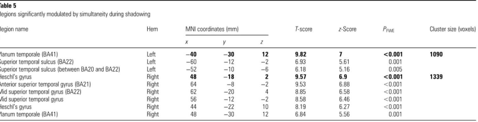
During SH, the brain areas modulated by simultaneity are
principally the superior temporal gyri, and a portion of the left
SMA (see Fig.
3
and Table
5
). A broader network is modulated
by simultaneity during SI (see Fig.
3
and Table
6
). In addition
to the superior temporal gyri, it includes 2 left frontal regions
(medial prefrontal and medial orbitofrontal cortices [mPFC
and mOFC]), alongside the putamen and the superior aspect of
the cerebellum bilaterally.
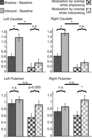
Dissociating the Roles of the Caudate and the Putamen
In order to establish whether there was a dissociation between
the roles of the caudate and the putamen during SH and
inter-pretation, an additional analysis was carried out. We selected
the voxels in the left and right caudate and putamen showing
the peak effect over the group for the contrast of interpretation
versus SH and for modulation by simultaneity during
interpret-ation. For each of the structures, we de
fined a region of interest
in the left and right caudate as the cluster showing signi
ficant
(
PFWE
< 0.05) activation for interpretation
–SH, and for the left
and right putamen as the region showing signi
ficant
modula-tion by simultaneity during interpretamodula-tion. The
first
eigenvari-ate of the T-statistic for activation in these regions was
extracted, subjectwise, for the tests: SH
–baseline, interpreting–
baseline, modulation by SH, and modulation by interpreting,
using Marsbar (
Brett et al. 2002
). These were then analyzed
using a 3-way repeated-measures ANOVA with the following
within-subject factors: structure (4 levels: left and right caudate
and putamen), condition (2 levels: s SH and interpretation)
and analysis type (2 levels: subtractive and modulation).
Language experience was included as a covariate of no
inter-est. A signi
ficant 3-way interaction was observed (degrees of
freedom Greenhouse
–Geisser corrected for nonsphericity
F1.382,56.049
= 4.267,
P = 0.032, partial η
2= 0.094), indicating a
difference in response in the different structures as a function
of condition and analysis type (see Fig.
4
). Post hoc
compari-sons show that in the right putamen, there was equal response
to interpretation and SH versus baseline, and signi
ficantly
greater modulation by the duration of overlap during
interpret-ation than during SH ( post hoc pairwise comparison:
T(42)
=
2.662,
P = 0.011). Similarly, in the left putamen, there was
equal response to interpretation and SH versus baseline, and
there was a marginally signi
ficant greater effect of simultaneity
on modulation during interpretation than during SH ( post hoc
pairwise comparison:
T
(42)= 1.974,
P = 0.055). (We note that
this difference is signi
ficant if we apply a directional
hypoth-esis of
“modulation by simultaneity is greater during
Table 3
Shadowing versus passive listening
Region name Hem MNI coordinates (mm) T-score z-Score PFWE Cluster size (voxels)
x y z
Postcentral gyrus (BA3) Left −46 −12 36 15.45 Inf <0.001 10868
Precentral gyrus (BA4) Left −56 0 22 13.35 Inf <0.001
Inferior frontal gyrus (pars opercularis) (BA44) Left −46 10 4 10.35 7.22 <0.001
Anterior insula Left −38 8 4 10.2 7.16 <0.001
Subthalamic nucleus Left −12 −18 4 10.11 7.12 <0.001
Posterior superior temporal gyrus (BA42) Left −56 −40 18 9.81 7 <0.001
Inferior frontal gyrus (pars opercularis) Left −54 14 8 9.58 6.9 <0.001
Mid superior temporal gyrus (BA22) Left −48 −30 6 9.4 6.83 <0.001
Superior temporal pole (BA38) Left −52 12 −6 9.34 6.8 <0.001
Globus pallidus Left −22 −6 −4 9.11 6.7 <0.001
Thalamus Right 12 −18 0 9.07 6.68 <0.001
Posterior superior temporal gyrus (BA22) Left −56 −26 6 8.97 6.64 <0.001
Inferior frontal gyrus (pars opercularis) (BA44) Left −48 12 16 8.75 6.53 <0.001
Planum temporale (BA41) Left −40 −32 10 8.56 6.45 <0.001
Anterior insula Left −30 22 6 8.17 6.26 <0.001
Globus pallidus Right 16 −2 −4 8.01 6.18 <0.001
Globus pallidus Right 22 −8 −2 7.98 6.17 <0.001
Brainstem white matter Left −8 −22 −10 7.94 6.15 <0.001
Putamen Left −18 2 10 7.94 6.14 <0.001
Superior temporal sulcus (BA22) Left −58 −40 8 7.93 6.14 <0.001
Central sulcus (BA4) Right 52 −8 26 15.37 Inf <0.001 5011
Central sulcus (BA4) Right 50 −10 40 13.95 Inf <0.001
Anterior insula Right 40 12 2 10.12 7.12 <0.001
Middle temporal gyrus Right 52 −24 6 9.85 7.01 <0.001
Middle temporal gyrus (BA22) Right 64 −28 12 8.26 6.3 <0.001
White matter subjacent to middle temporal gyrus Right 44 −26 −10 6.67 5.46 0.001
Superior temporal pole (BA38) Right 54 14 −16 5.46 4.71 0.027
Supplementary motor area (BA6) Left −6 0 64 15.23 Inf <0.001 3076
Anterior cingulate cortex (BA24) Left −6 10 42 9.83 7.01 <0.001
Anterior cingulate cortex (BA32) Right 10 12 38 9.52 6.88 <0.001
Cerebellum lobule VI Right 16 −62 −20 12.25 Inf <0.001 2692
Cerebellum lobule VI Left −14 −62 −20 11.55 7.66 <0.001
Vermis VI Left 0 −68 −14 8.81 6.56 <0.001
Cerebellum lobule VI Right 36 −54 −28 8.2 6.27 <0.001
Central sulcus (between BA3 and BA4) Left −18 −30 60 7.4 5.87 <0.001 64
Calcarine sulcus (BA17) Left −10 −74 12 6.58 5.41 0.001 353
Cerebellum lobule VIII Right 14 −66 −40 6.48 5.35 0.002 39
Cerebellum crus I Left −48 −60 −28 6.45 5.33 0.002 25
White matter [fronto-opercular fascicle] Right 28 −34 10 5.93 5.01 0.007 108
White mater [corticospinal tract] Right 22 −28 18 5.39 4.66 0.033
Calcarine sulcus (BA17) Right 14 −68 12 5.6 4.8 0.018 23
Precentral gyrus (BA3) Right 20 −30 62 5.58 4.79 0.02 10
Table lists maximum of 20 voxel-peaks per cluster, spaced a minimum of 8 mm apart. Bold rows indicate peak voxel within cluster. Hem, hemisphere;PFWE, familywise error-correctedP-value.
interpretation than during shadowing
” [i.e. using a one-tailed
test].)
Discussion
Simultaneous Interpretation
All the regions involved in SH are also involved in SI, re
flecting
the common linguistic and executive demands of the tasks.
Left inferior frontal gyrus regions additionally recruited during
interpretation over SH include pars triangularis, known for its
role in semantic processing (
Dapretto and Bookheimer 1999
;
Bookheimer 2002
), and pars orbitalis, implicated in semantic
memory and cognitive control of memory (
Badre and Wagner
2007
). Pre-SMA and dACC were also additionally recruited
during SI. Pre-SMA activation has been associated with tasks in
which there is response con
flict, and generation of complex
motor acts (
Picard and Strick 1996
;
Nachev et al. 2008
).
Pre-SMA and dACC are thought to play complementary roles in
action selection: The pre-SMA is involved in the selection and
initiation of action sets, and the dACC in monitoring the
outcome for errors (
Rushworth et al. 2004
). During SI, there is
a high level of competition between languages, and the
en-gagement of the pre-SMA is consistent with the need to handle
the resulting response con
flict. This analysis of the results is
supported by previous studies on language switching, which
have implicated the pre-SMA (
Abutalebi et al. 2008
).
The caudate nuclei were also engaged speci
fically during SI.
The left caudate nucleus has previously been reported to be
re-cruited in multilingual paradigms (
Crinion et al. 2006
;
Abutale-bi and Green 2008
;
Garbin et al. 2010
), and has been shown to
be implicated in multilingual language control by direct
elec-trical stimulation (
Wang et al. 2012
).
The left anterior insula and SMA have been associated with
a speech preparatory loop (
Riecker et al. 2005
). SI calls on
speech preparation to a greater extent than SH due to the
re-quirement to formulate the output
ab initio rather than simply
repeating the heard speech segments, and due to more
effort-ful speech output due to the competition between 2 languages
during the former, but not the latter, condition. The increased
anterior insula and dACC activation may also re
flect the
in-creased attentional demands and dif
ficulty of SI compared
with SH, managed by the cingulo-opercular system (
Petersen
and Posner 2012
).
The network of regions that we identi
fied during SI is
broader than previously reported. This could be due to our
larger sample size, or may be due to the fact that participants
in the present study were untrained, whereas the previously
studied participants were professional simultaneous
inter-preters with between 5 and 20 years of experience (
Rinne et al.
2000
;
Tommola et al. 2000
). Trained interpreters
’ expertise
may enable them to carry out the task ef
ficiently and effectively
while recruiting fewer brain regions than naïve participants,
consistent with many studies showing decreased brain
activa-tion when a task is more rehearsed and automated compared
with when it is more effortful and novel (
Ericsson et al. 2006
).
The regions we identify include many of those proposed by
Fabbro (1999)
to underlie SI. In addition to these, our
investi-gation reveals the involvement of brain regions involved in the
language- and executive-control requirements of the task.
The Neural Correlates of Simultaneity
An important distinction exists between the cognitive control
required to meet the overall demands of speci
fic task
instruc-tions, and the exercise of control on a moment-to-moment
basis while carrying out that task. This distinction is borne out
in the existing neuroimaging literature, which shows different
brain networks associated with task set maintenance over the
full length of experimental trials, or associated with the
initi-ation of responses and the adjustment of control on a
moment-to-moment basis (
Dosenbach et al. 2008
). Our
para-metric modulation analysis aimed to reveal the latter. During
periods of overlapping speech input and speech production,
the participants are concurrently engaged in processing the
Figure 2. Significant differences in BOLD response for the contrasts shadowing > passive listening (blue) and simultaneous interpreting > shadowing (red). Regions where both contrasts are significant (i.e., simultaneous interpreting > shadowing > passive listening) are also shown (magenta). Results are projected on a canonical single-subject MNI brain. Shadowing induces activation in a broad bilateral network encompassing temporal, inferior frontal, inferior parietal, motor, and subcortical regions including thalamus and globus pallidus. Interpretation additionally recruits further inferior frontal, motor, and basal ganglia regions. Bar charts indicate T-statistics averaged over the group at selected peak voxels in the named activation clusters, and error bars indicate standard error of the mean corrected to be appropriate for repeated-measures designs (Loftus and Masson 1994). Coordinates indicate plane of section (in MNI space) of the background images. pOp, pars opercularis; pOr, pars orbitalis; PTr, pars triangularis; aIns, anterior insula; lCd, left caudate; pSMA, presupplementary motor area; SMA, supplementary motor area; ACC, anterior cingulate cortex; Cru1, Crus 1 of cerebellum; CerVI, Cerebellum lobule VI; rCd, right caudate; STN, subthalamic nucleus; GP, globus pallidus.
Figure 3. Regions showing significant modulation of BOLD response as a function of the duration of overlapping speaking and listening during shadowing (blue) and interpretation (red) and both (magenta), projected on a the canonical single-subject MNI brain. During these periods of overlap, participants are concurrently engaged in processing input, producing an output, and monitoring that output for errors. This modulation analysis thus reflects the neural bases of dynamic, moment-to-moment cognitive control required within each task. During both shadowing and interpretation, there is significant modulation of superior temporal lobe activity by simultaneity. During shadowing, a portion of the SMA also displays significant modulation. During interpretation, a broader network of regions, constituting a putamen-cerebellar-prefrontal circuit is modulated by simultaneity. Coordinates indicate plane of section (in MNI space) of the background images.
Table 5
Regions significantly modulated by simultaneity during shadowing
Region name Hem MNI coordinates (mm) T-score z-Score PFWE Cluster size (voxels)
x y z
Planum temporale (BA41) Left −40 −30 12 9.82 7 <0.001 1090
Superior temporal sulcus (BA22) Left −60 −12 −2 6.93 5.61 0.001
Superior temporal sulcus (between BA20 and BA22) Left −52 −10 −6 6.18 5.16 0.005
Heschl’s gyrus Right 48 −18 2 9.57 6.9 <0.001 1339
Anterior superior temporal gyrus (BA21) Right 64 −8 −2 9.53 6.88 <0.001
Mid superior temporal gyrus (BA22) Right 62 −20 4 8.85 6.58 <0.001
Mid superior temporal gyrus Right 56 −12 −2 8.58 6.46 <0.001
Heschl’s gyrus Right 44 −22 10 8.19 6.27 <0.001
Planum temporale (BA41) Right 48 −30 12 6.84 5.56 0.001
Table lists maximum of 20 voxel-peaks per cluster, spaced a minimum of 8 mm apart. Bold rows indicate peak voxel within cluster. Hem, hemisphere;PFWE, familywise error-correctedP-value.
Table 4
Interpretation versus shadowing
Region name Hem MNI coordinates (mm) T-score z-Score PFWE Cluster size (voxels)
x y z
Presupplementary motor area (BA6) Left −6 14 60 10.94 7.44 <0.001 2833
Premotor cortex (BA6) Left −40 2 48 7.65 6 <0.001
Inferior frontal gyrus (pars triangularis) Left −48 20 24 7.56 5.95 <0.001
Inferior frontal gyrus (pars opercularis) (BA44) Left −46 12 30 7.08 5.69 <0.001
Superior frontal sulcus (BA6) Left −26 −2 54 6.46 5.34 0.002
Inferior frontal sulcus (BA45) Left −42 32 32 6.07 5.1 0.005
Anterior cingulate cortex (BA32) Right 14 24 38 5.84 4.95 0.009
Anterior cingulate cortex (BA32) Right 14 12 48 5.84 4.95 0.009
Superior frontal sulcus (between BA6 and BA8) Left −30 6 64 5.48 4.72 0.024
Anterior insula (BA47) Left −28 26 −2 7.62 5.98 <0.001 86
White matter [inferior fronto-occipital fasciculus] (BA47) Right 22 34 −2 6.73 5.49 0.001 375
White matter [anterior thalamic radiation] (BA47) Right 22 28 6 6.73 5.49 0.001
Head of the caudate Right 12 20 6 5.99 5.05 0.006
Head of the caudate Right 10 4 16 5.93 5.01 0.007
Head of the caudate Right 8 12 8 5.92 5.01 0.007
White matter [callosal body] Right 12 32 8 5.85 4.96 0.009
White matter [superior corona radiata] Left −20 4 30 6.03 5.07 0.005 72
Head of the caudate Left −14 0 22 5.94 5.02 0.007
Cerebellum crus I Right 48 −66 −28 5.87 4.97 0.008 12
Head of the caudate Left −8 20 4 5.83 4.95 0.009 19
Superior frontal sulcus Right 24 0 50 5.6 4.8 0.017 14
White matter [superior corona radiata] Right 22 4 28 5.59 4.79 0.018 22
Inferior frontal gyrus (between pars orbitals and pars triangularis) Left −52 22 −4 5.56 4.77 0.019 11
Table lists maximum of 20 voxel-peaks per cluster, spaced a minimum of 8 mm apart. Bold rows indicate peak voxel within cluster. Hem, hemisphere;PFWE, familywise error-correctedP-value.
input stream, producing an output, and monitoring that output
for semantic, syntactic and stylistic compatibility with the
source language input. By revealing brain areas that are more
heavily recruited during longer lasting periods of simultaneous
task execution, the modulation analysis re
flects the neural
bases of dynamic, moment-to-moment cognitive control
re-quired within each task. These very regions are likely to be
those that meet the greater linguistic and attentional control
re-quirements of the tasks during the more demanding periods of
simultaneity.
Simultaneity in both SH and SI modulated auditory regions
(the superior temporal gyri). Greater overlap of the input and
output speech streams results in a more complex auditory
input for a longer period of time. Consequently, there are
likely to be increased demands on auditory processing and on
auditory attention during periods of overlapping speech
per-ception and production.
During SI, we also found modulation of the left mPFC,
which has been implicated in multitasking. It has been
sug-gested that this region serves to balance attention between
stimulus-oriented
and
stimulus-independent
processes
(
Gilbert et al. 2006
), a role likely to be called upon more when
handling demands of processing the input stream, producing
speech in the target language, and monitoring output
simul-taneously as opposed to consecutively. The mOFC also
showed modulation by simultaneity and it may be involved in
language switching (
Wang et al. 2007
,
2009
), another key
com-ponent of interpretation.
During SI, activity in the putamen, bilaterally, was
modu-lated by simultaneity, with the response of the right putamen
showing a signi
ficant dissociation such that its response was
signi
ficantly more modulated by simultaneity during SI than
during SH, and with a strong trend for such a dissociation in
the left putamen. In both the left and right putamen, there was
no signi
ficant global difference in response to the 2 tasks. The
left putamen has been implicated in translation (
Klein et al.
1995
;
Price et al. 1999
), in second-language single-word
repeti-tion (
Klein et al. 1994
), and more recently it has been shown to
be engaged when multilingual individuals employ a language
that is not mastered in a native-like fashion (
Abutalebi et al.
2012a
). Although our participants produced speech in a
lan-guage in which they had an extremely high pro
ficiency, the
results can be reconciled by considering the putamen as the
source of ongoing suppression of speech production in the
in-appropriate language during SI, that is, while simultaneously
hearing sentences in that language. The pragmatics of speech
are such that when hearing one language it is normal to
respond with speech in that same language and, in naïve
indi-viduals, suppressing this prepotent response may call upon
the putamen substantially more than in experienced
inter-preters. Thus, we suggest that the role of the putamen in
poly-glot language control can be thought to be the suppression of
Table 6
Regions showing significant modulation of activity by simultaneity during interpretation
Region name Hem MNI coordinates (mm) T-score z-Score PFWE Cluster size (voxels)
x y z
Anterior superior temporal sulcus (BA22) Right 60 −4 −8 11.48 7.63 <0.001 3215
Heschl’s gyrus Right 44 −20 10 10.89 7.42 <0.001
Mid superior temporal gyrus (BA22) Right 52 −18 −2 9.44 6.84 <0.001
Planum temporale (BA21) Right 68 −22 2 9.04 6.67 <0.001
Mid superior temporal gyrus (BA22) Right 56 −22 10 8.66 6.49 <0.001
Posterior superior temporal gyrus (BA22) Right 64 −32 8 8.18 6.26 <0.001
Posterior superior temporal sulcus (BA21) Right 50 −38 4 7.15 5.73 <0.001
Deep sylvianfissure [between parietal operculum and planum temporale] (BA48) Right 34 −28 22 6.26 5.22 0.004
Mid superior temporal sulcus (BA22) Left −52 −30 4 9.93 7.05 <0.001 2599
Anterior superior temporal sulcus (BA22) Left −58 −14 −4 9.8 6.99 <0.001
Posterior superior temporal gyrus (BA22) Left −56 −36 8 8.16 6.25 <0.001
Heschl’s gyrus Left −48 −18 6 7.59 5.97 <0.001
Anterior superior temporal sulcus (BA38) Left −52 4 −8 7.2 5.76 <0.001
Planum temporale (BA42) Left −58 −34 16 6.44 5.32 0.002
Deep sylvianfissure [between parietal operculum and planum temporale] Left −34 −28 18 6.01 5.06 0.008
Anterior middle temporal gyrus (BA21) Left −52 −2 −18 5.39 4.66 0.043
Putamen Right 28 6 −4 8.29 6.32 <0.001 589
White matter [anterior limb of internal capsule] Right 20 16 6 7.3 5.81 <0.001
White matter [anterior limb of internal capsule] (BA0) Right 12 8 −4 7.21 5.76 <0.001
Anterior ventral paracingulate cortex (BA11) Left −8 32 −10 7.6 5.97 <0.001 67
Putamen Left −24 4 −4 7.33 5.83 <0.001 426
Putamen Left −22 10 2 6.82 5.55 0.001
White matter [anterior limb of internal capsule] Left −18 4 14 6.2 5.18 0.005
Putamen (BA0) Left −24 2 8 5.62 4.81 0.024
Cerebellum lobule IV/V (BA30) Right 22 −32 −24 6.65 5.45 0.001 83
Precentral gyrus (BA6) Right 30 −16 50 6.34 5.26 0.003 11
Cerebellum lobule VI (BA37) Left −28 −54 −24 6.31 5.24 0.003 85
Cerebellum lobule VI (BA19) Left −16 −62 −24 5.77 4.91 0.015
Medial superior frontal gyrus (BA10) Left −2 60 26 6.21 5.18 0.005 88
Dorsal anterior cingulate gyrus (BA32) Left −10 54 24 6.07 5.1 0.007
Cerebellum lobule VI Right 36 −62 −26 6.14 5.14 0.005 37
Cerebellum crus II Right 10 −82 −38 6.11 5.12 0.006 18
Cerebellum crus II Right 22 −78 −38 5.6 4.8 0.025
Superior parietal lobule (BA5) Left −16 −44 64 6.09 5.11 0.006 20
Cerebellum lobule IV/V Left −20 −36 −22 5.92 5.01 0.01 25
Inferior occipital cortex (BA18) Left −22 −94 −10 5.76 4.9 0.016 13
Table lists maximum of 20 voxel-peaks per cluster, spaced a minimum of 8 mm apart. Bold rows indicate peak voxel within cluster. Hem: hemisphere,PFWE: familywise error-correctedP-value.
a contextually dominant language. Taken together, our results
suggest a dissociation between the roles of the caudate and the
putamen in multilingual control.
The Central Role of the Dorsal Striatum in SI
A striking aspect of these results is the recruitment of the
dorsal striatum (the caudate nucleus and the putamen).
Al-though the basal ganglia are often discussed in terms of their
role in motor behavior, they play a central role in circuits
known to subserve multiple, nonmotoric aspects of cognition
such as attention, learning and memory, and executive
func-tions (
Saint-Cyr 2003
). It has been argued that the basal
ganglia and cerebellum interact in loops with cortical regions
to select and re
fine cortical patterns, essentially subtending the
ability to control action (
Houk et al. 2007
).
The linguistic roles of the basal ganglia have received some
concerted attention in the past (
Lieberman 2000
), and there is
increasing interest in basal ganglia language functions (
Kotz
et al. 2009
) particularly with regard to multilingualism (e.g.,
Friederici 2006
;
Abutalebi and Green 2007
,
2008
;
Abutalebi
et al. 2012a
;
Wang et al. 2012
;
Zou et al. 2012
). The basal
ganglia have previously been found to be involved in tasks
re-quiring bilingual language control, such as single-word
translation
(
Klein
et
al.
1995
;
Price
et
al.
1999
),
second-language word repetition (
Klein et al. 1994
,
2006
),
con-secutive interpretation (
Lehtonen et al. 2005
), and language
switching (
Price et al. 1999
;
Abutalebi and Green 2008
;
Garbin
et al. 2010
). Results have been equivocal as to the precise roles
of these structures. Some authors have reported that language
switching recruits the caudate nucleus (
Abutalebi and Green
2008
;
Garbin et al. 2010
), whereas others have reported
in-volvement of both caudate and putamen (
Price et al. 1999
).
Similarly, studies on translation have given rise to variable
results, with one showing recruitment of putamen (
Klein et al.
1995
) and, another, external globus pallidus (
Lehtonen et al.
2005
). Second-language word repetition has been reported to
recruit the left caudate (
Klein et al. 2006
) and the left putamen
(
Klein et al. 1994
). Other authors have not reported basal
ganglia involvement during language switching tasks (
Wang
et al. 2007
,
2009
) or, as previously noted, during SI (
Rinne
et al. 2000
;
Tommola et al. 2000
). Nevertheless, a central role
for the striatum in bilingual language control is supported
by case studies of subcortical polyglot aphasia, which have
reported that damage to the putamen or caudate in
multilin-gual individuals can result in involuntary language mixing or
switching (
Hervais-Adelman et al. 2011
;
Abutalebi et al.
2012a
).
The caudate nucleus and putamen are engaged by SI in our
study, and, frequently, by tasks explicitly demanding
multilin-gual control in previous work (see above). In nonlinguistic
domains, the caudate is typically described as being involved
in the selection of appropriate behavior as a function of
pre-dicted outcomes, while the putamen is associated with the
im-plementation and co-ordination of such behavior (
Grahn et al.
2008
). A compatible distinction between the caudate and
putamen is proposed by
Ali et al. (2010
), who examined the
neural correlates of a Stroop and Simon task in fMRI. They
con-cluded that the left caudate was implicated in the inhibition of
action plans triggered by incongruent words (in the Stroop
task), while the left putamen was implicated in response
change in both the Stroop (word interference) and Simon tasks
(spatial interference). These distinctions between the caudate
and putamen are consistent with our results and, together with
them, help to reconcile the existing literature on basal ganglia
structures in multilingualism.
We propose that the caudate nucleus is engaged in relatively
high-level monitoring and controlling of
“language set”
selec-tion, controlling which lexico-semantic system is
“live” at a
given moment. This concords with the notion that the caudate
is involved in determining context-appropriate behavior (e.g.,
Graybiel 1995
;
Chee 2006
). In contrast, we propose that the
putamen is engaged in moment-to-moment language output
control at what can be considered a lower level for example in
inhibiting the nontarget language in order to favor access to
ar-ticulatory motor representations required for articulation in the
appropriate language. The observed complementary
contribu-tions of the caudate and of the putamen during language
control re
flect an organizing principle of subcortical
involve-ment in the coordination of cognition, action, and learning
—
the caudate being implicated in planning and the putamen in
execution of actions.
The bilateral nature of the
findings we report here expand
those of several previous reports cited above, in which the left
striatum is principally implicated in tasks requiring
multilin-gual language control (
Klein et al. 1994
,
1995
,
2006
;
Price et al.
Figure 4. Bar charts showing contrast estimates of left and right caudate andputamen, for contrasts with baseline and for modulation by overlap for shadowing and interpretation. Values are averaged over participants and represent thefirst eigenvalue of the T-statistic in the regions defined as showing a significant effect for the contrast of interpretation–shadowing (the caudate nuclei) and modulation by overlap (the putamen, bilaterally). Error bars indicate ± 1 standard error of the mean, corrected to be appropriate for within-subjects comparisons (Loftus and Masson 1994).
1999
;
Lehtonen et al. 2005
;
Abutalebi and Green 2008
;
Garbin
et al. 2010
). There is also some indication from direct electrical
stimulation that the dominant striatum (typically the left
stri-atum in right-handed individuals) is involved in the control of
speech production, with the putamen more implicated in the
coordination of speech articulation, and the caudate involved
in inhibition and selection, in monolingual patients (
Gil Robles
et al. 2005
). A recent meta-analysis of neuroimaging studies of
language switching, however, indicates that both right and left
striatal structures are implicated in language switching (
Luk
et al. 2012
). Consistent with this, our data indicate that the
bilateral caudate nuclei and putamen are implicated in SI. Our
bilateral
findings may be due to the fact that owing to the large
number of participants that we tested, we had relatively higher
statistical power, or it may be because in contrast to most
previ-ous reports, we used a task that makes large, ongoing (as
opposed to momentary) demands on language control. The
findings herein reported demonstrate bilateral striatal
involve-ment in a demanding, continuous language control task. (It is
of particular interest to note that when including 7 left-handed
participants in our analysis [see Supplementary Tables 1
–4 and
Supplementary Figures 1
–3], the lateralization of these results
does not qualitatively change, suggesting that the language
control functions of these structures may not be substantially
affected by handedness. This
finding merits further
investiga-tion in the future.)
Our results provide new insights into the profound overlap
between the neural bases of extreme language control and those
of domain-general control of cognition and action. Indeed,
recent evidence suggests that experienced simultaneous
inter-preters display enhanced cognitive
flexibility compared even
with bilingual individuals (
Yudes et al. 2011
;
Stavrakaki et al.
2012
). The recruitment of similar fronto-subcortical-cerebellar
circuits during language and executive control provides
power-ful evidence that the continuous demands of language control in
the multilingual brain, and associated experience-dependent
plasticity, could underlie the nonlinguistic, executive advantages
that have been observed in bilingual individuals, advantages that
may also be protective in defying challenges posed by aging and
even disease.
Supplementary Material
Supplementary material can be found at: http://www.cercor.oxford journals.org/.
Funding
This work was supported by the Swiss National Science
Foun-dation (grant nos. PP00P3_133701 and 320030_122085).
Notes
We thank Alain Dagher for helpful comments on the manuscript, as well as 3 anonymous reviewers for their thoughtful suggestions. We also thank Frédéric Grouiller and Maria Pefkou for their assistance with data collection, and Sophie Hengl, Violeta Seretan, Magdalena Olivera-Tovar, and Carmen Delgado Luchner for their efforts in rating the participants’ output. Conflict of Interest: None declared.
References
Abutalebi J, Annoni JM, Zimine I, Pegna AJ, Seghier ML, Lee-Jahnke H, Lazeyras F, Cappa SF, Khateb A. 2008. Language control and lexical competition in bilinguals: an event-related fMRI study. Cereb Cortex. 18:1496–1505.
Abutalebi J, Della Rosa PA, Castro Gonzaga AK, Keim R, Costa A, Perani D. 2012a. The role of the left putamen in multilingual lan-guage production. Brain Lang. 125:307–315.
Abutalebi J, Della Rosa PA, Green DW, Hernandez M, Scifo P, Keim R, Cappa SF, Costa A. 2012b. Bilingualism tunes the anterior cingulate cortex for conflict monitoring. Cereb Cortex. 22:2076–2086. Abutalebi J, Green D. 2007. Bilingual language production: the
neuro-cognition of language representation and control. J Neurolinguis-tics. 20:242–275.
Abutalebi J, Green D. 2008. Control mechanisms in bilingual language production: neural evidence from language switching studies. Lang Cogn Process. 23:557–582.
Abutalebi J, Tettamanti M, Perani D. 2009. The bilingual brain: linguis-tic and non-linguislinguis-tic skills. Brain Lang. 109:51–54.
Ali N, Green DW, Kherif F, Devlin JT, Price CJ. 2010. The role of the left head of caudate in suppressing irrelevant words. J Cogn Neurosci. 22:2369–2386.
Aron AR. 2008. Progress in executive-function research: from tasks to functions to regions to networks. Curr Dir Psychol Sci. 17:124–129. Badre D, Wagner AD. 2007. Left ventrolateral prefrontal cortex and the
cognitive control of memory. Neuropsychologia. 45:2883–2901. Bialystok E, Craik FI, Luk G. 2012. Bilingualism: consequences for
mind and brain. Trends Cogn Sci. 16:240–250.
Boersma P, Weenink D. 2011. Praat: doing phonetics by computer. Version 5.2.23.
Bookheimer S. 2002. Functional MRI of language: new approaches to understanding the cortical organization of semantic processing. Annu Rev Neurosci. 25:151–188.
Botvinick MM, Cohen JD, Carter CS. 2004. Conflict monitoring and an-terior cingulate cortex: an update. Trends Cogn Sci. 8:539–546. Brett M, Anton JL, Valabregue R, Poline J-B. 2002. Region of interest
analysis using an SPM toolbox [abstract]. In. 8th International Con-ference on Functional Mapping of the Human Brain. Sendai, Japan. Available on CD-ROM in NeuroImage, Vol 16, No 2.
Chee MW. 2006. Dissociating language and word meaning in the bilin-gual brain. Trends Cogn Sci. 10:527–529.
Costa A. 2005. Lexical access in bilingual production. In: Kroll J, De Groot AMB, editors. Handbook of bilingualism: psycholinguistic approaches. Oxford, UK: Oxford University Press. p. 308–325. Costa A, Hernandez M, Costa-Faidella J, Sebastian-Galles N. 2009. On
the bilingual advantage in conflict processing: now you see it, now you don’t. Cognition. 113:135–149.
Crinion J, Turner R, Grogan A, Hanakawa T, Noppeney U, Devlin JT, Aso T, Urayama S, Fukuyama H, Stockton K et al. 2006. Language control in the bilingual brain. Science. 312:1537–1540.
Dapretto M, Bookheimer SY. 1999. Form and content: dissociating syntax and semantics in sentence comprehension. Neuron. 24:427–432.
Diamond J. 2010. The benefits of multilingualism. Science. 330:332–333.
Dijkstra T, van Heuven WJB. 2002. The architecture of the bilingual word recognition system: from identification to decision. Biling Lang Cogn. 5:175–197.
Dosenbach NU, Fair DA, Cohen AL, Schlaggar BL, Petersen SE. 2008. A dual-networks architecture of top-down control. Trends Cogn Sci. 12:99–105.
Eickhoff SB, Laird AR, Grefkes C, Wang LE, Zilles K, Fox PT. 2009. Coordinate-based activation likelihood estimation meta-analysis of neuroimaging data: a random-effects approach based on empirical estimates of spatial uncertainty. Hum Brain Mapp. 30(9):2907–2926. Ericsson KA, Charness N, Feltovich PJ, Hoffman RR. 2006. The Cam-bridge handbook of expertise and expert performance. New York (NY): Cambridge University Press.
Fabbro F, editor. 1999. The neurolinguistics of bilingualism: an intro-duction. Hove: Psychology Press.