Tissue-engineered vascular graft remodeling in a growing lamb
model: expression of matrix metalloproteinases
Ian Cummings
a,b, Sarah George
c, Jens Kelm
a,b, Doerthe Schmidt
a,b, Maximilian Y. Emmert
a,b,d,
Benedikt Weber
a,b, Gregor Zünd
a,band Simon P. Hoerstrup
a,b,d,*
a
Swiss Center for Regenerative Medicine, University Hospital and University Zurich, Zurich, Switzerland
b Department of Surgical Research, University Zurich, Zurich, Switzerland c Bristol Heart Institute, Bristol Royal Infirmary, Bristol, UK
d Clinic for Cardiovascular Surgery, University Hospital Zurich, Zurich, Switzerland
* Corresponding author. Tel: +41-1-2553801; fax: +41-1-2554369; e-mail: [email protected] (S.P. Hoerstrup). Received 10 December 2010; received in revised form 15 February 2011; accepted 21 February 2011
Abstract
OBJECTIVES: We have previously demonstrated the functionality and growth of autologous, living, tissue-engineered vascular grafts (TEVGs) in long-term animal studies. These grafts showed substantialin vivo tissue remodeling and approximation to native arterial wall characteristics. Based on this,in vitro and in vivo matrix metalloproteinase (MMP) activity of TEVGs is investigated as a key marker of matrix remodeling.
METHODS:TEVGs fabricated from biodegradable scaffolds ( polyglycolic-acid/poly-4-hydroxybutyrate, PGA/P4HB) seeded with autolo-gous vascular cells were cultured in static and dynamicin vitro conditions. Thereafter, TEVGs were implanted as pulmonary artery repla-cements in lambs and followed up for 2 years. Gelatin gel zymography to detect MMP-2 and -9 was performed and collagen content quantified (n = 5). Latent (pro) and active MMP-2 and -9 were detected.
RESULTS: Comparable levels of active MMP-9 and pro-MMP-2 were detected in static and dynamic culture. Higher levels of active MMP-2 were detected in dynamic cultures. Expression of MMP-2 and -9 was minimal in native grafts but was increased in implanted TEVG. Pro-MMP-9 was expressed 20 weeks post implantation and persisted up to 80 weeks post implantation. Collagen contentin vitro was increased in dynamically conditioned TEVG as compared with static constructs and was increasedin vivo compared with the corre-sponding native pulmonary artery.
CONCLUSIONS:MMPs are up-regulatedin vitro by dynamic culture conditions and could contribute to increased matrix remodeling, native analogous tissue formation and functional growth of TEVGs in vivo. Monitoring of MMP activity, for example, by molecular imaging techniques, may enable the non-invasive assessment of functional tissue quality in future clinical tissue-engineering applications.
Keywords:Matrix metalloproteinases• Tissue engineering • Vascular graft • Autologous • Growth • Cells
INTRODUCTION
Tissue-engineering technologies continue to address the critical need for living autologous tissue substitutes with growth poten-tial in congenital cardiac surgery. Applications of biological and engineering principles strive to construct tissues with characteris-tics of healthy native tissues from their cellular components. For reconstruction of the right-ventricular outflow tract, the use of homografts, xenografts, and polytetrafluoroethylene (PTFE) grafts remains the mainstay of surgical treatment options, despite vari-able results and degrees of success. The need, however, for sur-gical re-interventions is inevitable in many situations due to conduit failure as well as the inability of these conduits to grow with the child.
The feasibility of creating implantable large- and small-caliber
tissue-engineered vessels for systemic and low-pressure
applications has been demonstrated experimentally and in initial clinical trials. Functional arteries have been grown in vitro both using animal and human cells [1–3]. Recently, we have demon-strated that tissue-engineered vascular grafts (TEVGs) implanted as autologous, living, pulmonary artery replacements showed ex-cellent functionality and growth in long-term large animal follow-up studies covering the full biological growth cycle of the animal model [4]. Interestingly, these growing and functional tissue-engineered arteries demonstrated substantial remodeling over time, and, most importantly, structural approximation to native arterial wall tissue composition. The normal arterial wall consists of an intimal layer, a media layer, and an adventitial layer. Vascular remodeling and structural adaptation of tissue-engineered grafts in response to hemodynamic stresses based on constant cellular activity appear to be a prerequisite for growth and sustained functionality [4].
© The Author 2011. Published by Oxford University Press on behalf of the European Association for Cardio-Thoracic Surgery. All rights reserved. doi:10.1016/j.ejcts.2011.02.077
BASIC
S
Vascular smooth muscle cells (VSMCs) are the main cellular components of the arterial wall and provide vascular tone and the arterial wall extracellular matrix. Facilitating SMC growth, mi-gration, basement-membrane breakdown, angiogenesis, and other aspects of vascular remodeling necessitates an ongoing degradation and reorganization of extracellular matrix. First described in 1962 by Gross and Lapiere [5], 26 described matrix metalloproteinases (MMPs) contribute to extracellular matrix re-modeling. MMPs are zinc-dependent peptidases broadly classi-fied according to their substrate-degradation properties. They are synthesized in the latent form as zymogens and secreted as pro-enzymes ( pro-MMPs). Activation occurs via a process known as the cysteine-Zn+ switch [6], which enables proteolytic activity. In this study, we identified MMPs as important enzymes in TEVG remodeling. The functions of MMPs are not fully under-stood; however, they are of interest in physiological processes, such as pregnancy [7], and have also been reported to be asso-ciated with pathologic phenomena [8–10]. In vascular tissue en-gineering, MMP-related processes are expected to be of critical importance for tissue homeostasis and functional growth. With the aim of a more meticulous understanding of long-term re-modeling mechanisms, tissue-engineered autologous living pul-monary arteries were analyzed for MMP-2 and -9 expression as well as collagen content and compared with native arterial tissue. SMCs produce pro-MMP-2 in normal arterial wall and in culture, but active MMP-2 and pro- and active MMP-9 are absent [11–13]. By contrast, injured and diseased vessels under-going remodeling produce active MMP-2 and MMP-9 [11,12,14]. Consequently, in this study, we focused on the expression of the gelatinases MMP-2 and-9. MMP-2 has been shown to degrade native type IV, V, VI, and X collagen as well as denatured colla-gen, proteoglycans, elastin, and growth factors [15]. MMP-9 degrades gelatin, an irreversibly hydrolyzed collagen, and is expressed following injury or inflammatory stimulation [16]. In the light of these observations and the so-far limited under-standing with regard to tissue engineering [17], the long-term MMP expression in autologous, living TEVGs implanted as pul-monary artery replacements in a growing large animal model is investigated.
MATERIAL AND METHODS
The general approach to cell isolation, culture, and seeding has been described in detail [4,18]. All investigations performed in this work are based on tissue samples generated in a previous study focusing on functional performance of tissue-engineered arteries in a growing long-term animal model [4]. All procedures were conducted according to Swiss regulations on animal welfare and permission granted by the ethical committee (Permission Nr. 71/2002, 91/2004).
In vitro production of TEVGs
Bioabsorbable pulmonary artery scaffolds were fabricated from non-woven polyglycolic-acid mesh (PGA, thickness 1.0 mm, spe-cific gravity 70–90 mg cm−3, Cellon SA, Luxembourg) coated with poly-4-hydroxybutyrate (P4HB, molecular weight 1 × 10⁶, PHA 4400, Tepha Inc., Lexington, MA, USA). Based on biopsies of peripheral blood vessel (Swiss White Alpine lambs), endothe-lial cells were obtained using a collagenase-instillation technique.
Following isolation, they were cultured in tissue culture flasks (Nunc™, Roskilde, Denmark) with endothelial basal medium (EBM-2) medium supplemented with 10% lamb serum (GIBCO,
NY, USA). To obtain myofibroblasts, the remaining
de-endothelialized vessel segments were minced and cultured in
Dulbecco’s modified Eagle Medium (DMEM) or advanced
DMEM (GIBCO, NY, USA) supplemented with 10% lamb serum, 2 mM glutaMAX (GIBCO, NY, USA) and 50μg ml−1 gentamycin (GIBCO, NY, USA). After myofibroblast migration onto the dishes (after 5–7 days), the cells were serially passaged and expanded in a humidified incubator at 37 °C and 5% CO2. Sufficient cell
numbers for cell seeding were obtained in pure culture after 10–14 days.
Myofibroblasts (4.5–5.5 × 106cm−2) were sequentially seeded
onto the vascular scaffolds, followed by endothelial cell seeding (1 × 106 cm−2) phenotypically confirmed by von Willebrand
Factor (vWF) presence. Thereafter, the constructs were trans-ferred into a pulse duplicator system [18] and grown under
gradually increasing nutrient medium flow conditions
(50–550 ml min−1, 1 Hz) for 14 days. Control TEVGs were grown accordingly, but in static nutrient. Media were changed every 3–4 days.
In vivo implantation of TEVGs
The TEVGs were surgically implanted as replacements of the main pulmonary artery into lambs, as described in detail as pre-viously described inCirculation 2006 [4].
Computed tomography (CT)–angiography analysis
Computed tomography (CT)–angiography scans were performed under general anesthesia on a 64-slice scanner with a 0.37-s rotation time (Somatom Sensation 64™, Siemens, Forchheim, Germany). A bolus of Iodixanol (270 mg ml−1, Amersham Health, UK) was injected into a forearm vein at aflow rate of 4 ml s−1, using 2 ml of contrast agent per kg. Slices with a thickness of 0.75 mm (increment 0.5 mm) and a medium soft-tissue recon-struction kernel (B30f ) were used for evaluation.
Analyses of TEVG explants
Out of a total of 14 animals [4], a subgroup of five TEVGs samples was investigated as to MMP profiles, microstructure, and tissue composition. A representative longitudinal sample of each TEVG was fixed in 10% phosphate-buffered formalin (pH 7.0) and embedded in paraffin. Each cross section included native tissue proximal as well as distal to the entire length of the TE construct. Routine histological staining was by hematoxylin and eosin (H&E) and Masson’s trichrome. Immunohistochemical stainings were performed using the Ventana Benchmark auto-mated staining system (Ventana Medical Systems, Tucson, AZ, USA) with Ventana reagents for the entire process. Primary anti-bodies against the following antigens were applied: vWF( poly-clonal rabbit anti-vWF antibodies; all from DakoCytomation, Glostrup, Denmark), alpha smooth muscle actin (monoclonal mouse anti-αSMA, clone 1A4; Sigma), and desmin (monoclonal mouse anti-desmin, clone D33). Primary antibodies were detected with the Ventana iVIEW DAB detection kit, resulting in
a brown reaction product. Counterstaining was performed with hematoxylin. As an indicator for collagen formation, the hydro-xyproline content of lyophilized samples was determined, as described by Huszar et al. [19].
MMP-2 and -9 were detected by gelatin zymography, as pre-viously described [14]. Briefly, sodium dodecylsulfate (SDS) cell lysates were subjected to electrophoresis on 7.5% (w/v) polya-cryalamide gel electrophoresis (PAGE) gel containing 0.2% (w/v) SDS supplemented with 2 mg ml−1 gelatin. All gels were cali-brated with a high-molecular-mass protein standard mixture. In addition, for standardization, on each gel, an aliquot of a stand-ard conditioned medium collected from HT1080 cells (which secrete both MMP-2 and -9) was run.
Statistical analysis
Analysis of variance was performed to estimate the means and the significance of the categorical factors using Statistical Package for Social Sciences (SPSS) version 16 (SPSS, Inc., Chicago, IL, USA). A paired t-test was performed comparing native tissue to tissue-engineered samples. A p-value <0.05 was considered as significant.
RESULTS
Static and dynamically cultured TEVGs were analyzed and com-pared with tissue engineered and native in vivo samples at 20 (TE20,n = 1), 50 (TE50, n = 2), and 80 (TE80, n = 2) weeks post implantation. A macroscopic image of the tissue-engineered graft at time of implantation is shown in Fig. 1. Masson’s tri-chrome staining of a pre-implant conduit showed collagen within the extracellular matrix, as blue color. H&E staining showed layered tissue formation of the pre-implant conduit (Fig. 2(a) and (b)). As early as 20 weeks post implantation, the macroscopic external appearance of the pulmonary conduit was indistinguishable from native tissue (Fig.3).
Morphology
At the time of sacrifice, the tissue-engineered pulmonary artery graft was dissected out in toto. The TEVGs were carefully
Figure 1:Tissue engineered vascular graft (TEVG) after of surgical implant-ation as main pulmonary artery replacement.
Figure 2:(a) Trichrome Masson (TM) staining of pre-implant TEVG showing collagen staining in blue. (b) Hematoxylin and Eosin (H&E) staining of pre-implant TEVG showing layered tissue formation within the pre-implant TEVG (the bar represents 200μm).
BASIC
S
observed for signs of thrombus formation, calcification and obvious stenosis, or aneurysmal formation. There were no signs of morphological abnormalities in any of the animals included. The luminal layer appeared smooth and continuous with the native pulmonary artery and free of calcification or thrombus formation.
Tissue microstructure and composition
The triple-layered tissue-engineered pulmonary artery consisted of an intima, a media, and an adventitial layer reminiscent of native artery. At TE20, TE50, and TE80, there is an increasing vas-cular SMC distribution within the TEVGs, with a corresponding increased collagen deposition in the extracellular matrix with time (Fig.4).
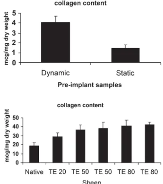
Collagen contentin vitro and in vivo, as measured by hydroxy-proline content, increased significantly in dynamically condi-tioned constructs as compared with static constructs (Fig. 5(a)) with the dynamically generated TEVGs having a hydroxyproline content of 4.08 ± 1.9μg mg−1 dry weight. The hydroxyproline content was significantly increased in tissue-engineered
constructs compared with native pulmonary artery (Fig. 5(b)).
Native pulmonary artery collagen content was
18.8 ± 3.6 μg mg−1 dry weight. At 20 weeks post implantation (TE20), the tissue-engineered conduit had a collagen content of 29.03 ± 4.14μg mg−1dry weight. As seen in Fig.5(b), the colla-gen content increases up to 42.3 ± 2.98μg mg−1 dry weight at TE80.
MMP activity
MMP expression was detected as clear bands on the blue back-ground of the stained gel, due to lysis of the gelatin. These bands correspond to the predicted molecular weights of MMP-2 and -9 and correspond with the positive control. MMP-2 and MMP-9 exist in both latent and active forms, with molecular weight of latent MMP-2 being 72 kDa and that of latent MMP-9 being 92 kDa. Comparable levels of active MMP-9 and pro-MMP-2 were detected in VSMCs in static and dynamic culture. However, higher levels of active MMP-2 were detected in TEVGs in dynamic cultures as compared with static culture (Fig.6). Expression of MMP-2 and MMP-9 was minimal in native grafts but was increased in implanted tissue-engineered grafts (Fig. 7). Both latent and pro-MMP-2 and -9 expression was observed as early as TE20 and persists up to TE80 (Fig.7).
DISCUSSION
More than 20 years ago,Weinberg and Bell produced a collagen-based vascular conduit by isolating aortic bovine endothelial,
Figure 4:Trichrome Masson (TM) staining of explanted TEVG at 20 weeks post implantation showing collagen staining in blue (The bar represents 200μm).
Figure 3:Macroscopic image of an explanted TEVG at 20 weeks post implant-ation. There is no evidence of thrombus or calcification and the TEVG shows a smooth luminal surface.
Figure 5: (a) Collagen content of static versus dynamic pre implant grafts. There is a significant difference between dynamic versus static samples (p = 0.05) (b). Collagen content of 5 TEVG at 20 (TE20), 50 (TE50) and 80 (TE80) weeks post implantation versus native (N) pulmonary artery. There is a significant difference between native and TE 50 (p < 0.05) and TE 80 (p < 0.05) samples.
smooth muscle, and adventitialfibroblast cells [20]. Although not implanted in vivo, these constructs showed sufficient burst strength and structural features of arterial tissue. Since these early days, tissue engineering has combined principles of cell biology with biomechanical engineering, leading to advances in thefield [21].
Various cell sources have been investigated for cardiovascular applications both in animal and in human models [1]. Shin’oka et al. used bone-marrow-derived cells intra-operatively seeded onto vascular scaffolds as pulmonary artery replacements in pediat-ric patients with adequate functionality [3]. The histological results elucidating the growth potential of these grafts are awaited. Others have used humanfibroblast cells in scaffold-free tissue-engineering applications for small-diameter blood vessels [22].
However, little is known regarding the post-implantation tissue-remodeling processes observed to a substantial degree in most experimental studies. It appears that, apart from the initially seeded cells, endogenous cell re-population phenomena con-tribute importantly to these remodeling processes comprising somatic growth and long-term functionality. With regard to future routine clinical use of the tissue-engineering technology, a meticulous understanding of the underlying remodeling mechanisms will be of crucial importance. Furthermore, safety parameters assessable in a non-invasive fashion after implant-ation may be a prerequisite for the safe translimplant-ation of clinical tissue engineering into clinical practice. It is anticipated that such assessments may be performed by molecular-imaging method-ologies, such as magnetic resonance imaging, which can monitor enzymatic activities of MMPs [23].
Ideal tissue-engineered conduits should show durability, absence of thrombus formation, resistance to infections, lack of immunological complications, and, most importantly, potential for growth. We have shown functional growth and approxima-tion of tissue-engineered pulmonary arteries to native tissue in animal studies. Long-term follow-up of animals with autologous ovine cells seeded onto a biodegradable scaffold and implanted
in the pulmonary position has confirmed both feasibility and functional growth of the tissue-engineered conduits based on CT and echocardiography data [4].
Improvements in bioreactor technology and physiological pre-conditioningin vitro have been shown to stimulate extracellular matrix formation in vitro. Collagen formation was significantly increased in the dynamic pre-implantation TEVG as compared with static culture. In our lamb model, as early as 20 weeks post-operatively, collagen formation in the tissue-engineered graft had already exceeded native pulmonary artery, with continued deposition leveling in at TE80.
The long-term role of MMPs in pulmonary artery tissue engin-eering in a growing animal model has been unknown so far. MMPs play a significant role in tissue remodeling and have been extensively investigated in pregnancy [7], as well as in pathological processes, such as hypertension [8], atherosclerosis [10], and aneurysmal formation [9]. All of these processes show degrad-ation of extracellular matrix and remodeling by MMP activity.
To date, there is only limited knowledge as to the role of MMPs in the remodeling of tissue-engineered cardiovascular structures [17], particularly with regard to MMP activity during somatic growth. Our results have confirmed MMP-2 and MMP-9 as key enzymes in tissue-engineered vascular remodeling with ongoing MMP-2 and MMP-9 activity up to TE80 weeks.In vitro, there was an up-regulation of active MMP-2 as a result of bio-mimetic, dynamic culture conditions. Once implanted, there appears to be constant recruitment of cells into the implanted tissue-engineered graft to maintain tissue homeostasis and sus-tained functionality [4]. The origin of these cells is under investi-gation and may include blood-born stem cells as well as cells from the peri-vascular environment [24]. Histologically, the media layer of the tissue-engineered graft showed fibroblasts and collagen deposition with a rich capillary network. This, to-gether with the hemodynamic stresses of the pulmonary circula-tion, would account in part for the increased activated MMP-2 and MMP-9 activity in the tissue-engineered graft. The enzymat-ic activity is ongoing throughout the TE80 period, even though there is complete degradation of the biodegradable polymer and therefore absence of foreign body phenomena in the graft. The MMP-2 activity in native tissue does not appear to vary sig-nificantly at the various time points, despite being subjected to the same hemodynamic stresses as the TEVGs. This implies that hemodynamic stresses alone do not account for the increased MMP activityin vivo, which rather reflects the adaptive tissue re-modeling activities resulting in the observed functionality and growth of the TEVGs. Therefore, MMP activity could be regarded and used as an indicator of ‘guided healing processes’. MMP-9 activity is markedly up-regulated in the tissue-engineered grafts in conjunction with a significantly increased collagen production as compared with native tissue. Histologically, a corresponding increase in VSMCs in tissue-engineered implants is observed with time.
Indeed, MMPs have been demonstrated to be linked with aneurysms and atherosclerotic processes [8–10]. However, both echocardiography and CT data did not display any aneurysmal formation or atherosclerotic signs as also confirmed by detailed histology [4]. To the contrary, functional growth with arterial wall structures approximating native arterial layers over time were observed. Thus, the increased MMP activity in TEVGs appears not to be attributed to these pathological processes, but the result of increased cellular recruitment, which use MMP-2 and -9 to migrate or invade the tissue-engineered graft similarly seen
Figure 7:MMP-2 and -9 expression in TEVG (TE) versus native (N) at 20, 50 and 80 weeks after implantation. The TE pulmonary artery (TE), site of anasto-mosis (TE/N) between TE and native as well as native (N) PA were subjected to gelatin zymography. Both active and pro(latent) MMPs are indicated. Figure 6: MMP-2 and -9 expression in vascular smooth muscle controls, dynamic (n = 2) and static (n = 2) culture conditions. Gelatinolytic zymogra-phy was performed to detect MMP-2 and MMP-9. Active and pro(latent) forms are indicated.
BASIC
S
in classical healing processes. In addition, there was no evidence of calcification, thrombus formation, or stenosis within the tissue-engineered graft, further suggesting the absence of patho-logical processes.
Our observations raise questions in terms of the differences which exist between tissue-engineered and native vascular grafts. The presence of increased MMP activity even in the long-term grafts suggests a sustained remodeling activity to maintain tissue homeostasis and functionality. Further studies will be necessary to evaluate the role of other classes of MMPs and the effects of inhi-biting MMP activity with regard to functionality, long-term growth potential, and performance of tissue-engineered vascular grafts. Based on a more detailed understanding of matrix remodeling, modern magnetic resonance and molecular imaging techniques may allow for non-invasive tracking of MMP activity in clinical set-tings [25]. Such methodologies enabling the definition of clinically relevant quality criteria may contribute to the safe translation of tissue-engineering technologies to routine clinical practice. In add-ition, this growing insight into the remodeling mechanisms involv-ing MMPs could help to specifically design intelligent scaffold materials. These may support remodeling phenomena within tissue-engineered constructs in the future by attaching transcrip-tion factors and/or cytokines to the starter matrices.
Conflict of interest: none declared.
REFERENCES
[1] Niklason LE, Gao J, Abbott WM, Hirschi KK, Houser S, Marini R, Langer R. Functional arteries grown in vitro. Science 1999;284:489–93. [2] Poh M, Boyer M, Solan A, Dahl SL, Pedrotty D, Banik SS, McKee JA,
Klinger RY, Counter CM, Niklason LE. Blood vessels engineered from human cells. Lancet 2005;365:2122–4.
[3] Shin’oka T, Matsumura G, Hibino N, Naito Y, Watanabe M, Konuma T, Sakamoto T, Nagatsu M, Kurosawa H. Midterm clinical result of tissue-engineered vascular autografts seeded with autologous bone marrow cells. J Thorac Cardiovasc Surg 2005;129:1330–8.
[4] Hoerstrup SP, Cummings Mrcs I, Lachat M, Schoen FJ, Jenni R, Leschka S, Neuenschwander S, Schmidt D, Mol A, Gunter C, Gossi M, Genoni M, Zund G. Functional growth in tissue-engineered living, vascular grafts: follow-up at 100 weeks in a large animal model. Circulation 2006;114:I159–66. [5] Gross J, Lapiere CM. Collagenolytic activity in amphibian tissues: a tissue
culture assay. Proc Natl Acad Sci USA 1962;48:1014–22.
[6] Van Wart HE, Birkedal-Hansen H. The cysteine switch: a principle of regulation of metalloproteinase activity with potential applicability to the entire matrix metalloproteinase gene family. Proc Natl Acad Sci USA 1990;87:5578–82.
[7] Merchant SJ, Davidge ST. The role of matrix metalloproteinases in vascu-lar function: implications for normal pregnancy and pre-eclampsia. BJOG 2004;111:931–9.
[8] Flamant M, Placier S, Dubroca C, Esposito B, Lopes I, Chatziantoniou C, Tedgui A, Dussaule JC, Lehoux S. Role of matrix metalloproteinases in early hypertensive vascular remodeling. Hypertension 2007;50:212–8. [9] Keeling WB, Armstrong PA, Stone PA, Bandyk DF, Shames ML. An
overview of matrix metalloproteinases in the pathogenesis and treat-ment of abdominal aortic aneurysms. Vasc Endovascular Surg 2005;39: 457–64.
[10] Zaltsman AB, Newby AC. Increased secretion of gelatinases A and B from the aortas of cholesterol fed rabbits: relationship to lesion severity. Atherosclerosis 1997;130:61–70.
[11] Zempo N, Kenagy RD, Au YP, Bendeck M, Clowes MM, Reidy MA, Clowes AW. Matrix metalloproteinases of vascular wall cells are increased in balloon-injured rat carotid artery. J Vasc Surg 1994;20:209–17. [12] Bendeck MP, Zempo N, Clowes AW, Galardy RE, Reidy MA. Smooth
muscle cell migration and matrix metalloproteinase expression after arterial injury in the rat. Circ Res 1994;75:539–45.
[13] Southgate KM, Davies M, Booth RF, Newby AC. Involvement of extracellular-matrix-degrading metalloproteinases in rabbit aortic smooth-muscle cell proliferation. Biochem J 1992;288:93–9.
[14] George SJ, Zaltsman AB, Newby AC. Surgical preparative injury and neointima formation increase MMP-9 expression and MMP-2 activation in human saphenous vein. Cardiovasc Res 1997;33:447–59.
[15] Sternlicht MD, Werb Z. How matrix metalloproteinases regulate cell behavior. Annu Rev Cell Dev Biol 2001;17:463–516.
[16] Whatling C, McPheat W, Hurt-Camejo E. Matrix management: assigning different roles for MMP-2 and MMP-9 in vascular remodeling. Arterioscler Thromb Vasc Biol 2004;24:10–11.
[17] Stock UA, Wiederschain D, Kilroy SM, Shum-Tim D, Khalil PN, Vacanti JP, Mayer JE Jr., Moses MA. Dynamics of extracellular matrix production and turnover in tissue engineered cardiovascular structures. J Cell Biochem 2001;81:220–8.
[18] Hoerstrup SP, Sodian R, Daebritz S, Wang J, Bacha EA, Martin DP, Moran AM, Guleserian KJ, Sperling JS, Kaushal S, Vacanti JP, Schoen FJ, Mayer JE Jr. Functional living trileaflet heart valves grown in vitro. Circulation 2000;102:III44–9.
[19] Huszar G, Maiocco J, Naftolin F. Monitoring of collagen and collagen fragments in chromatography of protein mixtures. Anal Biochem 1980; 105:424–9.
[20] Weinberg CB, Bell E. A blood vessel model constructed from collagen and cultured vascular cells. Science 1986;231:397–400.
[21] Langer R, Vacanti JP. Tissue engineering. Science 1993;260:920–6. [22] L’Heureux N, Dusserre N, Konig G, Victor B, Keire P, Wight TN, Chronos
NA, Kyles AE, Gregory CR, Hoyt G, Robbins RC, McAllister TN. Human tissue-engineered blood vessels for adult arterial revascularization. Nat Med 2006;12:361–5.
[23] Lancelot E, Amirbekian V, Brigger I, Raynaud JS, Ballet S, David C, Rousseaux O, Le Greneur S, Port M, Lijnen HR, Bruneval P, Michel JB, Ouimet T, Roques B, Amirbekian S, Hyafil F, Vucic E, Aguinaldo JG, Corot CFayad ZA. Evaluation of matrix metalloproteinases in atherosclerosis using a novel noninvasive imaging approach. Arterioscler Thromb Vasc Biol 2008;28:425–32.
[24] Zilla P, Bezuidenhout D, Human P. Prosthetic vascular grafts: wrong models, wrong questions and no healing. Biomaterials 2007;28:5009–27. [25] Rubbens MP, Driessen-Mol A, Boerboom RA, Koppert MM, van Assen
HC, TerHaar Romeny BM, Baaijens FP, Bouten CV. Quantification of the temporal evolution of collagen orientation in mechanically conditioned engineered cardiovascular tissues. Ann Biomed Eng 2009; 37:1263–72.