HAL Id: tel-01070644
https://tel.archives-ouvertes.fr/tel-01070644
Submitted on 2 Oct 2014HAL is a multi-disciplinary open access archive for the deposit and dissemination of sci-entific research documents, whether they are pub-lished or not. The documents may come from teaching and research institutions in France or abroad, or from public or private research centers.
L’archive ouverte pluridisciplinaire HAL, est destinée au dépôt et à la diffusion de documents scientifiques de niveau recherche, publiés ou non, émanant des établissements d’enseignement et de recherche français ou étrangers, des laboratoires publics ou privés.
To cite this version:
Hakima Flici. Neural stem cells plasticity and differentiation. Cellular Biology. Université de Stras-bourg, 2012. English. �NNT : 2012STRAJ118�. �tel-01070644�
ÉCOLE DOCTORALE DES SCIENCES DE LA VIE ET DE LA SANTE
Institut de Génétique et de Biologie Moléculaire et Cellulaire
THÈSE
présentée par :Hakima FLICI
soutenue le : 21 Septembre 2012pour obtenir le grade de :
Docteur de l’université de Strasbourg
Discipline/ Spécialité: Biologie du développement
Différenciation et plasticité des cellules
souches neurales
THÈSE dirigée par :
Mme GIANGRANDE Angela Docteur, université de Strasbourg RAPPORTEURS :
M. GONZALEZ Cayetano Professeur, Institut de Recerca Biomedica (IRB-Barcelona) M. REICHERT Heinrich Professeur, Biozentrum University of Basel
AUTRES MEMBRES DU JURY :
I would first like to thank my committee members, Prof. GONZALEZ Cayetano, Prof. REICHERT Heinrich, Prof. IMLER Jean-Luc, and Prof. DAVIDSON Erwin for their kindness and also for accepting to evaluate my thesis report.
I would also like to thank my advisor, Dr. Angela Giangrande, for giving me the opportunity to join her group at IGBM, where I have gained both scientific rigour and the ability to experimentally put my ideas into practice, and for her kind guidance and support.
I am thankful to the directors of IGBMC, Pr. MORAS Dino and Pr. POURQUIE Olivier for offering me the chance to be part of IGBMC community.
I also thank CERBM GIE and FRM for their financial support and the confidence they have given to my project.
I am grateful for the dedication of Dr. Pietro LANEVE. I have highly appreciated your near unrestricted availability and have greatly enjoyed our stimulating discussions. Oh, Pietro, they’re no words to say thanks for you. You were and you will be forever a precious example for me, giving without waiting for recompense. Many thanks for correcting the whole manuscript of my thesis, and for your continuous support.
I would also like to thank my first coach Dr. Orban KOMONIYI. You were a great teacher. I have highly appreciated working and discussing with you.
Thanks to the current and previous members of Giangrande lab: Orbán KOMONYI, Pietro LANEVE, Damiano PORCELLI, Sára BERZSĖNYI, Arun KUMAR, Ömer Faruk KARATAŞ, Francesca TAGLINI, Tripti GUPTA, Anna POPKOVA, Giulia PAPALINI, Céline DIEBOLD, Carole MURA, Iren KEREKES, Guillaume TREBUCHET, and Giuseppe D’AGOSTINO, H. Bahar SAHIN, Yoshihiro YUASA, Chethan REDDY. It was wonderful to work in a lab with people who are willing to help each other out, and even collect virgins for you on Saturday.
I am thankful for the help of Dr. Leila LAREDJ and Dr. Muhammad SHUAIB for helping me to correct the manuscript of my thesis.
Claude DELAPORTE and Monica BALLARINO for being helpful, lovely and humorous.
life.
For research support, I am indebted to all the members of the imaging facility, Marcel BOEGLIN, Marc KOCH, Yves LUTZ, Jean-Daniel FAUNY, Didier HENTSCH, Pascal KESSLER , Jean-Luc VONESCH, without whom I would not have able to perform my acquisitions. Similarly, the members of fly facility Nadine and Holly.
Furthermore, I would like to thank IGBMC Administration team for providing guidance and support over the course of my PhD.
It is impossible to advance alone, in research as elsewhere. A big thank to everyone who helped me during this period of my life.
Jelina, It was a great pleasure to share three continuous months behind you,… Keep in touch girl.
I would not have been able to complete this work without the emotional support of my family and friends, who closely shared both the frustrations and joys over the course of this work, but always provided support and strong encouragement, for which I am very grateful. Thank you Mom, Dad, Sofiane, Samir, Souad, Zineb, Dahmane, Ferrodja and Samy for your unending love and support.
I would also like to thank all my friends and family that I didn’t mention.
Finally, as we say in fresh “On garde toujours le meilleur pour la fin” So, thank you Aziz, for your love and continuous support.
Table of contents
LIST OF FIGURES ... 4
LIST OF PAPERS ... 5
ABBREVIATIONS ... 6
ABSTRACTS ... 11
1. En Français/ In French ... 11
2. En Anglais/In English ... 14
INTRODUCTION ... 16
1. The fundamental questions in developmental biology ... 16
2. Cell plasticity ... 18
2. 1. General aspects on cell plasticity ... 18
2. 2. Cell fate determinants and cell plasticity ... 19
2. 3. Stem cell plasticity research ... 20
2. 3. 1. Properties of stem cells ... 20
2. 3. 2. Drosophila embryonic NSCs as a model system to study cell plasticity ... 21
2. 4. Broad questions in cell plasticity ... 22
3. The development of embryonic nervous system ... 23
3. 1. The Development of Drosophila nervous system ... 23
3. 1. 1. General structure of the nervous system ... 23
3. 1. 2. Early neurogenesis in Drosophila embryos ... 25
3. 1. 3. Cells making Drosophila CNS ... 27
• Neuroblasts ... 27
• Glial cells ... 29
• Neurons ... 30
3. 2. The development of vertebrate nervous system ... 31
3. 2. 1. General structure of the nervous system ... 31
3. 2. 2. Early neurogenesis in vertebrate embryos ... 31
3. 2. 3. Cells of the nervous system ... 33
• Neural stem cells ... 33
• Neurons and glia ... 34
4. The mechanism regulating neurodevelopmental plasticity ... 36
4. 1. Regulation of NSC biology ... 36
4. 1. 1. Neural stem cell lineage types ... 36
4. 1. 2. Neural stem cell temporal gene cascade and cell fate specification ... 39
4. 1. 3. Neural stem cell mode of division ... 40
• Symmetric cell division in Drosophila ... 40
• Asymmetric cell division in Drosophila ... 41
• Neural stem cell mode of division in mammals ... 42
4. 1. 4. Neural stem cell quiescence ... 43
4. 1. 5. Neural stem cell apoptosis ... 48
4. 2. Epigenetic regulation of neurodevelopmental plasticity ... 48
4. 2. 1. Chromatin organization ... 48
4. 2. 2. Histone modifications ... 49
• Acetylation/Deacetylation ... 49
• Methylation/demethylation ... 59
• Complexes regulating chromatin organization ... 60
4. 2. 3. Epigenetic control of neural stem cells and their progenies ... 62
5. Gcm transcription factors ... 66
5. 1. gcm/Gcm in Drosophila ... 66
5. 1. 1. Background ... 66
5. 1. 2. Gliogenic potential of Gcm ... 67
• Background ... 67
5. 1. 3. gcm and hematopoiesis ... 71
5. 2. gcm/Gcm orthologs in vertebrates ... 72
5. 2. 1. GCM1 ... 72
5. 2. 2. GCM2 ... 73
5. 2. 3. GCM1 and GCM2 in the nervous system ... 73
5. 3. gcm gene regulation ... 74
5. 3. 1. Role of Gcm ... 74
5. 3. 2. Role of Gcm2 ... 75
5. 3. 3. Role of Prospero ... 75
5. 3. 4. Role of other factors ... 76
5. 4. Properties of the gcm mRNA ... 78
5. 5. Properties of Gcm protein ... 78
AIMS ... 80
MATERIALS AND METHODS ... 82
1. Methods for Drosophila melanogaster ... 82
2. Methods for S2 cells ... 83
RESULTS ... 85
1. 1st part. The gliogenic potential of Gcm and NSC plasticity ... 85
1. 1. Background ... 85
1. 2. Manuscript I. ... 87
1. 3. Summary of major findings ... 88
1. 3. 1. gcm overexpression is able to redirect NSCs to adopt the glial fate, a process that goes through an intermediate state ... 88
1. 3. 2. Age and mitotic state influence NSC plasticity ... 88
1. 3. 3. Ectopic glial cells display the same epigenetic marks as the endogenous ones ... 88
1. 4. Conclusion ... 89
1. 5. My contribution ... 89
2. 2nd part. Aging and NSC plasticity ... 91
2. 1. Introduction ... 91
2. 2. Manuscript II. ... 92
2. 3. Supporting data for manuscript II: PcG complex and NSC plasticity ... 93
2. 3. 1. Background ... 93
2. 3. 2. Results ... 93
2. 3. 3. Discussion ... 96
2. 4. Summary of the major discussed points ... 97
2. 4. 1. Temporal control of NSC plasticity ... 97
2. 4. 2. NSC plasticity and intermediate state ... 98
2. 4. 3. polycomb mutation promotes gcm-‐mediated cell fate conversion ... 98
2. 5. Conclusion ... 99
2. 6. My contribution ... 99
3. 3rd part. gcm/Gcm regulation and cell fate establishment ... 100
3. 1. Background ... 100
3. 2. Manuscript III. ... 101
3. 3. Supporting data for manuscript III: Interaction between the cell fate determinants Gcm and Twist ... 102
3. 3. 1. Background ... 102
3. 3. 2. Results ... 103
• twist activity controls negatively the gliogenic potential of gcm ... 103
• gcm and twist transcripts are reduced in gcm-twist co-gain of function embryos ... 104
3. 3. 3. Discussion ... 106
3. 4. Summary of the major findings ... 107
3. 4. 1. Repo positively controls gcm gene expression but negatively Gcm protein stability ... 107
3. 4. 4. Cell fate building goes though a transient metastable state ... 109
3. 4. 5. Twist counteracts gcm-‐mediated cell fate conversion ... 109
3. 5. Conclusion ... 110
3. 6. My contribution ... 110
4. 4th part. The Gcm/Glide protein visualized at last: novel hints on its metabolism and a marker for novel cell type ... 112
4. 1. Background ... 112
4. 2. Manuscript IV: ... 113
4. 3. Summary of the major findings ... 114
4. 3. 1. The Flag tag does not affect Gcm activity ... 114
4. 3. 2. GcmFlag allows the identification of new mechanisms regulated and regulating Gcm output ... 114
4. 3. 3. New cells expressing gcm ... 115
4. 4. Conclusion ... 115
4. 5. My contribution ... 115
DISCUSSION ... 117
1. Correct regulation of gcm in glial cells requires the interaction of multiple elements ... 117
1. 1. Positive autoregulatory and cross-‐regulatory loops ... 118
1. 2. Double-‐negative feedback from Repo and CBP ... 119
2. Mechanism regulating gcm to induce cell fate conversion ... 122
3. Cell plasticity and gcm-‐mediated cell fate conversion ... 124
3.1. Progressive loss of plasticity ... 124
3. 2. Cell cycle status and plasticity ... 125
3. 3. Intermediate fates ... 126
4. Role of epigenetic modifications in cell fate commitment ... 127
4. 1. Epigenetic regulation and cell fate choice ... 127
4. 2. Epigenetic regulation and cell fate commitment ... 128
CONCLUSION ... 131
LIST OF FIGURES
Figure 1. Progenitor cell self-renewal and differentiation contribute to tissue patterning and
tumorigenesis. ... 17
Figure 2. Structure of The Drosophila nervous system ... 23
Figure 3. Embryonic CNS and PNS axon pathways and pattern of glial cells. ... 24
Figure 4. The lateral inhibition process. ... 25
Figure 5: Drosophila neuroblasts map ... 26
Figure 6. NBs distribution in the developing Drosophila CNS. ... 28
Figure 7. Spatial distribution and classification of glial cells in the VNC. ... 30
Figure 8. ... 31
Figure 9. Adult neural stem cell niche. ... 34
Figure 10. Different modes of neuroblast division. ... 38
Figure 11. ... 43
Figure 12. Quiescent Drosophila NBs are characterized by an elongated shape and by the presence of a cytoplasmic process moving inward. ... 44
Figure 13. A model for the nutritional control of quiescent NB reactivation. ... 46
Figure 14. Representation of the different functional domains of CBP and p300. ... 53
Figure 15. Schematic illustration of CBP coregulator functions depending on the chromatin contexts. ... 59
Figure 16. The biochemical activity of the PcG complex. ... 62
Figure 17. gcm and gcm2 expression pattern at different embryonic stages. ... 67
Figure 18. Profile of embryonic glial cells in WT, gcm mutant, and gcm overexpressing embryos. ... 69
Figure 19. gcm-gcm2 complex and Gcm transcription factors. ... 75
Figure 20. Schematic representation of NB6-4 lineages. ... 77
Figure 21:gcm/Gcm regulation networks ... 121
LIST OF PAPERS
My thesis is based on the following manuscripts:
PAPER I: H. Flici, B. Erkosar, O. Komonyi, O. F. Karatas, P. Laneve and A. Giangrande (2011). In vivo Gcm/Glide-dependent conversion into glia depends on neural stem cell age but not division, triggering a chromatin signature conserved in vertebrate glia. Development, 138(19): 4167-78.
PAPER II: H. Flici and A. Giangrande (2012). Stem cell aging and plasticity in the Drosophila nervous system. Landes Bioscience, Fly, Volume 6, Issue 2.
PAPER III: H. Flici, O. Komonyi, P. Laneve, S. Berzsenyi, A. Giangrande. The homeobox transcription factor Repo collaborates with the histone acetyl transferase CBP to regulate the initiation and the maintenance of the glial fate and the cell fate choice of neuronal progenitors. In preparation.
PAPER IV: P. Laneve, C. Delaporte, G. Trebuchet, O. Komonyi, H. Flici, A. Popkova, G. D’Agostino, F. Taglini, I. Kerekes and A. Giangrande. The Gcm/Glide protein visualized at last: novel hints on its metabolism and a marker for novel cell type. Submitted to Developmental Biology.
ABBREVIATIONS
A A: abdominal AbdA: Abdominal‐A AbdB: Abdominal‐B Acetyl-CoA: acetyl-coenzyme A AD: transactivation domain Ada: transcriptional ADAptor Ago: ArchipelagoAgo1: Argonaute‐1
Akt: protein kinase B, PKB
A/P: Anterior/Posterior
Ascl1: Achaete-scute complex-like 1 ATAC: Ada two A containing complex ATP: Adenosine triphosphate
ATXN: ataxin
B β-gal: β-galactosidase
bHLH: basic Helix Loop Helix BMP4: Bone Morphogenic Protein 4 Brm: Brahma
BTB: Bric-à-brac Tramtrack Broad CBP FL: CBP full-length
CBP FLAD: CBP full-length acetyltransferase deficient
C
CARM1: coactivator-associated arginine
Cas: Castor CB: central brain CBG: cell body glia
CBP: cAMP response‐element binding protein
Cdc25: cell division cycle 25 Cdk2: Cyclin dependent kinase 2 CG: channel glia
CNS: central nervous system
CoREST: co‐repressor for element‐1 silencing transcription factor
CREB: cAMPresponse element-binding D
DAPI: 4',6‐diamidino‐2‐phenylindole DBD: DNA binding domain
dILP: Drosophila insulin-like peptides dInR: Drosophila insulin receptor Dlx2: distal-less homeobox 2 DNA: deoxyribonucleic acid Dpn: Deadpan
Dpp: Decapentaplegic E
EED: embryonic ectoderm development E(z): Enhancer of zeste
Elav: Embryonic lethal, abnormal vision ELP3: Elongation Protein 3
EMS: methanesulfonate En: Engrailed
ETS: erythroblast transformation specific ESC: Embryonic stem cell
Esc: Extra sex combs
EZH2: enhancer of zeste homolog 2 F
FACS: fluorescence activated cell sorting FGF-2: fibroblast growth factor-2
G
Gal4/80: GALactose metabolism 4/80 GB: glioblast
GBS: Gcm binding site
Gcm/Glide: Glial cells missing/glial cell deficient
Gcn5/PCAF: General control
nonderepressible 5/p300/CBP associated factor
GFAP: glial fibrillary acidic protein GFP: Green fluorescent protein
Gcm/Glide: Glial Cell Missing/Glial Cell Deficiency
GLAST: Glutamate astrocyte-specific
transporter
GMC: ganglion mother cell
GNAT: Gcn5‐related N‐Acetyltransferase Grh: Grainyhead
H
HAT: Histone acetyl‐transferase
HBO1: histone Acetyltransferase Binding to ORC-1
HD: Huntington’s disease HDAC: Histone deacetylase Hkb: Huckebein
HMT: histone methyltransferase Hox: Homeobox
Hkb: Hunchback HTLV-1: Human T cell
leukemia/lymphoma virus type 1
I IE: instability element IG: interface glia
IGF: insulin-like growth factor IgG: Immunolglobulin G
IHC: immunohistochemistry
INP: intermediate neural progenitor IP: Immunoprecipitation
IPC: intermediate progenitor cell ISN: intersegmental nerve
ISNG: intersegmental and segmental nerve root glia
Iswi: Imitation Switch J JmjC: Jumanji C domain
K K: lysine
L LG: longitudinal glia LGB: longitudinal glioblast Loco: Locomotion defects
LSD1: Lysine specific demethylase 1 Lz: Lozange
M MAM: methyl methacrylate
MAP2: microtubule-associated protein 2 MB: mushroom bodie
MEF2: Myocyte enhancing factor 2 MG: midline glia
Mira: Miranda
MLL: Mixed‐lineage leukemia MOF: Male absent on the first MORF: MOZ-related factor
MOZ: Monocytic leukemia zinc finger protein
mRNA: messenger ribonucleic acid Myc: v‐myc myelocytomatosis viral oncogene homolog
MYST: MOZ/YBF2/SAS2/Tip60 NCoR: Nuclear receptor co‐repressor
N
NAD: nicotinamide adenine dinucleotide NAP1: nucleosome assembly protein 1 NB: neuroblast
Neuro: Neurogenin
N-COR: Nuclear receptor corepressors NGB: neuroglioblasts
NRG: nerve root glia NS: nervous system NSC: Neural stem cell
NuRD: Nucleosome remodeling and deacetylase
O OL: optic lobe
P P300: protein of 300 kDa
PAF400: 400kDa PCAF associated factor (synonym of TRRAP)
Par-6: Partition-Defective 6 Pc: Polycomb
PC4: Positive coactivator 4 PcG: Polycomb group
PEST sequence: proline, glutamic acid, serine, threonine rich sequence
Ph: Polyhomeotic
PH3: phospho-Histone H3-Ser10
Pho: Polyhmomeotic
PHD: Plant Homeo Domain Pho: Pleiohomeotic
PI3K: phosphatidylinositol 3-kinase-Akt PIWI: P‐element induced wimpy testis PKMT: lysine HMT
PNS: peripheral nervous system Pnt: Pointed
PolyQ: poly-glutamine
PRC1: Polycomb repressor complex 1 PRC2: Polycomb repressor complex 2 PRMT: arginine HMT
Psc: Posterior sex combs Q
qPCR: quantitative polymerase chain reaction
R R: arginine
Rb: Retinoblastoma protein
REST/NRSF: RE1 silencing TF/neural-restrictive silencing factor
RHG: Reaper Hid, and/or Grim Repo: Reversed polarity
RFP: Red fluorescent protein RG: radial glia
RNA Pol II: RNA Polymerase II RNA: ribonucleic acid
RNAi: RNA interference
Rpd3: Reduced potassium dependency 3
Rpr: Reaper Hid, and/or Grim RTH: Rubinstein-Taybi
S S: serine
SAGA: Spt‐Ada‐Gcn5‐acetyl‐transferase Sbf1: SET binding factor 1
SC: Stem cell Sca: scabrous
Sce: sex combs extra
SET domain: Su(var)39, E(z) and trx domain
SGZ: subgranular zone Sin3: Switch independent
Sir2: Silent information regulator 2 SIRT: Sirtuin type protein
Slimb: Supernumerary limbs
SMRT: silencing mediator for retinoid and thyroid receptors
Sog: Short gastrulation
SOP: sensory organs precursor SPG: subperineurial glia Stg: String
STAT: Signal Transducer and Activator of Transcription
SVZ: subventricular zone
Su(z)12/SUZ12: suppressor of zeste 12 T
T: thoracic T: threonine
TAFII250 : TFIID 250 kDa TBP: TATA binding protein TF: transcription factor
TFIIB: Transcription factor II D Tip60: HIV‐1 Tat interacting protein TM3: Third multiple #3
TOR: target of paramycin Trx: Trithorax TrxG: Trithorax group Ts: temperature sensitive Ttk: Tramtrack Tyr: Tyrosine U
UAS: Uprtream activating sequence Ubx: Ubithorax
UTR: Untranslated Region
V VNC: ventral nerve cord VPA: valproic acid
VUM: Ventral Unpaired Median VZ: ventricular zone
W wg: wingless
YBF2/SAS3: Yeast binding factor 2/Something about silencing 3
Z Zn: zinc
ABSTRACTS
1. En Français/ In French
Différenciation et plasticité des cellules souches neurales
Le cerveau humain est un tissu en renouvellement constant où l’équilibre entre les cellules nouvellement formées et les cellules perdues est maintenu par la division asymétrique des cellules souches neurales (CSN), appelées aussi neuroblastes (NBs), produisant deux cellules filles différentes : l’une possédant la capacité de s’auto-renouveler et l’autre engagée dans la différenciation en neurones et en cellules gliales. Aujourd’hui, le défi fondamental de la neurobiologie est de comprendre la plasticité des CSNs, afin d’expliquer les mécanismes conduisant ces cellules à générer à la fois des neurones et des cellules gliales sains, mais aussi des cellules à identité ambiguë, pathologique. Ceci est particulièrement important lorsque l’on considère la réparation du système nerveux après une lésion ou une maladie en faisant appel aux cellules souches.
Mon projet de thèse porte sur la compréhension des mécanismes clés contrôlant l’élaboration du destin gliale et du destin neuronale in vivo, en utilisant comme modèle les CSNs de la corde ventrale de la drosophile (l’équivalent de la moelle épinière chez les vertébrés). Chez ce modèle, le facteur de transcription Glide/Gcm constitue le déterminant glial: son expression ectopique dans les précurseurs neunaux induit de la glie ectopique tandis que son absence induit la glie à adopter le destin neuronal. En utilisant le potentiel gliogénique de ce facteur dans les CSNs, nous avons déterminé plusieurs nouveaux processus impliqués dans la plasticité des CSNs.
Nous avons tous d’abord montré que les précurseurs neunaux peuvent être complètement redirigés pour adopter le destin glial et que cette conversion est stable et complète. Le processus de conversion du destin ne se manifeste pas uniquement par l’expression de marqueurs gliaux mais aussi par des changements spécifiques au niveau de la chromatine. En effet, durant nos analyses nous avons identifié le niveau d’acétylation de l’histone H3 au niveau de la lysine 9 (H3K9ac) comme une nouvelle marque qui distingue les neurones des cellules gliales, les neurones ont des niveaux élevés en H3K9ac contrairement au cellules gliales qui ont des niveau faibles de cette marque épigénétique. D’une manière intéressante, les cellules gliales ectopiques, qui se différencient normalement en neurones, ont
aussi des niveaux faibles en H3K9ac. En analysant le déroulement de la gliogenèse ectopique, nous avons aussi pu montrer que l’établissement du destin glial passe par une étape intermédiaire où les marqueurs de neuroblastes et les marqueurs gliaux sont co-exprimés.
L'on considère les cellules souches comme des précurseurs pouvant se diviser de manière indéfinie in vitro. Toutefois, in vivo, ces cellules prolifèrent activement lors du développement, puis entrent dans une phase quiescente ou dans un programme apoptotique chez l'adulte. Par des expériences d'expression conditionnelle dans des lignages de CSNs identifiés, nous avons pu montrer que la capacité des CSNs à se convertir en glie après expression forcée de Glide/Gcm décline avec l'âge et que lors de l'entrée en phase quiescente ou apoptotique, elles ne peuvent plus être converties. D’une manière surprenante, nous avons également montré que l’expression ectopique de Glide/Gcm dans les neurones post-mitotiques n’induit pas un changement du destin mais de l’apoptose, alors que les CSNs dont la division a été bloquée peuvent être converties.
Glide/Gcm est le facteur initiateur du destin glial par sa présence transitoire dans tous les précurseurs gliaux, par contre les mécanismes contrôlant sa cascade moléculaire reste fragmentaire. Nos analyses ont permis d’identifier de nouveaux facteurs impliqués dans ce processus et de comprendre comment ces facteurs peuvent affecter le destin des précurseurs générés par les CSNs. Il est connu que Glide/Gcm est capable d’induire l’expression de son propre gène et celui de sa cible directe, repo. A ceci nous avons rajouté une nouvelle voie qui est le pouvoir de Repo à induire l’expression de glide/gcm. D’une manière intéressante, nous avons aussi montré que la stabilité de la protéine Glide/Gcm est contrôlée par deux voies opposées, où Repo et dCBP (pour Drosophila Creb Binding Protein) jouent un rôle majeur. La première voie implique dCBP qui stabilise Glide/Gcm, induisant ainsi l’expression de ces cibles directes, y inclus glide/gcm et repo. La deuxième voie implique à la fois Repo et dCBP qui, ensemble, s’opposent à la progression de la première boucle en induisant la dégradation de Glide/Gcm. Ces voies suggèrent que la modulation quantitative de facteurs clés est cruciale, à des temps de développement précis, afin de garantir un destin glial complet. En déséquilibrant l’activité de ces facteurs, les cellules restent dans un état intermédiaire, où le destin glial et le destin neuronal se manifestent au même temps. Nous avons aussi pu montrer que cet état intermédiaire existe transitoirement dans les conditions physiologiques.
Nous pensons que l’établissement d’un destin cellulaire déclenche simultanément des voies opposées et que l’intégration des différents partenaires de ces voies est réalisée à des moments différents afin d’assurer l’initiation et la maintenance d’un destin cellulaire défini.
Un tel mécanisme pourrait être largement utilisé dans différentes cellules souches pour générer des types cellulaires distincts durant le développement.
2. En Anglais/In English
Neural stem cells plasticity and differentiation
The human brain is a tissue in constant renewal where the balance between newly formed and lost cells is maintained by the asymmetric division of neural stem cells (NSCs), also called neuroblasts (NBs) in flies, producing two different daughter cells: one having the ability to self-renew and the other engaged in the differentiation into several types of neurons and glial cells. Today, the fundamental challenge of neurobiology is to understand the plasticity of NSCs in order to explain the mechanisms leading these cells to generate both neurons and glia, and to clarify how in some cases the same cells could differentiate into pathologic cells. This is particularly important when considering the nervous system repair after injury or disease using stem cells.
My PhD project aims to unrevel key mechanisms underlying NSC plasticity in vivo, using as a model the Drosophila embryonic nerve cord (the equivalent of spinal cord in vertebrates). In this model, the Glial cell missing (Gcm/Glide) transcription factor constitutes the glial fate determinant; its overexpression induces ectopic glia while its loss forces glial precursors to adopt the neuronal fate. Using the gliogenic potential of Gcm/Glide in NSCs, we have identified several novel processes implicated in NSC plasticity.
We find that forced Gcm expression could completely redirected neural precursors toward the glial fate and that this conversion is stable and complete. This process of cell conversion is not only manifested by the expression of glial specific markers but also by specific changes at the chromatic level. Indeed, during our analysis we have identified the levels of acetylated histone H3 at lysine 9 residue (H3K9ac) as a novel mark to distinguish neurons from glia: neurons have high levels of H3K9ac compared to glia. Interestingly, ectopic glial cells that normally differentiate into neurons have also low levels of this epigenetic mark. Following the process of ectopic gliogenesis, we have also demonstrated that cell differentiation passes through an intermediate state where NSC markers are co-expressed with glial specific markers before glial fate consolidation.
The stem cells are considered as precursors that can divide indefinitely in vitro. However, in vivo, these cells actively proliferate during development then enter apoptotic or quiescent programs in the adult. Taking in consideration these two issues, we have demonstrated that NSC plasticity is age dependent as young NSCs are more competent to be converted into glia via Gcm than the old ones. In the same context, we have also shown that
investigations allowed us to show that the ectopic expression of Glide/Gcm in post-mitotic neurons does not induce a cell fate change but apoptosis, while NSCs whose division is blocked can be converted.
Gcm/Glide is the initiating factor of glial fate establishment by its transient presence in all glial precursors, however the molecular cascade that controls gcm/Gcm output is poorly understood. Our studies allowed us to identify new factors involved in Gcm molecular cascade control, and how these factors may affect the fate of NSCs. It is known that Gcm/Glide is able to induce the expression of its own gene and that of its direct target, repo. To this we added a new pathway that is the power of Repo to induce gcm/glide expression. Remarkably, we also showed that the stability of Gcm/Glide protein is controlled by two interlocked and opposing pathways, where Repo and dCBP (for Drosophila Creb Binding Protein) play a major role. The first track involves dCBP which stabilizes Gcm/Glide thereby inducing the expression of its direct targets, including gcm/glide and repo. The second path involves both Repo and dCBP, which together oppose the progress of the first loop by inducing Gcm/Glide degradation. These pathways suggest that the quantitative modulation of key factors is crucial, at precise developmental timing, to ensure a complete glial fate establishment. By disrupting the activity of these factors, the cells remain in an intermediate state, where the glial and neuronal markers are co-expressed. We also revealed that this intermediate state exists transiently during physiological conditions.
We speculate that cell fate choice between neurons and glia simultaneously triggeres opposing feedback loops, and the integration of different partners at different timings to control cell fate initiation and maintenance. Such mechanism is likely widely used throughout the different stem cell to generate cell type diversity in different tissues.
INTRODUCTION
INTRODUCTION
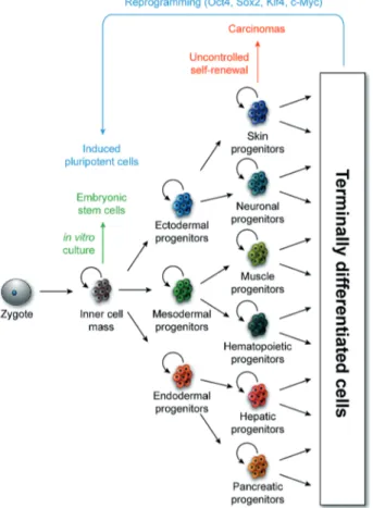
1. The fundamental questions in developmental biologyAccording to Aristotle, the first embryologist known to history, science begins with curiosity: “It is owing to wonder that people began to philosophize, and wonder remains the beginning of knowledge”. The development of an animal from a fertilized egg was a source of curiosity since antiquity. Therefore one of the fundamental issues in developmental biology was how does a single cell, the zygote give rise to a vast amount of different cell types (Figure 1). Developmental studies over the last few years have provided us with an understanding of cell families. The belief was that fertilized egg is a very plastic cell which divides and gives rise to embryonic stem cells (ESCs) that can progress in one direction along differentiation pathways, from totipotent, pluripotent or multipotent stem cells (SCs) to more differentiated cells: during this process cells became less and less plastic and somatic cell lineages do not differentiate across different embryonic-derived somatic lineages. Developmental biology is not limited to the study of the process by which a multicellular organism develops from one cell into an adult, but also to the mechanisms controlling cell replacement in adults, after injury or cell death.
Since all cells of a given organism contain the same DNA we need to understand how this same set of genetic instructions is regulated to produce this cell diversity and how cell plasticity is regulated. In addition to maternal and zygotic signaling molecules that initiate regulatory cascades, it is transcription factors (TFs) that act as switches to turn on or off gene expression or even modulate the precise expression output of a gene. Combinatorial control by multiple TFs working in performance can also confer cell type-specific regulation of target genes to produce specialized cell types. Interactions between multiple TFs at shared target genes result in complex gene regulatory networks, but understanding and unraveling these networks will help in understanding how different cell types arise and perhaps further the ability to direct cells to adopt a given fate. Towards this aim the basic question I have addressed in this study is how unique cell type, neural stem cell (NSCs) or neuroblast (NB), is specified during the construction of the embryonic central nervous system (CNS) of
Drosophila melanogaster. Understanding NSC plasticity is of fundamental importance, not
only for grasping development itself, but also for comprehending the pathogenesis of neurodevelopmental diseases, the initiation of neural tumors, and the therapeutic potential of
SCs. This is particularly important when considering the repair and regeneration of the nervous system (NS) after damage.
Figure 1. Progenitor cell self-renewal and differentiation contribute to tissue patterning and tumorigenesis.
Schematic representation of the self-renewal and differentiation of progenitor cells, in both normal and oncogenic contexts. Depicted derivatives of the primary germ layers are illustrative rather than
inclusive (Ari J. Firestone and James K. Chen,
2. Cell plasticity
In this section I will discuss successively the concept of cell plasticity and the innovative role of Drosophila NSCs in cell plasticity research.
2. 1. General aspects on cell plasticity
In general, cell plasticity refers to the ability of a cell to change its properties in response to intrinsic or extrinsic variations and the interactions between the two in order to generate cells with new properties. This process may be beneficial during development or when considering replacement of cells after injury, or problematic when taking in account their ability to generate cells with pathologic features. This concept is a necessary overall parameter in the definition of SC, which refers to the balance between self-renewal and differentiation but also to the ability of producing different cell populations (Lemischka, 2002).
The classical paradigm of cell plasticity, as described above, holds that all the cell lineages emerge from the most plastic cell known until now, the zygote, which gives rise to pluripotent cells during embryogenesis, and progressively to more restricted cells, in turn giving rise to the specialized cells of the different organs and tissues (Figure 1). In many tissues, self-renewing multipotent SCs are maintained in the adult and serve to replace cells that have a limited life span or to regenerate cells after injury or cell death
(Sell, 2004). Such SCs were believed to be restricted in their potential, and limited to generate the types of cells present in the tissue. Through these investigations the family tree for the generation of the major classes of cells in the body are provided. Thus it appears that plasticity
is lost progressively as development proceeds and the family trees of cells progress in one direction along these differentiation pathways and to be unable to switch tracks.
Cell plasticity
Dedifferentiation: it involves a terminally
differentiated cell reverting back to less differentiated stage from within its own lineage, allowing the cell to proliferate again before re-differentiating, leading to the replacement of those cells that have been lost
Transdifferentiation: this process sees cells
regressing to a point where they can switch lineages, allowing them to differentiate into another cell type.
Cell fate conversion: consists the changing the
fate of stem cells or their progeny from one fate to another
Reprogramming: aims to induce differentiated
cells into reverting to pluripotency. From here, they can differentiate into almost any cell type.
Specification: changes involved in the progressive diversification of the structure and function of cells. It concerns the acquisition of the characteristics that allow different cell types to perform their functions.
2. 2. Cell fate determinants and cell plasticity
The notion of progressive limitation of cell plasticity with development became completely blasphemous, when the role of TFs in lineage specification emerged, starting from 1980s, when Pr. Harold Weintraub’s and colleagues discovered that forced expression of MyoD, a TF that determines muscle cell fate, can induce myotube formation in a fibroblast cell line (Davis et al., 1987). Subsequently, cell fusion, consisting the combination of several uninuclear cells to form a multinuclear cell, and nuclear transfer, involving the injection of a defined nucleus in a cell lacking its own nucleus, have shown that the epigenome of differentiated cells can be remarkably plastic (Blau, 1989; Gurdon and Byrne, 2003; Gurdon and Melton, 2008; Hochedlinger and Jaenisch, 2002; Wilmut et al., 1997). Today, cell reprogramming can be obtained simply through ectopic expression or lose of function of defined TFs, known as cell fate determinants or master gene regulators (Davis et al., 1987; Graf and Enver, 2009; Kulessa et al., 1995). Using this strategy, the identity of differentiated cells can be fully reversed and it even makes it possible to produce pluripotent SCs-like from fully differentiated cells “reprogramming”, by simply expressing a cocktail of defined TFs
(Takahashi and Yamanaka, 2006).
The instructive role of TFs in lineage specification came from the diversity of SC progenitors; the best-studied example is provided by the hematopoietic SCs and their progenitors. For example, the overexpression of the erythroid-megacaryocyte-affiliate TF GATA1 forces macrophage precursors to express erythroid-megacaryocyte lineage markers and to repress the macrophage ones (Kulessa et al., 1995; Visvader et al., 1992). Conversely, the ectopic expression of PU.1 in an erythroid-megacaryocyte cell line induced its conversion into the monocytic lineage (Nerlov and Graf, 1998). The impact of TFs on lineage specification was also demonstrated in the NSCs progenitors. In invertebrates as well as in vertebrates, distinct types of neural progenitor cells generate neurons and glial cells, and in some cases common precursors are shared between the two differentiated cells (Fietz and Huttner, 2011; Fietz et al., 2010; Gotz and Huttner, 2005; Hansen et al., 2010; Lui et al., 2011; Reillo et al., 2011). Interestingly, a single TF specifying a given neural progenitor cell identity can also convert a defined type of neuronal progenitor cell from one type into another. Indeed, the misexpression of T-brain gene-2 forces radial glial (RG) cells, NSCs-like, to produce another type of progenitors, intermediate progenitor cells (IPCs) (Farkas et al., 2008; Sessa et al., 2008). In Drosophila CNS, my model of study, one TF Glial Cell Missing/Glial
able to force NSCs to adopt a glial fate at the expense of the neuronal one, when ectopically expressed in the whole neurogenic region (Bernardoni et al., 1998; Hosoya et al., 1995; Jones et al., 1995; Vincent et al., 1996).
The facility through which cell fates can be experimentally modified raises the question as to whether such events occur physiologically or in the context of disease. Arguably, in Mammals, Schwann cells possess a natural regenerative capacity called dedifferentiation. Following damages to the nerves they are associated with, Schwann cells dedifferentiate and proliferate (Chen et al., 2007). Another nice example is the transdifferentiation of ectodermal cells into mesodermal cells during gastrulation (Slack, 2007; Yang and Weinberg, 2008). In the case of cell plasticity and pathology, several types of metaplasia have been attributed to transdifferentiation (Slack, 2007), and epithelial mesenchymal transitions may be involved in the formation of metastatic breast cancers (Yang and Weinberg, 2008). Here, as during normal epithelial mesenchymal transitions, the activation of key TFs is essential, like Twist and Snail (Slack, 2007; Yang and Weinberg, 2008).
With the rapidly growing of lineage tracing tools I guess that many more physiological or pathological cell fate conversion events will be discovered in the future.
2. 3. Stem cell plasticity research
SC plasticity research is the most fascinating chapter in the history of biology. Traditionally restricted to the field of developmental biology, SC plasticity has become of increasing interest for biomedical research in more recent years. Indeed, the advances in SC research help us to understand the mechanisms underlying cancerougenesis and neuronal cell degeneration with aging. SC research aims to understand and treat such heavy pathologies. However, a major concern in protocols aiming on cell replacement is safety. This new concept was the subject of controversy because of the possibility of various experimental biases, including the possible presence of contaminating SCs, pluripotent SCs of any adult or cell fusion of SCs with differentiated cells that will be used as a treatment (Lakshmipathy and Verfaillie, 2005). Following this episode, more attention has been paid to the characterization of SCs, their fractionation and the study of their biology.
2. 3. 1. Properties of stem cells
SCs are the foundation for every organ, tissue and cell in the body and are characterized by unique defining properties. As previously mentioned, these cells are
proliferative precursor cells characterized by their ability to self-renew while generating a large number of progeny committed to differentiation. They can either divide symmetrically, producing two identical daughter cells, or asymmetrically producing one “identical” and one more differentiated daughter cell (Lin and Schagat, 1997). SCs are also this population of cells that face the consequence of aging, by changing their potential to differentiate into different cell types and controlling their cell cycling by entering cell cycle arrest, the quiescent phase, or dying under programmed cell death, also called apoptosis. These cells do not only exist during the embryonic life (ESCs), but also in major differentiated tissues of an adult organism (adult SCs), where they play a central role in tissue growth and maintenance
(Reya and Clevers, 2005; Yao et al., 2012).
2. 3. 2. Drosophila embryonic NSCs as a model system to study cell plasticity
Drosophila melanogaster is an extremely powerful model system for identifying and
analyzing complex biological processes in the context of a living organism. It was thoroughly demonstrated that the processes regulating fundamental aspects of animal development and physiology are well conserved, and that insights gained from studies in Drosophila can with high likelihood be transferred to other species. For example, developmental genes such as the Hox genes that play essential roles in setting up the vertebrate body axis were originally identified and well characterized in Drosophila. About 75 % of known human disease genes have a recognizable match in the genome of fruit flies (Reiter et al., 2001), and 50 % of fly protein sequences have mammalian orthologs. Today, Drosophila is used as a genetic model for several human diseases including neurodegenerative disorders. It is also used to study mechanisms underlying aging, immunity, diabetes and cancer, as well as drug abuse.
Specifically, the NSCs of Drosophila provide an excellent system to study the mechanisms regulating SC plasticity. As in mammals, the NSC generally called NBs divide to produce new SCs and daughter cells that go through a well-characterized cascade of differentiation steps to develop into neurons or glial cells. In the Drosophila embryonic CNS, we know the position and the identity of all NSCs, and their progenies. A variety of molecular markers and tools have been identified to study NSCs, as well as the neurons and glial cells. Most importantly, the genetic tractability of Drosophila allows for identifying genes regulating NSC function and plasticity (for more details see Introduction 4. 1.).
2. 4. Broad questions in cell plasticity
Cell fate transformations has changed the way that we view cell plasticity and how we can apply converted cells to regenerative medicine. However, several important questions should be first resolved before using these cells in therapy that I summarized in two questions:
1/ How can transcription factor induce a new program while repressing another? 2/ Is the identity of generated cells completely identical to the desired cell fate?
3. The development of embryonic nervous system
3. 1. The Development of Drosophila nervous system
The duration of Drosophila life cycle is influenced by the temperature; at 25°C, it takes around 10 days: one day of embryogenesis, which is divided into different seventeen stages where the majority of structures are generated, followed by three successive larval stages, which take around four days, and five days of pupal life where metamorphosis occurs to generate the adult fly.
3. 1. 1. General structure of the nervous system
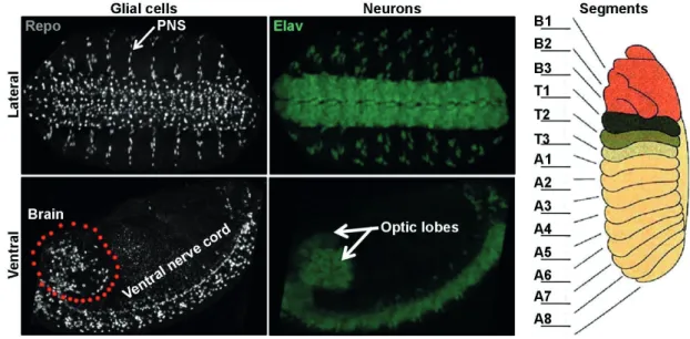
Drosophila NS is composed of three parts: the CNS which is composed of two parts:
the brain and the ventral nerve cord (VNC) (the equivalent of the spinal cord in vertebrates), the peripheral NS (PNS) (Figure 2, left panel) and the stomatogastric NS (Hartenstein et al., 1994). The stomatogastric NS and PNS will not be described.
Figure 2. Structure of The Drosophila nervous system
The left and middle panels show the profile of neurons (stained with Elav in green) and glia (stained with Repo in grey) in the nervous system of stage 16 embryos.
The right panel shows the position of the different segments (the cartoon showing the profile of segments is adapted from http://www.zoology.ubc.ca/~bio463/lecture_13.htm).
A large portion of nerve cells is found within the brain and the VNC. The brain contains two optic lobes (OL) and the central brain (CB), including the mushroom bodies
PNS, which entails all sensory neurons, as well as the neurons controlling the functions of various organs. Like the rest of the arthropod body, the CNS of Drosophila is segmented into subunits, called segments or neuromeres. The brain contains three segments B1, B2 and B3, which correspond to the prospective protocerebrum, deutocerebrum, and tritocerebrum, respectively. The VNC contains three thoracic (T1, T2 and T3), and eight abdominal (A1, A2, A3, A4, A5, A6, A7 and A8) segments (Figure 2, Right panel). Due to its simplicity the VNC was so far the principal model for the studies on the molecular mechanism behind cell fate specification in the Drosophila CNS, and henceforth it will also be the principal focus of my thesis work.
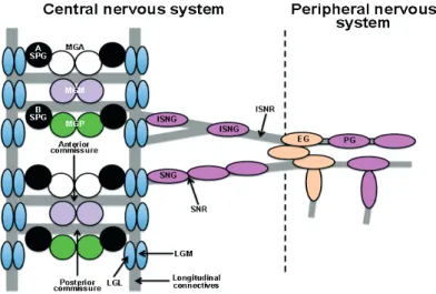
In the VNC, most neuronal axons are organized in a simple, ladder-like pattern. Within each segment, two horizontal commissural axon tracts (anterior and posterior commissures) cross the midline to connect the two longitudinal connectives, linking adjacent neuromeres to one another along the Anterior/Posterior (A/P) axis. Axon bundles and their associated glia constitute the neuropile. Exiting the longitudinal connectives at each side of the neuromeres there are two peripheral nerves, the segmental (SNR, at the level of the anterior commissure) and the intersegmental (ISNR, posterior to the posterior commissure) nerves, which are made up of peripherally projecting motor axons and centrally projecting sensory axons (Figure 3).
Figure 3. Embryonic CNS and PNS axon pathways and pattern of glial cells.
Midline glia (MG) surround anterior and posterior commissures, longitudinal glia (LGL and LGM, L for lateral and M for medial, respectively) arranged in two parallel rows along the longitudinal connectives. For more details see the text.
3. 1. 2. Early neurogenesis in Drosophila embryos
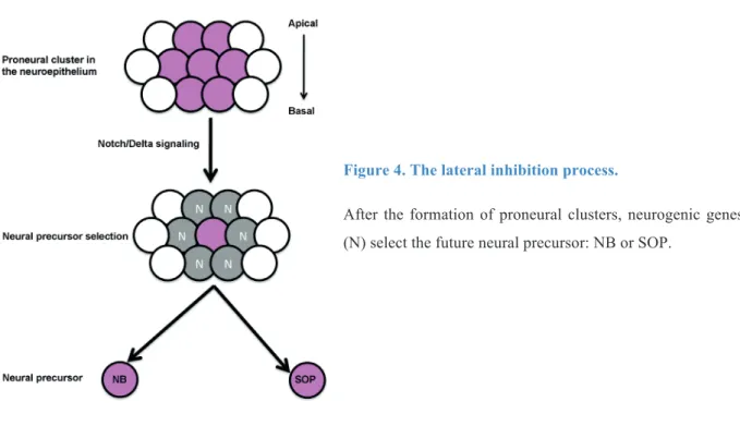
The embryonic life of Drosophila starts with the formation of three basic germlayers, the ectoderm, the mesoderm and the endoderm. The NS arises from the ectoderm, a layer that also gives the epidermis, the trachea and the hindgut. At the beginning of embryogenesis, the ectoderm is subdivided into a ventral neurogenic and a dorsal non-neurogenic region by the antagonistic activity of the secreted molecules Decapentaplegic (Dpp) and Short gastrulation (Sog) (Francois and Bier, 1995; Francois et al., 1994). The neurogenic ectoderm (neuroectoderm) starts as a simple epithelium sheet composed of proliferative cells. This region gives rise to the neural progenitors (NSCs or NBs) or sensory organs precursors (SOPs) and to the epidermal precursor cells.
Figure 4. The lateral inhibition process.
After the formation of proneural clusters, neurogenic genes (N) select the future neural precursor: NB or SOP.
The proneural genes control the position and the time at which groups of neuroectodermal cells, called proneural clusters, become competent to form a NSC (Ghysen and Dambly-Chaudiere, 1989), whereas the neurogenic genes control the cell interactions that prevent more than one cell in the group from developing into a NB, this process is called “lateral inhibition” (Figure 4) (Lehman, 1983). The proneural genes codes for TFs having a basic Helix Loop Helix (bHLH) domain (Campuzano et al., 1985; Villares and Cabrera, 1987) and they are represented by the three or the four members of Achate-Scute Complex: Achete, Scute and Letal of Scute (Cabrera et al., 1987; Martin-Bermudo et al., 1991). For the
which represents the receptor, and its ligand Delta. About 30 NBs delaminate from the neuroectoderm per thoracic and abdominal hemisegment (Broadus et al., 1995; Doe, 1992). The remaining cells of the neurogenic region remain superficial and generate the ventral epidermis.
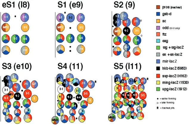
NBs delaminate from the surface in five successive waves (S1-S5) along the M-L and A-P axes in rows and columns in a stereotyped spatiotemporal pattern (Figure 5). NBs are given numerical designations according to their definitive position. The numbers consist of two indexes; the first index indicates the A-P position, and the second one indicates the M-L position of the NB. Thus, for example the NB6-4T is the fourth NB from the ventral midline in row 6 after the formation of all NBs (Figure 5). For more precision, “T” and “A” latters are added to distinguish the thoracic from the abdominal NBs at the same positions, respectively. Even if the name of some NBs is the same in the abdomen and the thorax, the progeny of these cells may differ. For example the thoracic NB6-4T generates neurons and glia, whereas the abdominal one generates only glia (Berger et al., 2005; Schmidt et al., 1997).
Figure 5: Drosophila neuroblasts map
The 30 NBs per hemisegment are generated in five sequential waves. Each NB is generated at a stereotyped time and position, and displays a unique expression profile of molecular markers.
By embryonic stage 9-11, approximately 75-80 NBs are formed. To build the CNS, each NB undergoes repeated asymmetric cell divisions to renew themselves while producing intermediate precursors, called ganglion mother cells (GMCs). The GMC divides once more to give two daughter cells that differentiate into neurons and/or glial cells (Chia et al., 2008; Matsuzaki, 2000; Urbach and Technau, 2004; Younossi-Hartenstein et al., 1996). As the neural progenitors produce progeny they change their competence over time in a step-wise manner, generating different types of cells at specific time points. One identified mechanism behind such competence transitions is the progenitor-intrinsic sequential expression of the so-called “temporal genes” (see Introduction 5. 1. 2).
The size of the NBs gradually decreases upon each division and towards the end of embryogenesis, some NBs stop dividing and enter a stage known as quiescence (Truman, 1990), whereas the rest are eliminated via programmed cell death (Prokop and Technau, 1991; Truman and Bate, 1988). The only NBs that do not undergo quiescence or programmed cell death at the end of embryonic life are the four OL/MB NBs, which generate very large lineages of 500 neurons each, and one less-well characterized V-L CB NB (Ito and Hotta, 1992; Lee et al., 1999; White and Kankel, 1978).
3. 1. 3. Cells making Drosophila CNS
Three cell types compose the CNS, the NBs, the neurons and the glial cells. The NBs are the founding cells of the CNS. Neurons are specialized in transmitting signals between different cell populations within the body, while glia provides insulation to neurons by controlling extracellular homeostasis and acting as NS immune cells.
• Neuroblasts
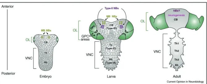
These cells are first generated during the embryonic life from the neuroectoderm. They are detectable in the VNC and the CB/MB, and serve to generate the CNS of the larva, which contributes to 10 % of neurons in the adult CNS. Contrarily to the VNC and the CB NBs, the OL NBs are generated during the larval life from the neuroepithelial placodes. During the postembryonic life, some reactivated embryonic NBs together with the larval NBs contribute to the generation of the remaining 90 % of adult neurons. (Reviewed by (Egger et al., 2008; Skeath and Thor, 2003; Sousa-Nunes et al., 2010)). See Figure 6 for the distribution of NBs at different developmental stages.
Figure 6. NBs distribution in the developing Drosophila CNS.
Representation of embryonic, late-larval and adult CNS, highlighting NBs (circles). The OL (green) is subdivided into the Glial Precursor Cell (GPC) areas and the Outer Proliferation Centre, which generates OPC NBs. For clarity, the Inner Proliferation Centre (IPC) is not shown. The CB/MB contains numerous Type-ID NBs, four Mushroom Body (MB) NBs and eight Type-II NBs. The VNC is subdivided into Thoracic (Th) segments and Abdominal (Ab) segments containing Type-ID and Type-IA NBs respectively. Note that no identifiable NBs are present in the adult CNS. Adapted from (Sousa-Nunes at al., 2010).
While adult NSCs appear to be common in vertebrates, the situation in Drosophila is much less clear. It was thought that the NBs that generate the CNS of adult Drosophila stop division, undergo apoptosis, or differentiate before eclosion (Bello et al., 2003; Ito and Hotta, 1992; Maurange et al., 2008; Truman and Bate, 1988). However, two recent reports identified small numbers of dividing cells in the adult brain and the majority of these cells express a glial marker, Reversed Polarity (Repo) (Kato et al., 2009; von Trotha et al., 2009). Interestingly, it was also reported in vertebrate that NSCs from the adult hippocampus might eventually differentiate into postmitotic astrocytes (type of glial cells), a process that would explain the loss of SCs and reduction in neurogenesis with age (Encinas et al., 2011). Evidence that astrocytes could hold the capacity to dedifferentiate into RG cells, and even immortalized cells, that could induce gliomas was produced by several labs (Dufour et al., 2009; Jiang and Uhrbom, 2012; Sharif et al., 2007). It was also reported that NSCs have astrocyte characteristics, (reviewed by (Bergstrom and Forsberg-Nilsson, 2012). All together, these results suggest that glial cells and adult NSCs may share the expression of some markers. Might Repo be a shared marker between glia and adult NBs in Drosophila brain? Is
it possible that adult NBs express other markers that are not yet identified? I believe that lineage tracers and the simplicity of Drosophila NS will soon resolve these questions.
• Glial cells
The term glia means "glue," a reflection of the fact that glial cells really do hold the brain together, occupying the space between neurons. In the developing CNS of the
Drosophila embryos, glial cells derive from two different germlayers. A small part of these
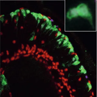
cells, midline glia, derives from mesectodermal progenitors and ensheath the commissural fiber tracts (Bossing and Technau, 1994; Menne and Klambt, 1994). The major part, the lateral glial cells, derives from the neurogenic region of the ectoderm. The first event in the determination of lateral glia is the transient expression of the TF Gcm (see Introduction 5.). Gcm is known to initiate the glial fate by activating downstream targets, which accomplish the differentiation and the maintenance of the glial fate. Among the Gcm target genes is repo, which codes for an homeodomain protein “Repo”, that is expressed in all lateral glial cells and is used as a general marker for these cells (Figure 7).
The lateral glial cells of the embryonic VNC are subdivided into three categories according to their association with the basic compartments of the CNS: the surface, the cortex, and the neuropile, (Figure 4) (Ito, 1995; Meyer et al., 1987). The group of surface-associated glia includes two subgroups: the subperineurial glia (SPG) that lie underneath the outer surface of the CNS, and the channel glia (CG), which are positioned along the dorsoventral channels, demarcating the borders between segmental neuromeres of the VNC. In the category of cortex-associated glia, that amalgamates between the neuronal cell bodies in the cortex, only one subtype is described in the embryonic VNC: the cell body glia (CBG). The third category, the neuropile-associated glia, includes the glial cells that are associated with axonal structures. Two subtypes were proposed in the embryonic VNC: the nerve root glia (NRG), which is further subdivided into intersegmental and segmental nerve root glia (ISNG and SNG, respectively), and the interface glia (IG), which are associated with the longitudinal connectives and are also called longitudinal glia (LG), (Beckervordersandforth et al., 2008; Ito, 1995). Using a combination of molecular markers, the NSCs generating each lateral glial cell have been identified (Beckervordersandforth et al., 2008).
Figure 7. Spatial distribution and classification of glial cells in the VNC.
Pattern of glial cells in an abdominal neuromere at embryonic stage 16. (A–C) Horizontal views of a preparation showing nuclear anti-Repo staining (anterior to the top; midline indicated by dashed line), and (A’–C’) corresponding cartoons at dorsal (A and A’), intermediate (B and B’), and ventral layers (C and C’) as indicated
by black frames in cartoons of frontal view (A’’–C’’; dorsal to the top) (Beckervordersandforth et al., 2008).
• Neurons
Three basic types of neurons are present in the NS of Drosophila: moterneurons, interneurons and neurosensory neurons. Moterneurons extend axonal projections out into the periphery to innervate the muscles. There are about 30 moterneurons per hemisegment. The interneurons extend axons within the CNS to innervate other neurons. To this class belong a total of about 300 interneurons, which can be subdivided into two subclasses: intersegmental interneurons, whose axon projections extend between segments within the CNS, local interneurons with axon projections terminate within their segment of origin in the CNS, and finally, neurosensory neurons which extend axons either out into the periphery or into the seat of the CNS to secrete neuropeptides and hormones. A total of about 10 cells have been identified in each hemisegment (Schmid et al., 1999).
3. 2. The development of vertebrate nervous system
The aim of this chapter is not to illustrate the vertebrate neurogenesis but to describe some structures and cell types that will help to understand the following parts. Similar mechanisms involved in fly and vertebrate neurogenesis will be underlined.
3. 2. 1. General structure of the nervous system
The NS of vertebrates has two main divisions: the CNS, consisting of the brain and the spinal cord, and the PNS. The brain consists of three major divisions, organized around the three chambers of the neural tube that develops early in embryonic life: the forebrain, the midbrain, and the hindbrain (Figure 8A).
Figure 8.
(A) Structure of vertebrate central
nervous system.
(B) The principal regions of the
embryonic and adult nervous system from which neural stem cells have been
isolated (Temple, 2001).
3. 2. 2. Early neurogenesis in vertebrate embryos
The vertebrate CNS derives from the neural plate, an epithelial sheet that arises from the dorsal ectoderm of the gastrula-stage embryo (Lee and Jessell, 1999). As in Drosophila, the vertebrate ectoderm is subdivided into neurogenic and non-neurogenic region by the antagonistic activities of two secreted molecules, Bone Morphogenic Protein 4 (BMP4) and Chordin, the orthologs of Dpp and Sog in Drosophila, respectively (Arendt and Nubler-Jung, 1997; De Robertis and Sasai, 1996; Lichtneckert and Reichert, 2005).