HAL Id: tel-02269303
https://tel.archives-ouvertes.fr/tel-02269303
Submitted on 22 Aug 2019HAL is a multi-disciplinary open access archive for the deposit and dissemination of sci-entific research documents, whether they are pub-lished or not. The documents may come from teaching and research institutions in France or abroad, or from public or private research centers.
L’archive ouverte pluridisciplinaire HAL, est destinée au dépôt et à la diffusion de documents scientifiques de niveau recherche, publiés ou non, émanant des établissements d’enseignement et de recherche français ou étrangers, des laboratoires publics ou privés.
transcription factor NusA
Xieyang Guo
To cite this version:
Xieyang Guo. Regulation of transcription : structural studies of an RNA polymerase elongation complex bound to transcription factor NusA. Genomics [q-bio.GN]. Université de Strasbourg, 2018. English. �NNT : 2018STRAJ071�. �tel-02269303�
UNIVERSITÉ DE STRASBOURG
ÉCOLE DOCTORALE des Sciences de la Vie et de la Santé
IGBMC – CNRS UMR 7104 – Inserm U 964
THÈSE
présentée par :
Xieyang GUO
Soutenue le: 4 Septembre 2018
pour obtenir le grade de : Docteur de l’université de Strasbourg
Discipline/ Spécialité : Biophysique et biologie structurale
(Biophysics and Structural biology)
Regulation of transcription:
Structural studies of an RNA polymerase elongation
complex bound to transcription factor NusA
Régulation de la transcription :
études structurales du complexe d’élongation de l'ARN
polymérase lié au facteur de transcription NusA
THÈSE dirigée par :
Dr. WEIXLBAUMER Albert Chargé de recherches, IGBMC, France RAPPORTEURS :
Prof. DARST Seth A. Professeur, The Rockefeller University, USA
Prof. MECHULAM Yves Directeur de recherches, École Polytechnique, France AUTRES MEMBRES DU JURY :
Dr. ROMBY Pascale Directeur de recherches, IBMC, France
2
Acknowledgements
First and foremost, I would like to express my sincere gratitude to my thesis advisor Dr Albert Weixlbaumer. You are a fantastic mentor to me, it has been an honor to be your first PhD student. Thank you for being supportive, encouraging and inspiring throughout the past four years. I could not have imagined having a better supervisor for my PhD study.
Besides my advisor, I would like to thank Professor Seth A. Darst, Professor Yves Mechulam, Dr Pascale Romby, Dr Valérie Lamour for accepting to be members of my thesis committee. Thank you all for taking the time to read and evaluate my thesis.
Special thanks goes to both past and current members of the Weixlbaumer lab. I want to thank Maria for always being there to help and to talk to whenever I needed; Claire for feeding us with baked goods and being my French interpreter making life here easier; Jinal for teaching me the fun of doing cloning; Chengjin for collaboration on NusA-NusG complexes; Charlotte, Ayesha, Kasimir and Qiancheng, for all the fun moments inside and outside of the lab; Dmitro, Sanjay and Moamen for all the discussion/advice. I will always cherish the time I spent with all of you in the lab.
I am indebted to all the people that have helped and advised me during my study. I want to thank Corinne for getting me started with cryo-EM; Julio for training me to use the microscope; Sasha, Gabor and Grigory for teaching me image processing; Patrick and James for the useful advice on EM project; Alastair, Morgan and Pierre for helping me at the crystallization platform; Adam for intriguing discussion and spreading your enthusiasm for science. I also want to thank Arnaud for all the endless discussion and sharing of our PhD
journey; Katka for being a great presentation rehearsal partner; Iskander and Irina for all the lunch shop talks and supports.
My time in Strasbourg was made enjoyable in large part due to the many friends and groups that became a part of my life. I want to thank my climbing group Sandrine, Alastair, Yassmine, Moyra, Renee, Jerome, Pierre, Matthias for all the fun times on the wall; my skiing party Iskander, Irina, Grigory, Arnaud, Katka, Melanie, Laurent, Stephanie, Pauline, Perrine for all the fantastic winters on the slope, my poster team Nicla, Katka, Robert, Syrine for all the interesting and successful sessions; my Chinese gang Wenjin, Changwei, Zhirong, Tanbing, Ruichen and Guoqiang for all the wonderful food gatherings; my skydiving buddies Nicolas and Benjamin for all the adrenaline rushes; my forest friend Gilles for the long hiking and cycling trips; as well as Anna, Alexey, Arantxa, Daniya, Jonathan, Karima, Löic, Lyuba, Marina, Matej, Pernelle for sharing all the great moments with me.
I am most grateful to my parents, my grandmother and my sister for their unconditional love and support, without which this work would never have been accomplished. Thank you for always believing in me and letting me follow my passion and dreams.
I want to dedicate this thesis to my beloved grandmother, who passed away before the completion of my study. I know you would be proud and I will forever be grateful for the knowledge and values you instilled in me.
4
Table of Contents
Acknowledgements ... 2 Table of Contents ... 4 Abstract ... 5 Résumé de thèse ... 6 List of Abbreviations ... 21 List of Figures... 22 Introduction ... 24 1. Overview of transcription...24 2. Transcriptional pausing ...282.1. Biological function of transcriptional pausing ...28
2.2. Current model for transcriptional pausing ...30
2.3. Mechanistic insights into class I pause ...33
2.4. In vitro experimental system to reconstitute a Class I paused complex ...35
3. RNA Polymerase ...37
3.1. Core architecture of RNAP ...37
3.2. Structure of bacterial RNAP ...38
3.3. Structure of eukaryotic RNAP II...39
3.4. Similarity between bacterial and eukaryotic RNAP structures ...41
3.5. Structural organization of the transcription elongation complex ...42
3.6. Structure of an elemental paused elongation complex ...44
4. Transcription elongation factors NusA ...45
4.1. Effects of NusA on transcription ...45
4.2. Structure of NusA and its interactions with the EC ...47
4.3. Role of NusA in lambda N-mediated antitermination ...50
4.4. NusA enhancement of hairpin pause and intrinsic termination ...51
Aims ... 54
Results ... 57
Structural basis for NusA stabilized transcriptional pausing (Guo et al., Molecular Cell, 2018) ...57
Discussion ... 94
Hairpin-stabilized transcriptional pausing ...94
RNAP translocation ...97
Transcription regulation by NusA ...98
A model for entering the his pause ... 102
Conclusions and Perspectives ... 106
Abstract
Transcriptional pausing by RNA polymerases (RNAPs) is a key mechanism to regulate gene expression in all kingdoms of life and is a prerequisite for transcription termination. The essential bacterial transcription factor NusA stimulates both pausing and termination of transcription, thus playing a central role. During my thesis, with an aim to understand the mechanistic basis of transcriptional pausing and its regulation by NusA, I carried out structural studies of a paused E. coli RNAP elongation complex at the well-characterized his pause site stabilized by NusA. My thesis work resulted in single-particle electron cryo-microscopy reconstructions of NusA bound to paused elongation complexes with and without a pause-enhancing hairpin in the RNA exit channel. The structures reveal four interactions between NusA and RNAP that suggest how NusA stimulates RNA folding, pausing, and termination. An asymmetric translocation intermediate of RNA and DNA converts the active site of the enzyme into an inactive state, providing a structural explanation for the inhibition of catalysis. Comparing RNAP at different stages of pausing provides insights on the dynamic nature of the process and the role of NusA as a regulatory factor.
6
Résumé de thèse
Introduction
L'ADN est transcrit en ARN par une protéine appelée ARN polymérase (RNAP) dans les trois règnes du vivant. Dans les bactéries, cinq sous-unités protéiques (α2ββ'ω) forment un core avec une architecture universellement conservée et abritent les sites fonctionnellement pertinents. La transcription est divisée en phases d'initiation, d'élongation et de terminaison.
Au cours de l'élongation, la RNAP se déplace le long de l'ADN, progressant à travers plusieurs états. Cependant, l'élongation est fréquemment interrompue par des états déconnectés non fonctionnels, qui sont en compétition avec l'addition de nucléotides. Ce sont des pauses transcriptionnelles. La mise en pause régule l'expression des gènes à plusieurs niveaux: (1) elle affecte les taux de synthèse de l'ARN et synchronise la transcription et la traduction (Landick et al., 1985); (2) elle facilite le repliement de l'ARN (Pan et al., 1999); (3) elle permet la liaison de facteurs de transcription aux complexes d'élongation (Artsimovitch and Landick, 2002); (4) elle joue un rôle dans la régulation par les riboswitches (Wickiser et al., 2005); (5) c'est une condition préalable à la terminaison (Gusarov and Nudler, 1999; Kassavetis and Chamberlin, 1981); et (6) il s'agit d'une étape importante et limitante de l'élongation précoce de nombreux gènes chez les métazoaires (Core and Lis, 2008). La pause est déclenchée par la séquence d'ADN sous-jacente et les études à l'échelle du génome ont permis d'identifier une séquence de pause consensus chez
E. coli. Les études cinétiques suggèrent que la pause se produit à travers au moins deux
(ePEC : elemental Pause Elongation Complex). Les pauses de classe I sont stabilisées par des structures naissantes d'ARN en épingle à cheveux dans le canal de sortie de la RNAP. Les pauses de classe II entraînent le recul de la RNAP le long de l'ADN (état "backtracked") (Artsimovitch and Landick, 2000). L'une des pauses de classe I les mieux caractérisées se trouve dans la région leader de l'opéron his de E. coli (his-pause). La his-pause synchronise la transcription et la traduction et un réarrangement de site actif a été proposé pour expliquer l’inhibition de l'addition de nucléotides (Toulokhonov et al., 2007). Les facteurs d'élongation de la transcription modulent davantage les pauses. L'un de ces facteurs essentiels, qui est conservé chez les bactéries et les archées, est appelé NusA (Ingham et al., 1999; Shibata et al., 2007). NusA a été étudié pendant plus de 40 ans et son gène a été identifié comme étant indispensable pour l'antiterminaison à médiation par la protéine N du phage lambda (Friedman et al., 1974). De même, NusA est un composant des complexes d’antiterminaison requis pour la transcription des ARNr (Vogel and Jensen, 1997). NusA a également été décrit comme facilitant le repliement de l'ARN (Pan et al., 1999). Cependant, le rôle le plus évident de NusA est de stimuler les pauses de classe I (stabilisées par une épingle à cheveux) et la terminaison intrinsèque et Rho-dépendante (Artsimovitch and Landick, 2000). Bien que des approches structurelles aient été utilisées pour faire la lumière sur les divers rôles de NusA dans le complexe avec la RNAP (Said et al., 2017; Yang et al., 2009), nous manquons d'information à haute résolution pour acquérir une connaissance mécanistique approfondie .
NusA est une protéine flexible avec plusieurs domaines (Worbs et al., 2001). Le domaine N-terminal (NusA-NTD) est nécessaire et suffisant pour améliorer la mise en pause (Ha et al., 2010). Il a été proposé une interaction avec la FTH ("flat-tip helix") de la sous-unité β et avec la boucle en épingle à cheveux de l'ARN réhaussant la pause (Ha et al., 2010;
8
Mah et al., 1999; Yang et al., 2009). Au centre de la protéine se trouvent trois domaines de liaison à l'ARN (S1 et deux domaines K-homologie, KH1 et KH2). Certaines espèces bactériennes, y compris E. coli, ont deux répétitions acides C-terminales (AR1, AR2). AR2 peut lier le domaine C-terminal de l'une des sous-unités α de la RNAP (α-CTD). Cette liaison libère une interaction auto-inhibitrice entre AR2 et KH1 et favorise la liaison de l'ARN à NusA (Mah et al., 2000; Schweimer et al., 2011).
La cristallographie aux rayons X a fourni des informations sur les intermédiaires en pause. Cela inclut les RNAP II eucaryotes à l’état "backtracked" et les RNAP bactériennes proposées comme étant en pause élémentaire (Cheung and Cramer, 2011; Wang et al., 2009; Weixlbaumer et al., 2013). Cependant, nous manquons d'une compréhension de la stabilisation de la pause à médiation par épingle à cheveux et de sa modulation par des facteurs de transcription. L'extrusion du site actif de l'extrémité 3 'de l'ARN interrompt la transcription dans une pause de classe II, mais nous ne savons pas ce qui stoppe la catalyse dans une pause de classe I.
Résultats
Ici, nous rapportons une structure cryo-EM de 3,6 Å d'un complexe d'élongation (CE) stabilisé par une épingle à cheveux lié au facteur essentiel d'élongation de la transcription NusA (hisPEC-NusA, Figure 1). Un sous-ensemble de particules nous a permis d'obtenir une reconstruction à 4,1 Å d'un complexe en pause sans épingle à cheveux et avec une liaison plus lâche de NusA (PEC-NusA). La structure hisPEC-NusA explique pourquoi la catalyse s'arrête à l'état de pause, donne un aperçu de sa nature dynamique et nous permet de proposer comment NusA prolonge la pause. La structure fournit également
une opportunité de spéculer sur la translocation de la RNAP et sur le rôle de NusA dans la terminaison de la transcription.
Figure 1(A) Vue d'ensemble de la structure cryo-EM hisPEC-NusA avec la RNAP en gris, l’ADNt en orange, l’ADNnt en bleu, l’ARN en noir et NusA en enveloppe jaune.
(B) La densité cryo-EM représentative (maille bleue) pour l'hybride ARN-ADN et l'ARN en épingle à cheveux est montrée avec un modèle de dessin animé superposé (ADNt orange, ADN nt bleu, ARN noir).
(C) La densité cryo-EM représentative (maillage bleu) pour le site actif a révélé des chaînes latérales du pont-hélice (rose) et des nucléotides de l'ADNt (orange) et de l'ARN (noir).
Notre structure a révélé quatre points d'interaction protéine-protéine entre le hisPEC et NusA (Figure 2A).
Premièrement, NusA-CTD interagit avec l’1-CTD de la RNAP en accord avec les résultats précédemment publiés (Figure 2A)(Mah et al., 2000; Schweimer et al., 2011).
Deuxièmement, NusA-NTD se lie au 2-CTD (Figure 2B). Cette interaction est cohérente avec les résultats des études de titration RMN (Drögemüller et al., 2015), sauf qu'elle a été interprétée comme une interaction entre la sous-unité ’ et la tête NusA-NTD (hélice α3, feuillets β3, β4 et partie N-terminale de β2). Nous avons confirmé la pertinence
10
fonctionnelle de cette interaction pour améliorer la pause par épingle à cheveux grâce à l’étude cinétique d’un mutant avec un délétion du -CTD. Il augmente vraisemblablement l'affinité globale et restreint la liberté conformationnelle de NusA-NTD.
Troisièmement, l'extrémité C-terminale de la sous-unité de la RNAP se lie entre les deux domaines KH de NusA (Figure 2A). En accord avec cette observation, la troncature des deux domaines KH affecte l'affinité de NusA pour la RNAP mais ne fonctionne pas chez E. coli (Ha et al., 2010). Fait important, la liaison de l'ARN aux domaines KH, mise en évidence dans les structures cristallines précédentes, nécessite probablement de rompre cette interaction (Beuth et al., 2005; Said et al., 2017).
Quatrièmement, le NusA-NTD lie le FTH de la RNAP, ce qui a également été établi par des expériences de pontage, de microscopie électronique à basse résolution (coloration négative), de RMN et de mutagenèse (Ha et al., 2010; Ma et al., 2015; Toulokhonov et al., 2001; Yang et al., 2009). En outre, la suppression de la FTH a aboli l'effet de renforcement de la pause de NusA-NTD (Ha et al., 2010; Toulokhonov et al., 2001). Notre structure est cohérente et fournit une image plus détaillée de l'interaction de FTH avec une poche hydrophobe dans NusA-NTD (Figure 2B). Les mutations R104A et K111A dans l'hélice NusA 4 ont conduit à une perte totale de l'activité NusA pour améliorer la pause (Ma et al., 2015). Bien que non résolu dans notre reconstruction, K111 pourrait interagir avec la FTH tandis que R104 pourrait interagir avec l'un des connecteurs reliant la FTH au domaine "flap" (Figure 2B).
Figure 2 (A) Représentation en surface de hisPEC-NusA. Sous-unité 1 de la RNAP (vert forêt), sous-unité 2 (vert clair), sous-unité (rose clair), flap-tip hélice β (cyan), NusA (jaune), ARN en épingle (noir) et ADN en amont et en aval (ADNtn, ntDNA bleu) sont indiqués. Les quatre points d'interaction sont (1) RNAP 1-CTD et NusA-AR2; (2) RNAP 2-CTD et NusA-NTD; (3) RNAP et NusA-KH1 / KH2; (4) RNAP FTH et NusA-NTD.
(B) Représentation graphique de l'interaction entre l'2-CTD (vert clair), la région β -flap (cyan) et la NusA-NTD (jaune). Les éléments de structure secondaire et les résidus identifiés dans les études mutationnelles sont étiquetés.
(C) Le potentiel de surface électrostatique de NusA au-dessus de la tige de l'épingle à ARN (noir) montre des régions chargées positivement (bleu). La boucle modélisée est représentée en gris.
12
La structure allongée et les domaines de NusA liés de manière flexible conduisent à un grand degré de liberté conformationnelle (Figure 3). En outre, NusA tourne par rapport à la RNAP autour d'un point de pivot proche de l'interaction FTH, cohérent avec la nature flexible de la FTH. De même, pour atteindre l'orientation de NusA observée dans un complexe d’antiterminaison lambda N-dépendant, il faut tourner d'environ 43° autour du même axe de rotation (Figure 3B)(Said et al., 2017)). Nous suggérons que l'interaction avec la FTH sert de point d'ancrage important pour fournir à NusA suffisamment de flexibilité pour interagir avec une pléthore de facteurs impliqués dans la mise en pause, la terminaison, l'antiterminaison et la réparation de l'ADN (Cohen et al., 2009; Said et al., 2017).
Figure 3 (A) La superposition de trois reconstructions montre que RNAP est stable, tandis que NusA et l'ADN en amont sont flexibles.
(B) Superposition de trois modèles d'orientations de NusA (reconstruction 2, 3, 4) avec NusA dans le complexe d'antiterminaison (PDB ID 5MS0) (Said et al., 2017). NusA doit effectuer une rotation de plus de 40 degrés de son EC pour obtenir le complexe d’antiterminaison. L'axe de rotation est en surbrillance.
En dehors de ces interactions, NusA fournit une surface de résidus chargés positivement connus pour lier l'ARN (Beuth et al., 2005; Said et al., 2017). Le canal de sortie de l'ARN s'élargit en un entonnoir chargé positivement à la surface de la RNAP, formé par le doigt de zinc de la sous-unité ', le '-dock et le -flap. NusA-NTD étend ce canal
chargé positivement en utilisant des résidus conservés. Les cavités formées par NusA peuvent accueillir l'ARN structuré et faciliter le repliement de l'ARN (Figure 2C). Cela expliquerait les effets de NusA sur la terminaison et le repliement de l'ARN co-transcriptionnel (Gusarov and Nudler, 2001; Pan et al., 1999). La boucle de l’épingle à cheveux est désordonnée mais la modélisation suggère que les cavités chargées positivement formées par NusA-NTD et S1 l'entoureraient (Figure 2C). Ceci explique comment NusA est capable de protéger les boucles de pause par épingle à cheveux du clivage par les RNases (Ha et al., 2010; Toulokhonov and Landick, 2003). En outre, les résidus du doigt de zinc de la sous-unité ', du '-dock, du Switch3 et le domaine C-terminal de la sous-unité forment une rainure chargée positivement à travers laquelle l'ARN peut être guidé le long de la surface chargée positivement de NusA.
La comparaison d'une reconstruction d'un hisPEC stabilisé par une épingle à cheveux sans NusA (Kang et al., 2018a) avec notre hisPEC-NusA ne montre que de modestes changements conformationnels dans la RNAP. Ceci suggère que la liaison de NusA à un complexe en pause avec une épingle à cheveux préformée stabilise majoritairement la conformation existante et n'induit pas de changements majeurs.
Enfin, en utilisant des ARN antisens, Hein et al. a remarqué que NusA stimule le taux de formation de duplex d'ARN dans le canal de sortie. La suppression de la FTH a eu le même effet (Hein et al., 2014). Ceci suggère que la liaison de NusA empêche la FTH d'interférer avec la formation de duplex.
14
Figure 4 (A) Illustration schématique des interactions polaires entre la RNAP et l'hybride ARN-ADN dans les états pré- (haut) et post-translocation (bas), et pour le hisPEC-NusA (milieu). Le mouvement hybride du hisPEC-NusA a été estimé en utilisant les fragments ribose du complexe pré- et post-translocation comme références. Les sucres de ribose sont représentés par des cercles, des bases et des phosphates représentés par des lignes. Les flèches indiquent les interactions polaires. Les résidus du Switch 2 sont soulignés.
(B) Comparaison de Switch 2 (vert), des hélices de la pince (gris), de la « lip loop » (bleu) et du pont hélice(rose) entre EC (transparent) et hisPEC-NusA (solide). Une superposition d'un hybride pré-transloqué modélisé (gris transparent) et de l'hybride hisPEC-NusA est également montrée (coloré). Dans le hisPEC-NusA, la « lip loop » s'est déplacée vers l'amont, fournissant de l'espace pour la paire de bases en - 10 Le Switchr 2, relié à la « lip loop » à travers les hélices de de la pince, s'est également déplacé vers l'amont mais a maintenu des contacts avec l'ADNt aval et les bases d'ARN en amont qu'il mettrait en contact à l'état pré-transloqué. Le pont-hélicet est légèrement plié.
(C) Comparaison du site actif entre la structure CE post-transloqué (hisPEC séquence modélisée basée sur PDB ID 6ALH, (Kang et al., 2017)) et le hisPEC-Nus A. Le site de liaison
au substrat est mis en évidence (rouge). dans un état post-transloqué (CE, à gauche), la base d'ADNt entrante suivante (C15) ne s'est pas encore logée dans le site actif du hisPEC-NusA en raison de l'hybride à moitié transloqué.
Nous proposons deux rôles pour NusA dans ce contexte: (1) En guidant l'ARN naissant le long de la surface chargée positivement, en stabilisant le FTH et en l'empêchant d'interférer avec la formation de duplex, NusA stimule la formation de structures en épingle à cheveux dans le canal de sortie de l'ARN; (2) NusA aide à former et à stabiliser la conformation en pause de la RNAP par des interactions protéine-protéine avec la RNAP et des interactions électrostatiques avec l’épingle à cheveux d’ARN, augmentant ainsi la durée de vie de la pause.
Nous avons utilisé le module de base structurellement rigide pour comparer les structures hisPEC-NusA aux structures de complexes d’élongation (Kang et al., 2017; Vassylyev et al., 2007b). Nous avons noté deux différences critiques pour l'hybride ARN-ADN dans la reconstruction hisPEC-NusA : (1) L'hybride ARN-ARN-ADN contient 10 paires de bases. La paire de bases terminales en amont de l'hybride ARN-ADN (position -10, ARN G20, ADN matrice (ADNt) C25) est décalée en amont par rapport à sa position dans des états pré- ou post-transloqués (Figure 4A). La boucle-couvercle, qui fait partie du module en pince de la RNAP, s'est déplacée vers l'amont à la suite des changements de conformation globaux et fournit de l'espace pour la paire de base en position -10 (Figure 4B). (2) La conformation globale de l'hybride ARN-ADN est différente de celle des CE. Dans le
hisPEC-NusA, les paires de bases hybrides sont inclinées à divers degrés. Le brin d'ARN
adopte un état post-transloqué, mais la base en position -10 (G20) est toujours associée à l'ADNt. D'autre part, l'ADNt n'a pas entièrement transloqué et apparaît dans une position intermédiaire, à demi-translocation (Figure 4). Le A16 de l'ADNt (position -1) s’est déplacé
16
entre le site de liaison au NTP (site i + 1) et le site i, mais était couplé à l'extrémité 3 'de l'ARN, qui s’est déplacé vers la position de post-translocation (site i) (Figure 4). La base d'ADNt suivante en aval (C15, position +1) est encore appariée au brin d'ADN non-matrice (ADNnt) (G25). Il ne peut pas entrer dans le site actif, en est séparé par un pont-hélice et ne peut donc lier aucun substrat NTP entrant. Le pont-hélice relie le module du noyau et le module "shelf" de la RNAP et est plus pliée dans le complexe hisPEC-NusA par rapport aux CEs suite aux
changements conformationnels globaux. Cette conformation de site actif arrête le cycle d'addition de nucléotides et fournit une explication structurelle pour l'inhibition de la catalyse (Figure 4C). Fait important, la même conformation hybride demi-transloquée est observée dans le hisPEC sans NusA par Kang et al. (Kang et al., 2018a). Ainsi, il ne s'agit pas d'un effet de liaison de NusA, mais il est probable qu'il soit caractéristique de tout état de pause stabilisé par épingle à cheveux. Kang et al. propose également que SI3 ne puisse pas adopter la position requise pour le repliement de la « trigger loop » dans la conformation hisPEC. Ceci constitue un obstacle supplémentaire pour la catalyse (Kang et al., 2018a).
Figure 5(A) Le plateau et le module de pince pivotent par rapport à leur position dans un CE (à gauche). L'étendue de la rotation est différente pour divers intermédiaires déterminés dans ce travail (PEC-NusA, hisPEC-NusA) et par (Kang et al., 2018a) (ePEC, son PEC). (B) Modèle pour RNAP entrant dans l'état en épingle à cheveux et NusA stabilisé à l'his-pause. La RNAP peut se convertir en un ePEC lorsqu'elle rencontre une séquence de pause avec un hybride ARN-ADN à moitié transloqué. Les changements de conformation dans le module de serrage et d'étagère peuvent être piégés par une formation en épingle à cheveux. La liaison de NusA induit des changements supplémentaires minimes et stabilise la conformation mise en pause (en haut). Alternativement, NusA peut lier un EC (conformation RNAP résultante inconnue) ou ePEC avec hybride demi-transloqué. RNAP adopte une conformation intermédiaire à la suite de la liaison NusA. La formation en épingle à cheveux (stimulée par NusA) conduit à la conformation finale de la RNAP en pause (en bas). Les schémas de site actifs sont montrés (notez qu'une CE pré-translocated a été modelée basée sur 6ALH).
18
Sur la base de nos résultats, nous proposons un modèle pour l’entrée en pause his lors de la translocation :
Immédiatement après la catalyse, la RNAP est pré-transloquée. Lorsqu'elle rencontre la pause his (ou toute séquence ressemblant à la pause consensus), la paire de bases en -10 dans l'hybride et la paire de bases en +1 dans le duplex d'ADN en aval sont des paires de bases GC. La fusion et donc la translocation complète sont retardées et la RNAP entre dans un état transitoire, intermédiaire, semi-transloqué, qui interrompt l'addition des nucléotides et correspond au ePEC (Figure 5). Une caractéristique qui distingue le ePEC d'un CE est l'hybride à demi-translocation, qui arrête l'addition de nucléotides. Des reconstructions à haute résolution d'un ePEC en l'absence de facteurs de transcription seront nécessaires pour confirmer que la pince et la rotation du module "shelf" ne sont pas corrélées avec l'hybride à moitié transloqué.
Les changements conformationnels globaux, y compris la rotation de la pince et du module "shelf" et l'expansion du canal de sortie, tels qu'observés dans la présente reconstruction, pourraient simplement se produire à la suite du mouvement brownien. La nucléation d'une épingle à cheveux pourrait bloquer le canal de sortie et stabiliser la RNAP dans l'état de pause et de demi-translocation. Une liaison supplémentaire de NusA augmenterait encore la durée de vie de l'état de pause (Figure 5B, en haut). Alternativement, NusA pourrait lier un CE ou un ePEC et induire un état intermédiaire. La présence de NusA stimulerait la formation d’épingle à cheveux et conduirait au même résultat final (Figure 5B, en bas). Des changements locaux concomitants tels que la torsion du pont-hélice, les mouvements du Switch 2 et la "lip loop" stabilisent la conformation hybride asymétrique.
Pour échapper à la pause, l'hybride ARN-ADN doit adopter un état post-transloqué afin qu'un substrat puisse se lier. Vraisemblablement, l'équilibre est fortement décalé à l'état de pause. Cependant, en présence de NTP, la compétition entre la liaison NTP à un état post-transloqué de courte durée et l'état de pause peut déterminer le taux d’échappement.
Conclusions
En résumé, nous avons reconstitué un CE fonctionnel en état de pause avec la RNAP de E. coli à l’aide de la pause his bien caractérisée et stabilisé par le facteur d'élongation de la transcription NusA. Le complexe montre comment la RNAP loge une épingle à cheveux d’ARN dans le canal de sortie, explique pourquoi la catalyse est inhibée, pourquoi une épingle à cheveux d’ARN naissant stabilise l'état de pause, comment NusA prolonge la pause, et comment cela peut aider la formation de la structure de l’ARN. Nous proposons également des hypothèses sur le mécanisme de translocation et le rôle de NusA sur les terminateurs intrinsèques.
Les perspectives de ce travail seront d'un grand intérêt : Comment NusA interagit-elle avec un CE ? Comment les facteurs d'élongation comme NusG peuvent-ils réduire la pause ? De futures études sont nécessaires pour répondre à ces questions et faire progresser notre compréhension de la régulation de la transcription.
20
Publications and Presentations
Publications
Guo, X., Myasnikov, A.G., Chen, J., Crucifix, C., Papai, G., Takacs, M., Schultz, P., and Weixlbaumer, A. (2018). Structural Basis for NusA Stabilized Transcriptional Pausing. Molecular Cell 69, 816–827.e4.
Presentations
Guo, X., Taking a break: How a transcription factor helps RNA polymerase to pause during transcription
2018 LMB-IGBMC Life Sciences Graduate Symposium, UK Poster presentation 2017 Gordon Research Seminar/Conference Nucleic Acid, USA Oral and poster presentation 2017 31st Rhine Knee Regional Meeting, France Oral presentation 2017 IGBMC Internal Seminar, France Oral presentation 2016 EMBO Practical Course: Cryo-EM and 3D Image Processing, Germany Oral presentation 2016 IGBMC Integrated Structural Biology Department Retreat, France Oral presentation
List of Abbreviations
AR – acidic repeatsbp – basepair BH – Bridge helix
CTD – C-terminal domain
Cryo-EM – Cryo electron-microscopy DSIF – DRB sensitivity-inducing factor EC – Elongation complex
ePEC – Elemental paused elongation complex FTH – flap-tip helix
hisPEC – his paused elongation complex
KH – K-homology miRNA – microRNA mRNA – messenger RNA
NELF – Negative elongation factor nt – nucleotide
NTD – N-terminal domain ntDNA – non-template DNA NTP – nucleoside triphosphate PEC – paused elongation complex RNAP – RNA polymerase
RNAP I – RNA polymerase I RNAP II – RNA polymerase II RNAP III – RNA polymerase III rRNA – ribosomal RNA
SI – Sequence insertion
snoRNA – small nucleolar RNA snRNA – small nuclear RNA tDNA – template DNA TL – Trigger loop tRNA – transfer RNA
22
List of Figures
Figure 1 – Reconstruction Cryo-EM de hisPEC-NusA
Figure 2 – Interactions entre NusA et hisPEC
Figure 3 – Hétérogénéité structurelle de NusA
Figure 4 – Comparaison hybride ARN-ADN entre ses structures PEC-NusA et EC
Figure 5 – Comparaison avec d'autres complexes en pause et modèle pour la pause his
Figure 6 – Schematics of transcription
Figure 7 – Consensus pause element
Figure 8 – Schematic representation of current model of transcriptional pausing
Figure 9 – Nucleic acid scaffold architecture for paused elongation complex reconstitution
Figure 10 – Conserved subunits of bacterial RNAP, archaeal RNAP, eukaryotic RNAP I, II and III
Figure 11 – Structural comparison of Thermus aquaticus RNAP and yeast RNAP II 4/7
Figure 12 – Structure of the bacterial elongation complex
24
Introduction
1. Overview of transcription
Transcription is the first and fundamental step in gene expression where genetic information stored in DNA is converted into RNA. A multi-subunit protein enzyme with a universally conserved core architecture called DNA-dependent RNA polymerase (RNAP) carries out transcription in all cellular organisms. The output of transcription are RNA molecules that can either act directly as functional RNA products (e.g. ribosomal RNA, rRNA; transfer RNA, tRNA; etc.) or serve as templates for protein synthesis in the second step of gene expression: translation (i.e. messenger RNA; mRNA). By regulating the process of transcription, the pattern of gene expression can be altered, thus enabling a cell to maintain homeostasis, enter or change developmental programs, or respond to different environmental conditions.
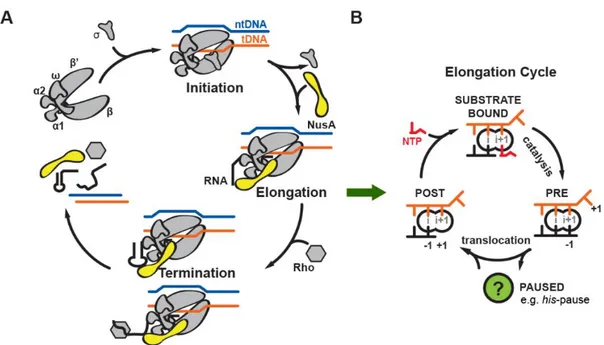
The transcription process can be divided into three distinct phases: initiation, elongation, and termination (Figure 6A).
Initiation of transcription begins when RNAP associates with transcription initiation factors to locate and bind to promoter elements on the DNA and initiate de-novo RNA synthesis using one DNA strand as a template. It involves a stressed intermediate with melting of the downstream DNA duplex and scrunching of the extended transcription bubble into RNAP while maintaining contacts of the initiation factors with promoter elements. The energy accumulated during the stressed intermediate is used to drive the breakage of interactions between initiation factors and promoter elements during promoter
escape, thus affecting the transition to elongation (Kapanidis et al., 2006; Revyakin et al., 2006; Straney and Crothers, 1987; Zuo and Steitz, 2015). When this transition fails, transcription initiation aborts, a common feature of most RNAPs during in vitro and in vivo transcription (Goldman et al., 2009; Murakami and Darst, 2003). Initiation is characterized by multiple rounds of abortive synthesis of short RNA products until RNAP has made a 13-15 nucleotide (nt) long RNA transcript, in which 9 nt pair with the DNA template in an RNA-DNA hybrid, to undergo promoter escape and transition to the elongation phase (Murakami and Darst, 2003).
Once RNAP escapes the promoter successfully, it quickly progresses into productive elongation where transcription elongation factors join and rapid RNA synthesis takes place. In a canonical elongation cycle, RNAP progresses through several states: (1) a post-translocated state (nascent RNA 3’-end occupies the so-called i-site); (2) a substrate bound state prior to catalysis (Nucleoside triphosphate (NTP) substrate base paired to the DNA template base in so-called i+1 site); and (3) a pre-translocated state after catalysis, where the RNA 3’-end has been extended and occupies the i+1-site. To complete the cycle, RNAP translocates along DNA by one base pair so the next substrate can enter the active site (Figure 6B).
Loading of the NTP substrate happens through at least two steps. First, the NTP substrate binds in a template-dependent manner to the open, inactive (‘pre-insertion’) state of the elongation complex (EC) where the trigger loop (TL) is unfolded. Secondly, folding of the TL into two more extended -helices and repositioning of the NTP substrate forms the closed, catalytically competent (‘insertion’) EC (Vassylyev et al., 2007b; Wang et al., 2006). The two-step mechanism of substrate loading may be universal for all RNAP, and
26
folding of the TL may be involved in other catalytic reactions by RNAP such as pyrophosphorolysis and pause escape (Vassylyev et al., 2007b; Zhang et al., 2010).
Figure 6 Transcription cycle (A) and elongation cycle (B) in bacteria. Template DNA (tDNA, orange), non-template DNA (ntDNA, blue), RNA (black), RNAP (gray), and NusA (yellow) are indicated.
RNAP is a highly processive enzyme and in order to stop transcription, an event called termination has to take place. In bacteria, two main pathways lead to termination. The first pathway is called Rho-dependent termination where a protein factor called Rho uses ATP hydrolysis to actively remove RNAP from the DNA template and releases the transcript. A second pathway, called intrinsic termination (or Rho-independent termination), happens when a hairpin secondary structure forms in the nascent RNA transcript, which destabilizes the EC and leads to dissociation of RNAP (Figure 6A)(Ray-Soni et al., 2016).
The regulation of transcription is a vital process in all living organisms. It is coordinated by transcription factors, cofactors, chromatin regulators and noncoding RNAs that interact with the regulatory sequences to finely tune the amount of RNA being produced through a variety of mechanisms. Mutations in the regulation pathway can lead to misregulation of gene expression, which are associated with many different diseases and syndromes including cancer, autoimmunity, neurological disorders, diabetes, cardiovascular disease, and obesity (Lee and Young, 2013). In principle, all three phases in the transcription process could be rate limiting and thus present potential targets for transcription factors to influence the rate. In the past, most studies have predominantly focused on transcriptional regulation that takes place before or during initiation where transcription factors modify the access of RNAP and general transcription factors to the promoter DNA (Svaren and Hörz, 1997), regulate and stabilize the formation of pre-initiation complexes (Stargell and Struhl, 1996), and control promoter escape before elongation starts (Liu et al., 2004). However, more and more studies indicate a potential paradigm shift and show that a lot of regulation takes place during elongation (Adelman and Lis, 2012).
28
2. Transcriptional pausing
2.1. Biological function of transcriptional pausing
It is known that RNAP moves along the DNA template in a discontinuous way during elongation as a result of frequent pauses. Transcriptional pausing describes a phenomenon where a fraction of RNAP temporarily halts nucleotide addition at distinct sites for a variable duration prior to resuming RNA synthesis. Both prokaryotes and eukaryotes control gene expression by modulating transcriptional pausing at specific sites on the DNA template. The frequent occurrence of pausing regulates gene expression on multiple levels (Landick, 2006).
Pausing plays a fundamental role in coupling transcription and translation by affecting the overall RNA synthesis rate in bacteria. During this process, pausing halts RNAP to allow a translating ribosome to catch up to RNAP and then release the enzyme from the paused state. To maintain the coupling of transcription and translation, it is also important for pausing to happen frequently enough to prevent exposing more than 100 nt of unstructured, ribosome-free mRNA, which is the minimal binding site for the Rho transcription termination factor (Landick et al., 1985).
Pausing facilitates the proper folding of nascent RNA transcripts. It has been shown that the folding pathway of a large ribozyme is significantly affected by the process of transcription and addition of the elongation factor NusA resulted in drastic changes in the folding pathway presumably through the alteration of the pausing pattern (Pan et al., 1999).
Pausing enables interaction or recruitment of transcription factors to RNAP by stalling RNAP at key locations. For example, during attenuation, pause sites in the leader regions of amino acid biosynthesis operons halt RNAP to allow ribosomes to be properly located to control the termination decision (Landick et al., 1985). Promoter-proximal pausing of RNAP is critical to ensure the binding of the bacteriophage lambda gene Q transcription antiterminator to RNAP and lambda Q-mediated antitermination (Yarnell and Roberts, 1992). Transcription factor RfaH is recruited to RNAP paused at ops sites in the leader regions of RfaH-regulated operons (Artsimovitch and Landick, 2002). Transcriptional pausing also plays a role in the proper regulation by the FMN riboswitch (Wickiser et al., 2005).
Pausing is a prerequisite for both Rho-dependent and intrinsic termination of transcription. During Rho-dependent termination, RNAP pauses at the terminators until Rho factor interacts with the EC and dissociates the nascent RNA transcript from it through its ATP-dependent helicase activity (Richardson, 2002). In the case of intrinsic termination, pausing of RNAP at the terminators ensures the proper folding of a nascent RNA secondary structure called terminator hairpin, which destabilizes the EC and leads to its dissociation (Gusarov and Nudler, 1999).
Pausing at a promoter-proximal site is an important, rate limiting step in the transcription for a majority of genes in eukaryotes (Core and Lis, 2008; Muse et al., 2007). Various studies have shown that pausing by RNA polymerase II (RNAP II) may facilitate the recruitment of Tat for regulation of HIV-1 transcription (Palangat et al., 1998), facilitate the activation of polyadenylation of the nascent RNA during transcription termination (Yonaha and Proudfoot, 1999), affect the splicing pattern of alternatively spliced mRNAs (la Mata
30
et al., 2003), contribute to the elongation barriers created by nucleosomes during chromatin transcription (Kireeva et al., 2005), and make RNAP II susceptible to abortive elongation (Sims et al., 2004).
Studying the connections of pausing to all these regulatory events as well as possible additional roles is crucial for understanding the regulation of gene expression.
2.2.Current model for transcriptional pausing
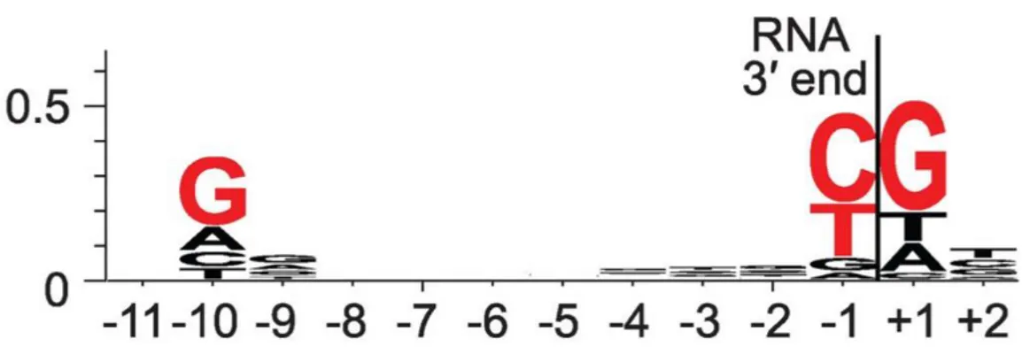
Pausing is triggered by the underlying DNA sequence and genome wide studies identified a consensus pause sequence in E. coli (Larson et al., 2014; Vvedenskaya et al., 2014). A consensus pause element has been proposed: G-10Y-1G+1, where position -1
corresponds to the position of the RNA 3’ end (Figure 7). The consensus pause element consists of a G at position +1, T or C at -1, and G at -10. Translocation involves breaking the DNA base pair at position +1 and breaking the RNA-DNA base pair at position -10. Each position of the consensus sequence is predicted to favor the pretranslocated state over the posttranslocated state, with -10 through effects on duplex stability, -1 through effects on active-centre binding, and +1 through both (Vvedenskaya et al., 2014).
Figure 7 Sequence logo for consensus pause element from NET-seq. Red, bases with ≥ 0.2 bit sequence-information content. Adapted from (Vvedenskaya et al., 2014).
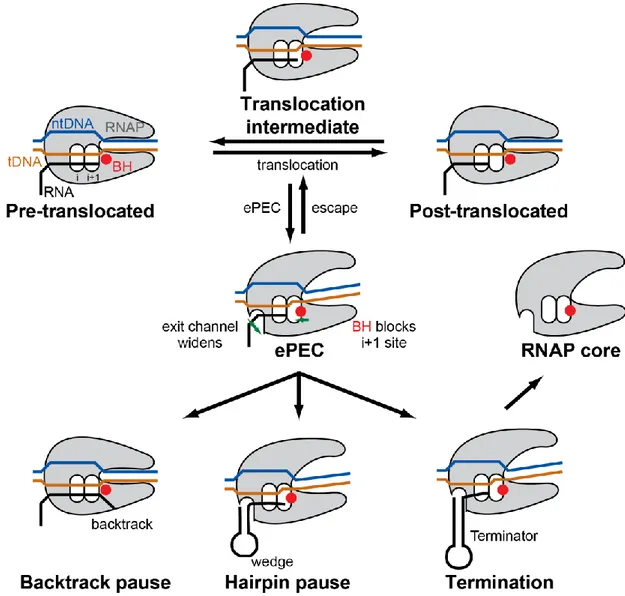
Pausing at a given template position can be characterized by two parameters: (1) a pause efficiency, which is the fraction of transcribing ECs that recognize and enter the paused state; and (2) a pause duration, which is the dwell time that an EC spends at the pause site (Landick, 2006). The consensus pause sequence is suggested to increase the probability of ECs to enter an elemental pause state (ePEC), and therefore increase pause efficiency.
The ePEC has been proposed as an intermediate state in pausing in which initial rearrangement of the RNAP active site inhibits nucleotide addition without changing the translocation register of RNAP (Herbert et al., 2006; Kireeva and Kashlev, 2009; Landick, 2006; 2009; Neuman et al., 2003). Single-molecule studies of transcription showed that bacterial RNAP pauses frequently (once every 100-200 basepair, bp) and the pauses last for 1-6 s on average (Adelman and Lis, 2012; Neuman et al., 2003). Neither the probability nor the duration of these ubiquitous pauses are affected by hindering or assisting forces on translocation of RNAP, ruling out involvement of backtracking or hypertranslocation in the elemental pause (Neuman et al., 2003). After entering the elemental pause, a fraction of RNAP will escape the pause state and resume active elongation while the rest will enter the
32
longer-lived pauses through at least two distinct mechanisms. Class I pauses, found in enteric bacteria, are stabilized by the interaction of a nascent RNA hairpin structure with RNAP to rearrange the active site conformation and render it inactive. Class II pauses, which may be more dominant in eukaryotes, cause RNAP to backtrack along DNA and RNA and to block the active site with nascent RNA threading past it (Figure 8) (Artsimovitch and Landick, 2000). In addition to the Class I and II pauses, RNAP can also have longer-lived pauses through interaction with regulatory proteins that further slowdown the rate of pause escape. For example, the ops pause is prolonged when RfaH interacts with the non-template DNA strand and stabilizes RNAP in a backtracked paused conformation (Artsimovitch and Landick, 2002; Kang et al., 2018b); likewise, NusA is able to stabilize the formation of a nascent RNA hairpin and enhance the Class I pause (Artsimovitch and Landick, 2000). Finally, the not-yet-transcribed DNA duplex downstream of the pause site can significantly influence pausing through interaction with RNAP (Palangat et al., 2004).
Figure 8 Schematic representation of current model of transcriptional pausing. During translocation, RNAP has an opportunity to enter ePEC involving an initial rearrangement of RNAP. From the ePEC: (1) the RNAP can enter a backtrack pause (Class II pause); (2) an RNA pause hairpin and form leading to hairpin pause (Class I pause); (3) a terminator hairpin can form leading to transcription termination. Adapted from (Weixlbaumer et al., 2013).
2.3.Mechanistic insights into class I pause
Extensive studies have been carried out to expand our understandings on the mechanistic basis of class I pauses stimulated by nascent RNA hairpin structures. One of the best-characterized class I pauses occurs in the leader region of the E. coli his operon (his pause). The his pause was discovered to be dependent on a nascent RNA hairpin structure and its
34
function is to synchronize translation and transcription during bacterial transcription attenuation of the his biosynthetic operon (Chan and Landick, 1989). The nascent RNA hairpin structure formed within the RNAP RNA exit channel is thought to stabilize the pause and enhance pause duration 10-fold or more (Toulokhonov et al., 2001). The his pause hairpin forms a 5 base-pair stem and 8 nt loop structure 11 or 12 nt upstream from the pause RNA 3’ end, and its ability to enhance pausing relies on its stability and position from the RNA 3’ end but not its sequence (Chan and Landick, 1993; Chan et al., 1997; Toulokhonov et al., 2001). Studies have shown that sequence elements in the DNA template downstream of the pause site also affect the efficiency of pausing, presumably by interaction with RNAP (Lee et al., 1990). Furthermore, transcription factors also play a role in modulating the his pause as it is stimulated by the NusA N-terminal domain (NTD) and suppressed by the RafH NTD (Artsimovitch and Landick, 2002; Ha et al., 2010; Kolb et al., 2014). It has been proposed that electrostatic interactions between the pause hairpin and RNAP, conformational changes in the flap tip and clamp of RNAP, and RNAP active site rearrangements predominantly involving spatially distant TL movements, result in the delayed elongation at the his pause site (Chan and Landick, 1993; Hein et al., 2014; Toulokhonov and Landick, 2003; Toulokhonov et al., 2007).
However, key questions regarding the pause mechanism remain to be answered. How is the active site rearranged to inhibit nucleotide addition in the his pause? In what way does the pause hairpin in the RNA exit channel stabilize the pause state? How does the transcription factor NusA enhance the pause? A high-resolution structure of a hairpin stabilized paused EC in complex with NusA is needed to answer these questions.
2.4. In vitro experimental system to reconstitute a Class I paused complex
One of the major challenges to deciphering the mechanism of pausing is the difficulty to assemble pure populations of the paused elongation complex (PEC) in vitro using DNA, RNA, and RNAP. Daube and von Hippel first reconstructed a functional EC in vitro by incubating RNAP with a nucleic acid scaffold comprising a double-stranded DNA duplex with an internal noncomplementary DNA “bubble” sequence and an RNA oligonucleotide that is partially complementary to the template DNA within the DNA bubble to form an RNA-DNA hybrid (Daube and Hippel, 1992). Kyzer et al. later demonstrated two methods to reconstitute a PEC: 1) direct reconstitution; and 2) a limited step reconstitution assay (Kyzer et al., 2007). Direct reconstitution uses a 29-nt RNA whose 3’-proximal 9-10 nt base pair to the template within an 11-nt noncomplementary bubble of a 39-bp duplex DNA; the RNA contains the 18-nt his pause RNA hairpin but lacked RNA upstream of the hairpin (Figure 9A). Limited step reconstitution is achieved similarly on the same DNAs with a 27-nt RNA whose 3’ end is 2 27-nt upstream of the pause site to allow labeling of only active ECs during RNA extension to reach the pause site (Figure 9B). PECs formed by either method recapitulated key features of the his pause including enhancement by NusA (Kyzer et al., 2007). In vitro reconstitution approaches of the his paused elongation complex (hisPEC) provide a valuable tool for functional and structural studies on the mechanism of class I pauses.
36
Figure 9 (A) Direct reconstitution and (B) limited step reconstitution nucleic acid scaffold architecture. RNA extension by RNAP is shown in lowercase type. Adapted from (Kyzer et al., 2007).
3. RNA Polymerase
RNAPs are essential to life, they are found in all living organisms and many viruses. In most prokaryotes, a single RNAP transcribes all types of RNA. While eukaryotes have three types of RNAP, each is responsible for the synthesis of a distinct subset of RNAs: RNA polymerase I (RNAP I) synthesizes rRNA, RNAP II synthesizes mRNA, micro RNA (miRNA), small nuclear RNA (snRNA), and small nucleolar RNA (snoRNA), and RNA polymerase III (RNAP III) synthesizes tRNA and 5S rRNA.
3.1.Core architecture of RNAP
RNAP is highly conserved in all three kingdoms of life. The universally conserved core architecture of RNAP consists of five subunits: (1) 160 kDa subunit (’ in bacterial RNAP; A in archaeal RNAP; RPA1, RPB1, and RPC1 in eukaryotic RNAP I, II, and III respectively), (2) 150 kDa subunit ( in bacterial RNAP; B in archaeal RNAP; RPA2, RPB2, and RPC2 in eukaryotic RNAP I, II, and III respectively), (3) 35 kDa subunit (1 in bacterial RNAP; D in archaeal RNAP; RPC5 in eukaryotic RNAP I, and III; RPB3 in eukaryotic RNAP II), (4) 10-35 kDa subunit (2 in bacterial RNAP; L in archaeal RNAP; RPC9 in eukaryotic RNAP I, and III; RPB11 in eukaryotic RNAP II), (5) 6 kDa subunit ( in bacterial RNAP; K in archaeal RNAP; RPB6 in eukaryotic RNAP I, II and III). Bacterial RNAP contains only the five conserved subunits with a total molecular mass of around 400 kDa while archaeal and eukaryotic RNAP contain additional subunits with a total molecular mass of around 500 kDa (Figure 10)(Ebright, 2000).
38
Figure 10 Conserved subunits of bacterial RNAP, archaeal RNAP, eukaryotic RNAP I, II and III. Adapted from (Ebright, 2000).
3.2.Structure of bacterial RNAP
The first structure of Thermus aquaticus RNAP core enzyme was solved by X-ray crystallography at 3.3 Å resolution (PDB accession code 1HQM), which marked a milestone in our structural understanding of the multi-subunit protein (Zhang et al., 1999). Bacterial RNAP has dimensions of 150Å x 115Å x 110Å and has a shape resembling a crab claw, with two pincers defining a 27 Å wide central groove to accommodate the double-stranded DNA, and a Mg2+ ion on the back wall of the groove for catalytic activity
(Figure 11A)(Zhang et al., 1999). The ’ subunit forms one pincer, while the subunit forms the other pincer. Both subunits form part of the base of the groove. The two subunits, despite having the same sequence, locate differently and they interact differently within RNAP: 1 is located closer to the groove and interacts with , while 2 is located further away from the groove and interacts with ’. Each subunit contains two domains: an N-terminal domain (NTD) that interacts with or ’ subunit, and a C-N-terminal domain (CTD) that interacts with upstream promoter DNA, activators, and repressors (Busby and Ebright, 1994). CTD is connected to NTD, and therefore to the rest of RNAP, through a
long and flexible linker, which explains the disordered density seen in X-ray crystallographic structures (Busby and Ebright, 1994; Zhang et al., 1999). The last subunit is located far away from the groove, near the base of the pincer formed by ’ (Zhang et al., 1999).
3.3.Structure of eukaryotic RNAP II
The first structure of yeast RNAP II 4/7 (lacking non-conserved subunits RPB4 and RPB7) was also solved by X-ray crystallography at 3.5 Å (PDB accession code 1EN0) providing a backbone model of the enzyme (Cramer et al., 2000), followed by a 2.8 Å atomic structure (PDB accession code 1I3Q) one year later (Cramer et al., 2001). Similar to bacterial RNAP, RNAP II also has a shape reminiscent of a crab claw with two pincers forming a central groove (Figure 11B) (Cramer et al., 2000; 2001; Zhang et al., 1999). Within yeast RNAP, the relative positions of the conserved subunits also match those in bacterial RNAP. Therefore in yeast RNAP, subunit RPB1 (counterpart of ’ subunit in bacterial RNAP) makes up one pincer and part of the base of the groove; subunit RPB2 (counterpart of subunit in bacterial RNAP) makes up the other pincer and part of the base of the groove; subunits RPB3 and RPB11 (counterpart of 1 and 2 subunits in bacterial RNAP, respectively) are located distal to the groove; subunit RPB6 (counterpart of subunit in bacterial RNAP) is located at the base of the pincer formed by RPB1. The non-conserved subunits RPB8, RPB9, RPB10 and RPB12 are located on the periphery of the structure while RPB5 is located distal to the groove.
40
Figure 11 (A) Structure of Thermus aquaticus RNAP at 3.3 Å resolution viewed from three different angles (Zhang et al., 1999). ’ is in orange; is in green; 1NTD is in cyan; 2NTD
is in blue; is in gray. The active site Mg2+ ion is shown as a white sphere. The activation
target within NTD is shown in yellow. (B) Structure of yeast RNAP II 4/7 at 3.0 Å resolution viewed from three different angles (Cramer et al., 2001). RPB1 is in orange; RPB2 is in green; RPB3 is in cyan; RPB11 is in blue; RPB6 is in gray. Non-conserved subunits RPB5, RPB8, RPB9, RPB10, and RPB12 are in red, aqua, violet, magenta, and pink,
respectively. The active site Mg2+ ion is shown as a white sphere. The activation target
3.4.Similarity between bacterial and eukaryotic RNAP structures
The overall structures of bacterial and eukaryotic RNAP are very similar, in addition, there are also similarities in the regions of important functions: (1) each RNAP contains a Mg2+ ion in the active center for catalytic activity; (2) each RNAP contains an equivalently
positioned 25 Å long, 15 Å wide channel that mediates exit of the nascent RNA during elongation (termed “RNA exit channel”) (Cramer et al., 2000; 2001; Korzheva, 2000; Naryshkin et al., 2000; Zhang et al., 1999); (3) each RNAP contains an equivalently positioned 20 Å long, 10 Å wide tunnel that mediates entry of NTPs to the active site during elongation (termed “secondary channel” in bacteria, or “funnel and pore” in eukaryotes) (Cramer et al., 2000; 2001; Fu et al., 1999; Korzheva, 2000; Zhang et al., 1999); (4) each RNAP contains an equivalently positioned activation target (“AT” in Figure 11) for interacting with transcription activator proteins during transcription activation (Busby and Ebright, 1999; Niu et al., 1996); (5) the similarity between bacterial RNAP and eukaryotic RNAP also extends to the folding topologies of the individual subunits (Figure 11) (Ebright, 2000). In addition to the similarities between bacterial and eukaryotic RNAPs, there is also striking structural conservation between archaeal and eukaryotic RNAPs (Hirata et al., 2008).
The remarkable degree of structural similarity between bacterial RNAP, archaeal RNAP, and yeast RNAP II implies a fundamental similarity in the mechanisms of transcription among all three kingdoms of life (Cramer et al., 2001; Hirata et al., 2008; Zhang et al., 1999). Therefore, structural and mechanistic insights we gain by studying
42
bacterial RNAP are likely to be directly relevant to understanding the process of transcription in all living organisms.
3.5. Structural organization of the transcription elongation complex
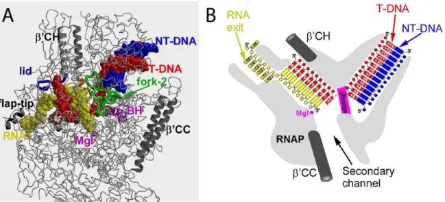
Previously mentioned structures of RNAP core enzyme were solved in the absence of templates, substrates or products (Cramer et al., 2000; 2001; Zhang et al., 1999). In an effort to understand the structural basis of transcription elongation, further studies were carried out aiming to obtain models of the structure of RNAP in complex with nucleic acids (Nudler et al., 1996), followed by structures of yeast RNAP II EC and Thermus thermophilus EC being solved by X-ray crystallography (Gnatt et al., 2001; Kettenberger et al., 2004; Vassylyev et al., 2007b; 2007a; Wang et al., 2006; Westover et al., 2004a; 2004b). These studies revealed several sets of protein-nucleic acids interactions that stabilize the EC: (1) downstream duplex DNA is bound within the enzyme entering through the main cleft; (2) about 9~10 nt of RNA are annealed to the template DNA forming a 9~10-bp RNA-DNA hybrid residing in the RNAP main channel, with the 3’ end of the RNA in the active site; (3) an additional ~5 nt of the nascent RNA upstream of the hybrid is threaded through the RNA exit channel (Figure 12).
Figure 12 Structure of the bacterial elongation complex. (A) The overall three-dimensional X-ray structure. (B) Schematic drawing showing position of nucleic acids within the EC. Adapted from (Vassylyev, 2009).
Structures of bacterial EC increase our mechanistic understanding of transcription elongation (Vassylyev et al., 2007a; 2007b). As seen in the structures, access to the RNAP active site through the main channel is blocked and it was first proposed by Zhang et al. that substrates enter through the widely open secondary channel (Zhang et al., 1999). A structure in the post-translocated state showed one nucleotide of template DNA at the active site available for base pairing with the incoming NTP one at a time. The downstream DNA duplex unwinds immediately downstream of the active site (register +2). The +2 downstream DNA base pair stacks on the fork 2, suggesting a crucial role of fork loop-2 in downstream DNA strand separation (Naji et al., loop-2008). At the upstream edge of the transcription bubble, the last base pair of the RNA-DNA hybrid (position -9) stacks on the ’-subunit “lid” loop, whereas the first displaced RNA base is trapped in the hydrophobic pocket formed by the -subunit switch-3 segment, suggesting a mechanism for RNA displacement (Kent et al., 2009; Toulokhonov and Landick, 2006). At the active site, folding of the ’-subunit TL into two -helices (trigger helices, TH) is shown to be vital for catalysis
44
during substrate NTP loading (Vassylyev et al., 2007b; Wang et al., 2006). The mechanisms of the downstream DNA and RNA-DNA hybrid strand separation, and substrate loading are most likely universally conserved amongst all multi-subunit RNAPs.
3.6.Structure of an elemental paused elongation complex
A widely accepted model for pausing is that upon encountering the multipartite pause signal, an EC first transforms through structural rearrangement into an inactive state ePEC, whose elemental conformational intermediate of the RNAP underlies all transcriptional pauses (Figure 8)(Herbert et al., 2006; Kireeva and Kashlev, 2009; Landick, 2006; 2009; Neuman et al., 2003). X-ray crystallographic structures of bacterial ePEC have been solved showing that, in this intermediate state, RNAP adopts a relaxed, open-clamp conformation (Weixlbaumer et al., 2013). In this conformation, the clamp opening may be the result of loss of contact between the downstream DNA duplex and RNAP during translocation; a kinked bridge-helix structure blocks the RNAP active site, providing an explanation for inhibition of nucleotide addition; a widened RNA exit channel provides space for the formation of an RNA pause hairpin that in turn prolongs the pause (Weixlbaumer et al., 2013). Even though all three ePEC structures of two bacterial RNAPs (Thermus
thermophilus and Thermus aquaticus) show the same open-clamp conformation, it remains
debatable whether this conformation is influenced by crystal packing interactions. The ePEC structure, although lacking an RNA hairpin, provided a framework for understanding the subsequent hairpin-stabilized paused complex, whose structure remained elusive.
4. Transcription elongation factors NusA
Transcription elongation factors are proteins and non-coding RNAs that alter the elongation properties of RNAP in various ways: some elongation factors stimulate elongation by suppressing pausing and increasing elongation rates or by promoting read through by RNAP at arrest sites; some elongation factors stimulate termination of transcription or antitermination; some elongation factors alter the activity or protein components of the elongation complex; some elongation factors bind to the nascent RNA transcripts or to the DNA to affect elongation; some elongation factors act via direct protein-protein interactions with either the RNAP or other accessory protein-proteins. A lot has yet to be discovered about the mechanism by which the elongation factors carry out their effects (Roberts et al., 2008).
NusA, short for N-utilizing substance A protein, is an essential multifunctional transcription elongation factor that is universally conserved among bacteria and archaea. NusA has been studied for over 40 years: the nusA gene was first identified as a requirement for phage lambda protein N-mediated antitermination (Friedman et al., 1974); the protein NusA was first referred to as the L factor when it was first discovered for its ability to stimulate -galactosidase synthesis in coupled in vitro transcription-translation assays (Kung et al., 1975); later the L factor was shown to affect termination and was identified as the product of the nusA gene, and hence termed the NusA protein (Greenblatt et al., 1980).
4.1.Effects of NusA on transcription
NusA is essential in wild-type E. coli. Isolation of a number of conditionally defective
46
al., 1986). However, in combination with other mutations that suppress Rho-dependent termination, bacteria with a nusA deletion can survive (Zheng and Friedman, 1994). Deletion of horizontally acquired genes from E. coli allows nusA and nusG deletions to survive (Cardinale et al., 2008). The gene nusG encodes the general elongation factor NusG, which is able to inhibit pausing and enhance Rho-dependent termination (Li et al., 1992; Roberts et al., 2008). The nusA and nusG deleted strains are more resistant to the Rho inhibitor bicyclomycin, implying that high activity of Rho is essential for cell survival because it suppresses the toxic activity of foreign genes (Cardinale et al., 2008). These results suggest that NusA and NusG are important for cell survival through their influences on Rho-dependent termination. NusG has been shown in vitro to stimulate Rho-dependent termination (Burns et al., 1999), which agrees with the reduced genomes study (Cardinale et al., 2008). However the role of NusA is less clear, with conflicting reports that it both promotes and inhibits Rho-dependent termination. A number of studies have shown that NusA inhibits Rho-dependent termination (Burns et al., 1998; Greenblatt et al., 1980; Kung et al., 1975; Lau and Roberts, 1985; Lau et al., 1982; Qayyum et al., 2016). On the other hand, some studies suggest that, similar to NusG, NusA also promotes Rho-dependent termination (Cardinale et al., 2008; Saxena and Gowrishankar, 2011).
Depending on the DNA sequence context and the presence or absence of auxiliary factors, NusA may exhibit opposite effects on transcription. Its most apparent effects are the enhancement of hairpin-induced pausing such as those caused by his or trp hairpins, which were discovered in the leader region of E. coli his or trp biosynthetic operons, respectively (Farnham et al., 1982; Kassavetis and Chamberlin, 1981; Landick and Yanofsky, 1984; Lau et al., 1983), and the enhancement of intrinsic termination during
transcription with purified RNAP (Schmidt and Chamberlin, 1987). On the contrary, NusA is an essential component of the phage lambda gene N antitermination complex including other Nus factors (NusG, NusB, NusE), and it is also involved in lambda gene Q mediated antitermination, both of which inhibit pausing (Nudler and Gottesman, 2002). NusA is a key regulator of transcriptional antitermination in bacterial rRNA operons (Berg et al., 1989). In addition to its roles in transcription elongation and termination, NusA is involved in transcription-coupled repair of DNA damage and stress-induced mutagenesis (Cohen and Walker, 2010; Cohen et al., 2009; 2010). Furthermore, NusA has been shown to accelerate the co-transcriptionsl folding of a ribozyme more than 10-fold by stimulating pausing of E.
coli RNAP (Pan et al., 1999). The diverse and contradictory effects of NusA on transcription
elongation and termination make it a very intriguing protein to study.
4.2.Structure of NusA and its interactions with the EC
E. coli NusA is a 55-kDa monomeric RNA binding protein. According to structures of
NusA from E. coli, Thermotoga maritima and Mycobacterium tuberculosis solved by X-ray crystallography and NMR (Beuth et al., 2005; Eisenmann et al., 2005; Gopal et al., 2001; Said et al., 2017; Schweimer et al., 2011; Worbs et al., 2001), this 495-amino acid protein has three distinct domains: (1) an N-terminal RNAP binding domain (NTD); (2) a middle portion comprising three globular RNA-binding domains S1, KH1 and KH2; (3) a C-terminal domain comprising acidic repeats 1 (AR1) and 2 (AR2) (Figure 13A). Except for the additional C-terminal extension AR1 and AR2 domains found in E. coli, other domains are highly conserved among the NusA proteins from different species. These structures revealed NusA to be a rod-shaped, elongated molecule.