CASE REPORT
Pediatric papillary tumors of the pineal region: to observe
or to treat following gross total resection?
Lucia Abela&Elisabeth Jane Rushing&Carmen Ares&
Ianina Scheer&Oliver Bozinov&Eugen Boltshauser&
Michael A. Grotzer
Received: 22 August 2012 / Accepted: 25 September 2012 / Published online: 6 October 2012 # Springer-Verlag Berlin Heidelberg 2012
Abstract
Introduction Papillary tumors of the pineal region (PTPR) are rare brain tumors characterized by frequent local recur-rences. Standardized treatment strategies are not yet defined. Case report We present the case of a 3-year-old girl diag-nosed with PTPR. Due to her young age, adjuvant radiother-apy was omitted after gross total tumor resection. Thirty-six months later, local tumor recurrence occurred. Considering
the possible risks of secondary surgery, the recurrent tumor was irradiated with proton radiotherapy. Three months later, the tumor showed near-complete remission.
Discussion Based on this experience and other pediatric case reports from the literature, local radiotherapy might be suggested also after complete tumor resection.
Keywords Brain tumor . Child . Proton beam irradiation . Outcome
Introduction
Tumors of the pineal region account for about 3–4 % of all brain tumors in childhood [4]. Papillary tumors of the pineal region (PTPR) represent a rare and histologically distinct sub-group of tumors originating in this area [3,10]. First described by Jouvet et al. in 2003 [10], PTPR have been included in the 2007 World Health Organization Classification of Tumors of the Nervous System as a separate tumor entity [15]. Among 70 PTPR reported to date, examples are recognized in both chil-dren and adults with ages ranging from 5 to 67 years (mean, 34 years) [14]. PTPR has a distinctive epithelial character and shows both solid and papillary architecture [6, 10, 14]. Immunohistochemical analysis is crucial to distinguish PTPR from morphological similar pineal tumors, mainly papillary ependymoma and choroid plexus papilloma [8]. PTPR express strong immunoreactivity for CK18, S100, NCAM, neuron-specific enolase (NSE), and vimentin [5,6]. Due to the specific localization of this entity in the region of the posterior com-missure and the distinctive immunohistochemical profile, PTPR is presumed to arise from the specialized ependyma of the subcommisural organ [10]. Recent ultrastructural studies have demonstrated that PTPR has concomitant ependymal, neuroendocrine, and secretory features, lending further support
L. Abela
:
M. A. GrotzerDepartment of Oncology,
University Children’s Hospital of Zurich, Zurich, Switzerland
E. J. Rushing
Institute of Neuropathology, University Hospital of Zurich, Zurich, Switzerland
C. Ares
Center for Proton Therapy, Paul Scherrer Institute, Villigen, Switzerland
I. Scheer
Department of Diagnostic Imaging,
University Children’s Hospital of Zurich,
Zurich, Switzerland O. Bozinov
Department of Neurosurgery, University Hospital of Zurich, Zurich, Switzerland
E. Boltshauser
Department of Neurology,
University Children’s Hospital of Zurich,
Zurich, Switzerland
M. A. Grotzer (*)
University Children’s Hospital,
Steinwiesstrasse 75, CH-8032 Zurich, Switzerland e-mail: Michael.Grotzer@kispi.uzh.ch Childs Nerv Syst (2013) 29:307–310 DOI 10.1007/s00381-012-1935-1
to this hypothesis [2]. Despite advances in immunohistochem-istry and cytogenetics, the biological behavior of PTPR is still not fully understood. Thus, uniform therapeutic guidelines have not yet been established. Here, we describe a case of PTPR in a 3-year-old girl with 3.5 years of clinical and neuro-radiological follow-up.
Case report
Clinical presentation
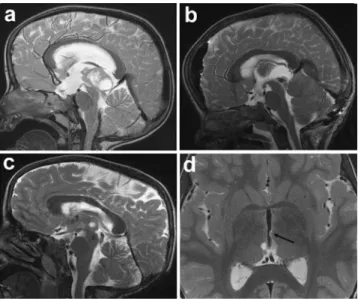
A 3-year-old girl presented to her pediatrician with a history of new-onset headaches accompanied by nausea, lasting for sev-eral hours a day. The neurological examination was normal and migraine was initially suspected. After symptoms persisted for 2 weeks, cranial magnetic resonance imaging (MRI) was per-formed. The MRI revealed a cerebral mass located in the pineal region measuring 2×2.8×3.5 cm (Fig. 1a). The tumor dis-played dorsal cystic structures and a ventral solid portion with inhomogeneous contrast agent enhancement leading to a com-pression of the tectum. The consecutive aqueductal stenosis resulted in occlusive hydrocephalus. A ventriculostomy of the third ventricle was performed to reduce intracranial pressure. Tumor markers such as AFP and beta-HCG in serum and cerebrospinal fluid (CSF) were within the normal range. Two weeks later, the patient underwent gross total tumor resection through a suboccipital supracerebellar approach. Postoperative imaging demonstrated complete resection (Fig.1b). On post-operative clinical examination, the girl was found to have a
vertical supranuclear gaze palsy, convergence deficit, and mid-dilated pupils (Parinaud’s syndrome). This functional deficit led to gait instability, particularly when climbing stairs.
Further treatment options were thoroughly discussed. Given the young age of the patient and macroscopically gross total tumor resection, the decision was made to omit adjuvant radiotherapy. The girl was followed at 3-month intervals by clinical examinations and cranial MRI. Control intervals were expanded to once every 6 months during the second postop-erative year. The clinical course was uneventful, except for persistent vertical gaze palsy. Local tumor recurrence was detected on routine follow-up cranial MRI after 3 years (Fig.1c). The solid and partially cystic recurrent tumor was located within the third ventricle dorsal inferior of the inter-thalamic adhesion without disturbance of the CSF circulation, approximately 1.8×0.7×1.3 cm in size. At this time, the patient did not show any additional neurological symptoms. The possibility of a second surgery was rejected considering the potential risks of additional neurological damage. Subsequently, it was decided to treat the recurrent tumor with proton beam irradiation, with a total dose of 54 Gy (RBE) in 30 fractions of 1.8 Gy (RBE) per fraction, five fractions a week. At first follow-up, 3 months after completion of thera-py, the MRI demonstrated near-complete remission (Fig.1d).
Histological diagnosis
Histopathological examination revealed a moderately cellular, epithelial tumor with solid and papillary growth patterns (Fig.2). Perivascular pseudorosette formation was a promi-nent feature; however, true rosettes could not be demonstrat-ed. The cytoplasm of the neoplastic cells varied from clear to amphophilic. Areas of necrosis were present and mitotic ac-tivity was moderate (4–6/10 HPF). Immunohistochemical analysis showed positive cytoplasmic staining for NSE and MAP2, while S100 protein was inhomogeneously expressed. Some tumor cells, mostly those forming papillary structures, displayed positivity for Pan-cytokeratin. Glial fibrillary acidic
Fig. 1 a Sagittal preoperative T2-weighted MRI showing a cerebral mass with cystic portions located in the pineal region. b Sagittal postoperative T2-weighted MRI demonstrates complete resection. c Local tumor recurrence 36 months after complete resection. d Axial T2-weighted MRI obtained after proton beam therapy. A small residual tumor is seen only on axial slices (arrow)
Fig. 2 Histopathological features of PTPR demonstrating solid and papillary growth pattern with perivascular pseudorosette formation
protein immunolabeling was minimal and restricted to peri-vascular areas, particularly adjacent to blood vessels. Staining for other neuronal markers like N-Neu, chromogranin A, neurofilament 200, neurofilament 70, and synaptophysin was negative. The Ki67 proliferation index was up to 10– 15 %.
Discussion
Grading criteria and therapeutic protocols for PTPR have not yet been established because of the rarity and relatively new diagnosis in the 2007 World Health Organization Classification of Tumors of the Nervous System. Considering its frequent local recurrence, PTPR is thought to correspond at least to WHO grade II/III. To gain further insight into tumor character-istics, genetic analysis has been performed by comparative genomic hybridization and gene expression studies [7]. Losses on chromosome 10 and 22q and gains on chromosomes 4, 8, 9, and 12 have been detected. Gene expression analysis has shown upregulation of genes such as ZH4, RFX3, TTR mRNA, and CGRP [5,8]. Clinical implication of these findings has not yet been elucidated.
Since its original description by Jouvet et al. in 2003, about 70 cases have been reported in literature [14, 16]. Among these patients, seven were children under 16 years of age (Table1) [1,6,12]. Clinical manifestations include headache, diplopia, dizziness, and vomiting [1,11,12,14]. Spinal dissemination is rare at the time of diagnosis. A key feature of PTPR is their high risk for local recurrence. Three out of six pediatric patients suffered from local tumor recur-rence, the others were lost to follow-up [1,6,12]. Poulgrain et al. investigated the 5-year- and 10-year progression-free survival of all reported cases to date, which has been esti-mated at 34.5 and 8.6 %, respectively [14]. Fèvre-Montange et al. performed univariate analysis to test the prognostic significance of clinical variables such as age, gender, tumor size, gross total tumor resection, and adjuvant radiotherapy [6]. The only clinical factor that tended to be associated with
overall survival and recurrence was the extent of resection [6].
Standardization of treatment regimen is not yet determined due to the lack of reliable clinical and biological predictors and the small number of cases with careful follow-up. Maximal surgical tumor resection is suggested as the first-line procedure. Radiotherapy and chemotherapy have been adopted as further treatment options [14]. Radiotherapy has been applied in about two thirds of all reported patients using different protocols and methods [14]. In the current report, radiotherapy was initially avoided because of the young age of the patient and total tumor resection. However, tumor relapse occurred 3 years after diagnosis, confirming the high risk of local recurrence in PTPR. With respect to the apparent high propensity of local recurrence, adjuvant radiotherapy might be applied in older children with gross total tumor resection. In younger children, the cerebral developmental vulnerability has to be taken into account. However, in case of recurrence, radiotherapy seems to be an effective therapy. Especially in young patients, proton beam irradiation needs to be consid-ered in order to reduce the integral dose reduction to the adjacent normal tissue [9,13,17,18].
References
1. Buffenoir K, Rigoard P, Wager M, Ferrand S, Coulon A, Blanc JL, Bataille B, Listrat A (2008) Papillary tumor of the pineal region in a child: case report and review of the literature. Childs Nerv Syst
24:379–384
2. Cykowski MD, Wartchow EP, Mierau GW, Stolzenberg ED, Gumerlock MK, Fung KM (2012) Papillary tumor of the pineal
region: ultrastructural study of a case. Ultrastruct Pathol 36:68–77
3. De Girolami U, Fevre-Montange M, Seilhean D, Jouvet A (2008)
Pathology of tumors of the pineal region. Rev Neurol 164:882–895
4. Drummond KJ, Rosenfeld JV (1999) Pineal region tumours in
childhood. A 30-year experience. Childs Nerv Syst 15:119–126,
discussion 127
5. Fevre-Montange M, Champier J, Szathmari A, Wierinckx A, Mottolese C, Guyotat J, Figarella-Branger D, Jouvet A, Lachuer J (2006) Microarray analysis reveals differential gene expression Table 1 Pediatric patients with PTPR. Treatment modalities and outcome
Case [reference]
Age (years) Sex Tumor
size (mm) Operation Adjuvant therapy Follow-up (months) Recurrence (months) Salvage therapy 1 [12] 1 3/12 M 10 IR CT, RT 15 NA NA 2 [6] 5 F 28 CR CT 24 21 CR, RT 3 [6] 11 F 30 IR, CR CT, RT 79 72 CR, CT, RT 4 [6] 13 M NA CR RT 5 NA NA 5 [1] 13 M 31 CR RT 15 NA NA 6 [6] 14 M NA CR NA NA NA NA 7 [6] 14 M 50 CR CT 102 44, 53, 88 RT
NA not available, M male, F female, CR complete resection, IR incomplete resection, CT chemotherapy, RT radiotherapy
patterns in tumors of the pineal region. J Neuropathol Exp Neurol
65:675–684
6. Fevre-Montange M, Hasselblatt M, Figarella-Branger D, Chauveinc L, Champier J, Saint-Pierre G, Taillandier L, Coulon A, Paulus W, Fauchon F, Jouvet A (2006) Prognosis and histopathologic features in papillary tumors of the pineal region: a retrospective multicenter study of 31 cases. J Neuropathol Exp Neurol 65:1004–1011 7. Gutenberg A, Brandis A, Hong B, Gunawan B, Enders C, Schaefer
IM, Burger R, Ostertag H, Gaab M, Krauss JK, Fuzesi L (2011) Common molecular cytogenetic pathway in papillary tumors of the
pineal region (PTPR). Brain Pathol 21(6):672–677
8. Hasselblatt M, Blumcke I, Jeibmann A, Rickert CH, Jouvet A, van de Nes JA, Kuchelmeister K, Brunn A, Fevre-Montange M, Paulus W (2006) Immunohistochemical profile and chromosomal imbal-ances in papillary tumours of the pineal region. Neuropathol Appl
Neurobiol 32:278–283
9. Hug EB (2004) Protons versus photons: a status assessment at the
beginning of the 21st century. Radiother Oncol 73(Suppl 2):S35–37
10. Jouvet A, Fauchon F, Liberski P, Saint-Pierre G, Didier-Bazes M, Heitzmann A, Delisle MB, Biassette HA, Vincent S, Mikol J, Streichenberger N, Ahboucha S, Brisson C, Belin MF, Fevre-Montange M (2003) Papillary tumor of the pineal region. Am J Surg Pathol 27:505–512
11. Junior GV, Dellaretti M, de Carvalho GT, Brandao RA, Mafra A, de Sousa AA (2011) Papillary tumor of the pineal region. Brain Tumor Pathol 28:329–334
12. Li J, Recinos PF, Orr BA, Burger PC, Jallo GI, Recinos VR (2011) Papillary tumor of the pineal region in a 15-month-old boy. J
Neurosurg Pediatr 7:534–538
13. Miralbell R, Lomax A, Bortfeld T, Rouzaud M, Carrie C (1997) Potential role of proton therapy in the treatment of pediatric me-dulloblastoma/primitive neuroectodermal tumors: reduction of the supratentorial target volume. Int J Radiat Oncol Biol Phys 38:477– 484
14. Poulgrain K, Gurgo R, Winter C, Ong B, Lau Q (2011) Papillary
tumour of the pineal region. J Clin Neurosci 18:1007–1017
15. Roncaroli F, Scheithauer BW (2007) Papillary tumor of the pineal region and spindle cell oncocytoma of the pituitary: new tumor
entities in the 2007 WHO Classification. Brain Pathol 17:314–318
16. Santoro A, D’Elia A, Fazzolari B, Santoro F, Antonelli M,
Giangaspero F, Brogna C, Lenzi J, Frati A, Salvati M (2011) Four-year clinical and neuroradiological follow-up of a papillary
tumor of the pineal region. Neurol Sci 33(4):931–935
17. St Clair WH, Adams JA, Bues M, Fullerton BC, La Shell S, Kooy HM, Loeffler JS, Tarbell NJ (2004) Advantage of protons com-pared to conventional X-ray or IMRT in the treatment of a pediatric patient with medulloblastoma. Int J Radiat Oncol Biol Phys 58:727–734
18. Yuh GE, Loredo LN, Yonemoto LT, Bush DA, Shahnazi K, Preston W, Slater JM, Slater JD (2004) Reducing toxicity from craniospinal irradiation: using proton beams to treat medulloblas-toma in young children. Cancer J 10:386–390