HAL Id: inserm-00135611
https://www.hal.inserm.fr/inserm-00135611
Submitted on 19 Mar 2007HAL is a multi-disciplinary open access
archive for the deposit and dissemination of sci-entific research documents, whether they are pub-lished or not. The documents may come from teaching and research institutions in France or abroad, or from public or private research centers.
L’archive ouverte pluridisciplinaire HAL, est destinée au dépôt et à la diffusion de documents scientifiques de niveau recherche, publiés ou non, émanant des établissements d’enseignement et de recherche français ou étrangers, des laboratoires publics ou privés.
Effect of chest X-rays on the risk of breast cancer among
BRCA1/2 mutation carriers in the international
BRCA1/2 carrier cohort study: a report from the
EMBRACE, GENEPSO, GEO-HEBON, and IBCCS
Collaborators’ Group.
Andrieu Nadine, Douglas Easton, Jenny Chang-Claude, Matti Rookus,
Richard Brohet, Elisabeth Cardis, Antonis Antoniou, Teresa Wagner, Jacques
Simard, Gareth Evans, et al.
To cite this version:
Andrieu Nadine, Douglas Easton, Jenny Chang-Claude, Matti Rookus, Richard Brohet, et al.. Effect of chest X-rays on the risk of breast cancer among BRCA1/2 mutation carriers in the international BRCA1/2 carrier cohort study: a report from the EMBRACE, GENEPSO, GEO-HEBON, and IBCCS Collaborators’ Group.. Journal of Clinical Oncology, American Society of Clinical Oncology, 2006, 24 (21), pp.3361-6. �10.1200/JCO.2005.03.3126�. �inserm-00135611�
Effect of chest X-rays on the risk of breast cancer among BRCA1/2 mutation carriers in the IBCCS Study
Nadine Andrieu1, Douglas F. Easton2, Jenny Chang-Claude3, Matti A. Rookus4, Richard Brohet4, Elisabeth Cardis5, Antonis C. Antoniou2, Teresa Wagner6, Jacques Simard7, Gareth Evans8, Susan Peock2, EMBRACE*, Jean-Pierre Fricker9, Catherine Nogues10, GENEPSO*, Laura Van’t Veer4, Flora E. Van Leeuwen4, GEO-HEBON*, the IBCCS collaborators group*, and David E. Goldgar5,11+
1 INSERM Emi00-06 et Service de Biostatistiques de l'Institut Curie, France
2 Cancer Research UK, Genetic Epidemiology Unit, Department of Public Health and Primary Care, University of Cambridge, UK.
3 Division of Clinical Epidemiology, German Cancer Research Center, Heidelberg, Germany, 4 The Netherlands Cancer Institute, Departments of Epidemiology (FL,MR) and Molecular Pathology (LV), Amsterdam, Netherlands.
5
International Agency for Research on Cancer, Lyon, France. 6
Department of Obstetrics and Gynecology, Universität für Frauenheilkunde, Vienna, Austria
7 Laboratoire de génomique des cancers, Laval University, Québec, Canada 8 Department of Genetics, St. Mary’s Hospital, Manchester, UK
9 Centre Paul Strauss, Strasbourg, France 10 Centre René Huguenin, Saint Cloud, France.
11 Department of Medical Informatics, University of Utah, Salt Lake City
Acknowledgements
This work was supported by NIH Award CA81203 to D.G. D.F.E. is a Principal Research Fellow of Cancer Research U.K. A.A. is supported by Cancer Research U.K. The
EMBRACE study is supported by Cancer Research U.K. and The British Council and Health Research Board Research Visits Scheme. D.F.E and D.G. received support for this work from the Canadian Institutes of Health Research through the INHERIT BRCAs research program. The IBCCS study has been supported by grants SI2.328176and SPC 2002482 from the Europe against Cancer Programme of the European Commission. The GENEPSO study is supported by the Fondation de France and the Ligue Nationale Contre le Cancer and appreciates the technical assistance of Ms Marie-Lise Manche Thévenot and Ms Claude Picard (Centre René Huguenin, Saint -Cloud, France). The GEO-HEBON study is supported by Dutch Cancer Society grant NKI98-1854. The authors also gratefully acknowledge the technical assistance of Ms. Helene Renard, Ms. Colette Bonnardel, and Mr. Othman Yaqoubi.
+
to whom correspondence should be addressed at David E. Goldgar
Genetic Epidemiology University of Utah
391 Chipeta Way, Suite D Salt Lake City, Utah 84108 801 581 5075(phone) 801 581 6052 (fax)
dgoldgar@genepi.med.utah.edu Running Head: X-Rays in BRCA Carriers
Abstract
PURPOSE: Women who carry germ-line mutations in the BRCA1 and BRCA2 genes are at greatly increased risk of breast cancer (BC). Numerous studies have shown that moderate to high doses of ionizing radiation are a risk factor for breast cancer. Because of the role of the BRCA proteins in DNA repair we hypothesized that BRCA carriers might be more sensitive to ionizing radiation than women in the general population. SUBJECTS AND METHODS: A retrospective cohort study of 1601 female BRCA1/2 carriers was performed. Risk of breast cancer from exposure to chest X-rays as assessed by questionnaire data was analyzed using a weighted Cox proportional hazards model. RESULTS: In this cohort, any reported exposure to chest X-rays was associated with an increased risk of BC (Hazard Ratio (HR)=1.54, P=.007). This risk was increased in carrier women aged 40 and younger (HR=1.97, P<.001), and in women born after 1949 (HR=2.56, P<.001), particularly those exposed only before age 20 (HR=4.64, P<.001 ). CONCLUSION: In our series of BRCA carriers we detected a relatively large effect on BC risk with a level of radiation exposure that is at least an order of magnitude lower than in previously studied medical radiation exposed cohorts. Although part of this increase may be attributable to recall bias, the observed patterns of risk in terms of age at exposure and attained age are consistent with those found in previous studies. If confirmed, the results have important implications for the use of X-ray imaging in young BRCA1/2 carriers.
Introduction
Exposure to ionizing radiation has been shown to be associated with a significant, but at low doses, usually modest increase in BC risk (reviewed recently in 1). Epidemiological studies of atomic bomb survivors such as the Life Span Study (LSS) and of medically irradiated populations show increased risks of female breast cancer, with relative risks ranging from near 1 to 4.3 at Gy2-11. Most of the information about patterns of risk over time comes from studies of populations who received relatively moderate to high doses of radiation to the breast. The relative risks of BC for women exposed to external doses of ionizing radiation in childhood and adolescence are substantially higher than for those exposed as adults.3-9 Results of a recent combined analysis of data from atomic bomb survivors and seven medically exposed cohorts3 indicate clearly that, while radiation exposure at any age increases breast cancer risk, the relative and absolute excess risks tend to decrease with increasing age at exposure3,5,7. The increased risk of BC starts to be observed 10 to 15 years after exposure, with relative risks decreasing as a function of attained age after
reaching a peak, usually between age 30 and 40.3,5,7 A study of children exposed repeatedly to X-rays for monitoring of the curvature of the spine for scoliosis, has suggested that
adolescence, when breast tissue is developing, is a vulnerable time for carcinogenic
exposures11. However, little, if any data are available on the risks due to routine, occasional X-rays in the general population.
The discovery of the BRCA1 and BRCA2 genes has facilitated the identification of a cohort of individuals at particularly high risk of this disease, with risks of breast cancer estimated to be 65% and 45% by age 70 years for BRCA1 and BRCA2 mutation carriers, respectively12 Identification of genetic or lifestyle factors that modify the risks conferred by BRCA mutations could allow more precise counseling of the individual woman at risk; identification of
avoidable modifiers of the genetic risk could provide at-risk women with a means of lowering their BC risk.
Accumulating evidence suggests a role for the BRCA proteins in various processes of DNA repair, including repair of double-strand breaks by homologous recombination13-14. Although these mechanisms have been primarily studied in BRCA nullizygous cells, there is now evidence that DNA damage repair in cells heterozygous for BRCA mutations is impaired. Indeed, studies have demonstrated a reduced fidelity of double-strand break end-joining in BRCA1+/- cells compared to controls15 and an increased radio-sensitivity and high level of micronuclei formation were observed in BRCA1+/- and BRCA2+/- lymphoblasts and
lymphocytes.16-17 We hypothesized that the presence of mutations in these genes could enhance the radiation-associated increase in breast cancer risk in young women following exposure to ionizing radiation. To our knowledge, this is the first epidemiological study to examine the effects of exposure to low-doses (<100 mGy) of ionizing radiation in this group of high-risk women.
Subjects and Methods
Study Group
The International BRCA1/2 Carrier Cohort Study (IBCCS) was initiated in 1997 as a collaborative European prospective study of women carrying a deleterious mutation in BRCA1 or BRCA2. In order to be eligible for the IBCCS subjects must be a carrier of a disease-predisposing mutation in either BRCA1 or BRCA2, over age 18, and have been counseled as to their mutation status. Details of the design and rationale of the study can be found in 18.
The present retrospective analyses were based on a sample of 1601 women with proven BRCA1 (1187, 74%) or BRCA2 (414, 26%) mutations that were recruited into the IBCCS study during the period 1997-2002. These women were recruited at European centers, with the exception of 88 subjects from Quebec, Canada. About two-thirds (1064/1601) of
subjects were participants in large ongoing national studies of BRCA carriers in the United Kingdom and Ireland (EMBRACE), Netherlands (GEO-HEBON), and France (GENEPSO). A standardized questionnaire was administered to each participant, primarily (~85% of
subjects) by mail. The research protocol was approved by the relevant ethics committees, and all participants provided written informed consent.
Exposure to chest X-rays (not including mammograms, which were assessed in a
subsequent part of the questionnaire) was assessed first as ever/never. For subjects that indicated such exposure, more specific information was requested relating to the number of such X-rays received before age 20 and after age 20 (0, 1-4, or >=5 X-rays in each of the two age periods). These two variables were combined to create a measure related to age at exposure (before age 20 only; after age 20 only; and one or more X-rays in both periods) and another related to level of ray exposure (at least one period with a maximum of 4 X-rays and no period with 5+ X-X-rays; at least one period with 5+ X-X-rays). In the EMBRACE study, subjects reported the total number of X-rays they had undergone, but did not provide information about age periods; thus the data from the UK and Ireland could only be included in the analysis of ever vs. never X-ray exposure, and are excluded from the more detailed analyses of exposure.
Statistical Methods
The data presented here were analyzed using a modified Cox proportional hazards model. Although standard Cox regression provides a valid test of the association between a risk factor and breast cancer, it may not give an unbiased estimate of the hazard ratio because the subjects in this study were taken from high-risk families qualifying for genetic testing. The disease status of the individuals may thus have affected the likelihood of ascertainment leading to an over-sampling of affected individuals. To correct for this potential bias, the Cox
regression analyses were performed using a weighted Cox regression approach19, where individuals are weighted according to gene, age group, and birth cohort (<1950, >1949) such that the observed BC incidence rates in the study sample are consistent with established BC risk estimates for BRCA1 and BRCA2 carriers12. This approach leads to estimates that are close to unbiased, but with some loss of power compared to the standard unweighted approach. There were a total of 65,675 individual person-years of observations, each corresponding to a single year of observation time starting at birth. All analyses were stratified by subjects’ year of birth (<1940, 1940-1949, 1950-1959, 1960+) and four country groupings roughly delineated along geographic lines, except for Quebec (group 1: Austria, Belgium, Germany, Holland, Hungary; group 2: Iceland, Denmark, Sweden; group 3: France, Spain, Italy, Quebec; group 4: United Kingdom, Ireland). As only parity and oophorectomy were significantly related to breast cancer in our data, the analyses were adjusted for these factors (parity: 0,1,2,3,4+; oophorectomy: yes/no) as time-dependent covariates. Because the dataset included multiple members of the same families, all analyses were performed using robust variance estimators clustering on family membership to account for familial correlations in the risk factors.20 Missing values were coded as an additional level to include as many subjects as possible for the adjustment factors. Of the 65,675 person years, 55,673 (85%) contributed data to the analysis of ever/never exposure and 36,046 (55%) were available for inclusion in the analysis of the more detailed exposures.
In order to better exclude potential survival bias resulting from some women being
interviewed a long time after their BC diagnosis, a second set of analyses was performed on individuals diagnosed/censored within the five years prior to or at their interview, with follow-up being counted only during this five year period, and with a new set of weights calculated only for this period. This ‘pseudo-incident’ cohort contained a total of 969 subjects (295 affected, mean age at diagnosis 43.3 years) and 4,070 total person-years. All statistical analyses were performed using the STATA version 8 statistical package (Stata Corporation, College Station TX).
Role of the funding source
The funding sources providing support for the IBBCS study had no role in the design, data collection, and analysis of the data described in this manuscript, and had no role in the writing of the manuscript and the decision to submit the paper for publication.
Results
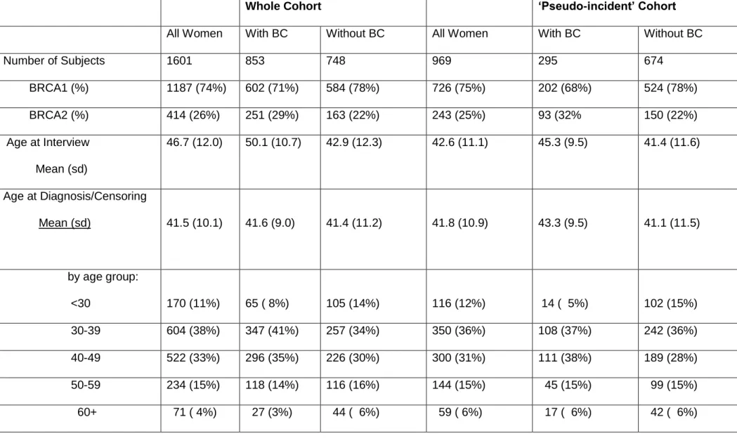
Characteristics of the entire and ‘pseudo-incident’ cohorts are described in table 1. Eight-hundred-seventy-nine women had been affected with BC at the time of their interview, though only 853 of these were considered as affected in this analysis as 26 cases had BC following a previous cancer (mostly ovarian). The remaining 748 women were censored at: age at diagnosis with ovarian cancer (122 subjects); age at diagnosis of another cancer (16); the age at which they underwent prophylactic bilateral mastectomy (31); or age at interview (579 subjects). The average age at censoring for the 748 subjects without BC was 41.4 years, similar to the age at diagnosis of the cases, although the age at interview was substantially older for the BC cases, reflecting the pattern of genetic testing among participants.
The estimated risks from both the standard unweighted and the weighted Cox regressions for ever/never exposure to X-rays are summarized in table 2. Overall, any exposure to chest X-rays was associated with a significant increase in risk in both the entire (HR=1.54, P=.007) and ‘pseudo-incident’ cohorts (HR=1.75, P=.020). Table 2 also shows results of these
analyses stratified by attained age (40 vs. >40), gene (BRCA1 vs. BRCA2), and year of birth (before 1950 vs. 1950+). Higher risks were found in women with attained age up to age 40 and in women born in 1950 or later, although these two categorizations are heavily confounded. In women born in 1950 or later, the relative risks of BC associated with any exposure to X-ray were particularly striking (HR=2.57, P=.002), while in the pseudo-incident
cohort young women had a particularly high risk (HR=2.75, P<.001). Table 3 describes the results of the different periods and levels of exposure. Although some power is lost due to the exclusion of the UK/Ireland subjects, in all analyses the estimated hazard ratios were significantly higher (all P<.05) among those reporting more than four X-rays in at least one age period than those reporting no more than 1-4 X-rays. Similarly, the hazard ratio in women who reported having X-rays only after age 20 was consistently lower that those who had X-rays before age 20. A markedly increased risk of BC was observed for women born after 1949 exposed only before age 20 compared to those never exposed (HR=4.64, 95% CI 2.2 – 10.9, P<.001). Comparable results for the ‘pseudo-incident’ cohort are shown in table 4 and also demonstrate the highly increased BC risk for women exposed before age 20
(HR=5.21), although the sample size is quite small for these analyses. We did not observe any clear differences in the effect of X-ray exposure between BRCA1 and BRCA2 carriers, with BRCA2 carriers (table 2) having a larger effect in the overall cohort, while BRCA1 carriers had a slightly larger effect in the ‘pseudo-incident’ cohort, although in this latter analysis the number of BRCA2 carriers is quite small.
Discussion
These results suggest that diagnostic ionizing radiation exposure from chest X-rays may be associated with a significant breast cancer risk among women who carry a deleterious germline mutation in the BRCA1 and BRCA2 genes. Although our measures of radiation exposure are imprecise in number and timing, it is certain that the ionizing radiation dose in these women from routine chest X-rays is at least an order of magnitude lower than that found in the other radiation-exposed cohorts studied to date. The average dose incurred in the scoliosis cohort is on the order of 100 mSv.11 A pooled analysis of eight radiation exposed cohorts3 estimated a relative risk of ~2.0 at a dose of 1 Gy (=1000mGy) assuming age at exposure of 25. Although dose estimates to the breast from an ordinary chest X-ray vary depending on time period, country and use of fluoroscopy, it is on the order of 0.5mGy2.
Thus even in women who reported a large number of X-rays, the total radiation dose to the breast is unlikely to exceed 10-20 mGy. This corresponds to a predicted relative risk of <1.02 based on the model cited above, which is substantially less than the lower 95% confidence limits from our analyses. Thus, a statistically significant increase in the risk of BC was seen in this population of BRCA1/2 carriers at a level of dose considerably lower than that at which increases have been noted in populations of other (i.e., largely non-carrier) cohorts.
These results must be interpreted with caution, however, as the magnitude of the observed effect is likely to be affected by differential recall of X-ray exposure between affected and unaffected women, and it will be important to confirm these results in prospective studies that avoid such biases. However, it is unlikely that recall bias entirely explains the observed increase: we found no risk associated with another exposure, alcohol consumption at age 20, that might also be subject to such recall bias (HR=1.06, 95% CI 0.8-1.3), nor did we see an effect of chest X-ray exposure in comparing women with ovarian cancer (n=124) to unaffected women (HR=1.26, 95%CI 0.8-2.1). Further, although the accuracy of the self-reported assessment of past diagnostic exposures could not be checked in our study, it is noteworthy that at least three studies have examined this issue by comparing
self-assessment to medical records21-23. All three reports show that while there was a certain amount of disagreement between medical records and interview exposure measures, this misclassification was largely non-differential between cases and controls.
Finally, the apparent relationships with increasing numbers of X-rays and age at exposure, and the consistency of the results in both the whole and ‘pseudo-incident’ cohorts, also lend credibility to the finding of an increased breast cancer risk following low doses of radiation in BRCA mutation carriers. Indeed, despite the crude measures of exposure as assayed in our questionnaire, the pattern of risks observed in our study is consistent with what is known about the effects of radiation on breast cancer in other exposed populations. In this study, X-ray exposure before age 20 was associated with a higher risk than a similar exposure
after age 20, and in general, higher numbers of X-rays were associated with a higher breast cancer risk. Surprisingly, women exposed during both age periods were at an intermediate risk, and as this is biologically implausible, further studies are needed to clarify this effect. We also found that the relative risk was greater in the period up to age 40, particularly in those only exposed before age 20, a pattern consistent with the reports on atomic bomb survivors5,7 and data observed in patients irradiated as treatment for Hodgkin’s disease.24 Moreover, when finer age groups are considered, we see a clear pattern, with estimated HRs of 2.26, 1.72, 1.36, and 1.04 for the risk periods <36, 36-40, 41-50 and >50
respectively. We also observed a strong effect of birth cohort, with no significant increased BC risk observed for women born before 1950. It should also be noted that it is only in the recent birth cohorts that substantial numbers of women report having no X-rays; half of the women reporting having had no X-rays were born in 1960 or later.
Based on the large increased risk of early onset breast cancer in the LSS study cohort and in cohorts of HD survivors, it has been postulated that the excess BC cases in this group may be occurring in a small genetically susceptible subset of the entire exposed population.1,24 Given the role of BRCA1 and BRCA2 in DNA double-strand break repair, BRCA1/2 mutation carriers are strong candidates for such susceptible individuals, with even a relatively modest increase in radiation-induced damage having a substantial impact on breast cancer risk. The results of this study appear to be entirely consistent with this hypothesis and at least one other study has reported an increased breast cancer risk among women with a family history of breast cancer following relatively low ionizing radiation exposure25. It should be noted however, that another study26 has tested a small series of breast cancer cases occurring following very high doses of radiation for treatment of Hodgkin’s disease for mutations in BRCA1 and BRCA2, and identified only a single BRCA2 germ-line mutation.
The results from this study raise potentially significant clinical considerations. The absolute risk of breast cancer by age 50 is of the order of 40% in BRCA1 carriers and 15% in BRCA2
carriers12. A two- to three-fold increased risk associated with exposure to chest X-rays, would imply that there are subgroups of women defined by their X-ray exposure history that are at substantially higher risk and others who are at reduced risk compared to the
population of carriers as a whole. If an increased risk were to be confirmed, however, these results would have implications regarding the appropriate use of medical imaging in carriers at young ages. Thus, in young members of BRCA families, a careful risk-benefit analysis is needed before deciding on a diagnostic procedure that delivers a relatively high dose of ionizing radiation exposure to the breast such as a CT scan, or multiple X-rays. The results presented here also raise the issue of the potential risks from mammographic screening, which is often used to screen BRCA carriers starting in their 30s. Unfortunately, the analysis of the effect of mammographic exposure on breast cancer risk is likely to be biased in retrospective studies due to its obvious relationship to diagnosis, and accordingly, a prospective study of mutation carriers with detailed mammographic exposure history with adjustment for confounding variables (e.g. family history) is a priority.
In conclusion, this study indicates a potentially important association between radiation exposure through chest X-rays and breast cancer risk in BRCA1/2 carriers, of greater magnitude than that seen in the general population. Although the magnitude of this risk cannot be evaluated with any precision due to the likely effects of recall bias, the pattern of risk with age and dose is similar to that in other radiation-exposed cohorts, which provides plausibility to these findings. Nevertheless, confirmation of these results with more detailed exposure data, preferably in a prospective study, is required before definite clinical
Table 1. Characteristics of the study population.
Whole Cohort ‘Pseudo-incident’ Cohort
All Women With BC Without BC All Women With BC Without BC
Number of Subjects 1601 853 748 969 295 674 BRCA1 (%) 1187 (74%) 602 (71%) 584 (78%) 726 (75%) 202 (68%) 524 (78%) BRCA2 (%) 414 (26%) 251 (29%) 163 (22%) 243 (25%) 93 (32% 150 (22%) Age at Interview Mean (sd) 46.7 (12.0) 50.1 (10.7) 42.9 (12.3) 42.6 (11.1) 45.3 (9.5) 41.4 (11.6) Age at Diagnosis/Censoring Mean (sd) 41.5 (10.1) 41.6 (9.0) 41.4 (11.2) 41.8 (10.9) 43.3 (9.5) 41.1 (11.5) by age group: <30 170 (11%) 65 ( 8%) 105 (14%) 116 (12%) 14 ( 5%) 102 (15%) 30-39 604 (38%) 347 (41%) 257 (34%) 350 (36%) 108 (37%) 242 (36%) 40-49 522 (33%) 296 (35%) 226 (30%) 300 (31%) 111 (38%) 189 (28%) 50-59 234 (15%) 118 (14%) 116 (16%) 144 (15%) 45 (15%) 99 (15%) 60+ 71 ( 4%) 27 (3%) 44 ( 6%) 59 ( 6%) 17 ( 6%) 42 ( 6%)
Year of Birth <1940 223 (14%) 151 (18%) 72 (10%) 73 (8%) 25 (8%) 48 (7%) 1940-1949 356 (22%) 232 (27%) 124 (17%) 152 (16%) 57 (19%) 95 (14%) 1950-1959 494 (31%) 296 (35%) 198 (26%) 291 (30%) 109 (37%) 182 (27%) 1960+ 528 (33%) 174 (20%) 354 (47%) 453 (47%) 104 (35%) 349 (52%) Country Groupa 1 358 (22%) 179 (21%) 179 (24%) 211 (22%) 53 (18%) 158 (23%) 2 171 (11%) 81 (10%) 90 (12%) 127 (13%) 45 (15%) 82 (12%) 3 539 (33%) 299 (35%) 231 (31%) 312 (32%) 106 (36%) 206 (31%) 4 542 (34%) 294 (34%) 248 (33%) 319 (33%) 91 (31%) 228 (34%)
Chest X-Ray: Ever/Never
None 398 (25%) 143 (17%) 255 (34%) 279 (29%) 46 (16%) 233 (35%)
>=1 970 (61%) 594 (70%) 376 (50%) 534 (55%) 199 (67%) 335 (50%)
missing 233 (15%) 116 (14%) 117 (16%) 156 (16%) 50 (17%) 106 (15%)
Table 2. Risk of breast cancer (HR) associated with any exposure to X-rays compared with never exposed, in the whole cohort and in the ‘pseudo-incident’ cohort.
Whole Cohort Pseudo-incident Cohort
Unweighted Weighted Unweighted Weighted
Pyrs1 BC2 HR3 (95% CI) HR (95% CI) Pyrs1 BC2 HR3 (95% CI) HR (95% CI) Whole Data Set
Ever vs. Never Exposure 55673 737 1.53*** (1.3 – 1.8) 1.54** (1.1 - 2.1) 4065 245 1.79*** (1.3 –2.5) 1.75* (1.1 –2.8) Stratified by : Attained Age 40 49436 392 1.69*** (1.3 – 2.2) 1.97*** (1.3 – 2.9) 2436 116 2.31*** (1.4 –3.7) 2.75** (1.4 –5.3) >40 5500 345 1.34* (1.0 –1.7) 1.27 (0.8 – 1.8) 1629 129 1.43 (0.9 –2.3) 1.35 (0.7 –2.6) Gene BRCA1 Carrier 41191 535 1.53*** (1.2 – 1.9) 1.42* (1.0 - 2.0) 3130 175 2.06*** (1.4 –3.1) 1.91* (1.1 - 3.3) BRCA2 Carrier 13947 202 1.68** (1.2 – 2.5) 2.33* (1.1 – 5.0) 935 70 1.39 (0.7 –2.7) 1.71 (0.4 –8.2) Year of Birth
<1950 23636 321 1.11 (0.8 – 1.5) 1.17 (0.8 – 3.3) 880 62 0.89 (0.5 – 1.7) - - 1950 3160 416 1.91*** (1.5 – 2.5) 2.57*** (1.8 – 3.7) 3185 183 2.13*** (1.5 – 3.1) - -
1 Number of person-years of observation (not including missing X-ray data)
2 Number of BC cases occurring in the specified cohort/subcohort (not including missing X-ray data) 3
Estimated Hazard Ratio, stratified by birth cohort and country group; analyses adjusted for parity and oophorectomy (yes/no). - weighted analysis not performed on ‘pseudo-incident’ five-year cohort because no cohort-specific weights were available. P-value for test HR=1: *<.05 ; ** P<.01 ; *** P<.001
Table 3. Risk of breast cancer (HR) associated with time and level of exposure to X-rays using the weighted Cox regression analysis of the whole cohort (UK/Ireland data excluded).
Whole Sample Attained Age <41 Born >1949
Ever Exposure Pyrs (BC) HR1 (95% CI) Pyrs (BC) HR1 (95% CI) Pyrs (BC) HR1 (95% CI)
Never Exposed 9882 (95) 1.00 9033 (60) 1.00 6342 (49) 1.00
1 X-Ray 26164 (377) 1.56* (1.0 – 2.4) 23162 (200) 1.86* (1.1 – 3.0) 14523 (224) 2.56*** (1.6 – 4.1) Age at X-ray Exposure
Never 9882 (95) 1.00 9033 (60) 1.00 6342 (49) 1.00
Before age 20 Only 2205 (34) 1.76 (0.9 – 3.4) 2002 (26) 2.61** (1.3 – 5.4) 1504 (25) 4.64*** (2.2 – 10.9) After age 20 Only 4291 (57) 1.32 (0.8 – 2.3) 3668 (20) 1.26 (0.6 – 2.7) 2054 (27) 1.70 (0.9 – 3.4) Before & after age 20 18365 (262) 1.60* (1.0 – 2.5) 16383 (144) 1.91* (1.1 – 3.2) 10380 (160) 2.50*** (1.5 – 4.1)
Level of X-ray Exposure
Never 9882 (95) 1.00 9033 (60) 1.00 6342 (49) 1.00
At least one period with 1-4 Xrays and no period with 5+ X-rays
At least one period with 5+ X-rays
15761 (250) 1.92** (1.2 – 3.0) 13862 (127) 2.39*** (1.4 – 4.0) 7727 (140) 3.57*** (2.1 – 6.0)
1 Adjusted for parity and oophorectomy (yes/no), stratified by country group and birth cohort. 2
Ever/Never result included to provide a comparison with result in table 2 including the UK/Ireland data. *
Table 4. Weighted Cox regression Analysis of combined X-ray exposure in the pseudo-incident cohort with follow-up beginning five years prior to interview. UK/Ireland data excluded.
Exposure Pyrs (BC) HR1 (95% CI)
Age at X-ray Exposure
Never 773 (28) 1.00
Before age 20 Only 137 (12) 5.21** (1.6 – 17.5) After age 20 Only 256 (26) 1.91 (0.9 – 4.1) Before & after age 20 978 (88) 1.98* (1.1 – 3.7)
Level of X-ray Exposure
Never 773 (28) 1.00 At least one period with
1-4 Xrays ; and no period with 5+ X-rays
705 (50) 1.76 (0.9 – 3.4)
At least one period with 5+ X-rays
666 (76) 2.69** (1.4 – 5.3)
1 Adjusted for parity and oophorectomy (yes/no) , stratified by country group and birth cohort. * P-value <.05 ; ** P<.01 ; *** P<.001
References
1. Ronckers CM, Erdmann CA, Land CE. Radiation and breast cancer: a review of current evidence. Breast Cancer Research 7:21-32, 2005.
2. United Nations Scientific Committee on the Effects of Atomic Radiation. Sources and Effects of Ionizing Radiation. New York: United Nations, 2000.
3. Preston DL, Mattsson A, Holmberg E, et al: Radiation effects on breast cancer risks: a pooled analysis of eight cohorts. Radiat Res 158:220-235, 2002.
4. Van Leeuwen FE, Klokman WJ, Stovall M et al: Roles of radiation dose,
chemotherapy, and hormonal factors in breast cancer following Hodgkin’s disease. J Natl Cancer Inst 95:971-980, 2003.
5. Tokunaga M, Land CE, Tokuoka S, et al: Incidence of female breast cancer among atomic bomb survivors, Hiroshima and Nagasaki, 1960-1985. Radiat Res 138:209-223, 1994.
6. Land CE, Hayakawa N, Machado et al : A case-control interview study of breast cancer among Japanese A-bomb survivors, I: main effects. Cancer Causes Control 5:157-165, 1994.
7. Land CE, Tokunaga M, Koyama K et al: Incidence of female breast cancer among atomic bomb survivors, Hiroshima and Nagasaki, 1950-1990. Radiat Res 160:707-717, 2003.
8. Hoffman DA, Lonstein JE, Morin MM, et al: Breast cancer in women with scoliosis exposed to multiple diagnostic x rays. J Natl Cancer Inst 81(17):1307-1312, 1989. 9. Boice JD JR, Preston D, Davis FG, et al: Frequent chest X-ray fluoroscopy and
breast cancer incidence among tuberculosis patients. Radiat Res 125:214-22, 1991. 10. Howe GR, McLaughlin J: Breast cancer mortality between 1950 and 1987 after
fluoroscopy cohort study and a comparison with breast cancer mortality in the atomic bomb survivors study. Radiat Res 145:694-707, 1996.
11. Doody MM, Lonstein JE, Stoval M et al : Breast cancer mortality after diagnostic radiography : findings from the US Scoliosis Cohort Study Spine 25:2052-2063, 2000.
12. Antoniou A, Pharoah PD, Narod S, et al: Average risks of breast and ovarian cancer associated with BRCA1 or BRCA2 mutations detected in case series unselected for family history: a combined analysis of 22 studies. Am J Hum Genet 72:1117-1130, 2003.
13. Venkitaraman AR: Cancer susceptibility and the functions of BRCA1 and BRCA2. Cell 108:171-182, 2002.
14. Powell SN, Kachnic LA: Roles of BRCA1 and BRCA2 in homologous recombination, DNA replication fidelity and the cellular response to ionizing radiation. Oncogene 22:5784-5791, 2003.
15. Baldeyron C, Jacquemin E, Smith J et al: A single mutated BRCA1 allele leads to impaired fidelity of double strand break end-joining. Oncogene 21:1401-1410, 2002.
16. Buchholz TA, Wu X, Hussain A et al : Evidence of haplotype insufficiency in human cells containing a germline mutation in BRCA1 or BRCA2. Int J Cancer 97:557-561, 2002. 17. Rothfuss A, Schutz P, Bochum S et al: Induced micronucleus frequencies in peripheral
lymphocytes as a screening test for carriers of a BRCA1 mutation in breast cancer families. Cancer Res 60:390-394, 2000.
18. The IBCCS Collaborators Group: The International BRCA1/2 Carrier Cohort Study: Purpose, Rationale, and Study Design. Breast Cancer Res 2000 (6) WEB -
http://breast-cancer-research.com/content/2/6/E010
19. Antoniou A, Goldgar D, Andrieu N, et al: A weighted cohort approach for analysing factors modifying disease risks in carriers of high risk susceptibility genes, Genetic Epidemiology 29:1-11, 2005.
20. Lin DY and Wei LJ: Robust inference for the Cox proportional hazards model. J Am Stat Assn 84:1074-1078, 1989.
21. Pagoda JM and Preston-Martin S: Radiation exposure from diagnostic imaging: Agreement between self-report and medical records. Health Physics 83:907-917, 2002.
22. Berrington de Gonzalez A, Ekboom A, Glass A, et al: Comparison of documented and recalled histories of exposure to diagnostic X-rays in case-control studies of thyroid cancer. Am J Epidemiol 157:652-663, 2003.
23. Preston-Martin S, Bernstein L, Maldonado AA, et al: Comparison of information from patient interviews and dental charts. Am J Epidemiol. 1985;121:430-439, 1985. 24. Dores GM, Metayer C, Curtis RE, et al: Second malignant neoplasms among
long-term survivors of Hodgkin's disease: a population-based evaluation over 25 years. J Clin Oncol. 20:3484-3494, 2002.
25. Hill DA, Preston-Martin S, Ross RK, et al.: Medical radiation, family history of cancer, and benign breast disease in relation to breast cancer risk in young women, USA. Cancer Causes Control 13:711-718, 2002.
26. Nichols KE, Heath JA, Friedman D, et al: TP53, BRCA1, and BRCA2 tumor
suppressor genes are not commonly mutated in survivors of Hodgkin’s disease with second primary neoplasms. J Clin Oncol 21:4505-4509, 2003.
Appendix A. Collaborators
IBCCS Collaborating Group
Vienna, Austria: Teresa Wagner, Verena Korn, Christine Fürhauser Odense, Denmark: Anne-Marie Gerdes
Budapest, Hungary: Edith Olah Reykjavik, Iceland: Jorunn Eyford Milan, Italy: Paolo Radice
Madrid, Spain: Javier Benitez, Ana Osorio
Madrid Spain: Trinidad Caldes, Miguel de la Hoya Szczecin, Poland: Jan Lubinski
Stockholm, Sweden: Brita Arver
Lund, Sweden: H. Olsson, Niklas Loman Quebec, Canada: Jacques Simard Brussels, Belgium: Catherine Sibille
GEO-HEBON Collaborating Centers
Center for Human and Clinical Genetics, Dept. of Clinical Genetics, Leiden University Medical Center: Christi van Asperen
Dept. of Clinical Genetics, Erasmus Medical Center, Rotterdam: Hanne Meijers-Heijboer Dept. of Clinical Genetics, Nijmegen Medical Center, Nijmegen: Nicoline Hoogerbrugge Family Cancer Clinic, The Netherlands Cancer Institute, Amsterdam: Senno Verhoef The Netherlands Foundation for the detection of hereditary tumours: Leiden, Hans Vasen Dept. of Medical Genetics, University Medical Center Utrecht, Utrecht: Margreet Ausems Dept. of Clinical Genetics and Human Genetics, VU University Medical Center, Amsterdam: Fred Menko
Dept. of Clinical Genetics, Maastricht University Medical Center, Maastricht: Encarna Gomez-Garcia
EMBRACE Collaborating Centers
Coordinating Centre, Cambridge: Susan Peock, Margaret Cook
North of Scotland Regional Genetics Service, Aberdeen: Neva Haites, Helen Gregory Northern Ireland Regional Genetics Service, Belfast: Patrick Morrison
West Midlands Regional Clinical Genetics Service, Birmingham: Trevor Cole, Carole McKeown
South West Regional Genetics Service, Bristol: Alan Donaldson East Anglian Regional Genetics Service, Cambridge: Joan Paterson Medical Genetics Services for Wales, Cardiff: Jonathon Gray
St James’s Hospital, Dublin & National Centre for Medical Genetics, Dublin: Peter Daly, David Barton
South East of Scotland Regional Genetics Service, Edinburgh: Mary Porteus, Michael Steel Peninsula Clinical Genetics Service, Exeter: Carole Brewer, Julia Rankin
West of Scotland Regional Genetics Service, Glasgow: Rosemarie Davidson, Victoria Murday
South East Thames Regional Genetics Service, London: Louise Izatt, Gabriella Pichert North West Thames Regional Genetics Service, Harrow: Huw Dorkins
Leicestershire Clinical Genetics Service, Leicester: Richard Trembath Yorkshire Regional Genetics Service, Leeds: Tim Bishop, Carol Chu Merseyside & Cheshire Clinical Genetics Service, Liverpool: Ian Ellis
Manchester Regional Genetics Service, Manchester: Gareth Evans, Fiona Lalloo, Andrew Shenton
North East Thames Regional Genetics Service, London: James Mackay, Anne Robinson Nottingham Centre for Medical Genetics, Nottingham: Susan Ritchie, Sandy Raeburn Northern Clinical Genetics Service, Newcastle: Fiona Douglas, John Burn
Department of Cancer Genetics, Royal Marsden Hospital: Ros Eeles
North Trent Clinical Genetics Service, Sheffield: Jackie Cook, Oliver Quarrell South West Thames Regional Genetics Service, London: Shirley Hodgson
Wessex Clinical Genetics Service, Southampton: Diana Eccles, Anneke Lucassen
GENEPSO Collaborating Centers
Coordinating Centre, Centre René Hugenin, Saint Cloud: Catherine Noguès, Emmanuelle Fourme, Rosette Lidereau, Denise Stevens
Paris, Institut Curie : Dominique Stoppa-Lyonnet, Marion Gauthier-Villars Villejuif, Institut Gustave Roussy : Agnès Chompret
Saint Cloud, Centre René Huguenin : Catherine Noguès Strasbourg, Centre Paul Strauss : Jean-Pierre Fricker Caen, Centre François Baclesse: Pascaline Berthet
Vandoeuvre-les-Nancy, Centre Alexis Vautrin: Elisabeth Luporsi Lyon, Centre Léon Bérard : Christine Lasset, Valérie Bonadona
Angers, Nantes: Centres Paul Papin, René Gauducheau and Catherine de Sienne : Alain Lortholary
Nice, Centre Antoine Lacassagne: Marc Frénay Dijon, Hôpital D'Enfants CHU: Laurence FAIVRE
Marseille, Institut Paoli-Calmettes: Hagay Sobol, François Eisinger, Laetitia HUIART Bordeaux. Institut Bergonié: Michel Longy
Reims, Institut Jean Godinot: Tan Dat Nguyen
Toulouse, Institut Claudius Regaud: Laurence Gladieff, Rosine Guimbaud Niort, CH Georges Renon: Paul Gesta
Lille, Centre Oscar Lambret: Philippe Vennin, Claude Adenis
Rouen, Hôpital Charles Nicolle, Centre Henri Becquerel : Annie Chevrier, Annick Rossi Clermont-Ferrand, Centre Jean Perrin: Yves-Jean Bignon
Rennes, Centre Eugène Marquis: Catherine Dugast Reims, Polyclinique Courlancy: Liliane Demange
Marseille, Hôpital de la Timone: Hélène Zattara-Cannoni Avignon, Clinique Sainte Catherine: Hélène Dreyfus Montpellier, CHU Arnaud Villeneuve: Mehrdad Noruzinia Limoges, CHRU Dupuytren: Laurence Venat-Bouvet Associated centres :
Nadine Andrieu (Inserm Emi00-06/Service de Biostatistique, Institut Curie, Paris), Catherine Bonaïti (Inserm U535, Villejuif), Claire Julian-Reynie (Inserm U379, Marseille), Florent de Vathaire (Inserm U605, Villejuif), Hagay Sobol (IPC, Inserm E-9939, Marseille)