HAL Id: hal-02540647
https://hal.umontpellier.fr/hal-02540647
Submitted on 11 Apr 2020
HAL is a multi-disciplinary open access
archive for the deposit and dissemination of
sci-entific research documents, whether they are
pub-lished or not. The documents may come from
teaching and research institutions in France or
abroad, or from public or private research centers.
L’archive ouverte pluridisciplinaire HAL, est
destinée au dépôt et à la diffusion de documents
scientifiques de niveau recherche, publiés ou non,
émanant des établissements d’enseignement et de
recherche français ou étrangers, des laboratoires
publics ou privés.
Effects of Exposure to a 128-mT Static Magnetic Field
on Glucose and Lipid Metabolism in Serum and Skeletal
Muscle of Rats
Miryam Elferchichi, Jacques Mercier, Marjorie Coisy-Quivy, Lore Metz,
Anne-Dominique Lajoix, René Gross, Hatem Belguith, Hafedh Abdelmelek,
Mohsen Sakly, Karen Lambert
To cite this version:
Miryam Elferchichi, Jacques Mercier, Marjorie Coisy-Quivy, Lore Metz, Anne-Dominique Lajoix, et
al.. Effects of Exposure to a 128-mT Static Magnetic Field on Glucose and Lipid Metabolism in
Serum and Skeletal Muscle of Rats. Archives of Medical Research, Elsevier, 2010, 41 (5), pp.309-314.
�10.1016/j.arcmed.2010.07.008�. �hal-02540647�
ORIGINAL ARTICLE
Effects of Exposure to a 128-mT Static Magnetic Field on Glucose and Lipid
Metabolism in Serum and Skeletal Muscle of Rats
Miryam Elferchichi,
a,b,cJacques Mercier,
b,cMarjorie Coisy-Quivy,
bLore Metz,
dAnne-Dominique Lajoix,
eRene Gross,
eHatem Belguith,
aHafedh Abdelmelek,
aMohsen Sakly,
aand Karen Lambert
b,caFaculte´ des Sciences de Bizerte, Laboratoire de Physiologie Inte´gre´e, Jarzouna, Tunisia bINSERM, ERI25, Muscle and Pathologies, Montpellier, France
cUniversite´ Montpellier I, Montpellier, France
dLaboratoire de Biologie des Activite´s Physiques et Sportives, Biologie Bat B, Universite Blaise Pascal, Les Ce´zeaux, Aubie`re, France eCNRS UMR5232, UM1, Centre for Pharmacology and Innovation in Diabetes, Montpellier, France
Received for publication June 24, 2010; accepted July 2, 2010 (ARCMED-D-10-00145).
Background and Aims. Increasing environmental pollution may participate in the growing incidence of metabolic disorders. Static magnetic fields (SMFs) are an emerging environmental health issue due to increased exposure in residential and commercial areas; however, their metabolic effects in serum and skeletal muscle are largely unknown. The aim of this study was to investigate the effect of SMF exposure on glucose and lipid metabolism in serum and skeletal muscles of rats.
Methods. Twelve 6- to 7-week-old male Wistar rats were randomly divided into two groups: rats exposed to 128 mT SMF and sham-exposed rats. This moderate-intensity exposure was performed for 1 h/day for 15 consecutive days.
Results. Animals exposed to 128 mT SMF displayed significant changes in both glucose (i.e., increases in plasma glucose and lactate and decrease in plasma insulin levels) and lipid (i.e., increases in plasma glycerol, cholesterol and phospholipids but not triglyceride levels) metabolism. During intraperitoneal glucose tolerance tests, SMF-exposed rats displayed significantly higher hyperglycemia compared to sham-exposed rats despite similar insulin levels in both groups. In tissues, SMF exposure induced significant alter-ations in enzyme activities only in glycolytic muscles and caused a significant decrease in quadriceps and liver glycogen content together with increased phospholipid levels. Conclusions. This study provides evidence that subacute SMF exposure of moderate inten-sity induces important alterations of glucose and lipid metabolisms, which deserve further investigations to evaluate long-term consequences. ! 2010IMSS. Published byElsevier Inc.
Key Words: Metabolism, Skeletal muscle, Static magnetic fields.
Introduction
The incidence of metabolic disorders grows among devel-oping nations. Several factors are known to promote metabolic alterations. These include endogenous factors characterized by the interplay of genes with insulin-related abnormalities
and metabolic deregulations, as well as exogenous factors such as sedentary life style, food intake and environmental exposure(1e3).
This latter factor may be implicated in the development of metabolic disorders(1,2,4). Electromagnetic fields (EMFs) and static magnetic fields (SMFs) are growing environmental pollutants due to increased exposure in both residential and working areas(5,6). To date there are few data on health effects of static magnetic fields.
In an attempt to address the biological effects of SMFs, it is useful to classify them as weak (!1 mT), moderate Address reprint requests to: Karen Lambert, INSERM ERI25, Baˆtiment
Crastes de Paulet, Hoˆpital A. de Villeneuve, 34 295 Montpellier Cedex 5 France; Phone: þ 33(4) 67 41 52 30; FAX: þ 33(4) 67 41 52 31; E-mail:
karen.lambert@univ-montp1.fr
0188-4409/$ - see front matter. Copyright! 2010 IMSS. Published by Elsevier Inc. doi: 10.1016/j.arcmed.2010.07.008
(1 mT to 1T), strong (1e5 T) and ultrastrong (O5 T). Until recently, the International Commission on Non-Ionizing Radiation Protection (ICNIRP) recommendations for occupational exposure were a whole-working-day time-averaged exposure limit of 200 mT. The acute exposure limit for head and trunk was 2T, which is the maximum for limiting vertigo, nausea and metallic taste (7). In 2009, SMF guidelines were modified(8). Now, the public limit is at 400 mT for occupational exposure and up to 8T exposure can be permitted in a controlled environment. However, moderate-intensity SMF exposure (generally |200 mT) has been shown to affect a wide variety of bio-logical systems in vitro(9e15). Another important and so far underestimated outcome is the impact of SMFs on in vivo metabolism. Indeed, the most recently published study deals with the effects of SMFs on bacteria metabolism(16)
and only few studies describe their effects in mammals. For instance, Gorczynska et al. (17) observed an increase in blood glucose associated with a reduced insulin secretion in rats exposed to a constant magnetic field of moderate intensity, 1 h/day for 10 days. Recently, Chater et al.(18)
reported an increase in glycemia after subacute exposure of pregnant rats to 128 mT, 1 h/day for 13 consecutive days. Skeletal muscles represent the most important metabol-ically active mass of the body and play a major role in the regulation of lipid and glucose metabolism. Therefore, skel-etal muscles may be sensitive to SMF exposure. Metabolic response is highly dependent on oxidative and glycolytic muscle fiber types with sensitivity to external stimuli related to muscle typology (3). In vitro, SMFs enhance skeletal muscle differentiation (19) and accelerate Ca2þ/ calmodulin-dependent myosin light-chain phosphorylation
(20). Moreover, SMFs alter ion transporters (9) and seem to affect muscle microcirculation (14,21,22). Finally, because SMFs are widely used in therapeutics for musculo-skeletal pain relief (23), it is crucial to dissect their effect on skeletal muscle homeostasis and to take into account muscle typology.
In the present study, our aim was to investigate whether subacute exposure to moderate-intensity SMFs could induce both systemic and local (i.e., in oxidative and glycolytic muscle and liver) alterations of glucose and lipid metabolism. Therefore, we compared various hormonal and metabolic parameters in male Wistar rats exposed or not to 128 mT SMF (1 h/day) for 15 days.
Materials and Methods Animals and Protocol
Animal care and experimental procedures were carried out in accordance with the guidelines set by the European Community Directives (86/609/EEC) and the protocol was approved by the Ethics Committee of the University of Montpellier I, France.
Twelve 6- to 7-week-old male Wistar rats were housed (n 5 3/cage) in a temperature-controlled room at 25"C with
a relative humidity of 80% and a 12:12 h light-dark cycle (lights on at 8 AM). Water and food were available ad libitum. Rats were randomly divided into two groups: rats exposed to 128 mT SMF (n 5 6, SMF) and sham-exposed rats (n 5 6, sham sham-exposed). Rats sham-exposed to 128 mT SMF and sham-exposed rats were placed in the electro-magnet for 1 h/day for 15 consecutive days. The exposure period always took place between 8 and 12 AM under standard light exposure and constant temperature.
Exposure System
We used an electromagnet (Model EM4-HVA, Lake Shore Cryotronic, Inc., Westerville, OH) and a magnet power supply (Model 647, Lake Shore Cryotronic, Inc.) with an air gap of 11 cm. This apparatus incorporates water-cooled coils and precision yokes that assure precise cap alignment and excellent field stability and uniformity when high power is required to achieve the maximum field capa-bility for the electromagnet. SMF intensity was measured and standardized over the total floor area of the plexiglas cage at 128 mT. SMF uniformity in the active exposure volume was #0.2% over 1 cm3
. The cage measured 20 $ 10 $ 20 cm. The two coils of the Lake Shore electromagnet were separated by a 12.1-cm gap. Exposed and sham-exposed rats (n 5 2/each time) were placed in the cage at the center of the uniform field area and exposed (or not) to 128 mT SMF.
Intraperitoneal Glucose Tolerance tests (IPGTT)
Two days before sacrifice, rats underwent an intraperitoneal glucose tolerance test (IPGTT) as previously described
(24). Briefly, after being fasted for 4 h, a glucose solution (2 g/kg body weight) was administered IP. Blood was collected after tail snipping at time 0 and 20, 40, 60, and 90 min after glucose administration for measurements of plasma glucose and insulin levels.
Blood Chemistry
Exposed and sham-exposed rats were sacrificed by decapita-tion in postprandial state. Blood samples were immediately centrifuged and plasma aliquots were frozen and stored at %80"C until assays.
Plasma glucose and glycerol/triglyceride concentrations were determined using enzymatic methods following the manufacturer’s instructions (Sigma 510, and Serum Triglycer-ides Determination Kit TR0100, Sigma, France). Insulin concentrations were determined using radioimmunoassay with125I-labeled insulin and a rat insulin antiserum to
deter-mine the level of rat insulin. The sensitivity of this radioimmu-noassay was 0.02 ng/ml (SRI-13K, Labodia, France). The colorimetric enzymatic test CHOD-PAP (Biomaghreb 310 Elferchichi et al./ Archives of Medical Research 41 (2010) 309e314
20111, Tunisia) was used for cholesterol quantification ac-cording to the manufacturer’s instructions. Lactate concentra-tion was determined according to the method of Gutmann and Wahlefeld (25) and plasma phospholipids were analyzed following the method developed by Shibuya et al.(26). Tissue Sampling
Immediately after sacrifice, the soleus (SOL; oxidative muscle) and the extensor digitorum longus (EDL; glycolytic muscle) of the hindlimb were removed, frozen in liquid nitrogen and stored at e80"C until use for enzymatic
activ-ities measurements. Quadriceps and liver biopsies were carried out to be used for determination of tissue glycogen, phospholipids, triglycerides and glycerol concentrations. Enzymatic Activities
Citrate synthase (CS) activity was measured at 412 nm and 30"C for 2.5 min as suggested by Srere(27).
3-Hydroxyac-yl-coenzyme A-dehydrogenase (HADH) and lactate dehy-drogenase (LDH) activities were measured at 340 nm for 10 min and 2.5 min, respectively. Results are expressed in micromoles/minute/g of tissue (mmol/min/g).
Muscle and Liver Glycogen Contents
Muscle and liver glycogen contents were determined from quadriceps and liver biopsies using the procedure described by Lo et al. (28). Values were calculated from a standard curve generated at the same time and expressed in mg glycogen/g tissue.
Data Presentation and Statistical Analysis
Data are presented as mean # SEM. Statistical significance of the differences between mean values was assessed by Student t-test or Mann-Whitney U test; differences within groups for the IPGTT values were assessed by analysis of variance (ANOVA) followed by Bonferroni post hoc tests. The level of significance was set at p !0.05.
Results
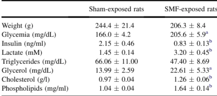
Effect of SMF on Body Weight and Metabolic Parameters SMF exposure did not induce an obvious phenotype; exposed rats were normal in terms of appearance, body weight and relative weight of tissues. However, SMF-exposed rats dis-played significant changes in metabolic parameters. Under postprandial conditions, SMF-exposed rats presented higher glycemia and lower insulinemia values than sham-exposed animals (Table 1). They also displayed a significant increase of plasma lactate compared to sham-exposed rats.
On the other hand, SMF exposure had no effect on the triglycerides level, whereas it strongly enhanced plasma glycerol, cholesterol and phospholipid concentrations.
Effect of SMF Exposure on Glucose Tolerance
To investigate whole-body glucose metabolism, IPGTT was performed in fasted animals 2 days before sacrifice. IP admin-istration of glucose resulted in an increase of plasma glucose and insulin concentrations in both groups (Figure 1). However, ANOVA analysis showed that overall, SMF-exposed rats had a significant higher glucose levels than sham-exposed animals ( p !0.01). These higher glucose levels in exposed rats were accompanied by insulin levels similar to those observed in non-exposed control animals.
Table 1. Basal metabolic and morphometric parameters in sham-exposed and SMF-sham-exposed rats
Sham-exposed rats SMF-exposed rats
Weight (g) 244.4 # 21.4 206.3 # 8.4 Glycemia (mg/dL) 166.0 # 4.2 205.6 # 5.9a Insulin (ng/ml) 2.15 # 0.46 0.83 # 0.13b Lactate (mM) 1.45 # 0.14 3.20 # 0.45b Triglycerides (mg/dL) 66.06 # 11.00 47.40 # 8.69 Glycerol (mg/dL) 13.99 # 2.59 22.61 # 5.33a Cholesterol (g/l) 0.97 # 0.04 1.26 # 0.06b Phospholipids (mg/ml) 1.04 # 0.04 1.64 # 0.14b n 5 6 in each group. ap !0.05.
bp !0.01 significantly different from sham-exposed rats.
Figure 1. Glucose (A) and insulin (B) levels during an intraperitoneal glucose tolerance test (IPGTT) in sham-exposed (-; n 5 5) and SMF exposed rats (,; n 5 6). Values are mean # SEM. There is a significant increase in hyperglycemia in SMF-exposed rat ( p !0.01) compared to sham-exposed rat and $: statistical difference ( p !0.01) at time 20 min between groups.
Effects on Metabolic Parameters in Quadriceps and Liver Biopsies after SMF Exposure
Because skeletal muscle and liver play a crucial role in glucose and lipid metabolism, we then analyzed metabolic parameters in these tissues. Glycogen content was strongly reduced both in quadriceps (by 44%) and liver (by 25%) biopsies of exposed rats compared with sham-exposed rats. We also noticed a significant increase in phospholipid content in both tissues after exposure to SMF (Table 2); in contrast, we did not find significant differences in hepatic and muscular levels of triglycerides and glycerol (Table 2). Enzymatic Activities in Oxidative and Glycolytic Muscle Biopsies after SMF Exposure
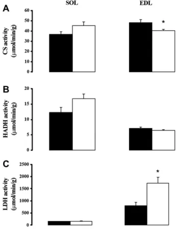
To further analyze the effect of SMF exposure on skeletal muscle, we studied the activities of enzymes involved in glycolytic and oxidative metabolism in both oxidative and glycolytic muscle. We therefore tested CS, HADH and LDH activities in the soleus (SOL; oxidative muscle) and in the extensor digitorum longus (EDL; glycolytic muscle) of both groups (Figure 2). SMF exposure did not affect CS, HADH and LDH activities in oxidative muscles. Conversely, in glyco-lytic muscles, SMF exposure reduced CS activity and increased LDH but had no effect on HADH activity.
Discussion
In this study we showed that exposure of normal Wistar rats to 128 mT SMF (1 h/day) for 15 days induced significant changes in lipid and glucose homeostasis. In accordance with previously published studies (17,18), we found that rats exposed to SMF displayed significantly higher glyce-mia and lower insulineglyce-mia. We were also able to show that exposure to SMF induced a significant increase in hyper-glycemia after IPGTT without insulin response alteration, suggesting a decrease in glucose tolerance possibly due to impaired glucose uptake after SMF exposure. The main tissues implicated in glucose uptake are skeletal muscle and liver. We did find a significant decrease in glycogen content in muscles and liver. This reduction may be caused
either by decreased glucose uptake or increased glycogen-olysis. Indeed, Abdelmelek et al. (29) reported higher norepinephrine levels in skeletal muscle of rats after SMF exposure at 128 mT, which could account for increased glycogen breakdown. Such a hypothesis cannot be excluded but, in our opinion, a decrease in glucose uptake is more likely to be responsible for the reduction of glycogen content in liver and skeletal muscles. Indeed, our exposed rats also presented higher plasma lactate concentrations and several studies have demonstrated a metabolic compe-tition between lactate and glucose uptake (30,31). In the presence of elevated plasma lactate, glucose uptake is Table 2. Liver and quadriceps parameters in sham-exposed and SMF-exposed rats
Liver Quadriceps Sham SMF Sham SMF Glycogen (mg/g tissue) 33.3 # 3.1 25.2 # 1.4a 5.7 # 0.5 3.2 # 0.3b Phospholipids (mg/g tissue) 16.7 # 0.6 20.4 # 0.3b 12.7 # 0.7 17.7 # 1.2b Triglycerides (mg/dL) 98.6 # 13.7 110.3 # 11.0 20.0 # 2.2 22.1 # 1.4 Glycerol (mg/dL) 103.0 # 12.1 117.7 # 9.9 19.0 # 1.7 20.6 # 1.3 n 5 6 in each group. ap !0.05.
bp !0.01 significantly different from sham-exposed rats.
Figure 2. (A) CS activity in oxidative muscle: soleus (SOL) and glycolytic muscle: extensor digitorum longus (EDL) in sham-exposed (-; n 5 6) and SMF exposed rats (,; n 5 6); (B) HADH activity and (C) LDH activity. Values are mean # SEM. *p !0.05 vs. sham-exposed rats. 312 Elferchichi et al./ Archives of Medical Research 41 (2010) 309e314
reduced and lactate is used as a metabolic substrate(31,32). This metabolic switch is often described in glucose homeo-stasis disorders such as diabetes (33e35)and our exposed rats also displayed lower plasma insulin levels, which could further favor the decrease in glucose uptake. Insulin levels were not different in fasted SMF-exposed rats and controls and also during glucose tolerance tests in the two groups. The reason for such a discrepancy between fasting and postprandial insulin levels (changes in insulin degradation and/or secretion) remains to be determined. SMF exposure has been shown to have an impact on impact on insulin secretion. Sakurai et al. (36) found an increase in insulin secretion after SMF exposure and, on the other hand, on isolated pancreatic islets, Hayek et al. (37) demonstrated a decrease in insulin secretion after SMF exposure. These alterations in insulin secretion may be due to the increase in reactive oxygen species (ROS), which are suggested to be important mediators of SMF effects(38).
Skeletal muscle is the main producer and consumer of lactate (39e41). The increase in LDH activity and the decrease in CS activity were observed only in glycolytic muscles. These data indicate that glycolytic muscles partic-ipate actively in the increase of plasma lactate under our conditions and emphasize the importance of muscle typology in metabolic responses.
Finally, we also found that after SMF exposure, glycerol, cholesterol and phospholipid contents were significantly increased in the plasma, whereas triglycerides remained unchanged. An excess of circulating lipids is often associ-ated with cardiovascular diseases and glucose metabolism deregulation (42,43). Adipose tissue is a likely candidate for the release of such lipids in the circulation because of lipid metabolism alterations. The higher plasma glycerol level in rats exposed to SMFs suggest an increased adipose tissue lipolysis is in accordance with the decreased plasma insulin levels. In addition, increases in circulating choles-terol and phospholipids suggest a higher turnover of plasma membrane constituents. Our results concerning lipid metab-olism alterations are in line with the signalling networks identified to respond to SMF exposure by Wang et al.
(44). Hashish et al.(45)found in liver a significant increase in lipid peroxidation associated with a decrease in the anti-oxidant GSH after SMF exposure, suggesting an increase in oxidative stress after such exposure. Alternate magnetic field exposure induces an accumulation of lipids in the plasma membrane of heart associated with a decrease in membrane fluidity (46). Moreover, Rosen et al. (11) sug-gested that changes to ion channel conductivity due to SMF exposure may result from the slow re-orientation of aligned groups of diamagnetic phospholipid molecules within the cell membrane. Thus, SMF exposure could alter membrane fluidity by changing phospholipids membrane composition and phospholipids properties. Consequently, SMF exposure could modify molecule flux into the cell membrane.
In conclusion, our findings at both systemic and tissue levels produce evidence for an impaired glucose homeostasis and a deregulated lipid metabolism after a moderate-intensity SMF exposure (1 h/day) for 15 days. These data question the safety of such exposures. Further investigations are necessary to evaluate long-term consequences of these metabolic alterations and their reversibility.
References
1. Chen H, Simar D, Lambert K, et al. Maternal and postnatal overnutri-tion differentially impact appetite regulators and fuel metabolism. Endocrinology 2008;149:5348e5356.
2. Chen H, Simar D, Morris MJ. Hypothalamic neuroendocrine circuitry is programmed by maternal obesity: interaction with postnatal nutri-tional environment. PLoS One 2009;4:e6259.
3. Lambert K, Py G, Robert E, et al. Does high-sucrose diet alter skeletal muscle and liver mitochondrial respiration? Horm Metab Res 2003;35: 546e550.
4. Havas M. Dirty electricity elevates blood sugar among electrically sensitive diabetics and may explain brittle diabetes. Electromagn Biol Med 2008;27:135e146.
5. Dini L, Abbro L. Bioeffects of moderate-intensity static magnetic fields on cell cultures. Micron 2005;36:195e217.
6. Genuis SJ. Fielding a current idea: exploring the public health impact of electromagnetic radiation. Public Health 2008;122:113e124. 7. Swanson J. Consultation on revisions to the European Directive on
occupational EMF exposure. J Radiol Prot 2009;29:291e292. 8. International Commission on Non-Ionizing Radiation Protection.
Guidelines on limits of exposure to static magnetic fields. Health Phys 2009;96:504e514.
9. Ite!gin M, Gu¨nay I, Lo!go!glu G, et al. Effects of static magnetic field on specific adenosine-5’-triphosphatase activities and bioelectrical and biome-chanical properties in the rat diaphragm muscle. Bioelectromagnetics 1995;16:147e151.
10. Jajte J, Grzegorczyk J, Zmy"slony M, et al. Effect of 7 mT static magnetic field and iron ions on rat lymphocytes: apoptosis, necrosis and free radical processes. Bioelectrochemistry 2002;57:107e111. 11. Rosen AD. Mechanism of action of moderate-intensity static magnetic
fields on biological systems. Cell Biochem Biophys 2003;39:163e173. 12. Rosen AD. Effect of a 125 mT static magnetic field on the kinetics of voltage activated Naþchannels in GH3 cells. Bioelectromagnetics
2003;24:517e523.
13. Rosen AD, Chastney EE. Effect of long term exposure to 0.5 T static magnetic fields on growth and size of GH3 cells. Bioelectromagnetics 2009;30:114e119.
14. Xu S, Okano H, Ohkubo C. Subchronic effects of static magnetic fields on cutaneous microcirculation in rabbits. Vivo 1998;12:383e389. 15. Yuge L, Kataoka K. Differentiation of myoblasts is accelerated in
culture in a magnetic field. Vitro Cell Dev Biol Anim 2000;36: 383e386.
16. Morrow AC, Dunstan RH, King BV, et al. Metabolic effects of static magnetic fields on Streptococcus pyogenes. Bioelectromagnetics 2007;28:439e445.
17. Gorczynska E, Wegrzynowicz R. Glucose homeostasis in rats exposed to magnetic fields. Invest Radiol 1991;26:1095e1100.
18. Chater S, Abdelmelek H, Pequignot JM, et al. Effects of sub-acute exposure to static magnetic field on hematologic and biochemical parameters in pregnant rats. Electromagn Biol Med 2006;25:135e144. 19. Coletti D, Teodori L, Albertini MC, et al. Static magnetic fields enhance skeletal muscle differentiation in vitro by improving myoblast alignment. Cytometry A 2007;71:846e856.
20. Pilla AA, Muehsam DJ, Markov MS, et al. EMF signals and ion/ligand binding kinetics: prediction of bioeffective waveform parameters. Bioelectrochem Bioenerg 1999;48:27e34.
21. Brix G, Strieth S, Strelczyk D, et al. Static magnetic fields affect capil-lary flow of red blood cells in striated skin muscle. Microcirculation 2008;15:15e26.
22. Xu S, Okano H, Ohkubo C. Acute effects of whole-body exposure to static magnetic fields and 50-Hz electromagnetic fields on muscle microcirculation in anesthetized mice. Bioelectrochemistry 2001;53: 127e135.
23. Pilla AA. Mechanisms and therapeutic applications of time-varying and static magnetic fields. In: Barnes FGB, ed. Handbook of Biolog-ical Effects of Electromagnetic Fields. 3rd Ed. Boca Raton: CRC Press; 2006. pp. 351e412.
24. Metz L, Vermaelen M, Lambert K, et al. Endurance training increases lactate transport in male Zucker fa/fa rats. Biochem Biophys Res Commun 2005;331:1338e1345.
25. Gutmann I, Wahlefeld M. L-(þ)-Lactate Determination with Lactate Dehydrogenase and NAD. New York: Academic Press; 1974. 26. Shibuya I, Honda H, Maruo B. Simplified colorimetry without
incinera-tion of phosphorus in phosphatides. Agric Biol Chem 1967;31:111e114. 27. Srere P. Citrate synthase. Methods Enzymol 1969;13:3e11. 28. Lo S, Russell JC, Taylor AW. Determination of glycogen in small
tissue samples. J Appl Physiol 1970;28:234e236.
29. Abdelmelek H, Molnar A, Servais S, et al. Skeletal muscle HSP72 and norepinephrine response to static magnetic field in rat. J Neural Transm 2006;113:821e827.
30. Miller BF, Fattor JA, Jacobs KA, et al. Lactate and glucose interac-tions during rest and exercise in men: effect of exogenous lactate infusion. J Physiol 2002;544:963e975.
31. Vettor R, Lombardi AM, Fabris R, et al. Substrate competition and insulin action in animal models. Int J Obes Relat Metab Disord 2000;24:S22eS24.
32. Lombardi AM, Fabris R, Bassetto F, et al. Hyperlactatemia reduces muscle glucose uptake and GLUT-4 mRNA while increasing (E1alpha) PDH gene expression in rat. Am J Physiol 1999;276:E922eE929. 33. Vettor R, Lombardi AM, Fabris R, et al. Lactate infusion in
anesthe-tized rats produces insulin resistance in heart and skeletal muscles. Metabolism 1997;46:684e690.
34. Py G, Eydoux N, Perez-Martin A, et al. Streptozotocin-induced dia-betes decreases rat sarcolemmal lactate transport. Metabolism 2001; 50:418e424.
35. Py G, Lambert K, Milhavet O, et al. Effects of streptozotocin-induced diabetes on markers of skeletal muscle metabolism and monocarbox-ylate transporter 1 to monocarboxmonocarbox-ylate transporter 4 transporters. Metabolism 2002;51:807e813.
36. Sakurai T, Terashima S, Miyakoshi J. Effects of strong static magnetic fields used in magnetic resonance imaging on insulin-secreting cells. Bioelectromagnetics 2009;30:1e8.
37. Hayek A, Guardian C, Guardian J, et al. Homogeneous magnetic fields influence pancreatic islet function in vitro. Biochem Biophys Res Commun 1984;122:191e196.
38. Okano H. Effects of static magnetic fields in biology: role of free radicals. Front Biosci 2008;13:6106e6125.
39. Bergman BC, Wolfel EE, Butterfield GE, et al. Active muscle and whole body lactate kinetics after endurance training in men. J Appl Physiol 1999;87:1684e1696.
40. Brooks GA. Lactate: link between glycolytic and oxidative metabo-lism. Sports Med 2007;37:341e343.
41. Hashimoto T, Brooks GA. Mitochondrial lactate oxidation complex and an adaptive role for lactate production. Med Sci Sports Exerc 2008;40:486e494.
42. Boden G, Shulman GI. Free fatty acids in obesity and type 2 diabetes: defining their role in the development of insulin resistance and beta-cell dysfunction. Eur J Clin Invest 2002;32:14e23.
43. Savage DB, Petersen KF, Shulman GI. Disordered lipid metabolism and the pathogenesis of insulin resistance. Physiol Rev 2007;87: 507e520.
44. Wang Z, Sarje A, Che P-L, et al. Moderate strength (0.23e0.28 T) static magnetic fields (SMF) modulate signaling and differentiation in human embryonic cells. BMC Genomics 2009;10:356e379. 45. Hashish AH, El-Missiry MA, Abdelkader HI, et al. Assessment of
biological changes of continuous whole body exposure to static magnetic field and extremely low frequency electromagnetic fields in mice. Ecotoxicol. Environ. Saf 2007;71:895e902.
46. Chernysheva ON. Status of the lipid phase of plasma membranes of the rat heart after repeated exposure to an alternate magnetic field of 50 Hz frequency. Kosm Biol Aviakosm Med 1990;24:30e31. 314 Elferchichi et al./ Archives of Medical Research 41 (2010) 309e314