HAL Id: inserm-01857861
https://www.hal.inserm.fr/inserm-01857861
Submitted on 17 Aug 2018
HAL is a multi-disciplinary open access
archive for the deposit and dissemination of
sci-entific research documents, whether they are
pub-lished or not. The documents may come from
teaching and research institutions in France or
abroad, or from public or private research centers.
L’archive ouverte pluridisciplinaire HAL, est
destinée au dépôt et à la diffusion de documents
scientifiques de niveau recherche, publiés ou non,
émanant des établissements d’enseignement et de
recherche français ou étrangers, des laboratoires
publics ou privés.
Aurélie Daumas, Julie Alingrin, Richard Ouedraogo, Patrick Villani, Marc
Leone, Jean-Louis Mege
To cite this version:
Aurélie Daumas, Julie Alingrin, Richard Ouedraogo, Patrick Villani, Marc Leone, et al.. MALDI-TOF
MS monitoring of PBMC activation status in sepsis. BMC Infectious Diseases, BioMed Central, 2018,
18 (1), pp.355. �10.1186/s12879-018-3266-7�. �inserm-01857861�
R E S E A R C H A R T I C L E
Open Access
MALDI-TOF MS monitoring of PBMC
activation status in sepsis
Aurélie Daumas
1,2*, Julie Alingrin
1,3, Richard Ouedraogo
1, Patrick Villani
2, Marc Leone
1,3and Jean-Louis Mege
1Abstract
Background: MALDI-TOF mass spectrometry (MS) on whole cells enables the detection of different cell types and cell activation. Here, we wondered whether this approach would be useful to investigate the host response in sepsis. Methods: Peripheral blood mononuclear cells (PBMCs) from patients with severe sepsis and healthy donors were analyzed with MALDI-TOF MS. PBMCs from healthy donors were also stimulated with lipopolysaccharide, peptidoglycan, CpG oligonucleotides, polyinosinic polycytidylic acid, and with heat-inactivated bacteria. Averaged spectra of PBMCs stimulated in vitro by different agonists were generated from the database using the Biotyper software and matching scores between each spectrum from patients and averaged spectra from the database were calculated.
Results: We show that the MALDI-TOF MS signature of PBMCs from septic patients was specific, compared with healthy controls. As the fingerprints observed in patients may be related to PBMC activation, PBMCs from healthy controls were stimulated with cytokines, soluble Pathogen-Associated Molecular Patterns (PAMPs) and heat-killed bacteria, and we created a database of reference spectra. The MALDI-TOF MS profiles of PBMCs from septic patients were then compared with the database. No differences were found between patients with documented infection (n = 6) and those without bacteriological documentation (n = 6). The spectra of PBMCs from septic patients matched with those of interferon-γ- and interleukin-10-stimulated PBMCs, confirming that sepsis is characterized by both inflammatory and immunoregulatory features. Interestingly, the spectra of PBMCs from septic patients without documented infection matched with the reference spectrum of PBMCs stimulated by CpG oligonucleotides, suggesting a bacterial etiology in these patients. Conclusions: Despite the limits of this preliminary study, these results indicate that the monitoring of functional status of PBMCs in peripheral blood by whole cell MALDI-TOF MS could provide unique opportunities to assess disease
progression or resolution in clinical settings.
Keywords: Sepsis, Mass spectrometry, MALDI-TOF, Mononuclear cells, IFN-γ, Interleukin-10, CpG oligonucleotides
Background
Sepsis is the combination of a systemic inflammatory re-sponse syndrome (SIRS) and infection [1]. Sepsis can pro-gress to severe sepsis and septic shock, with mortality rates of 25 to 30% and 40 to 70%, respectively [2,3]. The clinical manifestations of the early stages of sepsis are often similar to those of a patient with SIRS caused by sterile inflamma-tion [3], leading to frequent underappreciation of sepsis in clinical practice. The traditional approach to sepsis diagno-sis is based on the clinical signs and symptoms of sepdiagno-sis,
supported by relevant microbiological data. Unfortunately, up to 40% of the infections suspected in patients with sepsis are not microbiologically documented [4–6]. Consequently, physicians often use empiric antibiotic therapy, which has three major drawbacks: increased antibiotic resistance, patient toxicity, and elevated costs [7].
Two important challenges for physicians are to deter-mine if the patient is infected or not in the absence of microbiological documentation, and when to begin anti-microbial therapy [8–10]. Numerous molecules, such as procalcitonin, are unable to discriminate between sepsis and SIRS [11, 12]. Cytokine imbalance has been thought to be useful for defining sepsis. Indeed, the recognition of soluble Pathogen-Associated Molecular Patterns (PAMPs) such as lipopolysaccharide (LPS), peptidoglycan (PGN),
* Correspondence:aurelie.daumas@ap-hm.fr
1
Aix-Marseille Université, URMITE, IHU Méditerranée Infection, UMR CNR 7278, IRD 198, INSERM 1095, Marseille, France
2Service de Médecine Interne et Thérapeutique, Hôpital de la Timone,
Assistance Publique-Hôpitaux de Marseille, Marseille, France Full list of author information is available at the end of the article
© The Author(s). 2018 Open Access This article is distributed under the terms of the Creative Commons Attribution 4.0 International License (http://creativecommons.org/licenses/by/4.0/), which permits unrestricted use, distribution, and reproduction in any medium, provided you give appropriate credit to the original author(s) and the source, provide a link to the Creative Commons license, and indicate if changes were made. The Creative Commons Public Domain Dedication waiver (http://creativecommons.org/publicdomain/zero/1.0/) applies to the data made available in this article, unless otherwise stated.
Daumaset al. BMC Infectious Diseases (2018) 18:355 https://doi.org/10.1186/s12879-018-3266-7
CpG oligonucleotides (CpG-ODN), and polyinosinic poly-cytidylic acid (poly I:C) by pathogen recognition receptors (PRRs) induces the release of inflammatory cytokines, such as gamma interferon (IFN-γ) and the concomitant liberation of immunoregulatory cytokines, including inter-leukin (IL)-10 and IL-4. However, cytokine profiles are also unable to discriminate sepsis, and seem more related to SIRS severity than to sepsis [13].
MALDI-TOF mass spectrometry (MS) has emerged as a fast, reliable and inexpensive tool for bacterial identifi-cation and diagnosis [14, 15]. Interestingly, bacterial identification does not require previous fractionation steps [16]. Recently, we and others have applied the whole-cell MALDI-TOF MS technique to identify eukaryotic cells, including circulating cells [17–19]. Our team has also shown that whole-cell MALDI-TOF MS detects the multifaceted activation of monocyte-derived macrophages in response to various cytokines and bac-terial pathogens [20]. Portevin et al. [21] demonstrated recently that MALDI-TOF MS fingerprints distinguish human monocyte sub-populations activated by distinct microbial ligands.
Our goal was to analyze peripheral blood mononuclear cells (PBMCs) in septic patients through whole-cell MALDI-TOF MS. This approach enabled the detection of a specific PBMC signature in septic patients. The ana-lysis of the signature of healthy PBMCs stimulated with cytokines, soluble PAMPs and bacteria, frequently in-volved in sepsis, showed that the spectra of PBMCs from septic patients matched with those of PBMCs stimulated by IFN-γ, IL-10 and CpG-ODN. These findings evoked an infectious activation in septic patients regardless of documented or undocumented infection. Despite the limits of this preliminary study, this is the first report describing the use of a whole-cell MALDI-TOF MS ap-proach to identify PBMC activation in septic patients.
Methods
Ethics statement
The study was approved by the Ethics Committee of the Assistance Publique-Hôpitaux de Marseille, France. Blood was collected after informed and written consent of healthy donors and septic patients.
Patients and healthy controls
Patient recruitment was provided from an ancillary study to the project “De-escalation of Empirical Antibiotics in Severe Sepsis” (Comité de Protection des Personnes Sud Méditerranée No. 2011–002297-22). Twelve patients (aged 18 and over) were enrolled in the polyvalent inten-sive care unit (North Hospital, Marseille, France). A single blood sample was collected at the time of empirical anti-biotic initiation. Eligibility criteria were the presence of se-vere sepsis requiring empirical antimicrobial treatment.
Severe sepsis was defined as the criteria for SIRS and sus-pected infection with at least one organ failure. SIRS was defined by two or more of the following conditions: temperature > 38 °C or < 36 °C, heart rate > 90 beats per minute, respiratory rate > 20 breaths per minute or PaCO2 < 32 mmHg, white blood cell count >12G/l, < 4G/ l, or > 10% immature cells. Two patients with SIRS had
Staphylococcus aureus bacteremia, 4 had gram-negative
bacillus bacteremia and 6 had a strongly suspected infec-tion clinically but not microbiologically documented. PBMCs from healthy donors were isolated from leuko-packs (Etablissement Français du Sang).
Isolation and in vitro activation of PBMCs
Blood was collected in EDTA-containing tubes and PBMCs were isolated using Ficoll cushions (MSL, Euro-bio). After centrifugation, PBMCs were washed in sterile phosphate buffered saline (PBS) without Ca2+and Mg2+,
and 1 × 10⁶ cells were suspended in 10 μL of PBS and
frozen at− 80 °C for 2 to 3 days before analysis. In some experiments, PBMCs from healthy donors (1 × 10⁶cells in 6-well plates) were incubated in 2 mL of RPMI 1640 containing 10% fetal calf serum (FCS) and stimulated with 20 ng/mL IFN-γ (PeproTech), 20 ng/mL IL-4 or IL-10 (R&D Systems) for 18 h [22]. PBMCs were also
stimulated with LPS from Escherichia coli (1 μg/mL,
Sigma-Aldrich), peptidoglycan (PGN) from Bacillus
sub-tilis (10 μg/mL, Sigma-Aldrich), CpG-ODN (2 μg/mL,
InvivoGen) and poly I:C (25 μg/mL, InvivoGen). Finally, PBMCs were stimulated with heat-inactivated bacteria (10 bacteria per cell). Bacteria included oxacillin-sensitive S. aur-eus, community strain group B streptococcus, Pseudomonas
aeruginosa (ATCC 27853) and E. coli (ATCC 25922).
PBMCs stimulated for 18 h were pelleted in 10μl of PBS and frozen as unstimulated PBMCs for MALDI-TOF MS analysis.
MALDI-TOF MS analysis
After thawing of the PBMCs, 1 μL of cell suspension
was added to 1μL of matrix solution (saturated solution
of α-cyano-4-hydroxy-cynnamic acid in a mixture of
50% acetonitrile, 25% trifluoroacetic acid and water) as previously described [17, 20, 23]. The mixture was de-posited on the MALDI target. The evaporation that gradually took place at room temperature allowed the
formation of α-cyano-4-hydroxy-cynnamic acid crystals
containing the dispersed samples. Measurements were performed using an Autoflex II mass spectrometer (Bruker Daltonics, Wissembourg, France) equipped with a 337-nm nitrogen laser. Each sample was irradiated with a laser for desorption and ionization. Each spectrum resulted from the sum of positive ions obtained after 525 laser shots in differ-ent regions of the analyzed spot (automatic mode). All the positive-ion mass spectra were acquired in the linear mode
at an acceleration voltage of 20 kV in the delayed extraction mode. A signal-to-noise of 3.0 was selected to define peaks, with a maximum of 100 peaks per spectrum. Spectra were automatically acquired with a mass/charge (m/z) ranging from 2000 to 20,000 Da using FlexControl and FlexAnalysis 2.4 software (Bruker Daltonics). The x-axis of spectra repre-sented the m/z ratio (in daltons) of ionized molecules, and the y-axis indicated the intensity (relative abundance) of these ions.
Spectrum analysis
Analyses and graphical outputs were performed using the free and open source statistical analysis software R (version 2.13), along with specific analysis libraries
(MALDIquant) as previously described [20]. The gel
view representation indicates the reproducibility of the spectra. A hierarchical clustering with a ward algorithm for agglomeration and a dissimilarity matrix based on the Jaccard distance were used to classify the spectra. The MALDI Biotyper 3 software (Bruker Daltonics) was used to create an average reference spectrum for each PBMC sample, corresponding to at least 10 individual spectra. The Biotyper software realigns acquired spectra and automatically creates an average spectrum using default Biotyper software settings provided by the manu-facturer, and we created a database as previously
de-scribed [20]. The Biotyper software also allows the
identification of unknown spectra as shown in clinical samples by comparison with reference spectra, for the identification and classification of microorganisms [14]. The score values proposed by the manufacturer have been used for microorganism identification. The score values between 0.000 and 1.699 do not allow reliable mi-crobe identification; the values between 1.700 and 1.999 allow probable cell identification and score values higher than 2.0 are considered statistically significant; they allow the confident identification of different microbe species. We extended this method to assess PBMC acti-vation status in septic patients. As the score values pro-vided by Bruker Daltonics ranged from 0.000 to 2.000 when we used control samples and reference spectra, we considered that medians of matching scores higher than 1.5 allowed reliable identification of the activation profile of patient PBMCs and could be clinically relevant. Dif-ferences between conditions were tested with the Mann-Whitney U test and a cutoff value of 0.05 was chosen to consider a difference statistically significant.
Results
Signatures of PBMCs
As compared to phenotype studies that require panels of antibodies to characterize circulating cells, MALDI-TOF MS enables simple and rapid measurement of cell status. First, we wondered if PBMCs from healthy controls were
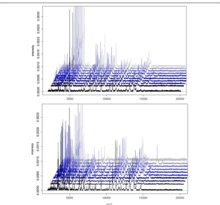
characterized by a specific whole-cell MALDI-TOF MS signature. The spectra of PBMCs from ten healthy do-nors were composed of numerous peaks ranging from 2000 to 15,000 Da, with a major peak at 4961 Da. Note that the spectra of the ten donors were highly reprodu-cible (Fig. 1a). Second, we found that the signatures of PBMCs from ten septic patients were similar but they were distinct from those of PBMCs from healthy con-trols. Because MALDI-TOF MS profiles were specific for PBMCs from healthy controls and septic patients,
the dendrogram constructed by Ouedraogo et al. [17]
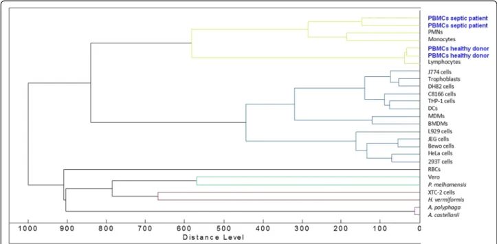
was implemented with reference spectra of PBMCs from two healthy donors and two septic patients. As expected, the PBMCs from the two healthy donors were similar and clustered with T lymphocytes and, to a lesser extent, with monocytes and polymorphonuclear cells (PMNs). Likewise, the PBMCs from the two septic patients were similar and formed a specific cluster. This cluster was close to that of monocytes and PMNs but was away from the T lymphocyte cluster (Fig.2). This specific pat-tern underlines the prominent role of the innate immune response in sepsis. Taken together, these results demonstrated that PBMC fingerprints distinguished patients with sepsis from healthy controls.
Peak characteristics of PBMCs in sepsis
We wondered if the analysis of individual peaks enables stratification of patients with sepsis, microbiologically documented or not. When the spectra were analyzed by comparing the presence or absence of peaks, we found that 10 peaks were present only in controls, 12 peaks were common to healthy donors and septic patients and 18 peaks were specific in septic patients (Table1). Tak-ing into account the limitations related to the limited number of patients in this preliminary study, among these 18 specific peaks, 15 were common to all septic patients independently of microbiological documenta-tion. Concerning the three other peaks, the peak at 5415 Da was found in patients with gram-negative bacil-lus bacteremia (4/4) and one patient without microbio-logical documentation. The peak at 3329 Da was present in 3/4 patients with gram-negative bacillus bacteremia and the same patient without bacteriological documenta-tion, suggesting that these two peaks could be associated with gram-negative bacillus bacteremia and that this patient without microbiological documentation could have gram-negative bacillus sepsis. Finally, the peak at 2942 Da was found in the two patients with S. aureus bacteremia and in one patient without microbiological documentation, suggesting that this peak could be
re-lated to S. aureus bacteremia (Table 1). Despite the
limits of the study, these results show that the signature of septic patients is very similar, regardless of docu-mented or undocudocu-mented infection.
Signatures of PBMCs activated in vitro
Because the differences between the signatures of healthy and septic PBMCs may be related to their activa-tion state, we stimulated PBMCs with various agonists to determine if PBMC activation results in specific pro-files. The stimulation of PBMCs with IFN-γ, LPS, IL-4 and IL-10 led to specific and reproducible responses, as shown using a virtual gel view representation (Fig. 3). The spectra from all stimulated samples were clearly separated from those of unstimulated PBMCs. Each type of stimulation led to specific fingerprints. The spectra from the PBMCs stimulated with inflammatory molecules (IFN-γ/LPS) clustered together. Similarly,
immunoregulatory cytokines induced the clustering of PBMCs. The existence of these three major clusters (un-stimulated, IFN-γ/LPS-, IL-4/IL-10-stimulated PBMCs) suggests that MALDI-TOF MS may be used to analyze the inflammatory response of PBMCs in the clinical setting.
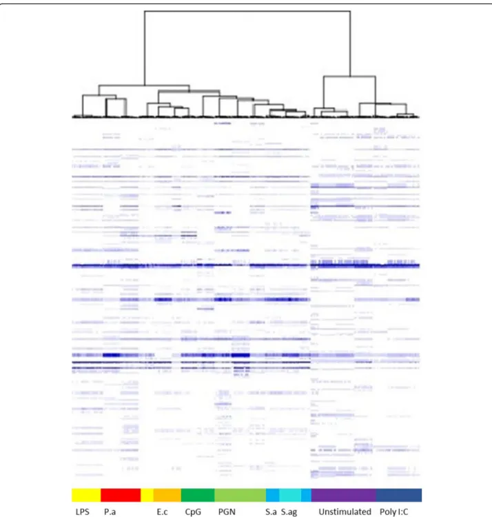
We also investigated the responses of PBMCs to PAMPs and bacteria often found in sepsis. For that pur-pose, we used unstimulated PBMCs as controls and we compared the fingerprints induced by PAMPs and bac-teria to these controls. Two very different clusters were clearly identified: one including unstimulated and poly I:C-stimulated PBMCs and the other with PBMCs
Fig. 1 MALDI-TOF MS spectra of PBMCs. The PBMCs (1 × 10⁶cells) from ten healthy donors (a) and ten septic patients (b) were suspended in
10μL of PBS, and 1 μL was deposited onto the MALDI target. Representative MALDI-TOF MS spectra are shown. MALDI-TOF MS spectra were analyzed using statistical analysis software R (version 2.13)
stimulated with bacteria and other PAMPs (Fig. 4). Among the bacterial signatures, we observed that the signatures induced by P. aeruginosa and E. coli (Gram negative bacteria) were coupled to the signature induced by LPS from E. coli. Similarly, the signatures induced by S. agalactiae group B and S. aureus (gram-positive bac-teria) were associated and close to the signature induced by PGN from B. subtilis. These results suggest that MALDI-TOF MS may be useful to assess a series of spe-cific responses of PBMCs to varied agonists.
Finally, we used matching scores to compare the differ-ent modes of PBMC activation after having created aver-aged spectra of stimulated PBMCs. The MALDI-TOF MS profiles of PBMCs from septic patients were then com-pared with the database. None of the septic patients matched with the averaged spectrum of healthy donors, while the spectra of septic patients significantly matched with IFN-γ and IL-10 spectra regardless of whether the in-fection was documented (n = 6) or not (n = 6), confirming that sepsis is characterized by both inflammatory and im-munoregulatory features (Fig. 5 and Additional file 1: Figure S1). It is noteworthy that the scores obtained by comparing the spectra of E. coli- and S. aureus-infected patients with the reference spectrum of E. coli- and S. aur-eus-stimulated PBMCs were significantly lower (p < 0.05) than those obtained with IL-10- and IFN-γ-specific spec-tra, respectively (see Fig.5a, b), suggesting the prominence of the activation profile as a specific response to
pathogens. Importantly, we found that the spectra of PBMCs from septic patients significantly (p < 0.05) matched with CpG-ODN, independently of a documented infection. Clearly, the activation profile found in the pa-tients with unknown infection was similar to that of the patients with documented infection (see Fig.5c), suggest-ing that these patients had a bacterial infection. Taken together, these results are consistent with that septic PBMCs were activated in a specific way and suggest a bac-terial infection in septic patients without documented infection.
Discussion
Sepsis is a frequent and serious complication in intensive care unit patients. Despite many years of active research to find effective and specific therapies, the only true treatment still relies on organ system support and effect-ive antimicrobial eradication with antibiotics and/or sur-gical intervention. An important factor in optimizing survival rates in these patients is the speed of diagnosis [2–5]. However, diagnosing sepsis is not always straight-forward, particularly in patients who have complex on-going disease processes. In addition, many of these patients received antimicrobial therapy that rendered microbial cultures negative. Hence, 30–40% of intensive care unit patients with severe sepsis had negative bacter-ial cultures [4–6]. Even when cultures are positive, re-sults take several days to become available, thus slowing
Fig. 2 Dendrogram representation of PBMCs. The dendrogram constructed by Ouedraogo et al. [17] was implemented with reference spectra of PBMCs from two healthy donors and two septic patients. The Biotyper (Bruker Daltonics) software was used to create an averaged spectrum for each patient, corresponding to at least 10 individual spectra. The averaged spectra were added to the database using the Biotyper software and the dendrogram creation method. Peripheral blood mononuclear cells (PBMCs); polymorphonuclear cells (PMNs); dendritic cells (DCs); monocyte-derived macrophages (MDMs); bone marrow-monocyte-derived macrophages (BMDMs); red blood cells (RBCs)
the diagnostic process. Many biomarkers have been pro-posed over the years, but the diagnostic value of these molecules remains uncertain [24,25].
We reasoned that a more integrated approach such as MALDI-TOF MS may be used to detect sepsis specific-ally in patients without microbiological documentation. As expected, we found that the signature of PBMCs from different healthy subjects was highly reproducible. It clustered with T lymphocytes but was largely different from the signature of numerous non-circulating cells. The signatures of PBMCs from septic patients largely differed from that of PBMCs from healthy controls. Interestingly, they clustered with the signature of mono-cytes and PMNs but not with T lymphomono-cytes. The absence of clustering with T lymphocytes may be related to the lymphopenia associated with systemic inflamma-tion syndromes [26]. This specific pattern underlines the prominent role of the innate immune response in sepsis.
We postulated that the fingerprints of septic patients may be related to specific activation of PBMCs. To assess the activation of PBMCs, we stimulated PBMCs from healthy controls with cytokines, PAMPs and bac-teria. First, we identified both inflammatory (IFN-γ and LPS) and immunoregulatory (IL-4 or IL-10) signatures in PBMCs. Second, we also found that gram-negative bacteria and LPS induced specific signatures compared to those induced by gram-positive bacteria and PGN. Interestingly, the signatures induced by bacterial PAMPs were separate from a poly I:C, a PAMP known to strongly stimulate type 1 interferon as do most of vi-ruses. This result might be useful in discriminating bac-terial and viral infections, such as in pneumonia, for which no clinical, radiological and laboratory data differ-entiate bacterial from viral pneumonia [27].
Finally, we attempted to relate in vitro data and the fingerprints of septic PBMCs. We clearly identified IFN-γ and IL-10 signatures in septic patients. This result is consistent with the natural history of sepsis, where both inflammatory and immunoregulatory responses are observed [28]. We did not detect the signals delivered in vitro by heat-inactivated bacteria and bacterial ligands such as LPS and PGN, even when microbiological infec-tion was documented. We can hypothesize that the lack of LPS and PGN signatures in sepsis may be related to anergy (endotoxin tolerance) [29]. In contrast, we found an intense CpG-ODN signature in septic patients, even in patients without microbiological documentation, sug-gesting the prominence of the activation profile as a spe-cific response to pathogens. The Biotyper score cutoffs of 1.5 can be considered as unacceptably low for func-tions such as microbial identification. However, no com-parison is possible because no score has so far been validated to discriminate specific responses of PBMCs to varied agonists. This is the first report describing the use
Table 1 Peak characteristics of PBMCs Healthy donors Gram-negative bacillus bacteremia S. aureus bacteremia Patients without documented infection 2165 2227 2302 2503 2617 2617 2617 2631 2631 2631 2646 2646 2646 2777 2795 2795 2795 2942 2942 3329 3329 3355 3355 3355 3363 3369 3369 3369 3369 3398 3398 3398 3426 3426 3426 3441 3441 3441 3441 3455 3467 3467 3467 3485 3485 3485 3485 3708 3708 3708 3881 3998 3998 3998 4323 4323 4323 4642 4935 4935 4935 4935 4961 4961 4961 4961 4983 4983 4983 4983 5023 5415 5415 6298 6298 6298 6342 6342 6342 6574 6574 6574 6574 7621 7621 7621 7762 7762 7762 7762 8561 8561 8561 8561 9285 9285 9285 9285 10,259 10,259 10,259 10,259 10,441 10,441 10,441 10,831 10,831 10,831 10,831
The PBMCs from healthy donors and septic patients were analyzed by MALDI-TOF MS
Them/z ratio of peaks is shown. The peaks that were common to septic patients are underlined
of a whole-cell MALDI-TOF MS approach to identify PBMC activation in septic patients. As the score values provided by Bruker Daltonics ranged from 0.000 to 2.000 when we used control samples and reference spec-tra, we considered that medians of matching scores higher than 1.5 allowed reliable identification of the acti-vation profile of patient PBMCs. Despite the choice of this score cutoff, we did not detect the signals delivered in vitro by heat-inactivated bacteria or bacterial ligands such as LPS and PGN in septic patients even with docu-mented microbiological infection. Obviously, we ob-served a distinct and reproductible IFN-γ, IL-10 and
CpG-ODN. The preliminary nature of the findings re-quires nevertheless confirmation of results.
To our knowledge, this is the first report describing the use of whole-cell MALDI-TOF MS analysis to iden-tify mass spectra that discriminate specific responses of PBMCs to varied agonists and to study functional and activation status of septic patients with and without doc-umented bacterial infection.
Conclusions
This study shows that MALDI-TOF MS of patient PBMCs is easy and fast to perform and may be
Fig. 3 Gel view representation of activated PBMCs. The PBMCs from healthy donors were stimulated with 20 ng/ml IFN-γ, IL-4, IL-10 or 1 μg/mL LPS for 18 h. The spectra were arranged in a pseudo-gel format using a gel view representation. Vertical axis refers to the m/z ratio. Spectra are classified according to the presence/absence of peaks. Unstimulated PBMCs are presented in white
considered as a routine method for the detection of sep-sis. The reproducibility and accuracy of this approach enables the analysis of several types of PBMC activation and shows a similar activation signature for septic patients with and without documented bacterial infec-tion. Consequently, this innovative approach may be
promising in helping physicians in the identification and prognosis of septic patients and/or their treatment. This proof of concept could easily be translated to clinical studies to monitor the functional status of PBMCs from patients under treatment and to study the activation sta-tus of PBMCs from patients suffering from systemic and
Fig. 4 Hierarchical clustering of activated PBMCs. PBMCs were stimulated with different agonists for 18 h. The results are shown as hierarchical clustering. Vertical axis refers to the m/z ratio. Spectra are classified according to the presence/absence of peaks. PBMCs stimulated with LPS from Escherichia coli are presented in yellow, with Pseudomonas aeruginosa (P.a) in red, Escherichia coli (E.c) in orange, CpG ODN in dark green, PGN in green, Staphylococcus aureus (S.a) in blue, Streptococcus agalactiae (S.ag) in turquoise, unstimulated PBMCs in purple and PBMCs stimulated with poly I:C in dark blue
chronic inflammatory disorders. However, the prelimin-ary nature of the findings requires confirmation of re-sults. As a next step, larger studies would confirm whether this new technique can improve the medical management of patients. High throughput monitoring of functional status of PBMCs in peripheral blood based on whole cell MALDI-TOF MS could provide unique op-portunities to monitor disease progression and reso-lution in clinical settings.
Additional file
Additional file 1:Figure S1. Comparison between in vitro and in vivo data. Averaged spectra of PBMCs stimulated in vitro by different agonists were generated from the database using the Biotyper software. The spectra (n = 16) from 2 other patients with gram-negative bacillus bacteremia, the second patients with S. aureus bacteremia and three other patients with undocumented infection were then compared with the averaged spectra of the database. Matching scores between each spectrum from patients and averaged spectra from the database are represented with circles. Horizontal lines represent the medians of matching scores; a value higher than 1.5 was considered significant and allowed confident identification of the activation status of PBMCs. (JPG 320 kb)
Abbreviations
CpG-ODN:CpG oligonucleotides; E. coli: Escherichia coli; IFN-γ: Gamma interferon; IL: Interleukin; LPS: Lipopolysaccharide; MS: Mass spectrometry; PBMCs: Peripheral blood mononuclear cells; PGN: Peptidoglycan; Poly I:C: Polyinosinic polycytidylic acid; PRRs: Pathogen recognition receptors; S. aureus: Staphylococcus aureus; SIRS: Systemic inflammatory response syndrome
Acknowledgements
We thank Christian Capo for his attentive reading of our manuscript. RO was financially supported by“Infectiopôle Sud” foundation.
Availability of data and materials
The datasets used and/or analysed during the current study are available from the corresponding author on reasonable request.
Authors’ contributions
AD, RO and JLM designed the study. AD, RO and JA carried out the experiments. AD, PV, ML and JLM carried out the statistical analysis and drafted the manuscript. All authors read and approved the final version of the manuscript.
Ethics approval and consent to participate
Patient recruitment was provided from an ancillary study to the project “De-escalation of Empirical Antibiotics in Severe Sepsis”. Written informed consent was obtained from the participants or their parents. The study was approved by the Ethics Committee Sud Méditerranée (No. 2011–002297-22). Confidentiality of the participants’ details was assured.
Consent for publication Not applicable.
Competing interests
The authors declare that they have no competing interests.
Publisher’s Note
Springer Nature remains neutral with regard to jurisdictional claims in published maps and institutional affiliations.
Author details
1
Aix-Marseille Université, URMITE, IHU Méditerranée Infection, UMR CNR 7278, IRD 198, INSERM 1095, Marseille, France.2Service de Médecine Interne et
Thérapeutique, Hôpital de la Timone, Assistance Publique-Hôpitaux de Marseille, Marseille, France.3Service d’Anesthésie et Réanimation polyvalente,
Hôpital Nord, Assistance Publique-Hôpitaux de Marseille, Marseille, France.
Received: 24 December 2017 Accepted: 23 July 2018
References
1. Singer M, Deutschman CS, Seymour CW, Shankar-Hari M, Annane D, Bauer M, Bellomo R, Bernard GR, Chiche JD, Coopersmith CM, Hotchkiss RS, Levy MM, Marshall JC, Martin GS, Opal SM, Rubenfeld GD, van der Poll T, Vincent JL, Angus DC. The Third International Consensus Definitions for Sepsis and Septic Shock (Sepsis-3). JAMA. 2016;315:801–10.
2. Annane D, Aegerter P, Jars-Guincestre MC, Guidet B. Current epidemiology of septic shock: the CUB-Rea network. Am J Respir Crit Care Med. 2003;168: 165–72.
3. Cohen J, Brun-Buisson C, Torres A, Jorgensen J. Diagnosis of infection in sepsis: an evidence-based review. Crit Care Med. 2004;32:S466–94. Fig. 5 Comparison between in vitro and in vivo data. Averaged spectra of PBMCs stimulated in vitro by different agonists were generated from the database using the Biotyper software. The spectra (n = 16) from four patients with E. coli bacteremia, two patients with S. aureus bacteremia and six patients with undocumented infection were then compared with the averaged spectra of the database. Scatter plots of one representative septic patient with E. coli infection (a), S. aureus infection (b) or without microbiological documentation (c) are presented. Matching scores between each spectrum from patients and averaged spectra from the database are represented with circles. Horizontal lines represent the medians of matching scores; a value higher than 1.5 was considered significant and allowed confident identification of the activation status of PBMCs. The nonparametric Mann-Whitney U test was used to compare scores with the averaged spectra of the database
4. Dellinger RP, Levy MM, Rhodes A, Annane D, Gerlach H, Opal SM, Sevransky JE, Sprung CL, Douglas IS, Jaeschke R, Osborn TM, Nunnally ME, Townsend SR, Reinhart K, Kleinpell RM, Angus DC, Deutschman CS, Machado FR, Rubenfeld GD, Webb S, Beale RJ, Vincent JL, Moreno R, Surviving Sepsis Campaign Guidelines Committee including The Pediatric Subgroup. Surviving Sepsis Campaign: international guidelines for management of severe sepsis and septic shock, 2012. Intensive Care Med. 2013;39:165–228.
5. Adrie C, Alberti C, Chaix-Couturier C, Azoulay E, De Lassence A, Cohen Y, Meshaka P, Cheval C, Thuong M, Troché G, Garrouste-Orgeas M, Timsit JF. Epidemiology and economic evaluation of severe sepsis in France: age, severity, infection site, and place of acquisition (community, hospital, or intensive care unit) as determinants of workload and cost. J Crit Care. 2005;20:46–58.
6. Levy MM, Fink MP, Marshall JC, Abraham E, Angus D, Cook D, Cohen J, Opal SM, Vincent JL, Ramsay G. 2001 SCCM/ESICM/ACCP/ATS/SIS international Sepsis definitions conference. Crit Care Med. 2003;3: 1250–6.
7. Birmingham MC, Hassett JM, Schentag JJ, Paladino JA. Assessing antibacterial pharmacoeconomics in the intensive care unit. Pharmacoeconomics. 1997;12:637–47.
8. Rivers E, Nguyen B, Havstad S, Ressler J, Muzzin A, Knoblich B, Peterson E, Tomlanovich M, Early Goal-Directed Therapy Collaborative Group. Early goal-directed therapy in the treatment of severe sepsis and septic shock. N Engl J Med. 2001;345:1368–77.
9. Dellinger RP. The Surviving Sepsis Campaign: where have we been and where are we going? Cleve Clin J Med. 2015;82:237–44.
10. Bochud PY, Bonten M, Marchetti O, Calandra T. Antimicrobial therapy for patients with severe sepsis and septic shock: an evidence-based review. Crit Care Med. 2004;32:S495–512.
11. Tang BM, Eslick GD, Craig JC, McLean AS. Accuracy of procalcitonin for sepsis diagnosis in critically ill patients: systematic review and meta-analysis. Lancet Infect Dis. 2007;7:210–7.
12. Adib-Conquy M, Monchi M, Goulenok C, Laurent I, Thuong M, Cavaillon JM, Adrie C. Increased plasma levels of soluble triggering receptor expressed on myeloid cells 1 and procalcitonin after cardiac surgery and cardiac arrest without infection. Shock. 2007;28:406–10.
13. Bozza FA, Salluh JI, Japiassu AM, Soares M, Assis EF, Gomes RN, et al. Cytokine profiles as markers of disease severity in sepsis: a multiplex analysis. Crit Care. 2007;11:R49.
14. Drancourt M. Detection of microorganisms in blood specimens using matrix-assisted laser desorption ionization time-of-flight mass spectrometry: a review. Clin Microbiol Infect. 2010;16:1620–5.
15. Ferreira L, Sánchez-Juanes F, González-Avila M, Cembrero-Fuciños D, Herrero-Hernández A, González-Buitrago JM, Muñoz-Bellido JL. Direct identification of urinary tract pathogens from urine samples by matrix-assisted laser desorption ionization-time of flight mass spectrometry. J Clin Microbiol. 2010;48:2110–5.
16. Singhal N, Kumar M, Kanaujia PK, Virdi JS. MALDI-TOF mass spectrometry: an emerging technology for microbial identification and diagnosis. Front Microbiol. 2015;6:791.
17. Ouedraogo R, Flaudrops C, Ben Amara A, Capo C, Raoult D, Mege JL. Global analysis of circulating immune cells by matrix-assisted laser desorption ionization time-of-flight mass spectrometry. PLoS One. 2010;27(5):e13691. 18. Buchanan CM, Malik AS, Cooper GJ. Direct visualisation of peptide
hormones in cultured pancreatic islet alpha- and beta-cells by intact-cell mass spectrometry. Rapid Commun Mass Spectrom. 2007;21:3452–8. 19. Munteanu B, von Reitzenstein C, Hänsch GM, Meyer B, Hopf C. Sensitive,
robust and automated protein analysis of cell differentiation and of primary human blood cells by intact cell MALDI mass spectrometry biotyping. Anal Bioanal Chem. 2012;8:2277–86.
20. Ouedraogo R, Daumas A, Ghigo E, Capo C, Mege JL, Textoris J. Whole-cell MALDI-TOF MS: a new tool to assess the multifaceted activation of macrophages. J Proteome. 2012;75:5523–32.
21. Portevin D, Pflüger V, Otieno P, Brunisholz R, Vogel G, Daubenberger C. Quantitative whole-cell MALDI-TOF MS fingerprints distinguishes human monocyte sub-populations activated by distinct microbial ligands. BMC Biotechnol. 2015;15:24.
22. Mehraj V, Textoris J, Ben Amara A, Ghigo E, Raoult D, Capo C, Mege JL. Monocyte responses in the context of Q fever: from a static polarized model to a kinetic model of activation. J Infect Dis. 2013;208:942–51.
23. Ouedraogo R, Daumas A, Capo C, Mege JL, Textoris J. Whole-cell MALDI-TOF mass spectrometry is an accurate and rapid method to analyze different modes of macrophage activation. J Vis Exp. 2013;26:50926. 24. Biteker FS, Çaylak SD, Sözen H. Biomarkers in sepsis. Am J Emerg Med. 2016;
34:924-5.
25. Biron BM, Ayala A, Lomas-Neira JL. Biomarkers for Sepsis: what is and what might be? Biomark Insights. 2015;10:7–17.
26. Markwart R, Condotta SA, Requardt RP, Borken F, Schubert K, Weigel C, Bauer M, Griffith TS, Förster M, Brunkhorst FM, Badovinac VP, Rubio I. Immunosuppression after sepsis: systemic inflammation and sepsis induce a loss of naïve T-cells but no enduring cell-autonomous defects in T-cell function. PLoS One. 2014;9:e115094.
27. Engel MF, Paling FP, Hoepelman AI, van der Meer V, Oosterheert JJ. Evaluating the evidence for the implementation of C-reactive protein measurement in adult patients with suspected lower respiratory tract infection in primary care: a systematic review. Fam Pract. 2012;29:383–93.
28. Hotchkiss RS, Karl IE. The pathophysiology and treatment of sepsis. N Engl J Med. 2003;348:138–50.
29. Peng Q, O’Loughin JL, Humphrey MB. DOK 3 negatively regulates LPS responses and endotoxin tolerance. PLoS One. 2012;7:e39967.