Potential of matrix-assisted laser
desorption/ionization time-of-flight mass
spectrometry (MALDI-TOF MS) for the
identification of freshwater zooplankton: a
pilot study with three Eudiaptomus
(Copepoda: Diaptomidae) species
NICOLETTA RICCARDI1, LARA LUCINI2, CINZIA BENAGLI2, MARTIN WELKER3, BARBARA WICHT2AND MAURO TONOLLA2,4* 1
CNR-INSTITUTE OF ECOSYSTEM STUDY, 28922VERBANIA PALLANZA,ITALY,2CANTONAL INSTITUTE OF MICROBIOLOGY,VIA MIRASOLE22A,CH-6500 BELLINZONA,SWITZERLAND,3ANAGNOSTEC GMBH,AM MU¨ HLENBERG11, 14476POTSDAM-GOLM,GERMANY AND4MICROBIAL ECOLOGY,MICROBIOLOGY UNIT, BIVEG DEPARTMENT,UNIVERSITY OF GENEVA, 30,QUAI ERNEST ANSERMET, 3000GENEVA,SWITZERLAND
*CORRESPONDING AUTHOR: [email protected]
Received October 7, 2011; accepted in principle March 2, 2012; accepted for publication March 6, 2012
Corresponding editor: Beatrix E. Beisner
The accurate identification of individuals in zooplankton samples is a crucial step in many plankton studies. Up to now, this has been done primarily by microscopic analysis of morphological characters, and new molecular methodologies are still relatively rarely applied. Another promising technology is matrix-assisted laser de-sorption/ionization time-of-flight mass spectrometry (MALDI-TOF MS), which has had a major impact in applied and systematic microbiology, where it is used for routine high throughput identification of bacteria and fungi. For the present study, we developed a protocol for the rapid acquisition of mass spectra from whole individual copepods. The final protocol enabled us to obtain mass spectra with more than 100 distinct peaks in the mass range of 2000 – 20 000 Da. A com-parison of the mass spectra of three species of Eudiaptomus showed that they could all be clearly discriminated, whereas the mass spectra of different developmental stages and sexes of each particular species were highly similar. Further, a discrim-ination of con-specific individuals from different habitats was achieved, at least partly, even without extensive optimization of the analytical and statistical proce-dures. These results indicate the feasibility of identifying copepods by a rapid and simple MALDI-TOF MS analysis, e.g. for population ecology studies.
KEYWORDS: Eudiaptomus; MALDI-TOF MS; identification
I N T RO D U C T I O N
The taxonomic discrimination of zooplankton species relies on subtle morphological characteristics, which makes routine identification of individuals very difficult
and laborious, and requires considerable experience (Mauchline, 1998;Thum and Derry, 2008;Jagadeesan et al., 2009). For example, copepod species identification generally involves careful dissection and examination of
minute morphological characters located on different parts of the male or the female, such as the female genital somite and the male mating appendages in cala-noid families/genera (Ranga Reddy, 1994;Dussart and Defaye, 1995), and/or the fourth leg coxa and the fifth pair of legs in cyclopoids and poecilostomatoids (Einsle, 1996; Karaytug, 1999; Bo¨ttger-Schnack and Schnack, 2009). Depending on which morphological feature proves most useful for species discrimination, taxonomic keys to species within each genus are frequently based on either male or female characters, while equally detailed descriptions of both sexes are rarely provided (Mauchline, 1998). For instance, taxonomic keys for the identification of Eudiaptomus species are generally based on the morphology of males due to the lack of unam-biguous morphological differences between females (Kiefer, 1968;Petkowski, 1983;Stella, 1984). This con-tributes to a recurring problem in routine zooplankton analysis, matching the females to the males of each species. Moreover, immature copepod stages cannot generally be identified to species (or even genus) level (MacManus and Katz, 2009;Bucklin et al., 2010) and are pooled under more or less generic categories that can even include the total number of copepod nauplii or copepodites in multi-species copepod assemblages.
Studies of zooplankton assemblages are therefore often limited by the impossibility of assessing the actual abundance of each species population, which affects biodiversity and ecological assessment. This applies to all groups of holozooplankton, including calanoid cope-pods, for which species identification is further compli-cated by the existence of several groups of closely related sibling species that can be difficult or impossible to distinguish using morphological characters (Thum and Derry, 2008). Since accurate taxonomic species identification at all life stages is critical for ecological studies, the use of molecular methods complementary to traditional morphological analysis is rapidly growing (MacManus and Katz, 2009). Molecular approaches, in-cluding DNA barcoding and community metagenomics, can assess species diversity (Bucklin et al., 2010;
Radulovici et al., 2010), reveal cryptic and new species (Belyaeva and Taylor, 2009), elucidate phylogenetic rela-tionships (Bucklin and Frost, 2009) and identify species from diapausing eggs (Briski et al., 2011). However, most of these methods are applied in ecological studies with a focus on evolutionary questions (Selkoe and Toonen, 2006;Costa and Carvalho, 2010;Yebra et al., 2011). For the rapid identification of individuals, techniques based on nucleotide sequences (still) have the disadvantage of requiring a number of steps, i.e. DNA extraction, poly-merase chain reaction, gel electrophoresis, etc. Another approach for fast, high throughput identification of
zooplankton specimens could be the use of matrix-assisted laser desorption/ionization time-of-flight mass spectrometry (MALDI-TOF MS), a technology that has had a major impact in many fields of the life sciences over the last two decades (Duncan et al., 2008; Karr, 2008;Seng et al., 2010;Welker, 2011).
MALDI-TOF MS of whole cells has been recently applied to the identification of bacteria and fungi in clinical, plant and veterinary microbiology (Seng et al., 2009;Kallow et al., 2010,Rezzonico et al., 2010). This technology takes advantage of the fact that whole cells can be analyzed after a simple single-step extraction procedure without any further preparation steps. The resulting mass spectra display peak patterns that have been shown to be phylogenetic markers in a mass range of 2 – 20 kDa (Wynne et al., 2009). For bacteria, or pro-karyotes in general, it has been shown that the peaks recorded in MALDI-TOF mass spectra can be assigned to ribosomal proteins (Teramoto, 2009; Kallow et al., 2010), thus representing biomarkers that can be used in taxonomic studies (Kroppenstedt et al., 2005) and also for routine identification (Freiwald and Sauer, 2009).
Among eukaryotes, it is primarily yeasts and moulds of importance in medical and food safety that have been analyzed by MALDI-TOF MS (Erhard et al., 2008;Marinach-Patrice et al., 2009). An equal potential for species discrimination and identification has been recognized, although the identity of the compounds detected (mostly proteins) is not fully understood: along with ribosomal proteins, it is mostly cell-wall-associated proteins that seem to be recorded (Hettick et al., 2008).
The excellent results obtained with microorganisms have prompted a number of studies to assess the poten-tial of MALDI-TOF MS for discriminating species of metazoa. The outcome of these studies is equally prom-ising, as they have shown that species discrimination based on mass spectral pattern is possible for nematodes (Perera et al., 2005a), various insect taxa (Perera et al., 2005b; Feltens et al., 2010; Kaufmann et al., 2011), bivalves (Lopez et al., 2005) and fish (Mazzeo et al., 2008;Volta et al., 2012).
In this context, MALDI-TOF MS analysis appears to be a promising tool for plankton ecology studies. Its po-tential in limnological studies has already been shown, for example, by the clonal typing of Microcystis (Cyanobacteria) colonies based on metabolomic profiles (Welker et al., 2007). In the present pilot study, we sought to explore the potential of mass spectral analysis for the taxonomic identification of zooplankton. We chose diaptomid copepods (Copepoda: Calanoida: Diaptomidae), the most diverse group of freshwater calanoids, with over 400 species in more than 50 genera (for a complete list of species, see http://www
.nmnh.si.edu/iz/copepod). We focused on three of the five species of Eudiaptomus recorded in Italian waters (Stoch, 2005) which are most representative of the cala-noid assemblages of the Northern Italian water bodies (Stella, 1984; Tavernini et al., 2003; Riccardi and Rossetti, 2007).
The aims of the study were, first, to develop a simple and straightforward protocol for mass spectral analysis of individual copepods, and secondly, to evaluate this technology in discriminating the three species as a pre-requisite for the MALDI-TOF MS-based identification of these organisms.
M E T H O D
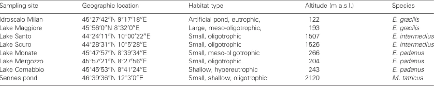
Sample collection and specimen identification
Eudiaptomus populations were collected with a 126 mm plankton net from six lakes and one artificial basin (Table I) between April and July 2009 and preserved in 95% ethanol until analysis. The specimens were not preserved in formalin because microbiological studies have demonstrated that formalin affects the acquisition of high-quality mass spectra due to protein denaturation and degradation. Eudiaptomus specimens were identified using the taxonomic literature of Dussart (Dussart, 1967), Stella (Stella, 1984), Kiefer (Kiefer, 1968,1978), Petkowski (Petkowski, 1983) and Einsle (Einsle, 1993). Reference specimens were examined for morphological differences, dissected in glycerine and stored in sealed slides. Species identification was made by examining the geniculate right antennule and the modified fifth legs of males, the shape of the genital somite and the morph-ology of the lateral wings of the fifth pediger and the fifth legs of females.
For mass spectral analysis, adults of both sexes were individually sorted from each sample under a dissecting microscope. Only individuals apparently free from epi-bionts and parasites were used for analysis. Because the copepodite stages of Eudiaptomus gracilis and Eudiaptomus
padanus are almost indistinguishable, we considered only copepodites of the third Eudiaptomus species (Eudiaptomus intermedius), which never co-occur with the other two species (Tavernini et al., 2003). Males, females and cope-podites of Mixodiaptomus tatricus were obtained from dif-ferent collections, analyzed and used as an outgroup.
Mass spectral analysis
The first part of the study involved optimizing the sample preparation procedure and the settings for data acquisition by MALDI-TOF MS. One hundred and two (102) specimens were used to compare the effects of sample preparation on analytical output and to define a protocol that was then applied to a further 122 speci-mens, which were analyzed and the data evaluated.
Three different matrices were tested: 2,4-dihydroxybenzoic acid (DHB), sinapinic acid (SA) and alpha-cyano-4-hydroxycinnamic acid (CHCA). Solid matrix compounds (75 mg mL21DHB, 40 mg mL21SA and 40 mg mL21CHCA, respectively) were dissolved in a mixture of acetonitrile, ethanol 96% and distilled water (30% each) acidified with 3% v/v trifluoroacetic acid.
We tested two protocols for protein extraction from whole individuals. In the first, individuals were placed in a reaction tube in 5 mL of matrix solution, and ex-traction was performed passively or by sonification for 5, 10, 15 and 30 s in a water bath. Of the resulting extract, 0.5 mL was deposited on a stainless steel target plate (FlexiMass, Kratos, Manchester, UK). In the second protocol, individual copepods were placed dir-ectly on the target plate and proteins were dirdir-ectly extracted on each sample spot by adding 0.5 mL of matrix solution.
Although the analysis of samples prepared according to the second protocol gave reproducible results with ac-ceptable quality of the mass spectra, this approach was not followed up due to the risk of contaminating the vacuum chamber by sample particles coming from the target plate during measurement. The extraction pro-cedure chosen was to submerge one single copepod in
Table I: Sampling sites of Eudiaptomus and Mixodiaptomus species analyzed
Sampling site Geographic location Habitat type Altitude (m a.s.l.) Species Idroscalo Milan 4582704200N 981701800E Artificial pond, eutrophic, 122 E. gracilis Lake Maggiore 458560000N 88320000E Large, meso-oligotrophic, 193 E. gracilis Lake Santo 4482401100N 1080002200E Small, oligotrophic 1507 E. intermedius Lake Scuro 4482803100N 108502800E Small, oligotrophic 1526 E. intermedius Lake Monate 4584705700N 883903400E Small, meso-oligotrophic 266 E. padanus Lake Mergozzo 4585702100N 882705600E Small, oligotrophic 204 E. padanus Lake Comabbio 4584505300N 884102400E Shallow, hypereutrophic 243 E. padanus Sennes pond 4683903600N 12830000E Small, shallow, oligotrophic 2120 M. tatricus
5 mL of the CHCA matrix solution for 10 min. During this time, the soft tissues and chitinous carapace dis-solved totally or partly. Sonication to enhance cell lysis and extraction did not lead to a significant improvement in peak intensity and peak number in mass spectra and was thus omitted.
A volume of 0.5 mL of the crude extract obtained was spotted in quadruplicates on the target plate and allowed to evaporate for 1 – 2 min at room temperature. Crystal formation on the sample position was controlled visually.
Protein mass fingerprints (Kallow et al., 2010) were obtained using an AXIMATM Confidence instrument
(Shimadzu-Biotech Corp., Kyoto, Japan), with spectrum acquisition in a linear, positive ion extraction mode at a laser frequency of 50 Hz and within a mass range of 2000 – 20 000 Da. The acceleration voltage was 20 kV, and the extraction delay time was 200 ns. Protein mass fingerprints were accumulated from 100 profiles, each in turn being accumulated from five laser pulse cycles with the auto-quality option turned on in the instru-ment’s software. The resulting raw spectra were thus accumulated from 500 laser pulse cycles and processed using the LaunchpadTM v. 2.8 software
(Shimadzu-Biotech Corp.) by smoothing with a filtering width of 50 channels and baseline subtraction with a filter width of 500 channels. Peak detection was performed with the threshold-apex peak detection method, applying the adaptive voltage threshold which follows the signal noise level. For each raw mass spectrum, a peak list was generated that included for each peak the apex m/z value, the mass deviations and the signal intensity. Calibration was performed for each target plate using the reference strain Escherichia coli K12 (GM48 genotype).
Data analysis and handling
Generated protein mass fingerprints ( peak lists) were imported into the SARAMISTM software (Spectral
Archive and Microbial Identification System, AnagnosTec GmbH) and analyzed using the following pre-setting parameters: mass range from 2000 to 20 000 Da, allowed mass deviation of 800 ppm. With these settings, a set of so-called supermasses was extracted from mass spectra of specimens belonging to the same species. These are mass signals that have been recorded, with the permitted analytical error range, with a fre-quency of at least 80%. The resulting pattern of super-masses represents a consensus mass spectra pattern for individual species.
The individual mass spectra were further analyzed by cluster analysis, applying a single link agglomerative
clustering algorithm in SARAMISTM. The clustering
was based on the absence or presence of peaks and not on their intensities.
R E S U LT S
For the present study, we focused on three species of Eudiaptomus which are the most widely distributed in Northern Italian lakes: (i) E. intermedius, an Eastern alpine—Illyric species (Stella, 1979), (ii) E. padanus (sub-species E. padanus padanus), one of the most common calanoids in Italian prealpine water bodies (Stella, 1984), and (iii) E. gracilis, which is broadly distributed throughout different continents (Dussart and Defaye, 2002) but which has started expanding in Northern Italy only recently (Riccardi and Rossetti, 2007). Copepod populations were sampled from water bodies representative of a wide range of environmental condi-tions (Table I). Eudiaptomus intermedius, a species typical of permanent water bodies in the Northern Apennines (Tavernini et al., 2003), was obtained from two oligo-trophic lakes where other diaptomid species are not present. Eudiaptomus padanus, an endemic species which is gradually being displaced by E. gracilis (Riccardi and Rossetti, 2007), was obtained from three lakes of differ-ent trophic degree, where it is still the only diaptomid present. The third species, E. gracilis, was the only dia-ptomid copepod in the eutrophic artificial Idroscalo res-ervoir, while it co-occurred with rare E. padanus individuals in the large, meso-oligotrophic Lake Maggiore. The sample of M. tatricus was collected from a high-altitude pond in the Alpine protected area of Fanes-Sennes-Braies (Dolomites), which sustains only one calanoid species (Marrone and Stoch, unpublished data).
This first approach to a MALDI-TOF MS-based identification of copepods involved comparing different matrices for sample deposition before the automated spectra acquisition procedure. Of the three different matrices tested (DHB, SA and CHCA), the last was chosen because it yielded the most reproducible results, most probably due to the homogeneous distribution of sample/matrix crystals on the target plate. The two other matrix compounds crystallize in larger needles, which form a heterogeneous distribution on the sample position, requiring a higher number of laser pulse cycles to yield a representative average mass spectrum (data not shown).
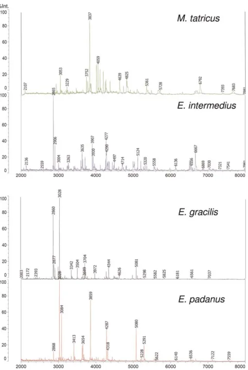
MALDI-TOF MS analysis of crude extracts of whole Eudiaptomus individuals using CHCA revealed complex mass spectral patterns in the m/z range of 2000 – 20 000 Da for all the species analyzed. Most
peaks of high intensity were recorded in the mass range between 2 and 6 kDa.
Quadruplicates of the samples yielded reproducible patterns from sample position to sample position, also from different target plates. Figure1shows typical spec-tral patterns of proteins extracted from Eudiaptomus and Mixodiaptomus specimens obtained in the automated
spectrum acquisition mode. Generally speaking, a number of some 10 – 15 peaks with a relative intensity exceeding 10% was recorded, along with some 100 further peaks of intermediate (1 – 10%) or low (,1%) intensity. This result is similar to those for mass spectral patterns of whole bacterial cells. The peak lists extracted from the raw spectra were taken for further data
Fig. 1. Comparison of protein mass spectra of organisms belonging to E. gracilis, E. intermedius, E. padanus and M. tatricus. Similar and diverging marker masses characteristic of these Eudiaptomus species are listed in Supplementary data, Table SI.
analysis, as provided by the Launchpad software. In consequence, replicate mass spectra of individual cope-pods were not identical, which was, however, not regarded as a major problem, as this is a well-known occurrence in the analysis of microorganisms. Our reasons for using this analysis to assess variability are in-trinsic to MALDI-TOF MS itself: heterogeneities occur during co-crystallization of matrix and analytes, and during ionization, they are not leveled out with a finite number of laser pulse cycles (Dreisewerd, 2003). As a consequence, it is primarily peaks of low intensity close to the threshold that cause analysis-to-analysis variabil-ity, while peaks of intermediate and high intensity were found to be generally stable.
Tentative protein assignment to individual peaks, using in silico database searches, e.g. the Tag-Ident pro-teomics tool or the ExPASy Sequence Retrieval System (http://us.expasy.org), was not possible due to the lack of genomic and proteomic data, not only for species of the genus Eudiaptomus but also for the entire Copepoda order. The peak patterns were therefore taken as bio-markers as such without further identification of the identities of individual peaks.
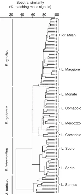
A comparison of spectral fingerprints of individuals belonging to E. gracilis, E. intermedius and E. padanus revealed species-specific differences in the peak patterns (Fig. 1). The mass spectra of the three Eudiaptomus species were distinctly different from those of M. tatricus, which produced a clearly separated clade in the dendro-gam (Fig.2).
Subsets of peaks characteristic of each species are shown in Supplementary data, Table SI. These peaks represent the m/z values of a set of prominent peaks detected in at least 80% of the individual mass spectra considered. A number of peaks were recorded for two or three species, while others appear to be unique for a single species. Such patterns of variably shared peaks have also been observed in studies on closely related bacterial species, where they are interpreted as varia-tions in homologous proteins due to amino acid exchanges caused by point mutations (Dieckmann et al., 2010).
Among the females, males and copepodites of indi-vidual species, differences between peak patterns were generally lower than between species (Fig. 2). This indi-cates that differential protein expression patterns of de-velopmental stages and sexes are only moderate and contribute to a lower degree of heterogeneity than the analytical variability itself. Thus, MALDI-TOF MS peak patterns are arguably stable mass fingerprints of individual taxonomic units, and can be applied to taxo-nomic identification independently of sex and develop-mental stage.
In the dendrogram computed from the mass finger-prints, the three species considered were clearly sepa-rated, irrespective of the origin of the samples (Fig. 2). Nevertheless, subclusters including individuals from a specific lake appeared for most species. This was found to be most pronounced for E. intermedius, with
E. gracilis E. padanus E. intermedius M. tatricus Idr. Milan L. Comabbio L. Scuro L. Santo L. Comabbio L. Mergozzo L. Maggiore L. Monate L. Sennes 20 40 60 80 100 Spectral similarity (% matching mass signals)
Fig. 2. Dendrogram based on the protein mass fingerprint patterns of all analyzed organisms (females, empty circle; males, full circle; copepodids, gray circle) using the single-link clustering algorithm implemented in SARAMIS.
individuals of populations from L. Scuro and L. Santo clearly separated in distinct clusters. For E. gracilis, only individuals from the Milan Idroscalo were grouped in a distinct cluster, while those from L. Maggiore appear to be more diverse, with smaller distinct subclusters. Lastly, individuals of E. padanus were grouped in several smaller subclusters not fully consistent with the indivi-duals’ origin.
D I S C U S S I O N
It must be emphasized that the results presented here are only a pilot study.
Nevertheless, clear differentiation between species was achieved, although all the settings and statistical algorithms used were originally designed for the analysis and identification of bacteria and fungi. This indicates that even such preliminary results as those presented here are contributing to the growing evidence of the re-liability of MALDI-TOF-based identification of multi-cellular organisms. Following promising pioneer work on nematodes and insects (Campbell, 2005; Perera, 2005a, b), MALDI-TOF protein profiling is currently applied to the identification of insect (e.g. Kaufmann et al., 2011) and fish species (Mazzeo et al., 2008;Volta et al., 2012). Our study appears to be the first applica-tion to crustacean zooplankton (i.e. copepods), as well as the first to use field-collected whole organisms for analysis. It is true that only wild specimens were used for the studies on fish, but the analyses were performed on muscle tissue samples. In contrast, laboratory-reared strains were analyzed for the insect studies referred to above. This probably resulted in a reduction in environmental-induced variations, a potential source of individual or between-strain variability and thus of an increase in the probability of inconsistent results (Campbell, 2005), which was expected to be a major concern in the application of this technique to the iden-tification of complex multicellular organisms. Potential sources of inconsistency might include sex, age and/or stage of development and all the differences in individ-ual diet and/or hosted biota (commensal or parasitic organisms). In fact, a considerable impact of the stomach blood content on the MALDI-TOF pattern of the haematophagous female midge Culicoides nubeculosus was observed (Kaufmann et al., 2011), while sex or age had negligible effects on the mass spectra of the fruit flies Drosophila melanogaster (Campbell, 2005) and C. nubeculosus (Kaufmann et al., 2011). Accordingly, we did not observe sex or age effects on the mass spectra of Eudiaptomus and Mixodiaptomus species. The above-mentioned studies on insects led to the conclusion that
the MALDI-TOF-based method is likely to be robust in the face of such variations, and can be used for the un-ambiguous identification even of very closely related species. Our results, though preliminary, seem to support this hypothesis. Indeed, even though we ana-lyzed wild specimens that were probably carrying differ-ent periphyton and had a differdiffer-ent gut contdiffer-ent, differences between individual mass spectra were not so large as to influence species discrimination. This sug-gests that an optimization of both mass spectral analysis and data processing for this particular type of sample is worth performing to exhaustively evaluate the potential of MALDI-TOF MS analysis for plankton studies. For instance, a detailed analysis of mass fingerprints may reveal biomarkers that can distinguish subpopulations or reveal cryptic species (e.g. Campbell, 2005). An indi-cation of this kind of potential seems to emerge from the subclustering distribution of the individuals within each of the Eudiaptomus species analyzed. However, testing this hypothesis demands first of all an accurate evaluation of the influence of the potential sources of “contamination” (e.g. gut content, endo- and ecto-parasites and commensalists) and of individual physio-logical status on mass spectra. For instance, spectra reproducibility within a population could be altered if larval stages (e.g. nauplii) are compared with post-metamorphic stages (copepodites). The extreme simpli-city of the MALDI-TOF technique aids in the rapid performance of such comparisons. In fact, the high degree of automation of the analytical procedure means that several hundred individuals can be prepared and measured per day, allowing the analysis of populations in statistically required numbers. In the present study, mass spectral analysis has not reached its limit of detec-tion, i.e. it should be feasible to analyze even smaller samples. Further studies will therefore include the possi-bility of analyzing nauplii and/or eggs by miniaturizing the sample preparation procedure.
This study suggests that MALDI-TOF MS analysis can provide an alternative or complementary tool for species identification and recognition in plankton re-search. While methods based on molecular biology gen-erally demand some a priori knowledge of the sample under investigation (e.g. for primer selection) and, more importantly, can be slow and laborious with results that may depend on experimental variables (e.g. DNA ex-traction, temperature-sensitive restriction), a mass spec-tral approach would be much more straightforward, requiring minimal time and relatively low costs. At the same time, it would be an accurate and sensitive tool not requiring any previous information of the samples to be analyzed, as is the case for all kinds of microor-ganisms. A prerequisite is the creation of a
taxonomically comprehensive molecular profile data-base for the known zooplankton species, to allow rapid and accurate identification of specimens and provide faster and more detailed characterization of species dis-tribution and diversity patterns.
S U P P L E M E N TA RY DATA
Supplementary data can be found online at http:// plankt.oxfordjournals.org.
AC K N O W L E D G E M E N T S
We are grateful to Giampaolo Rossetti, Giuseppe Alfonso, Federico Marrone and Fabio Stoch for E. inter-medius and M. tatricus specimens, and to Alice Porta and Dario Botturi for their help with field and laboratory work. We are also indebted to Hugh Mc Isaac and to two anonymous reviewers whose constructive criticism contributed to the amelioration of this manuscript. Our thanks to Sandra Spence for the English revision of the text.
F U N D I N G
BNF, Biomedical scientific found University of Bern (Switzerland) Project Nb 338313, research founds of Cantonal Institute of Microbiology Bellinzona (Switzerland) and CNR-Institute of Ecosystem Study, Pallanza (Italy).
R E F E R E N C E S
Belyaeva, M. and Taylor, D. J. (2009) Cryptic species within the Chydorus sphaericus species complex (Crustacea: Cladocera) revealed by molecular markers and sexual stage morphology. Mol. Phylogenet. Evol.,50, 534 – 546.
Bo¨ttger-Schnack, R. and Schnack, D. (2009) Taxonomic diversity and identification problems of oncaeid microcopepods in the Mediterranean Sea. Mar. Biodivers.,39, 131 – 145.
Briski, E., Cristescu, M. E., Bailey, S. A. et al. (2011) Use of DNA bar-coding to detect invertebrate invasive species from diapausing eggs. Biol. Invasions,13, 1325– 1340.
Bucklin, A. and Frost, B. W. (2009) Morphological and molecular phylogenetic analysis of evolutionary lineages within Clausocalanus (Crustacea, Copepoda, Calanoida). J. Crust. Biol.,29, 111 – 120. Bucklin, A., Hopcroft, R. R., Kosobokova, K. N. et al. (2010) DNA
barcoding of Arctic Ocean holozooplankton for species identifica-tion and recogniidentifica-tion. Deep-Sea Res. II,57, 40 – 48.
Costa, F. O. and Carvalho, G. R. (2010) New insights into molecular evolution: prospects from the Barcode of Life Initiative (BOLI). Theory Biosci.,129, 149 – 157.
Dieckmann, R., Strauch, E. and Alter, T. (2010) Rapid identification and characterization of Vibrio species using whole-cell MALDI-TOF mass spectrometry. J. Appl. Microbiol.,109, 199 – 211.
Dreisewerd, K. (2003) The desorption process in MALDI. Chem. Rev., 103, 395 – 425.
Duncan, M. W., Roder, H. and Hunsucker, S. W. (2008) Quantitative matrix-assisted laser desorption/ionization mass spectrometry. Brief. Funct. Genom. Proteom.,7, 355 – 370.
Dussart, B. (1967) Les Cope´podes des eaux continentales d’Europe Occidentale. I. Calanoı¨des et Harpacticoı¨des. Boube´e et Cie, Paris, France. Dussart, B. and Defaye, D. (2002) World Directory of Crustacea Copepoda of
Inland Waters. I—Calaniformes. Backuhys, Kerkwerve, Netherlands. Dussart, B. H. and Defaye, D. (1995) Introduction to the Copepoda.
In Dumont, H. J. F. (ed.), Guides to the Identification of the Microinvertebrates of the Continental Waters of the World. Vol. 7. SPB Academic Publishing, Amsterdam.
Einsle, U. (1993) Crustacea Copepoda Calanoida und Cyclopoida. Su¨bwa¨sser Fauna von Mitteleuropa. 8/4. Gustav Fischer Verlag, Jena, Germany. Einsle, U. (1996) Copepoda: Cyclopoida. Genera Cyclops, Megacyclops,
Acanthocyclops. In Dumont, H. J. F. (ed.), Guides to the Identification of the Microinvertebrates of the Continental Waters of the World. Vol. 7. SPB Academic Publishing, Amsterdam.
Erhard, M., Hipler, U. C., Burmester, A. et al. (2008) Identification of dermatophyte species causing onychomycosis and tinea pedis by MALDI-TOF mass spectrometry. Exp. Dermatol.,17, 365 – 371. Feltens, R., Go¨rner, R., Kalkhof, S. et al. (2010) Discrimination of
dif-ferent species from the genus Drosophila by intact protein profiling using matrix-assisted laser desorption ionization mass spectrometry. BMC Evol. Biol.,10, 95.
Freiwald, A. and Sauer, S. (2009) Phylogenetic classification and iden-tification of bacteria by mass spectrometry. Nat. Protoc.,4, 732 – 742. Hettick, J. M., Green, B. J., Buskirk, A. D. et al. (2008) Discrimination
of Aspergillus isolates at the species and strain level by matrix-assisted laser desorption/ionization time-of-flight mass spectrometry finger-printing. Anal. Biochem.,380, 276 – 281.
Jagadeesan, L., Perumal, P. and Thangaraj, L. (2009) Molecular iden-tification of marine calanoid copepod Paracalanus parvus. World J. Fish Mar. Sci.,1, 239 – 242.
Kallow, W., Erhard, M., Shah, H. N. et al. (2010) MALDI-TOF MS and microbial identification: years of experimental development to an established protocol. In Shah, H. N., Gharbia, S. E. and Encheva, V. (eds), Mass Spectrometry for Microbial Proteomics. Wiley, Chichester, UK, pp. 255 – 276.
Karaytug, S. (1999) Copepoda: Cyclopoida. Genera Paracyclops, Ochridacyclops and key to the Eucyclopinae. In Dumont, H. J. F. (ed.), Guides to the Identification of the Microinvertebrates of the Continental Waters of the World. Vol. 7. SPB Academic Publishing, Amsterdam. Karr, T. L. (2008) Application of proteomics to ecology and
popula-tion biology. Heredity,100, 200 – 206.
Kaufmann, C., Ziegler, D., Schaffner, F. et al. (2011) Evaluation of matrix-assisted laser desorption/ionization time of flight mass spec-trometry fro characterization of Culicoides nubeculosus biting midges. Med. Vet. Entomol.,25, 32 – 38.
Kiefer, F. (1968) Versuch einer Revision der Gattung Eudiaptomus Kiefer (Copepoda Calanoida). Mem. Ist. Ital. Idrobiol.,24, 9 – 160.
Kiefer, F. (1978) Das Zooplankton der Binnengewa¨sser. Freilebende Copepoda. Die Binnengewa¨sser. Band 26. Teil 2. E., Schweitzerbart’sche Verlag, Stuttgart, Germany.
Kroppenstedt, R. M., Mayilraj, S., Wink, J. M. et al. (2005) Eight new species of the genus Micromonospora, Micromonospora citrea sp. nov., Micromonospora echinaurantiaca sp. nov., Micromonospora echinofusca sp. nov., Micromonospora fulviviridis sp. nov., Micromonospora inyonensis sp. nov., Micromonospora peucetia sp. nov., Micromonospora sagamiensis sp. nov., and Micromonospora viridifasciens sp. nov. Syst. Appl. Microbiol.,28, 328 – 339.
Lopez, J. L., Abalde, S. L. and Fuentes, J. (2005) Proteomic approach to probe for larval proteins of the mussel Mytilus galloprovincialis. Mar. Biotechnol. (NY),7, 396 – 404.
MacManus, G. B. and Katz, L. A. (2009) Molecular and morpho-logical methods for identifying plankton: what makes a successful marriage? J. Plankton Res.,31, 1119– 1129.
Marinach-Patrice, C., Lethuillier, A., Marly, A. et al. (2009) Use of mass spectrometry to identify clinical Fusarium isolates. Clin. Microbiol. Infect.,15, 634 – 642.
Mauchline, J. (1998) The Biology of Calanoid Copepods. Advances in Marine Biology, Vol. 33. Academic Press, London.
Mazzeo, M. F., Giulio, B. D., Guerriero, G. et al. (2008) Fish authenti-cation by MALDI-TOF mass spectrometry. J. Agric. Food Chem.,56, 11071 – 11076.
Perera, M. R., Vanstone, V. A. and Jones, M. G. K. (2005a) A novel approach to identify plant parasitic nematodes using matrix-assisted laser desorption/ionization time-of-flight mass spectrometry. Rapid Commun. Mass Spectrom.,19, 1454 – 1460.
Perera, M. R., Vargas, R. D. F. and Jones, M. G. K. (2005b) Identification of aphid species using protein profiling and matrix-assisted laser desorption/ionization time-of-flight mass spectrometry. Entomol. Exp. Appl.,117, 243 – 247.
Petkowski, T. K. (1983) Calanoı¨des—Calanoida (Crustacea—Copepoda). Faune de Macedoine. V. Musee´ Histoire Naturelle de la Macedoine, Skopje. Radulovici, A. E., Archambault, P. and Dufresne, F. (2010). DNA
bar-codes for marine biodiversity: moving fast forward? Diversity, 2, 450 – 472.
Ranga Reddy, Y. (1994) Copepoda: Calanoida: Diaptomidae. Key to the genera Heliodiaptomus, Allodiaptomus, Neodiaptomus, Phyllodiaptomus, Eodiaptomus, Arctodiaptomus and Sinodiaptomus. In Dumont, H. J. F. (ed.), Guides to the Identification of the Microinvertebrates of the Continental Waters of the World. Vol. 7. SPB Academic Publishing, Amsterdam. Rezzonico, F., Vogel, G., Duffy, B. et al. (2010) Application of
whole-cell matrix-assisted laser desorption ionization-time of flight mass spectrometry for rapid identification and clustering analysis of Pantoea species. Appl. Environ. Microbiol.,76, 4497– 4509.
Riccardi, N. and Rossetti, G. (2007) Eudiaptomus gracilis in Italy: how, where and why. J. Limnol.,66, 64 – 69.
Selkoe, K. A. and Toonen, R. J. (2006) Microsatellites for ecologists: a practical guide to using and evaluating microsattelite markers. Ecol. Lett.,9, 615 – 629.
Seng, P., Drancourt, M., Gouriet, F. et al. (2009) Ongoing revolution in bacteriology: routine identification of bacteria by matrix-assisted laser desorption ionization time-of-flight mass spectrometry. Clin. Infect. Dis.,49, 543 – 551.
Seng, P., Rolain, J. M., Fournier, P. E. et al. (2010) MALDI-TOF mass spectrometry applications in clinical microbiology. Future Microbiol., 5, 1733– 1754.
Stella, E. (1979) Considerazioni biogeografiche sui Diaptomidi (Copepoda Calanoida) delle acque dolci italiane. Lav. Soc. Ital. Biogeo.,6, 315 – 328.
Stella, E. (1984) Crustacea—Copepoda: Calanoida d’acqua dolce. Calderini, Bologna.
Stoch, F. (2005) Crustacea Copepoda Calanoida. In Ruffo, S. and Stoch, F. (eds), Checklist e distribuzione della fauna italiana. Memorie del Museo Civico di Storia Naturale di Verona, 2. serie, Sezione Scienze della Vita, pp. 91 – 92.
Tavernini, S., Fratta, E., Sartore, F. et al. (2003). Distribution and ecology of calanoid species in relation to morphometric and chem-ical characteristics of lakes and ponds of the Northern Appennines (Italy). J. Limnol.,62, 28 – 34.
Teramoto, K. (2009) Rapid identification and classification of bacteria using ribosomal protein as biomarkers by matrix-assisted laser de-sorption/ionization-mass spectrometry. Bunseki Kagaku, 58, 971 – 972.
Thum, R. A. and Derry, A. M. (2008) Taxonomic implications for diaptomid copepods based on contrasting patterns of mitochondrial DNA sequence divergences in four morphospecies. Hydrobiologia, 614, 197 – 207.
Volta, P., Riccardi, N., Lauceri, R. et al. (2012) Characterization and separation of freshwater fish species by matrix-assisted laser desorp-tion/ionization-time of flight mass spectrometry (MALDI-TOF MS): a pilot study. J. Limnol.,71, doi: 10.4081/jlimnol.2012.e17. Welker, M. (2011) Proteomics for routine identification of
microorgan-isms. Proteomics,11, 1 – 11.
Welker, M., Sejnohova, L., von Do¨hren, H. et al. (2007) Seasonal shifts in chemotype composition of Microcystis sp. communities in the pela-gial and the sediment of a shallow reservoir. Limnol. Oceanogr.,52, 609 – 619.
Wynne, C., Fenselau, C., Demirev, P. A. et al. (2009) Top-down identi-fication of protein biomarkers in bacteria with unsequenced genomes. Anal. Chem.,81, 9633– 9642.
Yebra, L., Bonnet, D., Harris, R. P. et al. (2011) Barriers in the pelagic: population structuring of Calanus helgolandicus and C. euxinus in European waters. Mar. Ecol. Prog. Ser.,428, 135 – 149.