HAL Id: inserm-00410118
https://www.hal.inserm.fr/inserm-00410118
Submitted on 8 Aug 2011
HAL is a multi-disciplinary open access archive for the deposit and dissemination of sci-entific research documents, whether they are pub-lished or not. The documents may come from teaching and research institutions in France or abroad, or from public or private research centers.
L’archive ouverte pluridisciplinaire HAL, est destinée au dépôt et à la diffusion de documents scientifiques de niveau recherche, publiés ou non, émanant des établissements d’enseignement et de recherche français ou étrangers, des laboratoires publics ou privés.
Influence of gender, obesity, and muscle lipase activity
on intramyocellular lipids in sedentary individuals.
Cédric Moro, Jose Galgani, Lanchi Luu, Magdalena Pasarica, Aline Mairal,
Sudip Bajpeyi, Gerd Schmitz, Dominique Langin, Gerhard Liebisch, Steven
Smith
To cite this version:
Cédric Moro, Jose Galgani, Lanchi Luu, Magdalena Pasarica, Aline Mairal, et al.. Influence of gender, obesity, and muscle lipase activity on intramyocellular lipids in sedentary individuals.. Journal of Clinical Endocrinology and Metabolism, Endocrine Society, 2009, 94 (9), pp.3440-7. �10.1210/jc.2009-0053�. �inserm-00410118�
Influence of Gender, Obesity, and Muscle Lipase
Activity on Intramyocellular Lipids in Sedentary
Individuals
Cedric Moro, Jose E. Galgani, LanChi Luu, Magdalena Pasarica, Aline Mairal, Sudip Bajpeyi, Gerd Schmitz, Dominique Langin, Gerhard Liebisch,
and Steven R. Smith
Departments of Molecular and Experimental Endocrinology (C.M., M.P., S.B., S.R.S.), Human Physiology (J.E.G.), and Nutrition and Diabetes (L.L.), Pennington Biomedical Research Center, Baton Rouge, Louisiana 70808; Institut National de la Sante´ et de la Recherche Me´dicale Unite´ 858 (A.M., D.L.), Institut de Me´decine Mole´culaire de Rangueil, F-31432 Toulouse, France; Institut Fédératif de Recherche 31 (A.M., D.L.), Université Paul Sabatier, Universite´ de Toulouse, F-31432 Toulouse, France; Centre Hospitalier Universitaire de Toulouse (D.L.); F-31432 Toulouse, France; and Institute of Clinical Chemistry (G.S., G.L.), University of Regensburg, 93053 Regensburg, Germany
Context: Obesity and type 2 diabetes are associated with elevated intramyocellular lipids (IMCLs)
and insulin resistance.
Objective: We tested the hypothesis that skeletal muscle lipases activity could influence IMCL
content (including diacylglycerol and ceramides).
Design and Patients: The present study included 48 subjects with a wide range of age (19 – 68 yr)
and body mass index (20 – 45 kg/m2) who underwent skeletal muscle biopsy, dual-energy x-ray
absorptiometry and a hyperinsulinemic euglycemic clamp.
Main Outcome Measures: Insulin sensitivity by hyperinsulinemic clamp, and intramyocellular
tri-acylglycerol (IMTG), ditri-acylglycerol (DAG), and ceramides content, and tritri-acylglycerol and diacyl-glycerol hydrolase activities were measured in biopsies of vastus lateralis. IMCL was measured by
1H-magnetic resonance spectroscopy in a subgroup of 25 subjects. Multivariate regression analyses
were performed to identify the main predictors of IMCL.
Results: Body fat was the main predictor of IMTG independently of the method and the type
of muscle; IMTG concentration was higher in females vs. males and obese vs. nonobese subjects. Muscle DAG and ceramides concentrations were elevated in obese and type 2 diabetic subjects and were not related to body fat and fasting free fatty acids, whereas a direct association with the ratio of diacylglycerol hydrolase to triacylglycerol hydrolase activity (an index of incom-plete triacylglycerol hydrolysis) was observed, which explained 54 and 38% of the variance in
DAG and ceramides (P⬍ 0.001), respectively. DAG content was the main determinant of insulin
resistance.
Conclusions: These data suggest that intramyocellular DAG is an independent predictor of insulin
resistance in humans and that its levels correlate with lipolytic enzymes activity in skeletal muscle but not with markers of adiposity. (J Clin Endocrinol Metab 94: 3440 –3447, 2009)
ISSN Print 0021-972X ISSN Online 1945-7197 Printed in U.S.A.
Copyright © 2009 by The Endocrine Society
doi: 10.1210/jc.2009-0053 Received January 9, 2009. Accepted June 5, 2009. First Published Online June 16, 2009
Abbreviations: ATGL, Adipose triglyceride lipase; BMI, body mass index; DAG, diacylglyc-erol; DAGH, DAG hydrolase activity; FFA, free fatty acid; GDR, glucose disposal rate; IMCL, intramyocellular lipids; IMTG, intramyocellular triacylglycerol; MRS, magnetic resonance spectroscopy; PRESS, Point Resolved Spectroscopy; TAG, triacylglycerol; TAGH, TAG hy-drolase activity; T2DM, type 2 diabetes mellitus.
E n d o c r i n e C a r e
T
ype 2 diabetes mellitus (T2DM) is commonly associ-ated with disorders in lipid metabolism including el-evated plasma free fatty acid (FFA) concentration and ec-topic lipid deposition in multiple peripheral tissues such as skeletal muscle (1, 2). Ectopic lipids mainly accumulate as triacylglycerol (TAG). An inverse association between in-tramyocellular triacylglycerol (IMTG) content and pe-ripheral glucose disposal (measured by euglycemic hyper-insulinemic clamp) has been repeatedly reported (3–5). It has been proposed that reduced capacity for skeletal mus-cle fat oxidation, potentially due to mitochondrial dys-function could contribute to IMTG accumulation, lipo-toxicity, and insulin resistance (6, 7). However, evidence for a frank mitochondrial dysfunction in skeletal muscle in obesity and T2DM and its role on muscle lipid accumu-lation is lacking as recently reviewed (8, 9). Alternatively, elevated skeletal muscle FFA uptake could also contribute to elevated IMTG in obesity-associated insulin resistant states. It is still unclear at this point whether skeletal mus-cle FFA transport is increased in obese and type 2 diabetic subjects with studies showing either an increase (10) or no difference (11, 12) compared with lean controls. Thus, the cause of IMTG accumulation in sedentary subjects is poorly understood so far.It is now accepted that it is not IMTG per se that induces insulin resistance but rather some lipotoxic intermediates such as diacylglycerol (DAG) and ceramides, which alter insulin signaling and action (7, 13–15). We hypothesized first that IMTG accumulate mainly as a consequence of increased adiposity and second that a dysregulation of IMTG turnover and lipolysis in skeletal muscle could con-tribute to elevated DAG and ceramides content. Lipolysis is the main catabolic reaction leading to the hydrolysis of one molecule of TAG into three fatty acid molecules (16). High rates of lipolysis and FFA release could contribute to
de novo ceramides synthesis by generating palmitoyl-CoA
as shown in C2C12 myoblasts overexpressing adipose tri-glyceride lipase (ATGL) (17). In addition, an inability to completely hydrolyze TAG, due to an imbalance of TAG relative to DAG hydrolase activities, could contribute to increased DAG availability (18). To the best of our knowl-edge, there are currently no data available on the potential anthropometric and biochemical determinants of in-tramyocellular lipids (including DAG and ceramides) in humans. In the present study, we aimed first to identify the potential determinants of intramyocellular lipids (TAG, DAG, and ceramides) in sedentary humans and second to evaluate the role of skeletal muscle lipolysis as a determi-nant of markers of intramyocellular lipotoxicity (DAG and ceramides) and insulin sensitivity.
Subjects and Methods
Subjects
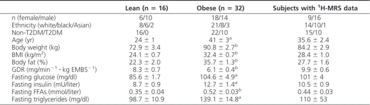
Forty-eight sedentary male (n⫽ 24) and female (n ⫽ 24) subjects were recruited in the study. Ten of 48 subjects had T2DM and were treated mainly with sulfonylurea, metformin, or diets. None of them were taking thiazolidinediones. The sub-jects had a wide range of body composition, age, insulin sensi-tivity, and metabolic status (Table 1). The subjects were recruited based on a sedentary lifestyle determined by activity index ques-tionnaire and were not enrolled in any structured sports activi-ties. The protocol was approved by the Institutional Review Board of the Pennington Biomedical Research Center, and all volunteers gave written informed consent. After completing the screening visit, total fat mass was measured on a dual-energy x-ray absorptiometer (QDR 4500A; Hologic Inc., Bedford, MA). The participants were asked to refrain from vigorous phys-ical activity for 48 h before presenting to the Pennington inpa-tient clinic and ate a weight-maintaining diet consisting of 35% fat, 16% protein, and 49% carbohydrate 2 d before the clamp and the muscle biopsy. The muscle biopsy was performed in the morning after a 10- to 12-h overnight fast and before the clamp. Samples of vastus lateralis weighing 60 –100 mg were obtained TABLE 1. Anthropometric and clinical characteristics of the subjects
Lean (nⴝ 16) Obese (nⴝ 32) Subjects with1H-MRS data
n (female/male) 6/10 18/14 9/16 Ethnicity (white/black/Asian) 8/6/2 21/8/3 14/10/1 Non-T2DM/T2DM 16/0 22/10 15/10 Age (yr) 24⫾ 1 41⫾ 3a 35.6⫾ 2.4 Body weight (kg) 72.9⫾ 3.4 90.8⫾ 2.7b 84.2⫾ 2.9 BMI (kg/m2) 24.1⫾ 0.7 32.4⫾ 0.7b 28.4⫾ 1.0 Body fat (%) 22.3⫾ 2.0 35.7⫾ 1.3b 27.7⫾ 1.6 GDR (mg/min⫺1䡠 kg EMBS⫺1) 8.3⫾ 0.7 6.1⫾ 0.4b 9.9⫾ 0.6 Fasting glucose (mg/dl) 85.6⫾ 1.7 104.6⫾ 4.9a 101⫾ 4
Fasting insulin (mU/liter) 8.7⫾ 0.9 12.7⫾ 1.4a 10.5⫾ 0.9
Fasting FFAs (mmol/liter) 0.35⫾ 0.04 0.52⫾ 0.03b 0.44⫾ 0.03
Fasting triglycerides (mg/dl) 98.7⫾ 10.9 139.1⫾ 14.8a 110⫾ 53
EMBS, Estimated metabolic body size.
aP⬍ 0.05.
b
P⬍ 0.01 vs. lean.
using the Bergstrom technique, blotted, cleaned, and snap frozen in liquid nitrogen (19).
Hyperinsulinemic euglycemic clamp
Insulin sensitivity was measured by hyperinsulinemic eu-glycemic clamp. After an overnight fast, insulin (80 mU/ m⫺2䡠 min⫺1) and 20% glucose were administered for 2 h to
maintain plasma glucose at 90 mg/dl. At this infusion rate, in-sulin has been previously shown to suppress more than 95% of hepatic glucose output in subjects with and without T2DM, and the glucose disposal is mainly dependent on skeletal muscle (20, 21). Plasma levels of glucose and insulin were measured in triplicate at 5-min intervals at baseline and during steady state from 95 to 120 min. Glucose disposal rate (GDR) expressed in mg/min⫺1was ad-justed for the estimated metabolic body size (kilograms of fat free mass⫹ 17.7) (22). Fat-free mass was calculated as the difference between body weight and total fat mass.
1
H-magnetic resonance spectroscopy (MRS)
Intramyocellular lipids (IMCL) were measured in the soleus and tibialis anterior muscles of the right calf by a1H-MRS
tech-nique on a GE Signa Excite 3T. 12.0-m5 build whole-body im-aging and spectroscopy system using the Point Resolved Spec-troscopy (PRESS) box technique (23). Measurements were acquired with volunteer lying in the supine position with right leg positioned inside a commercially made radiofrequency1
H knee coil with the knee in the extension and ankle in a neutral position. IMCL contents were determined from the average of the sum of three PRESS boxes in the soleus and one PRESS in the tibialis anterior. Summing the signals increases the signal to noise ratio. Peak positions and areas of interest were determined by time domain fitting using Java-based magnetic resonance user inter-face and a set of prior knowledge files (24, 25). Areas of all peaks were normalized to the corresponding internal water peak as previously described (3).
Lipase activity assays
TAG and DAG hydrolase activities were measured on muscle tissue homogenates as previously described (26). Briefly, triolein and 1(3)-mono-oleyl-2-O-mono-oleylglycerol were emulsified with phospholipids by sonication. Triolein is a triglyceride con-taining three oleic acid specifically used to determine TAG hy-drolase (TAGH) activity. 1(3)-mono-oleyl-2-O-mono-oleylg-lycerol is a DAG analog used to measure specifically the DAG hydrolase (DAGH) activity because it is not a substrate for monoacylglycerol lipase. Lipase activity data were normalized to total protein content determined in each sample and expressed in nanomoles per minute⫺1per milligram⫺1. We also calculated the ratio of DAGH to TAGH activity as a marker of complete TAG hydrolysis.
TAG and DAG determination by gas chromatography/mass spectrometry
Total lipids were extracted from frozen muscle tissue using the method of Folch et al. (27). The extracts were filtered and lipids recovered in the chloroform phase. TAG and DAG were isolated using thin-layer chromatography on Silica Gel 60 A plates developed in petroleum ether, ethyl ether, and acetic acid (80:20:1) and visualized by rhodamine 6G. The TAG and DAG band was scraped from the plate and methylated using BF3/
methanol as described by Morrison and Smith (28). The meth-ylated fatty acids were extracted with hexane and analyzed by gas chromatography using an HP 5890 gas chromatograph equipped with flame ionization detectors, an HP 3365 Chem-station, and a capillary column (SP2380, 0.25 mm⫻ 30 m, 0.25 m film; Supelco, Bellefonte, PA). Helium was used as a carrier gas. The oven temperature was programmed from 160 C to 230 C at 4 C/min. Fatty acid methyl esters were identified by com-paring the retention times to those of known standards. Inclusion of the internal standards, 20:1 (trieicosenoin) and 17:0 (dihep-tadecanoin), permits quantitation of the amount of TAG and DAG in the sample. The absolute quantity of total and each subspecies of TAG and DAG was calculated and expressed in microgram per milligram wet tissue weight.
Ceramides determination by electrospray ionization tandem mass spectrometry
Ceramides was quantified by electrospray ionization tandem mass spectrometry as previously described (29). Briefly, lipid extracts were prepared by the method of Bligh and Dyer (30) in the presence of non-naturally occurring Cer 14:0, Cer 17:0. Sam-ples were analyzed by direct flow injection on a Quattro Ultima triple-quadrupole mass spectrometer (Micromass, Manchester, UK) using a HTS PAL autosampler (Zwingen, Switzerland) and an Agilent 1100 binary pump (Waldbronn, Germany) with a solvent mixture of methanol containing 10 mMammonium
ac-etate and chloroform [3:1 (vol/vol)]. A flow gradient was per-formed starting with a flow of 55l/min for 6 sec followed by 30 l/min for 1 min and an increase to 250 l/min for another 12 sec. Ceramides was analyzed using a fragment of mass to charge ratio 264 with N-heptadecanoyl-sphingosine as internal stan-dard. Both ions [M⫹H]⫹ and [M⫹H-H
2O]⫹were used and
quantification was achieved by calibration lines generated by addition of Cer 16:0, 18:0, 20:0, 24:1, and 24:0 to tissue samples. Correction of isotopic overlap of ceramides species as well as data analysis was performed by self-programmed Excel macros according to the principles described previously (31). The abso-lute quantity of total and each subspecies of ceramides was cal-culated and expressed in nanomoles per milligram wet tissue weight.
Statistical analyses
All statistical analyses were performed using SAS 9.1 Service Pack 4 for Windows (SAS Institute Inc., Cary, NC), and figures were generated using GraphPad Prism 5.0 for Windows (GraphPad Software Inc., San Diego, CA). The relationships between in-tramyocellular lipid content (IMTG, DAG, and ceramides) and anthropometric and clinical variables were analyzed using Spearman rank correlations. Differences in intramyocellular lip-ids according to gender (female vs. male), obesity (obese vs. nonobese), and interactions were analyzed using the mixed model. Tukey-Kramer post hoc multiple comparison tests were performed to evaluate specific differences between groups. Sig-nificant variables were then included in multivariate stepwise regression analyses after ln transformation to achieve normal distribution to identify the best predictors of IMTG, DAG, and ceramides. All values in figures and tables are presented as mean⫾SEM. Statistical significance was set at P⬍ 0.05.
Results
Subject characteristics
The anthropometric and clinical characteristics of the population are presented in Table 1. As expected, the obese subjects had higher body weight, percentage of body fat, fasting glucose, FFAs, insulin, and triglycerides com-pared with the lean. Thus, the obese group had reduced whole-body insulin sensitivity as measured by clamp. The
obese group was slightly older and included 10 subjects with T2DM (Table 1).
Influence of gender and obesity on IMTG
A significant effect of obesity (P⫽ 0.0094) and gender
(P⫽ 0.0004) was found on total IMTG content, with no
interaction between gender and obesity (P⫽ 0.43) (Fig. 1). IMTG was higher in females vs. males (8.3⫾ 0.9 vs. 3.7 ⫾ 0.4g/mg, P ⬍ 0.0001) and obese vs. nonobese (6.8 ⫾ 0.7
vs. 2.7 ⫾ 0.4g/mg, P ⫽ 0.007). Similar findings were
observed irrespective to the IMTG subspecies (data not
shown). IMCL measured in soleus (P⫽ 0.0001) and
tib-ialis anterior (P⫽ 0.006) by1H-MRS was also elevated in obese vs. nonobese subjects in a subgroup of 25 subjects (including 15 nonobese and 10 obese).
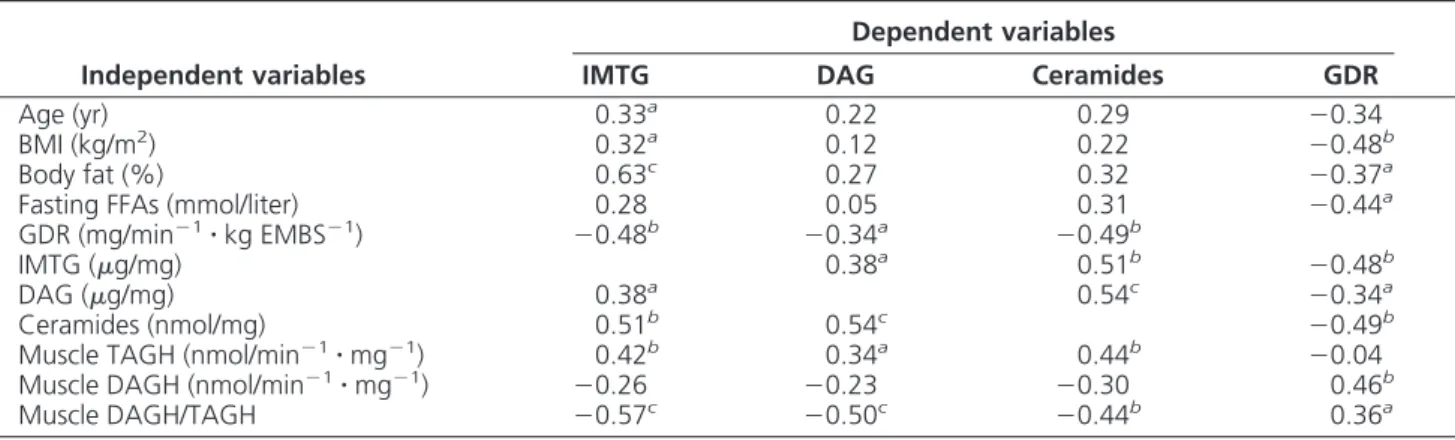
Determinants of IMTG
IMTG was positively correlated with age, body mass index (BMI), body fat, and skeletal muscle TAGH activity (Table 2). Similarly, IMCL both in soleus and tibialis an-terior was positively related to age, BMI, body fat, and fasting FFAs (data not shown). Of importance, IMTG was
positively correlated with intramyocellular DAG (r ⫽
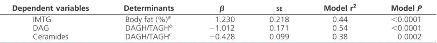
0.38, P⫽ 0.01) and ceramides (r ⫽ 0.51, P ⫽ 0.002). We next performed multivariate regression analyses to iden-tify the determinants of IMTG. In a model including age, body fat, gender, obesity, and skeletal muscle TAGH activity as independent variables, body fat was the best predictor of IMTG, explaining 44% of its variance (P⬍ 0.0001) (Table 3). Consistently, using the same regres-sion model, body fat was the main independent
deter-minant of IMCL measured by1H-MRS in tibialis
ante-rior (r2⫽ 0.30, P ⫽ 0.0054). Body fat remained the best predictor of IMTG in moderately obese subjects with
Lean Obese 0 5 10 15 Female Male To ta l I M T G (µ g /m g we t wt ) A B C # # Lean Obese 0.00 0.05 0.10 0.15 0.20 0.25 DAG 1 8 :1 (µ g/ m g w e t w t) Lean Obese 0.00 0.01 0.02 0.03 S a tu ra te d cer am id es (n m o l/ m g w e t w t) * * * * * *
FIG. 1. Gender difference in total IMTG (A), DAG containing 18:1
fatty acids (B), and saturated ceramide (C) content in lean and obese subjects. Statistical analyses were performed using two-way ANOVA.
*, P⬍ 0.05 when compared with lean; #, P ⬍ 0.05 when compared
with male.
TABLE 2. Spearman correlations of IMTG, DAG, ceramides, and insulin sensitivity, with various anthropometric and clinical variables
Independent variables
Dependent variables
IMTG DAG Ceramides GDR
Age (yr) 0.33a 0.22 0.29 ⫺0.34
BMI (kg/m2) 0.32a 0.12 0.22 ⫺0.48b
Body fat (%) 0.63c 0.27 0.32 ⫺0.37a
Fasting FFAs (mmol/liter) 0.28 0.05 0.31 ⫺0.44a
GDR (mg/min⫺1䡠 kg EMBS⫺1) ⫺0.48b ⫺0.34a ⫺0.49b
IMTG (g/mg) 0.38a 0.51b ⫺0.48b
DAG (g/mg) 0.38a 0.54c ⫺0.34a
Ceramides (nmol/mg) 0.51b 0.54c ⫺0.49b
Muscle TAGH (nmol/min⫺1䡠 mg⫺1) 0.42b 0.34a 0.44b ⫺0.04
Muscle DAGH (nmol/min⫺1䡠 mg⫺1) ⫺0.26 ⫺0.23 ⫺0.30 0.46b
Muscle DAGH/TAGH ⫺0.57c ⫺0.50c ⫺0.44b 0.36a
Value are given as the Spearman correlation coefficient.
a
P⬍ 0.05.
bP⬍ 0.01.
c
P⬍ 0.001.
BMI less than 35 kg/m2 (r2⫽ 0.40, P ⫽ 0.0001). The percentage of body fat was a better predictor of IMTG
in lean (r2 ⫽ 0.70, P ⫽ 0.0025) compared with obese
subjects (r2⫽ 0.42, P ⫽ 0.02).
Influence of gender and obesity on intramyocellular lipases activity
A significant and independent effect of gender (P ⫽
0.015) and obesity (P⫽ 0.037) was found on TAGH
ac-tivity, with no significant interaction between gender and obesity (P⫽ 0.10). TAGH activity was higher in females
vs. males (0.045 ⫾ 0.005 vs. 0.019 ⫾ 0.001 nmol/
min⫺1䡠 mg⫺1, P ⬍ 0.0001) and obese vs. nonobese
(0.035⫾ 0.004 vs. 0.021 ⫾ 0.003 nmol/min⫺1䡠 mg⫺1,
P⬍ 0.01) (Fig. 2A). DAG hydrolase activity and the ratio
of DAGH to TAGH activity were lower in obese vs.
nono-bese subjects (0.44 ⫾ 0.02 vs. 0.53 ⫾ 0.03 nmol/
min⫺1䡠 mg⫺1, P⬍ 0.0001 and 16.4 ⫾ 1.3 vs. 27.7 ⫾ 2.2
arbitrary units, P⬍ 0.0001, respectively). There was no gender difference in DAGH activity and the ratio of DAGH to TAGH activity and no interaction between obe-sity and gender (Fig. 2, B and C).
Influence of gender and obesity on
intramyocellular diacylglycerol and ceramides Intramyocellular DAG and ceramide levels were not different between males and females irrespective of their fatty acid profile. Intramyocellular DAG (0.33⫾ 0.05 vs.
0.13⫾ 0.01g/mg, P ⬍ 0.01) and ceramides (0.022 ⫾
0.002 vs. 0.015⫾ 0.002 nmol/mg, P ⫽ 0.02) content was elevated in obese vs. nonobese subjects. When we looked specifically into the fatty acid profile, we noticed a signif-icant increase in DAG containing oleic acid (18:1) (P⫽ 0.01) and saturated ceramide concentrations (P⬍ 0.0001) in obese vs. nonobese subjects (Fig. 1, B and C). We found a positive relationship between DAG and ceramides (r⫽
0.54, P⫽ 0.0009). DAG and ceramides were not
corre-lated with age, BMI, body fat, and fasting FFAs (Table 2). However, these two lipid species were correlated with the skeletal muscle TAGH activity and even stronger with the ratio of DAGH to TAGH activity (Table 2).
Determinants of intramyocellular DAG and ceramides
We next investigated the determinants of intramyocel-lular DAG concentration. In a model including obesity, gender, IMTG, ceramides, and the ratio of DAGH to TAGH activity as independent variables, the ratio of DAGH to TAGH activity was the strongest determinant of DAG, explaining more than half of the variance (r2⫽ 0.54, P⬍ 0.0001) (Table 3). Similarly, in a model in-cluding obesity, gender, IMTG, DAG, and the ratio of DAGH to TAGH activity as independent variables, cer-amide content was best predicted by the ratio of DAGH to TAGH (r2⫽ 0.38, P ⫽ 0.0002) (Table 3). To exclude
the confounding effect of T2DM on these relationships, we repeated the analyses in non-T2DM subjects only. The ratio of DAGH to TAGH activity remained the
major determinant of skeletal muscle DAG (r2⫽ 0.65,
P⬍ 0.0001) and ceramides (r2⫽ 0.48, P ⫽ 0.0002).
TABLE 3. Determinants of intramyocellular lipids in multivariate stepwise regression analyses
Dependent variables Determinants  SE Model r2 Model P
IMTG Body fat (%)a 1.230 0.218 0.44 ⬍0.0001
DAG DAGH/TAGHb ⫺1.012 0.171 0.54 ⬍0.0001
Ceramides DAGH/TAGHc ⫺0.428 0.099 0.38 0.0002
aIndependent variables included in the model were age, body fat, obesity, gender, and TAGH activity.
b
Independent variables included in the model were obesity, gender, ceramides, IMTG, and the ratio of DAGH to TAGH activity.
cIndependent variables included in the model were obesity, gender, DAG, IMTG, and the ratio of DAGH to TAGH activity.
Lean Obese 0.00 0.02 0.04 0.06 Female Male TA G H ac ti v it y (n mo l/ mi n /mg ) Lean Obese 0 10 20 30 40 DAG H /T A G H (a rb it ra ry u n it s ) Lean Obese 0.0 0.2 0.4 0.6 0.8 D A G H a c ti v ity (n m o l/m in /m g ) A B C * # # * * * *
FIG. 2. Gender difference in TAGH activity (A), DAGH activity (B),
and the ratio of DAGH to TAGH activity (C), in lean and obese subjects. Statistical analyses were performed using two-way
ANOVA. *, P⬍ 0.05 when compared with lean; #, P ⬍ 0.05 when
Relationship between intramyocellular lipids and insulin sensitivity
Insulin sensitivity was lower in obese vs. nonobese (P⫽ 0.01) subjects, with no significant effect of gender (P⫽ 0.78) and interaction (P⫽ 0.28). As shown in Table 2, insulin sensitivity was negatively correlated with anthro-pometric indices of fatness such as BMI, body fat, and fasting FFAs as well as to all three intramyocellular lipid species measured (Table 2). This finding was further
sup-ported when IMCL measured by 1H-MRS was also
in-versely related to insulin sensitivity in soleus (r⫽ ⫺0.48,
P ⫽ 0.02) and tibialis anterior (r ⫽ ⫺0.40, P ⫽ 0.06).
Importantly, the relationship between IMTG and insulin sensitivity was lost after adjustment for intramyocellular DAG content (r⫽ ⫺0.18, P ⫽ 0.36). Of interest, insulin sensitivity was positively correlated with the skeletal muscle DAGH activity and the ratio of DAGH to TAGH (Table 2).
Discussion
In the present study, we investigated for the first time in humans the determinants of intramyocellular lipids in-cluding the two insulin resistance-inducing lipid species DAG and ceramides and related these to insulin sensitiv-ity. We found that IMTG is primarily related to the per-centage of body fat in sedentary populations, whereas in-tramyocellular DAG and ceramides are mainly determined within the skeletal muscle by the ratio of DAGH to TAGH activity, a marker of lipolysis, and independent of adipos-ity. An imbalance of TAGH relative to DAGH activity might contribute to intramyocellular lipotoxicity and in-sulin resistance. These data suggest a previously undera-ppreciated link between skeletal muscle lipolysis, lipotox-icity, and insulin resistance.
The strength of the present study is that we measured simultaneously all three intramyocellular lipids species (TAG, DAG, and ceramides) in biopsy samples obtained from a well clinically phenotyped population. Of impor-tance, intramyocellular DAG and ceramides were mea-sured using gold standard mass spectrometry methods as previously discussed (29). We also investigated for the first time the hypothesis that skeletal muscle lipase activity might determine intramyocellular lipotoxicity and there-fore insulin resistance in the fasted condition. Alterna-tively, increased plasma FFA fractional extraction by the skeletal muscle during the postprandial condition and a mismatch between FFA uptake and oxidation could likely contribute to lipotoxicity and insulin resistance (8, 32). One limitation of the present study could be due to the direct measure of IMTG in vastus lateralis biopsies that could be contaminated by infiltrated adipocytes.
How-ever, in support of these findings, we observed similar associations between markers of adiposity and IMCL in a subgroup of subjects in which IMCL was measured by
1H-MRS in soleus and tibialis anterior. Another limitation
is that the present study may be underpowered to capture significant differences in intramyocellular DAG and cer-amides between groups, especially in subjects with T2DM. Therefore, we strictly focused our analysis on the effect of obesity and gender on intramyocellular lipids and lipase activity.
Since the late 90s, an inverse relationship between IMTG and insulin sensitivity has been reported by several independent groups (3–5). Later it was shown that this relationship is valid in sedentary populations but modified by the level of aerobic fitness. Indeed, IMTG is adapta-tively increased in the muscle of endurance-trained ath-letes to sustain muscle activity during long-lasting endur-ance activities and actually positively predicts insulin sensitivity (33). Several groups demonstrated increased IMTG content in obesity and T2DM and in athletes using various methodologies (11, 33, 34). Here we show that IMTG is mainly determined by the percentage of body fat in sedentary subjects, regardless of the technique used for its determination [i.e. biochemical (ex vivo) or spectro-scopic (in vivo)] and the characteristic of the muscle as-sessed (i.e. vastus lateralis, tibialis anterior, or soleus). This is consistent with a previous report in which IMTG content in tibialis anterior measured by1H-MRS was
pos-itively related to body fat (33) but in contrast with another report showing no association between IMTG measured from vastus lateralis biopsies and indices of adiposity in Pima Indians (4). The discrepancy might be due to the sample size, the methodology used to measure both body fat and IMTG in skeletal muscle, and/or study population characteristics. It has been hypothesized that increased intramyocellular lipid content could result from increased plasma FFA availability and/or reduced fat oxidation, possibly due to mitochondrial dysfunction (7). In the present study, IMCL measured by1H-MRS was positively
related to fasting FFAs. Increased adiposity and plasma FFA concentrations would therefore be sufficient to in-crease IMCL content unless skeletal muscle FFA uptake is reduced. This is unlikely because leg muscle FFA fractional uptake is either similar (6) or increased (35) in obese com-pared with lean individuals. It is then highly possible that IMTG mainly accumulate as a consequence of increased adiposity and plasma FFA availability.
In the present study, we show that intramyocellular DAG and ceramides were not related to any anthropo-metric parameters, possibly suggesting that lipotoxicity is mainly determined within the skeletal muscle indepen-dently of the degree of adiposity. Here we show that the
ratio of DAGH to TAGH activity, a marker of incomplete TAG hydrolysis, is a strong determinant of intramyocel-lular DAG and ceramide content, explaining 54 and 38% of the variance in both lipid species in our population, respectively. A low ratio of DAGH to TAGH activity is associated with elevated intramyocellular lipotoxic lipid species. Mechanistically, this could be driven by a com-bination of high rates of TAG hydrolysis and reduced DAG hydrolysis in obese subjects. Increased TAG hydro-lase activity may contribute to higher fatty acid release and
de novo ceramide synthesis by generating palmitoyl-CoA
(17). In addition, reduced DAG hydrolase activity con-tributes to a lower DAG turnover rate and therefore in-creased DAG availability. Recent studies indicate the pres-ence of ATGL in human skeletal muscle (36, 37). These studies suggest that ATGL might play an important role in the regulation of IMTG besides hormone-sensitive lipase. Thus, a reduced DAGH to TAGH activity ratio in obese subjects is consistent with the observation of Jocken et al. (37) showing a reduced hormone-sensitive lipase protein expression and lower forearm glycerol release in obese insulin-resistant subjects. Our study provides a common mechanism of synthesis of DAG and ceramides through the activity of skeletal muscle lipases. This observation requires further functional investigation.
We next examined the relationship between intramyo-cellular lipids and insulin sensitivity. All three lipid classes (TAG, DAG, and ceramides) were negatively correlated with insulin sensitivity. IMTG irrespective of the fatty acid profile, DAG (18:1), and total saturated ceramide content were significantly elevated in obese. This is in agreement with other studies that have shown a link between elevated intramyocellular DAG and ceramide content and insulin resistance in humans (15, 38). There was an independent effect of obesity on all three lipid species. Interestingly, the association between IMTG and insulin sensitivity was lost after adjustment for intramyocellular DAG, suggesting that this bioactive lipid is more mechanistically associated with insulin resistance. For instance, FFA-induced insulin resistance during lipid infusion studies seems to depend mainly on DAG-mediated Ser phosphorylation of insulin receptor susbtrate-1 and inhibition of insulin signaling (14, 15). Insulin resistance, which occurs in response to high saturated fat diets and glucocorticoids, depends more on ceramides (39). Thus, ceramide synthesis is not affected by mono- or polyunsaturated fat (13). The difference be-tween those studies might depend on the nature of the fatty acid cocktail used to prepare lipid infusion or diets. How-ever, it is still largely unknown how these lipotoxic inter-mediates accumulate and whether they contribute to in-sulin resistance simultaneously. In the present study, DAG and ceramides were both moderately related to each other
and to IMTG. However, we found that only DAG was a significant determinant of insulin sensitivity, suggesting that DAG could be more mechanistically related to skel-etal muscle insulin resistance in humans as previously discussed (7).
In conclusion, the data show that IMTG might mainly accumulate as a consequence of higher adiposity, whereas markers of intramyocellular lipotoxicity (DAG and cer-amides) are determined within the skeletal muscle. The data show that intramyocellular DAG is a significant de-terminant of insulin resistance in sedentary humans and that its levels are in large part determined by the activity of lipolytic enzymes within the skeletal muscle independently of adiposity. The next step should focus on mechanistic studies to investigate the functional relationship between skeletal muscle lipolysis, intramyocellular lipotoxicity, and insulin resistance. It will also be important to deter-mine whether primary defects in skeletal muscle lipolysis can cause insulin resistance.
Acknowledgments
We are very grateful to Shantele Thomas, Diana Albarado, and Kori Murray for outstanding technical support. A special thank you also goes to the study participants.
Address all correspondence and requests for reprints to: Cedric Moro, Ph.D., Pennington Biomedical Research Center, 6400 Perkins Road, Baton Rouge, Louisiana 70808. E-mail: cedric.moro@pbrc.edu.
This work was supported by pilot and feasibility Grant 340-40-0123 from the Biomedical Research Center (to C.M.), Grant 2003-34323-14010 from the U.S. Department of Agriculture, Grant P30-DK072476 from the Clinical Nutrition Research Unit, National Institutes of Heath (NIH) (to S.R.S.), NIH Grant T32-DK064584 (to L.L.), and the Commission of the European Communities (Integrated Project HEPADIP, http://www.hepadip. org/), Contract LSHM-CT-2005-018734 (to D.L.). We also thank the Hormone Assay and Analytical Services Core and Vanderbilt Diabetes Research and Training Center supported by NIH Grant DK20593 for TAG and DAG analyses. J.E.G. is sup-ported by a fellowship from the International Nutrition Founda-tion/Ellison Medical Foundation.
Disclosure Summary: The authors have nothing to disclose.
References
1. McGarry JD 2002 Banting lecture 2001: dysregulation of fatty acid metabolism in the etiology of type 2 diabetes. Diabetes 51:7–18 2. DeFronzo RA 2004 Pathogenesis of type 2 diabetes mellitus. Med
Clin North Am 88:787– 835, ix
3. Krssak M, Falk Petersen K, Dresner A, DiPietro L, Vogel SM, Rothman
DL, Roden M, Shulman GI 1999 Intramyocellular lipid concentrations
are correlated with insulin sensitivity in humans: a 1H NMR spectros-copy study. Diabetologia 42:113–116
Jenkins AB, Storlien LH 1997 Skeletal muscle triglyceride levels are
inversely related to insulin action. Diabetes 46:983–988
5. Perseghin G, Scifo P, De Cobelli F, Pagliato E, Battezzati A, Arcelloni
C, Vanzulli A, Testolin G, Pozza G, Del Maschio A, Luzi L 1999
Intramyocellular triglyceride content is a determinant of in vivo in-sulin resistance in humans: a 1H–13C nuclear magnetic resonance spectroscopy assessment in offspring of type 2 diabetic parents. Di-abetes 48:1600 –1606
6. Kelley DE, Goodpaster B, Wing RR, Simoneau JA 1999 Skeletal muscle fatty acid metabolism in association with insulin resistance, obesity, and weight loss. Am J Physiol 277:E1130 –E1141 7. Morino K, Petersen KF, Shulman GI 2006 Molecular mechanisms of
insulin resistance in humans and their potential links with mito-chondrial dysfunction. Diabetes 55(Suppl 2):S9 –S15
8. Galgani JE, Moro C, Ravussin E 2008 Metabolic flexibility and insulin resistance. Am J Physiol Endocrinol Metab 295:E1009 – E1017
9. Holloszy JO 2009 Skeletal muscle “mitochondrial deficiency” does not mediate insulin resistance. Am J Clin Nutr 89:463S– 466S 10. Bonen A, Parolin ML, Steinberg GR, Calles-Escandon J, Tandon
NN, Glatz JF, Luiken JJ, Heigenhauser GJ, Dyck DJ 2004
Triacyl-glycerol accumulation in human obesity and type 2 diabetes is as-sociated with increased rates of skeletal muscle fatty acid transport and increased sarcolemmal FAT/CD36. FASEB J 18:1144 –1146 11. Bruce CR, Anderson MJ, Carey AL, Newman DG, Bonen A, Kriketos
AD, Cooney GJ, Hawley JA 2003 Muscle oxidative capacity is a better
predictor of insulin sensitivity than lipid status. J Clin Endocrinol Metab 88:5444–5451
12. Pelsers MM, Tsintzas K, Boon H, Jewell K, Norton L, Luiken JJ,
Glatz JF, van Loon LJ 2007 Skeletal muscle fatty acid transporter
protein expression in type 2 diabetes patients compared with over-weight, sedentary men and age-matched, endurance-trained cyclists. Acta Physiol (Oxf) 190:209 –219
13. Chavez JA, Knotts TA, Wang LP, Li G, Dobrowsky RT, Florant GL,
Summers SA 2003 A role for ceramide, but not diacylglycerol, in the
antagonism of insulin signal transduction by saturated fatty acids. J Biol Chem 278:10297–10303
14. Dresner A, Laurent D, Marcucci M, Griffin ME, Dufour S, Cline
GW, Slezak LA, Andersen DK, Hundal RS, Rothman DL, Petersen KF, Shulman GI 1999 Effects of free fatty acids on glucose transport
and IRS-1-associated phosphatidylinositol 3-kinase activity. J Clin Invest 103:253–259
15. Itani SI, Ruderman NB, Schmieder F, Boden G 2002 Lipid-induced insulin resistance in human muscle is associated with changes in
diacylglycerol, protein kinase C, and IB-␣. Diabetes 51:2005–2011
16. Arner P, Langin D 2007 The role of neutral lipases in human adipose tissue lipolysis. Curr Opin Lipidol 18:246 –250
17. Watt MJ, van Denderen BJ, Castelli LA, Bruce CR, Hoy AJ, Kraegen
EW, Macaulay L, Kemp BE 2008 Adipose triglyceride lipase
regu-lation of skeletal muscle lipid metabolism and insulin responsive-ness. Mol Endocrinol 22:1200 –1212
18. Moro C, Bajpeyi S, Smith SR 2008 Determinants of intramyocel-lular triglyceride turnover: implications for insulin sensitivity. Am J Physiol Endocrinol Metab 294:E203–E213
19. Bergstrom J 1975 Percutaneous needle biopsy of skeletal muscle in physiological and clinical research. Scand J Clin Lab Invest 35:609 – 616
20. Bonadonna RC, Groop L, Kraemer N, Ferrannini E, Del Prato S,
DeFronzo RA 1990 Obesity and insulin resistance in humans: a
dose-response study. Metabolism 39:452– 459
21. Campbell PJ, Mandarino LJ, Gerich JE 1988 Quantification of the relative impairment in actions of insulin on hepatic glucose produc-tion and peripheral glucose uptake in non-insulin-dependent diabe-tes mellitus. Metabolism 37:15–21
22. Lillioja S, Bogardus C 1988 Obesity and insulin resistance: lessons learned from the Pima Indians. Diabetes Metab Rev 4:517–540
23. Larson-Meyer DE, Smith SR, Heilbronn LK, Kelley DE, Ravussin E,
Newcomer BR 2006 Muscle-associated triglyceride measured by
computed tomography and magnetic resonance spectroscopy. Obe-sity (Silver Spring) 14:73– 87
24. Naressi A, Couturier C, Devos JM, Janssen M, Mangeat C, de Beer
R, Graveron-Demilly D 2001 Java-based graphical user interface for
the MRUI quantitation package. Magma 12:141–152
25. Rico-Sanz J, Thomas EL, Jenkinson G, Mierisova S, Iles R, Bell JD 1999 Diversity in levels of intracellular total creatine and triglycer-ides in human skeletal muscles observed by (1)H-MRS. J Appl Physiol 87:2068 –2072
26. Langin D, Dicker A, Tavernier G, Hoffstedt J, Mairal A, Ryden M,
Arner E, Sicard A, Jenkins CM, Viguerie N, van Harmelen V, Gross RW, Holm C, Arner P 2005 Adipocyte lipases and defect of lipolysis
in human obesity. Diabetes 54:3190 –3197
27. Folch J, Lees M, Sloane Stanley GH 1957 A simple method for the isolation and purification of total lipides from animal tissues. J Biol Chem 226:497–509
28. Morrison WR, Smith LM 1964 Preparation of fatty acid methyl esters and dimethylacetals from lipids with boron fluoride-methanol. J Lipid Res 5:600–608
29. Liebisch G, Drobnik W, Reil M, Trumbach B, Arnecke R, Olgemoller
B, Roscher A, Schmitz G 1999 Quantitative measurement of different
ceramide species from crude cellular extracts by electrospray ionization tandem mass spectrometry (ESI-MS/MS). J Lipid Res 40:1539–1546 30. Bligh EG, Dyer WJ 1959 A rapid method of total lipid extraction and
purification. Can J Biochem Physiol 37:911–917
31. Liebisch G, Lieser B, Rathenberg J, Drobnik W, Schmitz G 2004 High-throughput quantification of phosphatidylcholine and sphin-gomyelin by electrospray ionization tandem mass spectrometry cou-pled with isotope correction algorithm. Biochim Biophys Acta 1686: 108 –117
32. Corpeleijn E, Saris WH, Blaak EE 2009 Metabolic flexibility in the development of insulin resistance and type 2 diabetes: effects of lifestyle. Obes Rev 10:178 –193
33. Thamer C, Machann J, Bachmann O, Haap M, Dahl D, Wietek B,
Tschritter O, Niess A, Brechtel K, Fritsche A, Claussen C, Jacob S, Schick F, Haring HU, Stumvoll M 2003 Intramyocellular lipids:
anthropometric determinants and relationships with maximal aer-obic capacity and insulin sensitivity. J Clin Endocrinol Metab 88: 1785–1791
34. Goodpaster BH, He J, Watkins S, Kelley DE 2001 Skeletal muscle lipid content and insulin resistance: evidence for a paradox in en-durance-trained athletes. J Clin Endocrinol Metab 86:5755–5761 35. Kelley DE, Simoneau JA 1994 Impaired free fatty acid utilization by
skeletal muscle in non-insulin-dependent diabetes mellitus. J Clin Invest 94:2349 –2356
36. Alsted TJ, Nybo L, Schweiger M, Fledelius C, Jacobsen P,
Zimmermann R, Zechner R, Kiens B 2009 Adipose triglyceride lipase
in human skeletal muscle is upregulated by exercise training. Am J Physiol Endocrinol Metab 296:E445–E453
37. Jocken JW, Roepstorff C, Goossens GH, van der Baan P, van Baak
M, Saris WH, Kiens B, Blaak EE 2008 Hormone-sensitive lipase
serine phosphorylation and glycerol exchange across skeletal muscle
in lean and obese subjects: effect of-adrenergic stimulation.
Dia-betes 57:1834 –1841
38. Adams 2nd JM, Pratipanawatr T, Berria R, Wang E, DeFronzo RA,
Sullards MC, Mandarino LJ 2004 Ceramide content is increased in
skeletal muscle from obese insulin-resistant humans. Diabetes 53: 25–31
39. Holland WL, Brozinick JT, Wang LP, Hawkins ED, Sargent KM,
Liu Y, Narra K, Hoehn KL, Knotts TA, Siesky A, Nelson DH, Karathanasis SK, Fontenot GK, Birnbaum MJ, Summers SA 2007
Inhibition of ceramide synthesis ameliorates glucocorticoid-, sat-urated-fat-, and obesity-induced insulin resistance. Cell Metab 5:167–179