HAL Id: hal-02322706
https://hal.archives-ouvertes.fr/hal-02322706
Submitted on 2 Jun 2020
HAL is a multi-disciplinary open access
archive for the deposit and dissemination of
sci-entific research documents, whether they are
pub-lished or not. The documents may come from
teaching and research institutions in France or
abroad, or from public or private research centers.
L’archive ouverte pluridisciplinaire HAL, est
destinée au dépôt et à la diffusion de documents
scientifiques de niveau recherche, publiés ou non,
émanant des établissements d’enseignement et de
recherche français ou étrangers, des laboratoires
publics ou privés.
Vincent El Ghouzzi
To cite this version:
Dhruti Patwardhan, Shyamala Mani, Sandrine Passemard, Pierre Gressens, Vincent El Ghouzzi. STIL
balancing primary microcephaly and cancer. Cell Death and Disease, Nature Publishing Group, 2018,
9 (2), �10.1038/s41419-017-0101-9�. �hal-02322706�
R E V I E W A R T I C L E
O p e n A c c e s s
STIL balancing primary microcephaly and
cancer
Dhruti Patwardhan
1,2, Shyamala Mani
1,3, Sandrine Passemard
1,4, Pierre Gressens
1,5and Vincent El Ghouzzi
1Abstract
Cell division and differentiation are two fundamental physiological processes that need to be tightly balanced to achieve harmonious development of an organ or a tissue without jeopardizing its homeostasis. The role played by the centriolar protein STIL is highly illustrative of this balance at different stages of life as deregulation of the human STIL gene expression
has been associated with either insufficient brain development (primary microcephaly) or cancer, two conditions resulting
from perturbations in cell cycle and chromosomal segregation. This review describes the recent advances on STIL functions in the control of centriole duplication and mitotic spindle integrity, and discusses how pathological perturbations of its finely tuned expression result in chromosomal instability in both embryonic and postnatal situations, highlighting the concept that common key factors are involved in developmental steps and tissue homeostasis.
Facts
● STIL is a cell cycle-regulated protein specifically
recruited at the mitotic centrosome to promote the duplication of centrioles in dividing cells.
● Complete loss of STIL results in no centrosomes, no
cilia, and is not compatible with life.
● By contrast, residual or increased expression of STIL
is viable but alters the centriole duplication process leading to either impaired or excessive centrosome formation.
● Genetic mutations in human STIL result in either
residual expression or stabilization of STIL at the centrosome both leading to mitotic spindle defects and primary microcephaly (MCPH7).
● Abnormally high expression of STIL in differentiated
tissues triggers centrosomal amplification and is associated with an increased metastatic potential in multiple cancers.
Open questions
● Centrosome amplification is seen both in cancer and
in MCPH phenotype. How is the context important in determining the phenotype?
● Is presence of STIL in the centrosome important in
determining cell fate?
● STIL has several binding partners. To what extent is
the STIL phenotype due to the independent functions of these binding partners?
Introduction
The developing brain appears particularly sensitive to centrosome dysfunction, which is also associated with a wide range of cancers. The centrosome is a cytoplasmic organelle built around microtubule-based core compo-nents called centrioles. This review focuses on the STIL gene that encodes a regulatory protein necessary for centriole biogenesis and is expressed in many cell types. The structure and function of STIL is described followed by an account of two phenotypes that have been asso-ciated with STIL dysfunction, autosomal recessive pri-mary microcephaly (MCPH), and cancer. Reasons for these different phenotypes are discussed in relation with
© The Author(s) 2018
Open Access This article is licensed under a Creative Commons Attribution 4.0 International License, which permits use, sharing, adaptation, distribution and reproduction in any medium or format, as long as you give appropriate credit to the original author(s) and the source, provide a link to the Creative Commons license, and indicate if changes were made. The images or other third party material in this article are included in the article’s Creative Commons license, unless indicated otherwise in a credit line to the material. If material is not included in the article’s Creative Commons license and your intended use is not permitted by statutory regulation or exceeds the permitted use, you will need to obtain permission directly from the copyright holder. To view a copy of this license, visithttp://creativecommons.org/licenses/by/4.0/.
Correspondence: El Ghouzzi Vincent (vincent.elghouzzi@inserm.fr)
1PROTECT, INSERM, Université Paris Diderot, Sorbonne Paris Cité, Paris, France 2
Centre for Neuroscience, IISC Bangalore, India
Full list of author information is available at the end of the article Patwardhan Dhruti, Mani Shyamala, Gressens Pierre, and El Ghouzzi Vincent contributed equally to this work.
Edited by A. Verkhratsky
centriole duplication and mitotic checkpoints that operate in different cellular contexts to deal with aneuploidy and chromosomal instability.
STIL structure
The human STIL gene was initially identified in a common chromosomal rearrangement in T-cell acute lymphoblastic leukemia and named SCL/TAL1
Inter-rupting Locus (SIL/STIL)1. It is predicted to encode five
isoforms. Isoform1 (NP_001041631.1; henceforth referred to as STIL) is a 1288-amino-acid protein; isoform2 has 1287 amino acids (NP_001269865.1) and differs from
isoform1 by a missing serine875; isoforms3–5
(NP_001269866.1, NP_001269867.1, NP_001269868.1) have several amino acids missing but their significance is unknown. STIL contains conserved regions and interacts
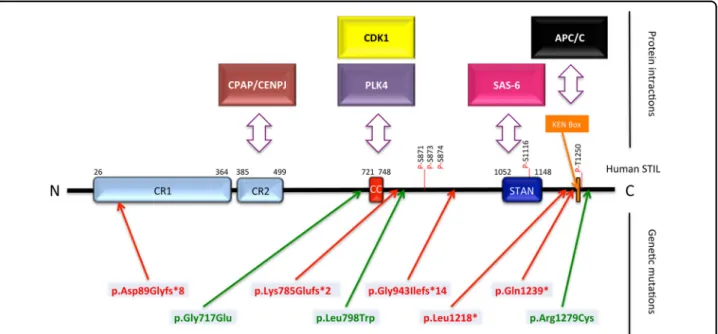
with several proteins (Fig.1). CR2 (amino acids 385–499)
is a proline-rich domain that includes the conserved PRXXPXP motif, which interacts with the Centrosomal P4.1-Associated Protein (CPAP/CENPJ). The coiled-coil
domain (amino acids 721–748) is important both for
Polo-Like Kinase 4 (PLK4) and Cyclin Dependent Kinase
1 (CDK1 also known as CDC2)/CyclinB binding2,3and for
STIL oligomerization4. The STAN domain (amino acids
1052–1148) mediates the binding of the centriole protein
SAS-62. At the C terminus is the KEN box involved in
Anaphase-Promoting-Complex/Cyclosome
(APC/C)-mediated degradation of STIL5. The C terminus of STIL
can also interact with conserved components of the Hedgehog signaling such as Suppressor-of-fused homolog
(SUFU)6and GLI17.
STIL function in centriole duplication
The centriole is an evolutionarily conserved structure consisting of a ninefold symmetric cartwheel with micro-tubule triplets, and thought to have been present in the Last
Eukaryotic Common Ancestor8. The centrosome has two
centrioles orthogonal to each other, surrounded by proteins constituting the pericentriolar material (PCM). Both the centriolar core and the PCM nucleate microtubules, which are important for positioning the mitotic spindles and imparting polarity and asymmetry to the cell. During cell division the centrioles duplicate only once; STIL and its
interactors are central to this process, ensuring thefidelity
of this unique duplication, thereby minimizing
chromo-some instability9. STIL transiently associates with PLK4
and SAS-6 to form the core module for centriole
dupli-cation10. PLK4 is related to the Polo-kinase family of
serine/threonine kinases and initiates centriole
forma-tion11,12, while SAS-6, a coiled-coil protein that
self-assembles into the ninefold symmetry gives the carthweel
its shape13,14. STIL and PLK4 are initially maintained at
low levels in non-dividing and differentiated cells15: STIL
forms dimers or tetramers through its CC domain and is degraded in the cytoplasm by APC/C and its co-activator
CDC20, through recognition of the KEN motif4,5. STIL
Fig. 1 Conserved regions, functional domains, and genetic mutations in the human STIL protein. Blue and orange boxes represent regions of the protein that are highly conserved across species (CR conserved region, CC Coiled-Coil domain, STAN STIL/Ana2 domain, KEN Box Conserved Lys-Glu-Asn residues). Double-headed arrows indicate known interactions with STIL. Known phosphorylated residues in 871, 873, 874, 1116, and 1250 are indicated by a red line. Human mutations predicted to truncate the protein are displayed in red and missense mutations are in green
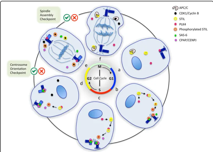
level in the cytoplasm rises in G1 because its degradation
by APC/C is prevented by the absence of CDC205. PLK4
recruits STIL at the base of the parental centriole through the CC domain and phosphorylates the STAN domain at
the late G1/G1-S transition16–18. Phosphorylated STIL
then promotes its binding to the C-terminal region of SAS-6 and recruits it to the outside wall of the
cen-triole2,17. This core module along with CPAP, also
recruited by STIL to the centriole, starts the assembly of
the cartwheel and centriole duplication19,20. At early S
phase, the centrosome-associated protein ROTATIN was recently shown to associate with STIL and contribute to
building full-length centrioles21. At S–G2 transition, the
centrioles are duplicated but remain close to each other, potentially preventing reduplication. Remarkably, CDK1, transiently expressed at late G2 and during M phase, competes with PLK4 for binding to the CC domain of STIL, preventing PLK4 from recruiting STIL until mitosis
is completed3,16,22. The spindle assembly checkpoint, a
highly conserved mechanism also called mitotic check-point, prevents the degradation of CDK1 during mitosis by the inhibition of APC/C-CDC20 until all chromosomes
are properly segregated23. CDK1 is thought to trigger
progressive dissociation of STIL and SAS-6 from early mitotic centrosomes, thereby initiating cartwheel dis-assembly during M phase and ensuring that centriole
biogenesis occurs only once before cell divides5. Upon
activation, APC/C-CDC20 degrades CDK1 for entry into anaphase and again marks STIL for degradation until next the G1 phase. Thus, by prometaphase there is no STIL at the centrosome and by anaphase there is no STIL in the cytoplasm either until upon mitotic exit APC/C is blocked
and STIL levels builds up once again in G1 (Fig.2).
STIL function in SHH signal transduction
SHH is a morphogen involved in patterning,
prolifera-tion, and survial of neural stem cells during development24.
SHH binds to its receptor Patched thereby relieving its inhibition on Smoothened and resulting in the activation of GLI transcription factors. That GLI proteins are the main downstream effectors for SHH signaling is borne out by the fact that activated GLI can rescue the loss-of-function of
SHH and lead to proliferation and increased cell survival25.
The initialfindings that STIL participates in the control of
SHH signaling came from Stil−/− mouse embryos, which
showed a marked reduction of Patched and Gli1
expres-sion26. Interestingly, Stil−/− mutants also lack primary
cilia27, a structure present in almost all cell types, which is
assembled beneath the plasma membrane, by the protru-sion of the microtubule-based axoneme. During interphase, the mother centriole and its appendages constitute the
basal body, from which the axoneme elongates28,29. STIL
requirement for cilia formation likely stems from its function in supporting centriole biogenesis and stability.
Receptors for SHH are abundant on cilia membranes allowing cilia to act as receivers of signals and as platforms
where downstream effectors can be modified30
. Thus, cilia, whose presence depends on STIL, are required for activity
of the SHH pathway31.
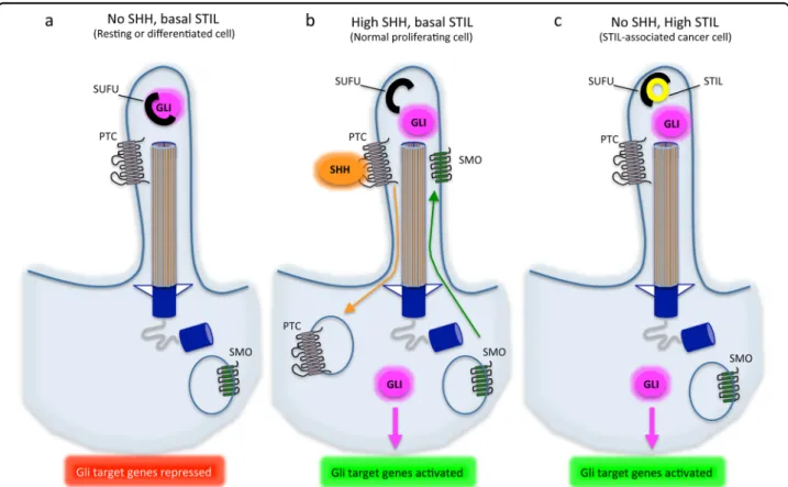
STIL also interferes with the SHH pathway in a direct manner, by interacting with SUFU. SUFU acts as a negative regulator of SHH by tethering GLI1 in the cytoplasm. STIL binds to SUFU in the cytoplasm of pancreatic cancer cells, thus releasing GLI1 for
tran-scription of SHH-downstream genes6. In PC12 cells, STIL
interacts with the SUFU/GLI1 complex, and its down-regulation results in a decrease in both SHH signaling and
cell proliferation7. A similar correlation has been
evi-denced in zebrafish retina cells, further suggesting that STIL plays a role in cell proliferation through the SHH
pathway32(Fig. 3).
Consequences of STIL deregulation
Given the centrality of STIL in centrosome duplica-tion and cilium biogenesis, dereguladuplica-tion of STIL is
predictably profound. Stil−/− embryos die at
midgesta-tion with axial midline defects, resulting from aberrant
SHH signaling26. MEFs derived from Stil−/− embryos
show a marked decrease in mitotic index, and Stil
knockdown results in the absence of identifiable
cen-trosomes during interphase and multiple spindle poles
with disrupted γ-tubulin signals during mitosis33, as
well as in the loss of primary cilia27. Conversely, STIL
overexpression causes centrosome amplification,
resulting in a star-like pattern around the parental centriole, consistent with a role of STIL in the onset of procentriole formation. Accordingly, this star-like structure is positive for the centriolar proteins CP110
and centrin20,34. Mutations within the various domains
of STIL give predictable phenotypes. When the CR2 domain or the CC domain or the STAN domain is
deleted, there is no centrosomal duplication16,18,
whereas removal of the KEN box leads to centrosome
amplification due to the inability of STIL to be
degra-ded5. However, both overduplication and lack of
duplication of centrosomes lead to abnormal mitotic spindle assembly and consequently increase the chances of abnormal chromosomal segregation and aneuploidy. Another consequence of STIL deregulation is the indirect effect on interacting partners. STIL binding to PLK4 supresses PLK4 auto-inhibition, thereby allowing its trans-phosphorylation and protecting activated PLK4
from degradation35. Conversely, depletion of STIL leads
to a marked accumulation of PLK4 in an inactive con-formation. Therefore, changes in STIL levels have
immediate consequences on PLK4 activation levels10,17.
STIL also negatively regulates Chfr, an E3 ligase that blocks mitotic entry in response to mitotic stress.
No direct interaction between STIL and Chfr has been shown, but STIL expression results in an increase of Chfr auto-ubiquitination, thereby promoting its proteasomal degradation and proper progression of cells through
mitosis33. Data support a role of Chfr in defective mitotic
progression associated with reduced activation of
CDK1/CYCLIN B and centrosomal abnormalities caused
by the lack of STIL33. Thus, STIL deregulation likely
impacts the activation level of CDK1/CYCLIN B and the timing of entry into mitosis, through its regulation of Chfr.
STIL expression during human brain development
STIL is expressed in both fetal and adult tissues.
How-ever, its expression levels fluctuate with the cell cycle,
making it difficult to detect in whole tissue, especially if
the cells are not synchronized. Indeed, it is detected more easily in cancer cell lines and tissue where it is
over-expressed36,37. The BrainSpan Atlas (http://www.
brainspan.org/) and the BrainCloud database (http://
braincloud.jhmi.edu) show its expression pattern during
brain development. The association of STIL with cell proliferation is borne out by its expression pattern during fetal stages. At 15 postconceptional weeks STIL is strongly expressed in the ventricular and subventricular zones of the forebrain, the ganglionic eminence and the rostral migratory stream, and less expressed in intermediate zone, subplate, cortical plate, marginal zone, and sub-granular layer of the forebrain. This pattern persists at 21 postconceptional weeks, although the expression is Fig. 2 STIL regulation during the cell cycle. Six phases of the cell cycle are represented. a Early G1 phase: STIL levels are low in the cytoplasm and STIL is absent at the centrosome. b, c G1–S and S phases: STIL levels are high in the cytoplasm and STIL starts being associated with the centrosome. PLK4 interacts and phosphorylates STIL. STIL recruits SAS-6 and CPAP to the centrosome, contributing to the assembly of the cartwheel. The procentriole starts elongating in S phase. d G2 phase: the two centrosomes begin to move apart. CDK1/CYCLIN B is active. Centrosome orientation checkpoint. e The nuclear envelope breakdown occurs, CDK1 binds to STIL and moves it to the cytoplasm, there is no more STIL at the centrosome, cartwheel disassembles. Spindle assembly checkpoint. f Anaphase, APC/C is fully active, cytoplasmic STIL is degraded. The cell nucleus or chromosomes with their mitotic spindle are represented in blue. Centrioles are shown in dark blue. STIL is represented by a yellow donut surrounded by a red circle when phosphorylated. PLK4 is represented by a red dot, CDK1/CYCLIN B by a black dot, SAS-6 by a small green hexagon, CPAP/CENPJ by a small pink hexagon, and APC/C by a black and white C-shape. Centrosome orientation and spindle assembly checkpoints are indicated by green/red tick boxes
reduced in the subventricular zone. The expression of PLK4, SAS-6, and CPAP also broadly shows this pattern of expression, although the expression of SAS-6 and STIL diverges in some regions of the cortical plate. In the cerebellum, STIL, PLK4, and SAS-6 but not CPAP are expressed in the external granule layer and regions of the rhombic lip. However, none of these genes are expressed in the transient Purkinje cell cluster, the ventricular matrix zone of the cerebellum or in the migratory streams of the hindbrain. Microdissection experiments at different
developmental stages confirmed that STIL expression is
high in the ventricular zone and low in the subplate and
cortical zones (http://www.brainspan.org/). Further,
microdissected diencephalon did not have STIL (http://
www.blueprintnhpatlas.org/). This suggests that (i) the
structural components of the cartwheel STIL-SAS-6-PLK4 is by no means obligatory for centrosome duplica-tion in all cells and (ii) STIL is present in specific popu-lations of dividing cells.
STIL mutations and primary microcephaly
Microcephaly (small brain size) is indirectly diagnosed by a head circumference smaller than the age-specific and gender-adjusted mean by more than 2 standard deviations (S.D.s) at birth. Primary microcephaly refers to hereditary microcephalies already detectable in utero. Most of them are autosomal recessive and include (i) isolated forms called MicroCephaly Primary Hereditary (MCPH), (ii) forms associated with growth retardation, called
micro-cephalic dwarfism. Most of STIL mutations identified in
patients are associated with a MCPH phenotype and STIL
is recognized as MCPH738–40. However, a few patients
exhibit criteria of microcephalic dwarfism, as short stature
has been reported41. So far, eight mutations in STIL have
been described in 37 patients all showing severe
micro-cephaly (−4 to −10 S.D.). Five mutations are splice42,43
,
deletion, nonsense43, or duplication44 that predict the
production of truncated proteins, while three mutations are
missense substitutions44–46 (Fig. 1). Despite the different
Fig. 3 STIL and SHH signaling at the cilium. A cilium and its axonemal structure are represented in three different contexts of expression of SHH and STIL. SMO = SMOOTHENED receptor, PTC = PATCHED receptor. a In resting or differentiated cells, there is no SHH and low STIL expressed in the cilium. The PTC receptor is expressed at the membrane while the SMO receptor is degraded in the cytoplasm. SUFU binds and tether GLI proteins in the cytoplasm, blocking GLI-dependent transcription. b When SHH is expressed, it binds to its receptor PTC inducing its internalization and degradation in the cytoplasm. This allows SMO expression at the membrane of the cilium and leads to GLI derepression. c When STIL is abnormally highly expressed during interphase, it becomes abundant in the cilium where it binds to SUFU, releasing GLI proteins and thus activating GLI-mediated transcription even in the absence of SHH
Table 1 List of mutations identi fi ed in the human STIL gene, and their functional consequence Mut ation (nt) Mut ation (aa) Domain affected (protei n) Functional co nsequence Head OFC (S.D.) Brain MRI data Refe rences c.453 + 5 G > A (sp lice dono r) p.Asp8 9Glyfs*8 (trunca ting) All d omains No func tion al protein − 9t o − 10 Short cc, simpl ifi ed gyration, loba r HPE 42 c.215 0 G > A p.Gly7 17Glu (missen se) CC dom ain Presum ably inab ility of STIL to oligom erize and interac t w ith PLK4 — decr eased centr iole duplication capacity − 7t o − 8 Parti al cc ag enesis, simpl ifi ed gyration, lob ar HP E 4 , 45 c.235 4_2 355dupGA p.Lys785Gl ufs*2 (trunca ting) Phosphor ylation sites, STAN domain, KEN box Removes critic al PLK 4 phospho rylation sites 26 cm at birth Cc agen esis, simpli fied gyration, lob ar HP E 44 c.239 2 T > G p.Leu798Trp (missen se) — ND NA NA 46 c.282 6 + 1 G > A (sp lice dono r) p.Gly9 43Ilefs*14 (like ly truncat ing) STAN dom ain, KEN box Not able to dup licate centrosome s since they can not recruit SAS6 − 5N A 27 , 43 c.365 5del G p.Val 1219* (initially repo rted as p.Leu128 *; truncat ing) KEN box Resistance to A PC/C-mediated degradation − 4t o − 10 NA 5 , 18 , 27 , 43 c.371 5 C > T p.Gln1 239* (trunca ting) KEN box Resistance to A PC/C-mediated degradation − 7t o − 8N A 18 , 27 , 43 , 95 c.383 5 C > T p.Arg1279C ys (missen se) C term inus ND 26 cm at birth Cc agen esis, simpli fied gyration, lob ar HP E 44
kinds of mutations, the various domains affected (Table1), and the report of lobar holoprosencephaly and partial agenesis of the corpus callosum in addition to
micro-cephaly in some cases42,44,45, no clear genotype–phenotype
correlation has come out so far. However, the fact that these mutations trigger a phenotype compatible with life (although very severe) is surprising since genetic ablation of STIL is embryonically lethal early during embryogenesis in
mice and fish26,47. This suggests that STIL mutations in
human do not result in a complete loss-of-function, or are
partially compensated by other genes. Thefirst hypothesis
is supported by thefindings that two C-terminal nonsense
mutations (p.Val1219* and p.Gln1239*), which result in the
loss of the KEN box43, do not affect the centrosomal
localization of STIL nor its functionality but rather abolish
its degradation by APC/C in late mitosis5. Removal of the
KEN box thus causes a strong accumulation of the mutant
protein, resulting in centrosome amplification5
. Alter-natively, microcephaly in MCPH7 patients can result from a decrease of STIL levels. This was illustrated by rescue experiments in U2OS cells showing that the pGly717Glu mutation induces a reduced but non-null activity of STIL
on centriole duplication45. Another mutation, inducing
exon 5 skipping and frameshift in the STIL sequence, is
likely close to a null mutation as all domains are predicted
to be lost42. However exon skipping was only partial in the
patient analyzed, suggesting some residual activity of STIL, and providing an explanation for the fact that this mutation is compatible with life and the idea that MCPH7 micro-cephaly can result from a decreased STIL activity. There-fore, both accumulation and impairment of STIL protein levels during cell cycle affect centriole regulation and result
in microcephaly (Fig.4).
STIL deregulation and cancer
Centrosomes are essential for chromosomal stability, and abnormalities of their number, or structure affect cell
division22. Considering the central role of STIL in
main-taining centrosome integrity in highly proliferating cells, STIL has been found upregulated in several cancers of bad prognosis, including lung cancer, colon carcinoma,
pros-tate adenocarcinoma36, and ovarian cancers48. Moreover,
STIL expression is associated with an increased metastatic
potential in multiple cancers49. As expected, STIL
upre-gulation is associated with a high histopathological
mitotic index in tumors36 and presumably affects the
formation of mitotic spindles, as well as SHH signaling and the function of its interactors.
Fig. 4 Consequences of deregulating STIL expression levels. STIL expression levels arefinely regulated both during development and in differentiated tissues. While total absence of STIL is lethal during development, high STIL levels due to random somatic mutations in differentiated tissues result in aneuploidy and/or high GLI-mediated transcription that can lead to cancer. Congenital deregulation of STIL (both a too high and a too weak expression) leads to microcephaly by either reduced or amplified centrosome duplication, highlighting the importance of a tightly regulated STIL expression
Although cells can proliferate and microtubule can nucleate in their absence, centrosomes are obligatory for
controlling spindle orientation50. Loss of this control
leads to spindle defects, especially in highly polarized cells, where misoriented spindles influence daughter cell positioning, possibly causing a disruption in tissue
mor-phology51. Several tumor suppressor genes such as APC,
or VHL, are important for stabilizing microtubules and
maintaining spindle orientation51. Similarly,
over-expression of genes such as STIL could act as oncogenes and lead to cancer by promoting spindle defects, although direct evidence of a link between oncogene activation and spindle misorientation is lacking. STIL overexpression, which results in supernumerary centrosomes, could lead
to cancer in inducing chromosomal instability10,52.
The role of STIL in promoting SHH signaling could be another pathway to account for its association with can-cer. Following the initial observation that SHH over-expression results in the development of basal cell
carcinomas in mice53, a large body of literature has
implicated the SHH pathway in development of various
cancers including a subset of medulloblastomas54,55. STIL
overexpression in pancreatic adenocarcinoma was shown to de-repress GLI1 from SUFU-mediated control and this
phenotype was reversed when STIL was downregulated6.
Thus, during the process of carcinogenesis, the increased expression of STIL promotes the transcriptional activity of GLI1, which is no longer regulated quantitatively. GLI1 upregulates genes that promote sustained proliferation, cell death resistance, stemness, angiogenesis, and genomic
instability, which are hallmarks of cancer56. Therefore,
increase in STIL expression leading to uncontrolled GLI1 de-repression likely represents a crucial step toward cancer progression.
MCPH and cancer—two conditions associated with
abnormal centrosome duplication
STIL loss-of-function results in embryonic lethality in
mice, fish, and most likely in human as well. MCPH7
phenotype reflects that perturbing the tuning of STIL
expression levels is compatible with life but triggers severe patterning phenotypes during development, including brain growth. Thus, microcephaly caused by STIL muta-tions can result from either too less centrosome
dupli-cation or too much centrosome amplifidupli-cation (Fig.4). In
both cases, the integrity of the mitotic spindle is com-promised and cell divisions have a greater chance of chromosomal instability. Similarly, the association of STIL upregulation with various cancers reflects a deregulation of STIL expression levels in specific tissues contributing to genomic instability. What results in MCPH rather than cancer could be the difference in mitotic checkpoints in
different contexts13,57. Centrosome orientation
check-point ensures that the centrosomes are duplicated and
appropriately positioned before entry into mitosis and several centrosomal proteins such as CNN and SAS-6 are
part of this checkpoint58. This checkpoint may not be
present in all cells at all developmental stages but rather in those cases where the cell division plane has important
outcomes59. In germline stem cells, for example, the
division plane determines the distribution of cell fate
determinants in daughter cells, helding cells at the G2–M
phase until their mitotic spindles are properly oriented58.
STIL downregulation results in disorganized mitotic spindles resulting in a loss of spindle orientation
con-trol47. In cortical development, STIL mutations leading to
spindle orientation defects likely result in a depletion of cortical progenitors due to cell death or premature
dif-ferentiation and give rise to a MCPH phenotype57(Fig.4).
PLK4 overexpression also results in centrosome amplifi-cation and aneuploidy leading to a reduction in brain size
due to cell death60. Apoptosis inhibition in this
back-ground causes the accumulation of aneuploid cells unable
to proliferate efficiently, leading to premature neuronal
differentiation60, whereas in a P53−/− context, PLK4
overexpression results in aneuploidy and skin cancer61.
The spindle assembly checkpoint is the next checkpoint once cells have entered mitosis and formed spindles. In the absence of bipolar spindles, cells are held-up in
pro-metaphase and do not proceed to anaphase58, which
results in increased apoptosis62. However, cells can
increase their time in mitosis, thus adapting to this checkpoint and can assemble bipolar spindles in the absence of centrosomes or in the presence of super-numerary centrosomes. First in the absence of centro-some duplication cells manage to have bipolar spindles even though there is no centriole attached to one of the
poles34. Second is a phenomena of centrosome clustering,
wherein multiple centrosomes are clustered to enable the
formation of bipolar spindles, a phenomenon whose ef
fi-ciency could be tissue-specific60. These divisions often
result in aneuploidy. When centrosome integrity is com-promised, cells usually arrest in G1 and do not re-enter cell cycle after a prolonged cytokinesis to avoid
chromo-somal instability22and in many cases proliferation in the
absence of centrioles additionally requires the suppression
of P5317. Upregulation of STIL giving rise to aneuploidy
could play a major role in providing an evolving genome to help the cells adapt to the changing environment of the cancer and escape normal checkpoints. In the developing cortex, abnormal mitotic spindles due to STIL mutations would result in cell fate switches rather than cell hyper-proliferation. However, that patients whose STIL muta-tion is clearly associated with centrosome amplificamuta-tion could be at risk of developing cancer cannot be ruled out.
Seventeen MCPH loci have been described so far40,
several of which also play a role in cancer. MCPH1 is an early DNA damage response (DDR) protein and MCPH1
deletions have been identified as a risk for breast cancer63
. Centrosome amplification is commonly seen following
DNA damage and can arise as part of the DDR64. The
serine-threonine kinase CHK1 plays a major role in DDR by halting cell division in G2/M until repair proteins are
recruited at lesion sites. In this context, MCPH1 de
fi-ciency potentiates CHK1 activity and increases
centro-some amplification65. Thus, compromising mitotic
checkpoints results in cell division in the presence of
abnormal centrosomes63,65. How CHK1 overactivation
leads to centrosome amplification is not fully understood,
but it is thought to activate CDK2, a cyclin-dependent kinase required for centrosome duplication, likely through
activating its phosphorylation66,67. WD repeat-containing
protein62 (WDR62), whose deficiency is associated with
MCPH268–70, is required for maintaining spindle and
centrosome integrity. Its overexpression, coincident with centrosome amplification, is also seen in lung
adeno-carcinomas and ovarian cancers71,72. WDR62 interacts
physically during the cell cycle with the abnormal spindle-like microcephaly-associated protein (ASPM), whose
deficiency causes the most frequent primary microcephaly
(MCPH5)73. Here too, increased ASPM levels cause
tumor growth and are seen in medulloblastomas while its
reduction causes a decrease in tumor proliferation74.
CDK5RAP2 (MCPH3) is also involved in the DDR; it functions by arresting cells before mitotic entry due to imparied centrosomes but also interacts with BUB1 and MAD2, which are important in spindle activation
check-point75,76. Interestingly, a correlation between
cen-trosomal abnormalities, aneuploidy, and cytogenic risk profile is seen in acute myeloid leukemia, where gene expression profiling has revealed the differential expres-sion of genes encoding centrosomal and mitotic spindle
proteins77. Among these are pericentrin, a scaffold protein
that anchors many other proteins at the centrosome78and
NuMA, which associates with dynein and microtubules to create localized pulling forces, thus regulating the correct
assembly and positioning of the mitotic spindle79. The
role of such factors points to mitotic centrosomal
abnormalities as important component of cancer
progression.
STIL could also have an indirect effect on cancer, as a downstream effector of PLK4. Increased PLK4 expres-sion has been reported in malignancies such as colorectal
cancer80, pediatric medulloblastoma81, and breast
tumors82. Its transient overexpression leads to
centro-some overduplication and in a P53−/− background that
inhibits apoptosis, it results in aneuploidy and
sponta-neous skin cancer61. Another recent study has
convin-cingly shown that PLK4 overexpression leads to
spontaneous tumors in several organs83. It remains to be
seen whether this phenotype is dependent on STIL expression. STIL binds to PLK4 in the cytoplasm and
therefore STIL expression levels could impact PLK4
cytoplasmic activity17, which functions to remodel the
cytoskeleton and may be important for cancer invasion and metastasis as its depletion is correlated with an
increase in E-cadherin expression and less metastasis84.
CYCLIN B is also frequently found elevated in primary breast cancer, esophageal squamous cell carcinoma, laryngeal squamous cell carcinoma, and colorectal
car-cinoma85–89and its expression indicates a bad prognosis
and is correlated with the malignancy of gynecological
cancers90. Downregulation of STIL decreases CDK1/
CYCLIN B activity, prevents G2–M transition, and
cau-ses inhibition of tumor growth in vivo91. Conversely,
increasing STIL could promote CDK1/CYCLIN B activity and indirectly participate in CYCLIN B-dependent proliferation in tumor cells. Absence of STIL also results in an upregulation of Chfr, and lowers PLK1, resulting in the activation of the CDC25c phos-phatase. This pathway could thus control the entry of cells into mitosis independent of its obligatory role in
centriole duplication33.
Conclusion
STIL mutations in MCPH show that centrosomes and cilia are essential for normal brain development. During evolution, one way that the cortex has undergone expansion is likely linked to mechanisms controlling spindle orientation and keeping the balance between
asymmetric and symmetric cell divisions92,93. A change in
the nature of spindle orientation control has been pro-posed to account for the population of subventricular zone progenitors in human development and based on the strong expression of STIL in the ventricular zone, one may speculate that STIL is important for controlling spindle orientation in giving rise to subventricular zone’s
cells94. Deficiency of several centrosomal proteins results
in MCPH, and centrosomal proteins may thus be a key to discovering the mechanism of expansion of cortical
area38. In addition, the involvement of STIL in cancer
shows that the result of spindle defects can be vastly different depending on the stage of development and the tissue involved that probably is partly due to differential responses to mitotic checkpoint mechanisms. STIL intersects several pathways, and lowering its level could block the effects of PLK4 overexpression, CDK1/CYCLIN B activation, and GLI1 signaling, and reduce proliferation even in the presence of aneuploidy. Thus, centrosomal proteins may be a new class of targets for cancer. Acknowledgments
This work was supported by the Institut National pour la Santé et la Recherche Médicale (INSERM), the Centre National de la Recherche Scientifique (CNRS), the Université Paris 7, the DHU PROTECT, the Indo-French Centre for the Promotion of Advanced Research“CEFIPRA” (grant no 4903-02 to S.M., V.E.G., P.G., and S.P.), and grants from the French National Research Agency
(ANR-13-RARE-0007-01 to S.P., P.G., and V.E.G. (ERA-NET E-Rare 2013, EuroMicro project), and ANR-16-CE16-0024- 01 to V.E.G. (PRC GENERIQUE 2016, MicroGol project)). S.M. was supported by a grant from Curadev Pharma.
Author details
1
PROTECT, INSERM, Université Paris Diderot, Sorbonne Paris Cité, Paris, France.
2Centre for Neuroscience, IISC Bangalore, India.3Curadev Pharma, B 87, Sector
83, Noida, UP 201305,, India.4AP HP, Hôpital Robert Debré, Service de Génétique Clinique, Paris, France.5Centre for the Developing Brain, Division of
Imaging Sciences and Biomedical Engineering, King’s College London, King’s Health Partners, St. Thomas’ Hospital, London, UK
Competing interests
The authors declare that they have no competingfinancial interests. Publisher's note
Springer Nature remains neutral with regard to jurisdictional claims in published maps and institutional affiliations.
Received: 29 August 2017 Revised: 4 October 2017 Accepted: 23 October 2017
References
1. Aplan, P. D. et al. Involvement of the putative hematopoietic transcription factor SCL in T-cell acute lymphoblastic leukemia. Blood. 79, 1327–1333 (1992).
2. Ohta, M. et al. Direct interaction of Plk4 with STIL ensures formation of a single procentriole per parental centriole. Nat. Commun. 5, 5267 (2014). 3. Zitouni, S. et al. CDK1 prevents unscheduled PLK4-STIL complex assembly in
centriole biogenesis. Curr. Biol. 26, 1127–1137 (2016).
4. David, A. et al. Molecular basis of the STIL coiled coil oligomerization explains its requirement for de-novo formation of centrosomes in mammalian cells. Sci. Rep. 6, 24296 (2016).
5. Arquint, C. & Nigg, E. A. STIL microcephaly mutations interfere with APC/C-mediated degradation and cause centriole amplification. Curr. Biol. 24, 351–360 (2014).
6. Kasai, K., Inaguma, S., Yoneyama, A., Yoshikawa, K. & Ikeda, H. SCL/TAL1 interrupting locus derepresses GLI1 from the negative control of suppressor-of-fused in pancreatic cancer cell. Cancer Res. 68, 7723–7729 (2008). 7. Sun, L. et al. Characterization of the human oncogene SCL/TAL1 interrupting
locus (Stil) mediated Sonic hedgehog (Shh) signaling transduction in pro-liferating mammalian dopaminergic neurons. Biochem. Biophys. Res. Commun. 449, 444–448 (2014).
8. Carvalho-Santos, Z., Azimzadeh, J., Pereira-Leal, J. B. & Bettencourt-Dias, M. Evolution: tracing the origins of centrioles, cilia, andflagella. J. Cell Biol. 194, 165–175 (2011).
9. Funk, L. C., Zasadil, L. M. & Weaver, B. A. Living in CIN: mitotic infidelity and its consequences for tumor promotion and suppression. Dev. Cell 39, 638–652 (2016).
10. Arquint, C. & Nigg, E. A. The PLK4-STIL-SAS-6 module at the core of centriole duplication. Biochem. Soc. Trans. 44, 1253–1263 (2016).
11. Bettencourt-Dias, M. et al. SAK/PLK4 is required for centriole duplication and flagella development. Curr. Biol. 15, 2199–2207 (2005).
12. Habedanck, R., Stierhof, Y. D., Wilkinson, C. J. & Nigg, E. A. The Polo kinase Plk4 functions in centriole duplication. Nat. Cell Biol. 7, 1140–1146 (2005). 13. Kitagawa, D. et al. Spindle positioning in human cells relies on proper centriole
formation and on the microcephaly proteins CPAP and STIL. J. Cell Sci. 124, (Pt 22), 3884–3893 (2011).
14. van Breugel, M. et al. Structures of SAS-6 suggest its organization in centrioles. Science 331, 1196–1199 (2011).
15. Bauer, M., Cubizolles, F., Schmidt, A. & Nigg, E. A. Quantitative analysis of human centrosome architecture by targeted proteomics andfluorescence imaging. EMBO J. 35, 2152–2166 (2016).
16. Kratz, A. S., Barenz, F., Richter, K. T. & Hoffmann, I. Plk4-dependent phosphor-ylation of STIL is required for centriole duplication. Biol. Open 4, 370–377 (2015).
17. Moyer, T. C., Clutario, K. M., Lambrus, B. G., Daggubati, V. & Holland, A. J. Binding of STIL to Plk4 activates kinase activity to promote centriole assembly. J. Cell Biol. 209, 863–878 (2015).
18. Vulprecht, J. et al. STIL is required for centriole duplication in human cells. J. Cell Sci. 125(Pt 5), 1353–1362 (2012).
19. Cottee, M. A. et al. Crystal structures of the CPAP/STIL complex reveal its role in centriole assembly and human microcephaly. eLife 2, e01071 (2013). 20. Tang, C. J. et al. The human microcephaly protein STIL interacts with
CPAP and is required for procentriole formation. EMBO J. 30, 4790–4804 (2011).
21. Chen, H. Y. et al. Human microcephaly protein RTTN interacts with STIL and is required to build full-length centrioles. Nat. Commun. 8, 247 (2017). 22. Pihan, G. A. Centrosome dysfunction contributes to chromosome instability,
chromoanagenesis, and genome reprograming in cancer. Front. Oncol. 3, 277 (2013).
23. Sivakumar, S. & Gorbsky, G. J. Spatiotemporal regulation of the anaphase-promoting complex in mitosis. Nat. Rev. Mol. Cell Biol. 16, 82–94 (2015). 24. Choudhry, Z. et al. Sonic hedgehog signalling pathway: a complex network.
Ann. Neurosci. 21, 28–31 (2014).
25. Cayuso, J., Ulloa, F., Cox, B., Briscoe, J. & Marti, E. The Sonic hedgehog pathway independently controls the patterning, proliferation and survival of neuroe-pithelial cells by regulating Gli activity. Development 133, 517–528 (2006). 26. Izraeli, S. et al. The SIL gene is required for mouse embryonic axial
develop-ment and left-right specification. Nature 399, 691–694 (1999).
27. David, A. et al. Lack of centrioles and primary cilia in STIL(−/−) mouse embryos. Cell Cycle 13, 2859–2868 (2014).
28. Ishikawa, H. & Marshall, W. F. Ciliogenesis: building the cell’s antenna. Nat. Rev. Mol. Cell Biol. 12, 222–234 (2011).
29. Tanos, B. E. et al. Centriole distal appendages promote membrane docking, leading to cilia initiation. Genes Dev. 27, 163–168 (2013).
30. Scholey, J. M. & Anderson, K. V. Intraflagellar transport and cilium-based sig-naling. Cell 125, 439–442 (2006).
31. Bangs, F. & Anderson, K. V. Primary Cilia and Mammalian Hedgehog Signaling. Cold Spring Harb. Persp. Biol. 9, pii: a028175 (2017).
32. Sun, L. et al. Transcription of the SCL/TAL1 interrupting Locus (Stil) is required for cell proliferation in adult Zebrafish Retinas. J. Biol. Chem. 289, 6934–6940 (2014).
33. Castiel, A. et al. The Stil protein regulates centrosome integrity and mitosis through suppression of Chfr. J. Cell Sci. 124(Pt 4), 532–539 (2011). 34. Arquint, C., Sonnen, K. F., Stierhof, Y. D. & Nigg, E. A. Cell-cycle-regulated
expression of STIL controls centriole number in human cells. J. Cell Sci. 125(Pt 5), 1342–1352 (2012).
35. Arquint, C. et al. STIL binding to Polo-box 3 of PLK4 regulates centriole duplication. Elife 4 (2015).
36. Erez, A. et al. Sil overexpression in lung cancer characterizes tumors with increased mitotic activity. Oncogene 23, 5371–5377 (2004).
37. Izraeli, S. et al. Expression of the SIL gene is correlated with growth induction and cellular proliferation. Cell Growth Differ. 8, 1171–1179 (1997).
38. Kaindl, A. M. et al. Many roads lead to primary autosomal recessive micro-cephaly. Prog. Neurobiol. 90, 363–383 (2010).
39. Passemard, S. et al. Expanding the clinical and neuroradiologic pheno-type of primary microcephaly due to ASPM mutations. Neurology 73, 962–969 (2009).
40. Verloes, A., Drunat, S., Gressens, P., Passemard, S. Primary autosomal recessive microcephalies and seckel syndrome spectrum disorders. In: Pagon R.A., Adam M.P., Ardinger H.H., Wallace S.E., Amemiya A., Bean L.J.H., et al. (eds). GeneRe-views(R): Seattle (WA) (1993).
41. Darvish, H. et al. A clinical and molecular genetic study of 112 Iranian families with primary microcephaly. J. Med. Genet. 47, 823–828 (2010).
42. Kakar, N. et al. STIL mutation causes autosomal recessive microcephalic lobar holoprosencephaly. Hum. Genet. 134, 45–51 (2015).
43. Kumar, A., Girimaji, S. C., Duvvari, M. R. & Blanton, S. H. Mutations in STIL, encoding a pericentriolar and centrosomal protein, cause primary micro-cephaly. Am. J. Hum. Genet. 84, 286–290 (2009).
44. Bennett, H. et al. A prenatal presentation of severe microcephaly and brain anomalies in a patient with novel compound heterozygous mutations in the STIL gene found postnatally with exome analysis. Pediatr. Neurol. 51, 434–436 (2014).
45. Mouden, C. et al. Homozygous STIL mutation causes holoprosencephaly and microcephaly in two siblings. PLoS ONE 10, e0117418 (2015).
46. Papari, E. et al. Investigation of primary microcephaly in Bushehr province of Iran: novel STIL and ASPM mutations. Clin. Genet. 83, 488–490 (2013). 47. Pfaff, K. L. et al. The zebrafish cassiopeia mutant reveals that SIL is required for
mitotic spindle organization. Mol. Cell Biol. 27, 5887–5897 (2007).
48. Rabinowicz, N. et al. Targeting the centriolar replication factor STIL synergizes with DNA damaging agents for treatment of ovarian cancer. Oncotarget 8, 27380–27392 (2017).
49. Ramaswamy, S., Ross, K. N., Lander, E. S. & Golub, T. R. A molecular signature of metastasis in primary solid tumors. Nat. Genet. 33, 49–54 (2003).
50. Yamashita, Y. M. & Fuller, M. T. Asymmetric centrosome behavior and the mechanisms of stem cell division. J. Cell Biol. 180, 261–266 (2008). 51. Pease, J. C. & Tirnauer, J. S. Mitotic spindle misorientation in cancer--out of
alignment and into thefire. J. Cell Sci. 124(Pt 7), 1007–1016 (2011). 52. Nigg, E. A. Centrosome aberrations: cause or consequence of cancer
pro-gression? Nat. Rev. Cancer 2, 815–825 (2002).
53. Oro, A. E. et al. Basal cell carcinomas in mice overexpressing sonic hedgehog. Science 276, 817–821 (1997).
54. Kool, M. et al. Molecular subgroups of medulloblastoma: an international meta-analysis of transcriptome, genetic aberrations, and clinical data of WNT, SHH, Group 3, and Group 4 medulloblastomas. Acta Neuropathol. 123, 473–484 (2012).
55. Northcott, P. A. et al. Medulloblastomics: the end of the beginning. Nat. Rev. Cancer 12, 818–834 (2012).
56. Kasai, K. GLI1, a master regulator of the hallmark of pancreatic cancer. Pathol. Int. 66, 653–660 (2016).
57. Wang, G., Jiang, Q. & Zhang, C. The role of mitotic kinases in coupling the centrosome cycle with the assembly of the mitotic spindle. J. Cell Sci. 127(Pt 19), 4111–4122 (2014).
58. Venkei, Z. G. & Yamashita, Y. M. The centrosome orientation checkpoint is germline stem cell specific and operates prior to the spindle assembly checkpoint in Drosophila testis. Development 142, 62–69 (2015).
59. Chen, C., Inaba, M., Venkei, Z. G. & Yamashita, Y. M. Klp10A, a stem cell centrosome-enriched kinesin, balances asymmetries in Drosophila male germline stem cell division. eLife 5, pii:e20977 (2016).
60. Marthiens, V. et al. Centrosome amplification causes microcephaly. Nat. Cell Biol. 15, 731–740 (2013).
61. Sercin, O. et al. Transient PLK4 overexpression accelerates tumorigenesis in p53-deficient epidermis. Nat. Cell Biol. 18, 100–110 (2016).
62. Novorol, C. et al. Microcephaly models in the developing zebrafish retinal neuroepithelium point to an underlying defect in metaphase progression. Open Biol. 3, 130065 (2013).
63. Mantere, T. et al. Targeted next-generation sequencing identifies a recurrent mutation in MCPH1 associating with hereditary breast cancer susceptibility. PLoS Genet. 12, e1005816 (2016).
64. Mullee, L. I. & Morrison, C. G. Centrosomes in the DNA damage response--the hub outside the centre. Chromosome Res. 24, 35–51 (2016).
65. Antonczak, A. K. et al. Opposing effects of pericentrin and microcephalin on the pericentriolar material regulate CHK1 activation in the DNA damage response. Oncogene 35, 2003–2010 (2016).
66. Bourke, E., Brown, J. A., Takeda, S., Hochegger, H. & Morrison, C. G. DNA damage induces Chk1-dependent threonine-160 phosphorylation and acti-vation of Cdk2. Oncogene 29, 616–624 (2010).
67. Bourke, E. et al. DNA damage induces Chk1-dependent centrosome ampli fi-cation. EMBO Rep. 8, 603–609 (2007).
68. Bilguvar, K. et al. Whole-exome sequencing identifies recessive WDR62 mutations in severe brain malformations. Nature 467, 207–210 (2010). 69. Nicholas, A. K. et al. WDR62 is associated with the spindle pole and is mutated
in human microcephaly. Nat. Genet. 42, 1010–1014 (2010).
70. Yu, T. W. et al. Mutations in WDR62, encoding a centrosome-associated protein, cause microcephaly with simplified gyri and abnormal cortical architecture. Nat. Genet. 42, 1015–1020 (2010).
71. Shinmura, K. et al. WDR62 overexpression is associated with a poor prognosis in patients with lung adenocarcinoma. Mol. Carcinog. 56, 1984–1991 (2017).
72. Zhang, Y. et al. Overexpression of WDR62 is associated with centrosome amplification in human ovarian cancer. J. Ovarian Res. 6, 55 (2013). 73. Jayaraman, D. et al. Microcephaly proteins Wdr62 and Aspm define a mother
centriole complex regulating centriole biogenesis, apical complex, and cell fate. Neuron 92, 813–828 (2016).
74. Williams, S. E. et al. Aspm sustains postnatal cerebellar neurogenesis and medulloblastoma growth in mice. Development 142, 3921–3932 (2015). 75. Barr, A. R., Kilmartin, J. V. & Gergely, F. CDK5RAP2 functions in centrosome to
spindle pole attachment and DNA damage response. J. Cell Biol. 189, 23–39 (2010).
76. Zhang, X. et al. CDK5RAP2 is required for spindle checkpoint function. Cell Cycle 8, 1206–1216 (2009).
77. Neben, K. et al. Gene expression patterns in acute myeloid leukemia correlate with centrosome aberrations and numerical chromosome changes. Oncogene 23, 2379–2384 (2004).
78. Delaval, B. & Doxsey, S. J. Pericentrin in cellular function and disease. J. Cell Biol. 188, 181–190 (2010).
79. di Pietro, F., Echard, A. & Morin, X. Regulation of mitotic spindle orientation: an integrated view. EMBO Rep. 17, 1106–1130 (2016).
80. Macmillan, J. C., Hudson, J. W., Bull, S., Dennis, J. W. & Swallow, C. J. Com-parative expression of the mitotic regulators SAK and PLK in colorectal cancer. Ann. Surg. Oncol. 8, 729–740 (2001).
81. Sredni, S. T. & Tomita, T. The polo-like kinase 4 gene (PLK4) is overexpressed in pediatric medulloblastoma. Child’s Nervous Syst. 33, 1031 (2017).
82. Marina, M. & Saavedra, H. I. Nek2 and Plk4: prognostic markers, drivers of breast tumorigenesis and drug resistance. Front. Biosci. 19, 352–365 (2014). 83. Levine, M. S. et al. Centrosome amplification is sufficient to promote
spon-taneous tumorigenesis in mammals. Dev. Cell 40, 313–322 (2017). e315. 84. Kazazian, K. et al. Plk4 promotes cancer invasion and metastasis through Arp2/
3 complex regulation of the actin cytoskeleton. Cancer Res. 77, 434–447 (2017).
85. Dong, Y., Sui, L., Watanabe, Y., Sugimoto, K. & Tokuda, M. Clinical relevance of cyclin B1 overexpression in laryngeal squamous cell carcinoma. Cancer Lett. 177, 13–19 (2002).
86. Hassan, K. A. et al. Cyclin B1 overexpression and resistance to radiotherapy in head and neck squamous cell carcinoma. Cancer Res. 62, 6414–6417 (2002).
87. Li, J. Q. et al. Cyclin B1, unlike cyclin G1, increases significantly during colorectal carcinogenesis and during later metastasis to lymph nodes. Int. J. Oncol. 22, 1101–1110 (2003).
88. Takeno, S. et al. Prognostic value of cyclin B1 in patients with esophageal squamous cell carcinoma. Cancer 94, 2874–2881 (2002).
89. Winters, Z. E. et al. Subcellular localisation of cyclin B, Cdc2 andp21(WAF1/CIP1) in breast cancer. association with prognosis. Eur. J. Cancer 37, 2405–2412 (2001).
90. Androic, I. et al. Targeting cyclin B1 inhibits proliferation and sensitizes breast cancer cells to taxol. BMC Cancer 8, 391 (2008).
91. Erez, A. et al. The SIL gene is essential for mitotic entry and survival of cancer cells. Cancer Res. 67, 4022–4027 (2007).
92. Florio, M. & Huttner, W. B. Neural progenitors, neurogenesis and the evolution of the neocortex. Development 141, 2182–2194 (2014).
93. Lancaster, M. A. & Knoblich, J. A. Spindle orientation in mammalian cerebral cortical development. Curr. Opin. Neurobiol. 22, 737–746 (2012).
94. LaMonica, B. E., Lui, J. H., Hansen, D. V. & Kriegstein, A. R. Mitotic spindle orientation predicts outer radial glial cell generation in human neocortex. Nat. Commun. 4, 1665 (2013).
95. Feinstein, M. et al. VPS53 mutations cause progressive cerebello-cerebral atrophy type 2 (PCCA2). J. Med. Genet. 51, 303–308 (2014).


