Fea
tured
a
r
ticle
Mast cell-derived mediators promote murine
neutrophil effector functions
Fatma Doener
1*
, Anastasija Michel
1*
, Sebastian Reuter
2, Pamela Friedrich
1, Livia Böhm
1,
Manfred Relle
3, Laura Codarri
4, Stefan Tenzer
1, Matthias Klein
1, Tobias Bopp
1, Edgar Schmitt
1,
Hansjörg Schild
1, Markus Philipp Radsak
1, Christian Taube
5, Michael Stassen
1*
and
Marc Becker
1*
1Institute for Immunology, University Medical Center, Mainz, Germany2Department of Pulmonary Medicine, III. Medical Clinic of the University Medical Center, Johannes Gutenberg University
Mainz, Mainz, Germany
3I. Medical Clinic, University Medical Center, Mainz, Germany
4Institute of Experimental Immunology, University of Zurich, Zurich, Switzerland 5Department of Pulmonology, University Medical Center, Leiden, Netherlands Correspondence to: Michael Stassen; E-mail: stassenm@uni-mainz.de
*These authors contributed equally to this work. Received 14 February 2013, accepted 4 April 2013 Abstract
Mast cells are able to trigger life-saving immune responses in murine models for acute inflammation. In such settings, several lines of evidence indicate that the rapid and protective recruitment of neutrophils initiated by the release of mast cell-derived pro-inflammatory mediators is a key element of innate immunity. Herein, we investigate the impact of mast cells on critical parameters of neutrophil effector function. In the presence of activated murine bone marrow-derived mast cells, neutrophils freshly isolated from bone marrow rapidly lose expression of CD62L and up-regulate CD11b, the latter being partly driven by mast cell-derived TNF and GM-CSF. Mast cells also strongly enhance neutrophil phagocytosis and generation of reactive oxygen species. All these phenomena partly depend on mast cell-derived TNF and to a greater extend on GM-CSF. Furthermore,
spontaneous apoptosis of neutrophils is greatly diminished due to the ability of mast cells to deliver antiapoptotic GM-CSF. Finally, we show in a murine model for acute lung inflammation that neutrophil phagocytosis is impaired in mast cell-deficient Kit W-sh/Kit W-sh mice but can be restored
upon mast cell engraftment. Thus, a previously underrated feature of mast cells is their ability to boost neutrophil effector functions in immune responses.
Keywords: cell activation, inflammation, lung, mast cells, neutrophils, rodent
Introduction
Polymorphonuclear neutrophils are the most abundant leukocyte population (50–70%) in the peripheral blood of humans and make important contributions to the innate host defense against pathogenic micro-organisms such as fungi and bacteria. They participate in the early innate immune response by rapidly migrating into inflamed tissue, where they employ their potent effector functions such as phagocytosis, the release of anti-microbial substances, reactive oxygen species (ROS) and inflammatory media-tors. The effector functions of neutrophils are mainly medi-ated and regulmedi-ated via cell-surface receptors (i.e. fMLP receptors and TLRs) (1–3). However, under inflammatory conditions and also in homeostasis, it is important that the activation and survival of neutrophils is tightly controlled since the release of cytotoxic substances by neutrophils
can easily cause collateral damage of adjacent healthy tissue (4).
Apoptosis is an important mechanism of safely eliminating neutrophils during the resolution of inflammation. In paral-lel with the activation of neutrophil effector functions, many inflammatory mediators also regulate cell survival by alter-ing apoptosis (5, 6). The local control of neutrophil activation and survival is highly relevant for all inflammatory conditions, from microbial infections to sterile inflammations such as gout (7) or rheumatoid arthritis where neutrophils make a signifi-cant contribution. Therefore, it is important to understand the underlying mechanisms to control local neutrophil activity.
With the advent of mast cell-deficient mice and selective engraftment with in-vitro-generated bone marrow-derived mast cells (BMMC), it was shown that these cells are critical
For permissions, please e-mail: journals.permissions@oup.com
doi:10.1093/intimm/dxt019
for the initiation of acute inflammatory responses in diverse experimental settings. In several models for acute inflammation in mice, the rapid recruitment of neutrophils turned out to be initiated by mast cells, which are predominantly localized at possible entry sites of noxious substances (8–11).
With regard to the underlying mechanisms, the influx of neutrophils in immune-complex-mediated peritonitis in mice was reported to partly depend on mast cell-derived leukot-rienes (LT) as potent chemoattractants (12).
In addition, mast cell-derived TNF was shown to be crucial for the recruitment of neutrophils in this model and also in late-phase cutaneous anaphylaxis (13, 14), contact hyper-sensitivity reactions (CHSR) (15), Th17 cell-dependent lung inflammation (16), formation of cutaneous granulomas (17), experimental autoimmune encephalomyelitis (18) and IL-33-induced inflammation (19). The unique ability of mast cells to store and immediately release TNF on demand is essential for the rapid onset of inflammatory reactions (20, 21).
A cornerstone in this context was the observation that mast cells and mast cell-derived TNF initiate the life-saving influx of neutrophils in mouse models for acute bacterial infections (22, 23). In murine infectious peritonitis, it was published that besides TNF, mast cell-derived LT, mouse mast cell protease 6 and the chemokine macrophage inflammatory protein 2 (MIP-2) MIP-2 are critical for a rapid and protective influx of neutrophils (24–27). In a murine model for CHSR, it was shown that mast cell-derived TNF acts on TNFRI-expressing endothelial cells mediating the recruitment of neutrophils to sites of inflammation (28). In addition, mast cell-derived MIP-2 acts synergistically with TNF by establishing a chemot-actic gradient for neutrophil extravasation and migration (15). The ability of mast cells to participate in innate immunity is based on the expression of a host of receptors leading to the activation of these cells independently of IgE, including most TLRs and complement receptors (29, 30).
Herein, we investigate the impact of murine BMMC and mast-cell-derived TNF and GM-CSF on critical effector functions of freshly isolated neutrophils. Activated BMMC modulate the expression of activation markers on neutrophils, trigger genera-tion of ROS, promote phagocytosis and reduce spontaneous apoptosis. Importantly, in mast cell-deficient mice, neutrophil phagocytosis is impaired in a model for acute lung inflammation.
Methods
Mice
Genetically mast cell-deficient Kit W-sh/Kit W-sh mice on a
C57BL/6 background (31–33) were initially obtained by Marcus Maurer (Department of Dermatology, Charite, Berlin, Germany). Mice deficient for TNF (34) were kindly provided by Kerstin Steinbrink (Department of Dermatology, Mainz, Germany). TNF-deficient animals were adequately backcrossed to the C57BL/6 strain and have been used as bone marrow and mast cell donors in several of our prior publications (35, 36). Mice deficient for GM-CSF (37) on a C57BL/6 background were provided by L.C. (Institute of Experimental Immunology, Zürich, Switzerland). Animal procedures were conducted in accordance with the institu-tional guidelines.
Generation and activation of BMMC and reconstitution of mast cell-deficient mice
BMMC were generated from bone marrow according to standard procedures (38). For stimulation via their FcεRI receptors, BMMC were sensitized with the IgE anti-DNP Ab A2 for 48–72 h (39), washed and then cross-linked using plate-bound 2,4-dinitrophenyl human serum albumin (DNP-HSA) (2.5 μg ml−1; Sigma-Aldrich, Seelze, Germany) for
24 h. Kit W-sh/Kit W-sh mice were systemically given intravenous
injections of 5 × 10⁶ BMMC for at least 12 weeks before the
experiments were conducted. To assess reconstitution effi-ciencies, lungs were fixed by inflation (1 ml), immersed in 4% formalin or Carnoy’s solution and embedded in paraf-fin. Tissue sections were used for metachromatic mast cell stainings with toluidine blue (40). Slides were examined in a blinded fashion with a microscope (BX40; Olympus, Hamburg, Germany). For the assessment for mast cell num-bers in each slide, mast cells were counted by a blinded investigator in five different fields and in each field the lung area was measured using an image analysis system (Soft Imaging System; Olympus). Numbers of mast cells are expressed per square centimeter.
Flow cytometric analyses
Flow cytometry studies were performed on FACSCanto and LSR II analyzer running FACSDiva software (BD, Heidelberg, Germany).
Isolation and activation of neutrophils
The Ly-6Ghigh-expressing granulocyte subpopulation was
isolated from bone marrow by positive selection magnetic cell separation (MACS) using the Miltenyi Biotec mouse Anti-Ly-6G MicroBead Kit according to the manufacturer’s instruc-tions (Miltenyi, Bergisch Gladbach, Germany). The purity of the isolated cells was at least 96% according to the expres-sion of Gr-1 and CD11b.
Neutrophil receptor expression was determined by flow cytometry using fluorochrome-conjugated monoclonal antibodies.
2.5 × 10⁵ neutrophils were incubated with 100 ng ml−1 LPS
(Escherichia coli serotype 055:B5, Sigma-Aldrich) or with 2.5 × 10⁵ activated mast cells or mast cell-conditioned
super-natants. After 5 h, the stimulation was stopped by washing cells with ice-cold FACS buffer (PBS + 0.5% BSA + 0.1% NaN3). Then, cells were re-suspended in FACS buffer with added antibodies and incubated at 4°C for 15 min. The panel of monoclonal antibodies (eBioscience, Frankfurt, Germany) was PE-Cy7 anti-CD11b (clone M1/70), FITC anti-CD62L (clone MEL-14), APC Gr-1 (clone RB6-8C5) and anti-CD16/CD32 (clone 93). Cells were washed twice with cold FACS buffer and were immediately analyzed using flow cytom-etry. Results are expressed as normalized geometric mean of fluorescence intensity on unstimulated neutrophils, which is a quantitative reflection of the number of CD11b or CD62L subset present on Ly-6G-positive cells.
ROS assessment
The amount of intracellular ROS was identified by flow cytom-etry using 2',7'-dichlorofluorescein diacetate (DCFH-DA; Sigma-Aldrich).
When applied on the cells, the non-ionic, non-polar, color-less DCFH-DA crosses cell membranes and is hydrolyzed enzymatically by intracellular esterases to non-fluorescent DCFH. In the presence of ROS, DCFH is oxidized to highly fluorescent dichlorofluorescein (DCF) (41). Therefore, intra-cellular DCF fluorescence can be used as an index to quan-tify the overall ROS in cells.
2.5 × 10⁵ neutrophils were incubated with 0.5 µM fMLP
(Merck, Darmstadt, Germany) or with previously generated supernatant from 2.5 × 10⁵ mast cells activated overnight.
After defined time points, the experiment was stopped by washing cells with ice-cold FACS buffer (PBS + 0.5% BSA). Then, cells were re-suspended in FACS buffer with APC anti-Gr-1 (clone RB6-8C5), anti-CD16/CD32 (clone 93) and 5 µM DCFH-DA, and incubated at room temperature for 20 min avoiding direct light. Cells were washed twice with ice-cold FACS buffer and were immediately analyzed by flow cytometry. Results are expressed as normalized geometric mean of fluorescence intensity on unstimulated neutrophils.
In vitro phagocytosis assay
Neutrophil phagocytosis was quantified by ingestion of PC red fluorescent polystyrene microspheres (diameter 1 µm; Fluoresbrite® polychromatic red microspheres; Polysciences, Eppelheim, Germany). Microspheres were opsonized with 20 µg ml−1 IgG mouse antihuman antibody
(clone 4C9) for 1 h at 37°C and subsequently were washed three times with PBS.
Aliquots of 0.25 × 10⁶ freshly purified neutrophils were
pre-incubated with supernatants from BMMC or with BMMC in co-culture in IMDM supplemented with 5% FCS (inactivated at 56°C) for 2 h at 37°C. Then, cells were incubated with microspheres (1 µl of suspension with 4.55 × 10¹⁰ particles
per milliliter) for 30–45 min at 37°C. Cells were harvested and washed three times in cold FACS buffer. After staining with APC-conjugated anti-Gr-1 (clone RB6-8C5; BD) and PE-Cy7-labeled anti-CD11b (clone M1/70; eBioscience), cells were fixed with Cytofix (BD) and analyzed by FACS (Canto II; BD).
In vivo phagocytosis assay and bronchoalveolar lavage
Mice were anesthetized (Ketamin-ratiopharm®/Rompun 2%; Ratiopharm, Ulm, Germany) and challenged intra-nasally with 1 µg LPS in combination with 1.5 × 10⁸ IgG-opsonized
PC red fluorescent polystyrene microspheres. Eight hours later, animals were sacrificed and lungs were lavaged via the tracheal tube with PBS (1 ml). Numbers of bronchoalveolar lavage (BAL) cells were counted by trypan blue dye exclu-sion. Cells were then stained, fixed in 1% paraformaldehyde in PBS and analyzed by FACS.
Annexin V and propidium iodide apoptosis assays
Aliquots of 0.25 × 10⁶ freshly purified neutrophils were
incu-bated with supernatants from BMMC or with BMMC in co-culture for 20–24 h at 37°C. Cells were harvested and stained with APC-conjugated anti-Gr-1 (clone RB6-8C5; BD). After washing twice in PBS, cells were stained with FITC-labeled Annexin V according to the manufacturer’s instructions (BD). Cells were stained with 50 µg ml−1 propidium iodide (PI;
Sigma Aldrich) and immediately analyzed by FACS.
Alternatively, DNA fragmentation was measured by flow-cytometric analysis of PI-stained nuclei (42).
Results
Mast cells modulate the expression of activation markers on neutrophils and trigger neutrophil oxidative burst
In order to investigate the influence of mast cells on neutro-phil effector functions, we established a co-culture system using Ly-6G-positive MACS-purified neutrophils from murine bone marrow and in-vitro-generated BMMC. Prior to the onset of the experiments, BMMC were sensitized with monoclonal anti-DNP IgE. In co-culture with freshly isolated neutrophils, BMMC were activated upon cross-linking of IgE with the antigen, DNP-HSA. In pilot experiments, it was confirmed that neutrophils do not respond to either IgE-loaded BMMC or their supernatants or to the antigen, DNP-HSA (data not shown).
As depicted in Fig. 1, we first analyzed the expression of the activation markers CD11b and CD62L on the neutrophil surface using FACS analyses. Freshly isolated resting neutro-phils and neutroneutro-phils activated by LPS in a range from 1 to 1000 ng ml−1 served as references. According to the
expecta-tions, resting neutrophils constitutively express intermediate levels of CD11b and high levels of CD62L on their surface, and the expression of both molecules is inversely regulated by LPS. Activation of neutrophils is accompanied by the rapid translocation of CD11b from latent intracellular granule-asso-ciated stores to the plasma membrane, whereas CD62L is down-regulated by shedding (43). Compared with untreated neutrophils, the expression levels of CD11b and CD62L are unimpaired in the presence of mast cells loaded with IgE but dramatically altered upon cross-linking of IgE with the respec-tive antigen. Relarespec-tive changes in the expression of both mol-ecules following activation of mast cells exceed the effects of LPS on neutrophils, which was used as a positive control.
Modulation of CD11b and CD62L surface expression can also be achieved using culture supernatants from activated BMMC, implying that soluble mast cell mediators are mainly responsible for this phenomenon. Pro-inflammatory stimuli are known activators of neutrophil function and mast cell-derived TNF has already been shown to exert pleiotropic effects on different cell types and inflammatory responses (30). For this reason, we included mast cells derived from TNF- and GM-CSF-deficient mice in our studies. It can be concluded from the data shown in Fig. 1 that mast cell-derived TNF is partly, but not solely, responsible for this mast cell-mediated increase in CD11b expression, whereas TNF has hardly any effect on down-regulation of CD62L.
Conversely, up-regulation of CD11b on neutrophils is com-pletely abrogated using conditioned medium derived from activated GM-CSF-deficient BMMC although lack of this cytokine does not impair the decrease of CD62L. It should be noted that all the effects of mast cell derived cytokines on neutrophils described herein were also verified using recom-binant TNF and GM-CSF (data not shown).
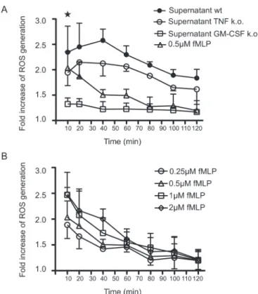
Along the same line, we examined the ability of neutrophils to mediate an oxidative burst, a key event for the elimina-tion of invading and ingested micro-organisms. Activaelimina-tion of the NADPH oxidase complex leads to the generation of ROS
including H2O2, which can be measured using the fluores-cent dye DCF (44). On the basis of our observations shown above, we used conditioned medium from BMMC in order to investigate the impact of mast cells on the generation of ROS by neutrophils.
In Fig. 2A, incubation of neutrophils with supernatants derived from activated wild-type BMMC leads to a strong production of intracellular ROS seen as a rapid and transient increase in fluorescence intensities. As a positive control for these kind of experiments, we chose fMLP, a known activator of ROS generation, in concentrations ranging from 0.25 to 2 μM (Fig. 2B). However, the impact of mast cells on the generation of ROS equals that of fMLP at the highest concentrations (1 and 2 μM) and the oxidative burst initiated by mast cell-derived supernatants appears more sustained compared with fMLP. Furthermore, it can be seen that the oxidative burst mainly depends on mast cell-derived GM-CSF and although to a lesser extent on TNF.
Mast cells prolong the survival of neutrophils
In order to investigate the impact of mast cells on sponta-neous apoptosis of neutrophils, we incubated neutrophils in the presence of conditioned medium derived from activated BMMC (Fig. 3). Following 24 h incubation of neutrophils in medium, more than 40% of the cells bind annexin V, a sign for early apoptosis. A minor fraction of late apoptotic cells is in addition also positive for PI staining. The addition of superna-tant derived from wild-type BMMC activated by cross-linking of IgE substantially decreases spontaneous apoptosis of
neutrophils (Fig. 3A and B). However, the antiapoptotic effect is completely abrogated using supernatants from GM-CSF-deficient BMMC (Fig. 3C and D). In contrast to this striking impact of mast cell-derived GM-CSF, TNF-deficient BMMC do not alter spontaneous neutrophil apoptosis (Fig. 3A and B). In these experiments, supernatants derived from IgE-loaded BMMC without cross-linking by antigen had no effect on neu-trophil apoptosis (data not shown).
To support these findings, we also measured the DNA con-tent in PI-stained neutrophil nuclei. As depicted in Fig. 3E, following 48-h incubation of neutrophils in medium, 50% of nuclei appear as a broad hypodiploid peak. The presence of conditioned medium derived from either activated wild-type or TNF-deficient mast cells strongly reduces the number of hypodiploid nuclei. Yet, supernatant from GM-CSF-deficient BMMC has hardly any effect on DNA fragmentation. Thus, both methods independently show that mast cell-derived GM-CSF can strongly delay the apoptosis of neutrophils.
Neutrophil phagocytosis is enhanced by mast cells
Ingestion and killing of microbes is a key element of neu-trophil function. Thus, we next analyzed the ability of mast cells to promote neutrophil phagocytosis. This process can be quantitatively assessed by measuring the uptake of flu-orescent polystyrene microspheres as depicted in Fig. 4. Without additional activation, about one-third of neutrophils take up beads, yet the relatively low fluorescence intensities indicate that most cells ingest only a very small number of beads (Fig. 4A). In the presence of LPS, both the numbers of cells that have taken up beads and their fluorescence
Fig. 1. Activated mast cells modulate the expression of activation markers on neutrophils. Freshly isolated neutrophils were incubated in co-culture in a ratio of 1:1 with IgE-loaded or IgE-loaded and cross-linked BMMC from wild-type mice. Additionally, neutrophils were treated with conditioned medium (50 vol%) derived from either activated wild-type BMMC (supernatant wt) or BMMC deficient for TNF or GM-CSF, respectively. Freshly isolated neutrophils and neutrophils treated with increasing doses of LPS served as references. After 5 h, CD62L and CD11b expression was measured by FACS as geometric mean fluorescence intensities. High expression of CD62L and constitutive low-level expression of CD11b on freshly isolated neutrophils were set to 1, respectively. Shown are the means (+ standard deviation) from at least three experiments. * indicates P < 0.05 and *** P < 0.001 as determined by one-way analysis of variance implemented in the Prism 5.0 software. n. s., not significant.
intensities strongly increase. However, phagocytosis can be further increased using supernatants derived from activated BMMC. Under these conditions, almost all neutrophils are loaded with numerous beads. In the absence of mast cell-derived TNF, the percentage of bead-positive neutrophils remains unchanged, yet the number of ingested particles per cell decreases (Fig. 4B). Finally, the lack of mast cell-derived GM-CSF leads to a strong decrease of both param-eters. However, even in the absence of GM-CSF, the residual effect of mast cell-conditioned medium on neutrophil phago-cytosis is still comparable with LPS. The enhancement of par-ticle uptake is a very strong effect as displayed in Fig. 4C
where conditioned medium derived from activated mast cells was diluted. Even at a dilution of 1:32, particle engulfment is significantly elevated above untreated levels. Additionally, we also varied the ratio of BMMC:neutrophils in co-culture experiments and got comparable results. At a ratio of 1:32, neutrophil particle uptake was still significantly increased (data not shown).
Thus, activated mast cells strongly boost neutrophil phago-cytosis and it appears likely that, besides TNF and GM-CSF, other mast cell-derived mediators are able to promote this process.
Fig. 2. Activated mast cells initiate the generation of ROS in neu-trophils. (A) Intracellular ROS generation in neutrophils was ana-lyzed by flow cytometry at the indicated time points following addition of conditioned mast cell medium derived from the geno-types described above. 0.5 μM fMLP was included as a reference. (B) Different concentrations of fMLP were used and the generation of ROS was recorded. Shown are the means (± SD) from three experiments. Mean fluorescence intensity of freshly isolated rest-ing neutrophils was arbitrarily set to 1. * indicates P < 0.05 as determined by one-way analysis of variance implemented in the Prism 5.0 software.
Fig. 3. Mast cells prolong the survival of neutrophils.(A) Freshly iso-lated neutrophils were incubated for 24 h with or without additional conditioned medium (50 vol%) derived from activated TNF-deficient BMMC or their congenic littermates. Spontaneous apoptosis was measured using staining with PI and annexin V. (B) Means (+ standard
Mast cells boost neutrophil phagocytosis in a model for acute lung inflammation
In order to investigate whether mast cells also boost the func-tion of neutrophils in vivo, we chose a model of LPS-induced acute lung inflammation. Besides wild-type and mast cell-deficient Kit W-sh/Kit W-sh mice, mast cell-deficient mice were
also engrafted with BMMC derived from wild-type, TNF- and GM-CSF-deficient mice 12 weeks prior to the experiment. Successful engraftment of Kit W-sh/Kit W-sh mice was ensured
by staining lung sections derived from all animal groups for the presence of mast cells (Fig. 5, lower panel).
For the induction of pulmonar inflammation, LPS was given intra-nasally in combination with fluorescent polysty-rene microspheres. Eight hours later, cellular infiltrates were recovered by lavage of the lungs and analyzed for the pres-ence of neutrophils. As depicted in Fig. 5 (upper panel), application of LPS induces a strong influx of neutrophils into the lungs, which is comparable in all experimental groups. This mast cell-independent recruitment of neutrophils is most likely due to the fact that in the lungs different cell types, including structural cells, express TLR4 and are able
to initiate an innate immune response (45). Phagocytosis of fluorescent beads by neutrophils can be visualized by FACS (Fig. 5, middle panel). Most importantly, the absence of mast cells is accompanied by a reduction in neutrophil phagocytosis, measured as a drop in fluorescence intensi-ties. This reduction in neutrophil phagocytosis can be cor-rected through the selective repair of mast cell-deficiency with mast cells derived from wild-type or TNF-deficient mice. Obviously, the observed effect of mast cell-derived TNF on neutrophil phagocytosis in vitro (Fig. 4) can be compensated for by additional sources for TNF in the lungs, most likely bronchiolar epithelial cells and alveolar macrophages (46). This assumption is supported by our observation that TNF levels in BAL do not significantly differ between mast cell-deficient and wild-type mice at 8 h after application of LPS (data not shown).
However, engraftment of sash mice with GM-CSF-deficient BMMC is insufficient to restore neutrophil phagocytosis, indi-cating that mast cells serve as a non-redundant source for this cytokine in this model. Unfortunately, we were not able to measure GM-CSF in BAL using ELISA. In summary, this result corroborates our data generated in vitro and empha-sizes the influence of mast cells on neutrophil phagocytosis
in vivo.
Discussion
It is increasingly being recognized that mast cells are able to induce and to regulate inflammatory responses in diverse experimental settings. These cells are strategically located
Fig. 4. Neutrophil phagocytosis is enhanced by mast cells. (A) Neutrophils were incubated for 2 h with PE-labeled microspheres under the con-ditions shown, washed and FACS analyzed for the uptake of fluorescent particles. (B) Means (+ SD) of three experiments like the representative shown in (A). (C) Neutrophils were treated as described above but only conditioned medium derived from activated wild-type mast cells was diluted as indicated. Means (± standard deviation) from three experiments. *** P < 0.001; n. s., not significant.
deviation) from four experiments like the one shown before. (C) Performed as described above but to investigate the effect of GM-CSF-deficient BMMC on neutrophil apoptosis. (D) Means (+ standard deviation) of three experiments. (E) Neutrophils were treated as described in (A) but incubated for 48 h. Following PI stain-ing, apoptotic nuclei appear as a broad smear of low fluorescence intensity. Means (± SD) from three experiments are given. ** P < 0.01; *** P < 0.001; n. s., not significant.
at the borders between host and environment, that is skin and mucosal surfaces, and can rapidly respond to a vari-ety of stimuli even in the absence of IgE antibodies (30, 47). Activation of mast cells can promote the rapid recruitment of neutrophils that are able to combat harmful intruders using oxidative and non-oxidative mechanisms but these pro-cesses can also play a key role in the development of inflam-matory and autoimmune diseases (48). Further, in the context of intercellular communication, recent studies revealed a bidirectional cross-talk between mast cells and eosinophils, termed the allergic effector unit, augmenting the activation of both cells (49, 50).
Our observations reported herein demonstrate that mast cells are able to boost important effector functions of neu-trophils, that is phagocytosis and generation of ROS. This is partly due to the de novo production of TNF and GM-CSF, whereas histamine, which is rapidly released from granu-lar stores, had no effect on neutrophils (data not shown). It was reported that mast cells are able to store TNF that can be released rapidly upon stimulation, but it should be noted that BMMC used for the experiments described herein and other mast cell lines do not contain preformed TNF (20). It has been known for a long time that mast cells are able to produce GM-CSF (51) and our results show prominent effects of mast cell-derived GM-CSF on all parameters of neutrophil activity examined including its strong antiapop-totic action. A variety of inflammatory signals can extend the lifespan of neutrophils. TNF is also known to regulate neu-trophil apoptosis but opposing effects have been reported in that it can either accelerate or delay programmed cell death, depending on the concentration. High concentrations of TNF promote apoptosis, whereas low concentrations are antiapoptotic. This dual effect of TNF is likely due to differen-tial effects on the antiapoptotic pathways in neutrophils, that is accelerated turnover of the antiapoptotic protein Mcl-1 at high concentrations and increased expression of another antiapoptotic molecule, BFL-1, at low concentrations of this cytokine (52).
However, in our in vitro system, mast cell-derived GM-CSF is solely responsible for delaying apoptosis of neutrophils. This antiapoptotic effect of mast cells might support efficient anti-microbial activity in inflamed tissues.
Besides mast cell-derived cytokines, we have good evi-dence that mast cell-derived lipid mediators also strongly promote the activation of neutrophils (data not shown) but additional work is needed to identify these lipids.
To corroborate our in vitro findings, we used a model for acute lung inflammation following instillation of LPS. Inhalation of LPS leads to the rapid recruitment of neutrophils and also activates mast cells (53, 54). In agreement with another report (16), we found that LPS-induced airway neutrophilia is not influenced by the lack of mast cells but importantly, the ability of neutrophils to phagocytose is impaired in the absence of mast cells.
Fig. 5. Mast cells promote neutrophil phagocytosis in a model for acute lung inflammation. One microgram LPS in combination with 1.5 × 10⁸ fluorescent polystyrene microspheres were applied
intra-nasally in anesthetized mice of the indicated groups. Eight hours later, BAL was recovered and neutrophils were counted (upper panel). FACS analyses were performed to quantify neutrophils that have undergone phagocytosis of PE-labeled microspheres. Geometric mean fluorescence intensities (MFI) are shown. * indicates P < 0.05
and *** P < 0.001 as determined by unpaired t-test implemented in the Prism 5.0 software (middle panel). Lung sections were stained for the presence of mast cells using toluidine blue (lower panel). Representative of at least two experiments with 4–5 mice per group.
Taken together, our results suggest that mast cells are involved in the local control of neutrophil activation and sur-vival and thus could have important implications for the role of mast cells in innate immunity but also in allergic and autoim-mune diseases.
Funding
Deutsche Forschungsgemeinschaft [STA984/1–2 to M.S., TA275/4-1 to C.T. and M.S and STA984/4-1 to M.S. and M.P.R.].
References
1 Segal, A. W. 2005. How neutrophils kill microbes. Annu. Rev.
Immunol. 23:197.
2 Witko-Sarsat, V., Rieu, P., Descamps-Latscha, B., Lesavre, P. and Halbwachs-Mecarelli, L. 2000. Neutrophils: molecules, functions and pathophysiological aspects. Lab. Invest. 80:617.
3 Nathan, C. 2006. Neutrophils and immunity: challenges and opportunities. Nat. Rev. Immunol. 6:173.
4 Savill, J., Dransfield, I., Gregory, C. and Haslett, C. 2002. A blast from the past: clearance of apoptotic cells regulates immune responses. Nat. Rev. Immunol. 2:965.
5 Kennedy, A. D. and DeLeo, F. R. 2009. Neutrophil apoptosis and the resolution of infection. Immunol. Res. 43:25.
6 Simon, H. U. 2003. Neutrophil apoptosis pathways and their mod-ifications in inflammation. Immunol. Rev. 193:101.
7 Schiltz, C., Lioté, F., Prudhommeaux, F. et al. 2002. Monosodium urate monohydrate crystal-induced inflammation in vivo: quan-titative histomorphometric analysis of cellular events. Arthritis
Rheum. 46:1643.
8 Qureshi, R. and Jakschik, B. A. 1988. The role of mast cells in thioglycollate-induced inflammation. J. Immunol. 141:2090. 9 Wershil, B. K., Murakami, T. and Galli, S. J. 1988. Mast
cell-dependent amplification of an immunologically nonspecific inflam-matory response. Mast cells are required for the full expression of cutaneous acute inflammation induced by phorbol 12-myristate 13-acetate. J. Immunol. 140:2356.
10 Ramos, B. F., Qureshi, R., Olsen, K. M. and Jakschik, B. A. 1990. The importance of mast cells for the neutrophil influx in immune complex-induced peritonitis in mice. J. Immunol. 145:1868. 11 Zhang, Y., Ramos, B. F. and Jakschik, B. A. 1991. Augmentation
of reverse arthus reaction by mast cells in mice. J. Clin. Invest. 88:841.
12 Ramos, B. F., Zhang, Y., Qureshi, R. and Jakschik, B. A. 1991. Mast cells are critical for the production of leukotrienes responsi-ble for neutrophil recruitment in immune complex-induced perito-nitis in mice. J. Immunol. 147:1636.
13 Zhang, Y., Ramos, B. F. and Jakschik, B. A. 1992. Neutrophil recruitment by tumor necrosis factor from mast cells in immune complex peritonitis. Science 258:1957.
14 Wershil, B. K., Wang, Z. S., Gordon, J. R. and Galli, S. J. 1991. Recruitment of neutrophils during IgE-dependent cutaneous late phase reactions in the mouse is mast cell-dependent. Partial inhi-bition of the reaction with antiserum against tumor necrosis factor-alpha. J. Clin. Invest. 87:446.
15 Biedermann, T., Kneilling, M., Mailhammer, R. et al. 2000. Mast cells control neutrophil recruitment during T cell-mediated delayed-type hypersensitivity reactions through tumor necrosis factor and macrophage inflammatory protein 2. J. Exp. Med. 192:1441.
16 Nakae, S., Suto, H., Berry, G. J. and Galli, S. J. 2007. Mast cell-derived TNF can promote Th17 cell-dependent neutrophil recruit-ment in ovalbumin-challenged OTII mice. Blood 109:3640. 17 von Stebut, E., Metz, M., Milon, G., Knop, J. and Maurer, M. 2003.
Early macrophage influx to sites of cutaneous granuloma forma-tion is dependent on MIP-1alpha /beta released from neutrophils recruited by mast cell-derived TNFalpha. Blood 101:210. 18 Sayed, B. A., Christy, A. L., Walker, M. E. and Brown, M. A. 2010.
Meningeal mast cells affect early T cell central nervous system
infiltration and blood-brain barrier integrity through TNF: a role for neutrophil recruitment? J. Immunol. 184:6891.
19 Enoksson, M., Möller-Westerberg, C., Wicher, G. et al. 2013. Intraperitoneal influx of neutrophils in response to IL-33 is mast cell-dependent. Blood 121:530.
20 Gordon, J. R. and Galli, S. J. 1990. Mast cells as a source of both preformed and immunologically inducible TNF-alpha/cachectin.
Nature 346:274.
21 Gordon, J. R., Burd, P. R. and Galli, S. J. 1990. Mast cells as a source of multifunctional cytokines. Immunol. Today 11:458. 22 Echtenacher, B., Männel, D. N. and Hültner, L. 1996. Critical
pro-tective role of mast cells in a model of acute septic peritonitis.
Nature 381:75.
23 Malaviya, R., Ikeda, T., Ross, E. and Abraham, S. N. 1996. Mast cell modulation of neutrophil influx and bacterial clearance at sites of infection through TNF-alpha. Nature 381:77.
24 Malaviya, R. and Abraham, S. N. 2000. Role of mast cell leukot-rienes in neutrophil recruitment and bacterial clearance in infec-tious peritonitis. J. Leukoc. Biol. 67:841.
25 Huang, C., Friend, D. S., Qiu, W. T. et al. 1998. Induction of a selective and persistent extravasation of neutrophils into the peri-toneal cavity by tryptase mouse mast cell protease 6. J. Immunol. 160:1910.
26 Thakurdas, S. M., Melicoff, E., Sansores-Garcia, L. et al. 2007. The mast cell-restricted tryptase mMCP-6 has a critical immuno-protective role in bacterial infections. J. Biol. Chem. 282:20809. 27 Mercer-Jones, M. A., Shrotri, M. S., Heinzelmann, M., Peyton, J.
C. and Cheadle, W. G. 1999. Regulation of early peritoneal neu-trophil migration by macrophage inflammatory protein-2 and mast cells in experimental peritonitis. J. Leukoc. Biol. 65:249.
28 Kneilling, M., Mailhammer, R., Hültner, L. et al. 2009. Direct cross-talk between mast cell-TNF and TNFR1-expressing endothelia mediates local tissue inflammation. Blood 114:1696.
29 Stassen, M., Hültner, L. and Schmitt, E. 2002. Classical and alternative pathways of mast cell activation. Crit. Rev. Immunol. 22:115.
30 Heib, V., Becker, M., Taube, C. and Stassen, M. 2008. Advances in the understanding of mast cell function. Br. J. Haematol. 142:683.
31 Lyon, M. F. and Glenister, P. H. 1982. A new allele sash (Wsh) at the W-locus and a spontaneous recessive lethal in mice. Genet.
Res. 39:315.
32 Wolters, P. J., Mallen-St Clair, J., Lewis, C. C. et al. 2005. Tissue-selective mast cell reconstitution and differential lung gene expression in mast cell-deficient KitW-sh/KitW-sh sash mice. Clin.
Exp. Allergy 35:82.
33 Grimbaldeston, M. A., Chen, C. C., Piliponsky, A. M., Tsai, M., Tam, S. Y. and Galli, S. J. 2005. Mast cell-deficient W-sash c-kit mutant Kit W-sh/W-sh mice as a model for investigating mast cell biology in vivo. Am. J. Pathol. 167:835.
34 Pasparakis, M., Alexopoulou, L., Episkopou, V. and Kollias, G. 1996. Immune and inflammatory responses in TNF alpha-defi-cient mice: a critical requirement for TNF alpha in the formation of primary B cell follicles, follicular dendritic cell networks and germinal centers, and in the maturation of the humoral immune response. J. Exp. Med. 184:1397.
35 Heib, V., Becker, M., Warger, T. et al. 2007. Mast cells are crucial for early inflammation, migration of Langerhans cells, and CTL responses following topical application of TLR7 ligand in mice.
Blood 110:946.
36 Reuter, S., Heinz, A., Sieren, M. et al. 2008. Mast cell-derived tumour necrosis factor is essential for allergic airway disease. Eur.
Respir. J. 31:773.
37 Stanley, E., Lieschke, G. J., Grail, D. et al. 1994. Granulocyte/ macrophage colony-stimulating factor-deficient mice show no major perturbation of hematopoiesis but develop a characteristic pulmonary pathology. Proc. Natl. Acad. Sci. U. S. A. 91:5592. 38 Stassen, M., Valeva, A., Walev, I. and Schmitt, E. 2006. Activation
of mast cells by streptolysin O and lipopolysaccharide. Methods
Mol. Biol. 315:393.
39 Eshhar, Z., Ofarim, M. and Waks, T. 1980. Generation of hybrido-mas secreting murine reaginic antibodies of anti-DNP specificity.
40 Blumenkrantz, N. and Asboe-Hansen, G. 1975. A selective stain for mast cells. Histochem. J. 7:277.
41 LeBel, C. P., Ischiropoulos, H. and Bondy, S. C. 1992. Evaluation of the probe 2’,7’-dichlorofluorescin as an indicator of reactive oxygen species formation and oxidative stress. Chem. Res.
Toxicol. 5:227.
42 Nicoletti, I., Migliorati, G., Pagliacci, M. C., Grignani, F. and Riccardi, C. 1991. A rapid and simple method for measuring thy-mocyte apoptosis by propidium iodide staining and flow cytom-etry. J. Immunol. Methods 139:271.
43 Kishimoto, T. K., Jutila, M. A., Berg, E. L. and Butcher, E. C. 1989. Neutrophil Mac-1 and MEL-14 adhesion proteins inversely regu-lated by chemotactic factors. Science 245:1238.
44 Freitas, M., Lima, J. L. and Fernandes, E. 2009. Optical probes for detection and quantification of neutrophils’ oxidative burst. A review. Anal. Chim. Acta 649:8.
45 Hammad, H., Chieppa, M., Perros, F., Willart, M. A., Germain, R. N. and Lambrecht, B. N. 2009. House dust mite allergen induces asthma via Toll-like receptor 4 triggering of airway structural cells.
Nat. Med. 15:410.
46 Skerrett, S. J., Liggitt, H. D., Hajjar, A. M., Ernst, R. K., Miller, S. I. and Wilson, C. B. 2004. Respiratory epithelial cells regulate lung inflammation in response to inhaled endotoxin. Am. J. Physiol.
Lung Cell. Mol. Physiol. 287:L143.
47 Stassen, M., Hültner, L., Müller, C. and Schmitt, E. 2002. Mast cells and inflammation. Arch. Immunol. Ther. Exp. (Warsz) 50:179. 48 Kneilling, M. and Röcken, M. 2009. Mast cells: novel clinical
per-spectives from recent insights. Exp. Dermatol. 18:488.
49 Elishmereni, M., Alenius, H. T., Bradding, P. et al. 2011. Physical interactions between mast cells and eosinophils: a novel mecha-nism enhancing eosinophil survival in vitro. Allergy 66:376. 50 Elishmereni, M., Bachelet, I., Ben-Efraim, A. H., Mankuta, D. and
Levi-Schaffer, F. 2013. Interacting mast cells and eosinophils acquire an enhanced activation state in vitro. Allergy 68:171. 51 Wodnar-Filipowicz, A., Heusser, C. H. and Moroni, C. 1989.
Production of the haemopoietic growth factors GM-CSF and inter-leukin-3 by mast cells in response to IgE receptor-mediated acti-vation. Nature 339:150.
52 Cross, A., Moots, R. J. and Edwards, S. W. 2008. The dual effects of TNFalpha on neutrophil apoptosis are mediated via differential effects on expression of Mcl-1 and Bfl-1. Blood 111:878.
53 Nigo, Y. I., Yamashita, M., Hirahara, K. et al. 2006. Regulation of aller-gic airway inflammation through Toll-like receptor 4-mediated modifi-cation of mast cell function. Proc. Natl. Acad. Sci. U. S. A. 103:2286. 54 Murakami, D., Yamada, H., Yajima, T., Masuda, A., Komune, S.
and Yoshikai, Y. 2007. Lipopolysaccharide inhalation exacerbates allergic airway inflammation by activating mast cells and promot-ing Th2 responses. Clin. Exp. Allergy 37:339.