Transient decrease of serum iron after acute
erythropoietin treatment contributes to hepcidin
inhibition by ERFE in mice
To coordinate iron supply with hemoglobin (Hb) syn-thesis, erythropoietin (EPO)-stimulated erythroid precur-sors release erythroferrone (ERFE), a soluble protein that suppresses the expression of hepcidin (Hamp).1Hepcidin is a short liver peptide that controls iron entry into the circulation by blocking the sole iron exporter ferroportin.2 ERFE is essential in stress erythropoiesis since the Erfe knockout mice fail to suppress hepcidin after phlebotomy and show a delayed recovery from anemia.1
Increased erythropoiesis and iron deficiency suppress hepcidin via ERFE-dependent and ERFE-independent mechanisms, respectively, culminating in chromatin remodeling due to reversible loss of the histone activation marks H3K4me3 and H3K9ac.3
Hepcidin expression in hepatocytes is mainly regulated by the Bone Morphogenetic Protein (BMP)-Son of Mother Against Decapentaplegic (SMAD) pathway. Increased plasma iron in the form of diferric transferrin (holoTF), measured in vivo as increased transferrin satura-tion, activates the signaling that is initiated by the ligand BMPs and requires the co-receptor hemojuvelin (HJV).
The mechanisms by which ERFE represses hepcidin are still unclear. Mounting evidence indicates that ERFE requires some basal BMP-SMAD activity,4but excessive activation of the BMP-SMAD pathway prevents its sup-pressive effects on hepcidin.5In Tmprss6 KO mice, with an inappropriately active BMP-SMAD signaling, hepcidin levels are high, despite elevated EPO and ERFE levels.6 The resistance of Tmprss6 KO mice to ERFE is rescued by pharmacologic down-regulation of the pathway.5 We thus hypothesized that EPO itself might attenuate the BMP-SMAD pathway to favor the ERFE regulatory function.
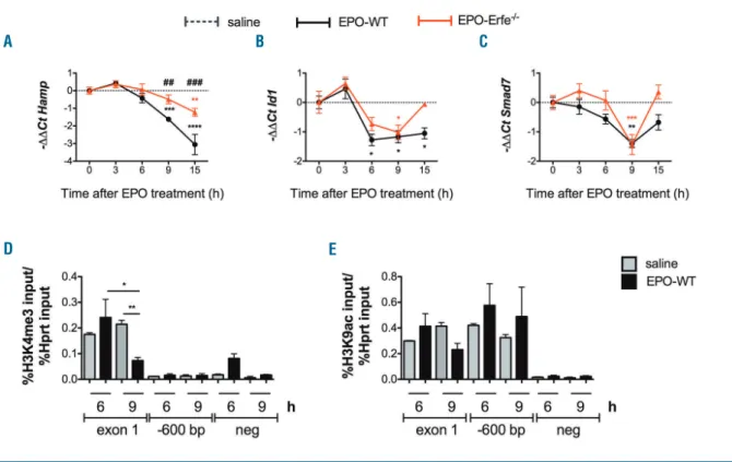
To investigate whether the liver BMP-SMAD pathway is downregulated by EPO, C57BL/6N male wild type (WT) mice were treated with a single 200 U/mouse intraperitoneal injection of EPO or saline and sacrificed at 3, 6, 9 and 15 hrs after treatment. Consistent with previ-ous reports,1
Erfe expression is upregulated both in bone
marrow and spleen of EPO-treated mice (Online
Supplementary Figure S1A and S1B) and Hamp expression
is inhibited at 9 hrs post EPO (Online Supplementary Figure
S1C), paralleled by a downregulation of Id1 (Online
Supplementary Figure S1D) and Smad7 (Online
Supplementary Figure S1E). However, at 15 hours the
pathway is active (increased Id1 and Smad7) while hep-cidin is inhibited. Although Erfe expression is upregulated at 3 and 6 hrs post-EPO, Hamp expression is not decreased until 9 hrs, when the pathway is attenuated. Altogether, these results demonstrate that the BMP-SMAD pathway is inhibited shortly after EPO injection. To determine the contribution of ERFE at short time points after EPO, we next analyzed the response of Erfe–/–
mice to EPO. As expected, Hamp was only slightly reduced in Erfe–/– animals 15 hrs post-EPO (Figure 1A).
Interestingly, Id1 (Figure 1B) and Smad7 (Figure 1C) expression are also decreased in Erfe-deficient mice 9 hours after injection, suggesting an EPO-dependent but ERFE-independent inhibition of the pathway.
Previous studies have indicated that suppression of
Hamp expression 3-4 days after EPO treatment leads to a
reversible loss of the histone activation marks H3K4me3 and H3K9ac at the Hamp locus (Online Supplementary
Figure S2). We sought to assess whether chromatin
remodeling may contribute to hepcidin inhibition in WT mice at short time points after EPO. Consistent with the repression of hepcidin (Online Supplementary Figure S1C), chromatin immunoprecipitation (ChIP)–quantitative PCR (qPCR) assays demonstrated a reduction in H3K4me3 (Figure 1D) but not in H3K9ac (Figure 1E) at the Hamp locus 9 hrs, but not at 6 hrs after EPO. This indicates that loss of H3K4me3 at the hepcidin promoter occurs early after EPO administration to repress hepcidin even in the presence of H3K9ac.
At early time points (6 and 9 hrs) after EPO treatment, liver and spleen iron content are unchanged (data not
shown), whereas, serum iron (Online Supplementary Figure S1F), TS (Online Supplementary Figure S1F), Id1 and Smad7
(Online Supplementary Figures S1D and S1E) are signifi-cantly reduced, in agreement with similar recently reported results.7Serum iron (Figure 2A) and TS (Figure 2B) are decreased also in the absence of ERFE. However, since hepcidin suppression is blunted, their levels remain low in Erfe-deficient mice at 15 hours (Figure 2A and 2B). In contrast, in WT mice, both parameters increase between 9-15 hrs, stimulating the expression of Id1 and
Smad7 (Figure 1B and 1C, Online Supplementary Figure S1D and S1E). We thus surmise that the ERFE effect on
haematologica 2019; 104:e87
L
ETTERS TO THE
E
DITOR
Table 1. Membrane TFR1 (CD71) expression in bone marrow and spleen-derived cells at different stages of maturation.
Subpopulations
UT
3hr
6hr
3hr vs. UT
6hr vs.UT
3hr vs.6hr
BM I 835.2±170.9 2014±180.7 3682±355.9 * *** ** II 3401±538.4 8473±953.1 10895±683 ** ** ns III 3020±66.23 5245±484.7 7562±102.9 ** **** ** IV 1950±525.4 2511±213.2 3862±144.6 ns * ns V 3013±558.5 2762±289.1 3312±354.7 ns ns ns SP II 3500±257.1 8220±929.5 10027±734.8 *** **** ns III 2997±199.4 5569±600 7977±841.4 ns ** ns IV 3063±649.3 3064±338.9 3931±481.9 ns ns ns V 3467±693.6 2794±299.3 3660±329.3 ns ns nsC57BL/6N wild type mice (3 mice for each time point) were treated with EPO (200 U/mouse) or saline and sacrificed at 3 and 6 hrs post-injection. BM and spleen cells were isolated and analyzed for Ter119/CD44 expression. Ter119 was gated and analyzed with respect to FSC and CD44 surface expression for subpopulation composition (gated cluster I-V) (see Online Supplementary Figure S2A and S2B). Membrane CD71/TFR1 expression in the BM and spleen derived erythroid populations was evaluated as mean fluorescent intensity (MFI). Unpaired two-tailed Student t-test was used for significance calculation. ns: not significant; *P<0.05; **P<0.01; ***P<0.001; ****P<0.0001. BM: bone marrow; SP: spleen.
hepcidin is uncoupled from the iron effect on the BMP-SMAD pathway.
Circulating iron is uptaken mainly by maturing ery-throid cells through binding of circulating holo-TF to transferrin receptor 1 (TFR1) and internalization of the complex. Mirciov and colleagues described an increase in
Tfr1 mRNA expression as a potential cause of reduction
of circulating diferric TF.7
Bone marrow (BM) and spleen erythroblasts of EPO or saline treated WT mice were analyzed by flow cytometry using Ter119 and CD44 antibodies staining and forward scatter to separate cells at distinct stages of terminal dif-ferentiation: proerythroblasts (I), basophilic erythroblasts (II), polychromatic erythroblasts (III), orthochromatic erythroblasts and immature reticulocytes (IV), and mature red cells (V) (data not shown). We show that despite unchanged proliferation and differentiation of bone marrow (BM) (Online Supplementary Figure S3A) and spleen derived erythroid cells (Online Supplementary
Figure S3B) surface TFR1 protein is increased after EPO
treatment in immature erythroid cells both in the BM and spleen of WT mice (Table 1). The same approach was used to isolate erythroid cell population in Erfe–/– mice
and WT animals (Online Supplementary Figure S4A). Irrespective of unchanged proliferation and differentia-tion of erythroid cells (Online Supplemental Figure S4B), TFR1 increase was observed in the BM of Erfe–/– and WT
at 6 hrs after EPO (Online Supplementary Figure S4C).
To confirm that the early downregulation of hepcidin is driven by EPO-induced decrease of holoTF, WT mice were treated with EPO and 1 hr later received intra-venous human holo transferrin (holoTF) (0.3 g/kg) to augment circulating iron8 and sacrificed at 6, 9 and 15 hrs after EPO. Human holoTF injection does not interfere with Erfe expression in BM (Figure 2C) and spleen (Figure 2D) erythroid cells. Since Erfe is an EPO target gene, we also assume that the erythroid response to EPO is com-parable in the two groups of mice. Liver iron content is unchanged after holoTF injection (data not shown). However, downregulation of Hamp (Figure 2E) and Id1 (Figure 2F) mRNA expression is delayed after EPO-holoTF combinatory treatment compared with mice treated with EPO only. This confirms that decreased cir-culating iron mitigates the BMP-SMAD pathway.
We have shown that BMP-SMAD pathway attenuation is required for hepcidin inhibition.5 While we were preparing this manuscript, it was reported that serum diferric transferrin is decreased early after EPO with a consequent reduction of SMAD1/5/8 phosphorylation in the liver.7 The reduction of serum iron and TS was ascribed to increased Tfr1 expression by EPO in BM and spleen.7
We confirm this mechanism showing that TFR1 protein is highly expressed on the surface of erythroid precursors before any increase in Tfr1 mRNA levels (data
not shown). The consequent iron uptake of the acutely
expanded erythropoiesis overcomes the capacity of iron
haematologica 2019; 104:e88
L
ETTERS TO THE
E
DITOR
Figure 1. Time course analysis of the effect of a single EPO injection on hepcidin and BMP-SMAD target genes in WT and Erfe–/–mice and analysis of
chro-matin modifications at the hepcidin promoter locus 6 and 9 hours after EPO. C57BL6/J wild type and Erfe-deficient male mice were treated with EPO (200 U/mouse) or saline (6-9 mice for each time point) and sacrificed at different time points. The liver expression of hepcidin (Hamp) (A) and the BMP-SMAD target genes Id1 (B) and Smad7 (C) was analyzed by qRT-PCR using Hprt1 as housekeeping gene. Changes in the expression levels were represented as –ΔΔCt (saline– EPO). Data shown are means ± s.e.m and were compared for each genotype at each time point to values at t = 0 (***P<0.001, **P<0.01) by one-way ANOVA and between WT and Erfe–/–mice (###P<0.001, #P<0.05) by two-tailed Student t-test. ChIP-PCR assessing H3K4me3 (D) and H3K9ac (E) status at 2 regions
around the Hamp promoter and a negative region was performed on liver tissue from C57BL6/N wild type mice taken 6 or 9 hours after EPO (200U/mouse) (black bars) or saline (grey bars) treatments (3 mice for each time point). Three-way ANOVA with Tukey’s correction for multiple comparisons was used for sig-nificance calculation. *P<0.05; **P<0.01.
A
B
C
release from absorption and store sites, causing a tran-sient reduction of serum iron and TS levels until hepcidin is suppressed.
Taken together, our data demonstrate an iron-depen-dent and ERFE-indepeniron-depen-dent mechanism of hepcidin reg-ulation by EPO, which is crucial for efficient inhibition of hepcidin by ERFE. EPO treatment rapidly increases the expression of TFR1 on plasma membrane of erythroid precursors. We hypothesize that upregulation of mem-brane TFR1 by EPO results in a strong and acute increase in iron uptake by maturing erythroblasts, as observed in EPO-treated human (K562) and murine erythroleukemic (MEL) cells,9thus reducing the levels of circulating iron and TS. The (acute) expansion of erythropoiesis over-comes the capacity of iron release from absorption and recycling until hepcidin is suppressed. The mechanism is ERFE-independent since the same decrease of circulating iron is observed shortly after EPO injection in Erfe–/–
mice, followed by a downregulation of BMP-SMAD
tar-get genes, including hepcidin. We also show that con-comitant to the early (9 hrs) hepcidin decrease, Hamp locus undergoes epigenetic modification with loss of H3K4me3, involved in facilitating gene transcription,10 but not of H3K9ac. Taken together with previous data,3 these findings indicate the time course of loss of chro-matin activation marks, which closely mirror reductions in gene expression.
The holoTF injection counteracts the early decrease of serum iron, delays the downregulation of hepcidin, fur-ther supporting that the transient drop of iron is essential for ERFE function on hepcidin. In summary, the EPO effect on hepcidin expression is biphasic. The immediate and transient effect on serum iron that allows the early repression of BMP-SMAD pathway and hepcidin repres-sion is Erfe independent; the late, ERFE-mediated effect, persists until the increased iron needs are met. Our find-ings are a further example of the tight crosstalk between erythroid cells and iron homeostasis.
haematologica 2019; 104:e89
L
ETTERS TO THE
E
DITOR
Figure 2. Circulating iron is reduced after EPO treatment both in WT and Erfe–/–mice, and human
holo-transferrin injection prevents the downregulation of the BMP-SMAD pathway by EPO and attenu-ates the hepcidin suppressive effect of ERFE. Serum iron (A) and transferrin saturation (B) were ana-lyzed in C57BL/6J wild type and
Erfe-deficient mice (6-9 mice for
each time point) treated with a sin-gle EPO injection (200 U/mouse) and expressed as a difference between the EPO-treated and the saline-treated values. Data shown are means ± s.e.m and were com-pared for each genotype at each time point to values at t=0 (*P<0.05; **P<0.01) by one-way ANOVA and between WT and Erfe–/–
mice (###P<0.001) by two-tailed
Student t-test. EPO-treated C57BL/6N wild type mice (3-4 mice for each time point) were adminis-tered intravenously with human holo-transferrin (HoloTF) and sacri-ficed at 6, 9 and 15 hrs post-EPO.
Erfe expression was evaluated in
the bone marrow (C) and spleen (D) by qRT-PCR and normalized on
Gapdh expression. Liver hepcidin
(Hamp; E) and Id1 (F) mRNA levels were analyzed by qRT-PCR. Hprt1 was used as housekeeping gene. Data are shown as means ± s.e.m. Two way-ANOVA was used to com-pare EPO and EPO-hHoloTF treated mice (ns: not significant; **P<0.01; ****P<0.0001). Two-tailed t-test was used to calculate statistically significant differences of EPO treat-ment at different time points com-pared to t=0; ns: not significant;
#P<0.05; ###P<0.001.
A
B
C
D
Irene Artuso,1,#Mariateresa Pettinato,1,2Antonella Nai,1,2 Alessia Pagani,1Ugo Sardo,3Benjamin Billoré,3
Maria Rosa Lidonnici,4 Cavan Bennett,5Giacomo Mandelli,4 Sant-Rayn Pasricha,5,6Giuliana Ferrari,4Clara Camaschella,1 Léon Kautz3 and Laura Silvestri1,2
1Regulation of Iron Metabolism Unit, Division of Genetics and Cell Biology, IRCCS San Raffaele Scientific Institute, Milan, Italy; 2 Vita-Salute San Raffaele University, Milan, Italy; 3IRSD, Université de Toulouse, INSERM U1220, INRA U1416, ENVT, UPS, France; 4SR-Tiget Unit, IRCCS San Raffaele Scientific Institute, Milan, Italy; 5The Walter and Eliza Hall Institute of Medical Research, Parkville, Victoria, Australia and 6Department of Medical Biology, The University of Melbourne, VC, Australia
IA and MP contributed equally to this work
#Present address: Cellular and Molecular Immunology, Department of Biotechnology and Biosciences, University of Milano-Bicocca, Milano, Italy
Acknowledgments: we are indebted to Marieke von Lindern (Sanquin, Amsterdam, The Netherlands) for the kind gift of human holotransferrin and to Chloé Latour (Inserm, Toulouse, France) for tech-nical assistance.
Funding: this work was supported in part by: Fondazione Telethon Grant n° GGP15064 to Laura Silvestri; ANR-16-ACHN-0002-01 and by European Research Council grant 715491 to Léon Kautz; the Telethon SR-TIGET Core Grant to Giuliana Ferrari; the National Health and Medical Research Council, Australia, Early Career Fellowship GNT1035339 to Sant-Rayn Pasricha. Antonella Nai was supported by the European Hematology Association Josè-Carreras Junior Research fellowship.
Correspondence: LAURA SILVESTRI - [email protected] doi:10.3324/haematol.2018.199810
Information on authorship, contributions, and financial & other disclo-sures was provided by the authors and is available with the online version of this article at www.haematologica.org.
References
1. Kautz L, Jung G, Valore EV, Rivella S, Nemeth E, Ganz T. Identification of erythroferrone as an erythroid regulator of iron metabolism. Nat Genet. 2014;46(7):678-684.
2. Nemeth E, Tuttle MS, Powelson J, et al. Hepcidin regulates cellular iron efflux by binding to ferroportin and inducing its internalization. Science. 2004;306(5704):2090-2093.
3. Pasricha SR, Lim PJ, Duarte TL, et al. Hepcidin is regulated by pro-moter-associated histone acetylation and HDAC3. Nat Comm. 2017;8(1):403.
4. Wang CY, Core AB, Canali S, et al. Smad1/5 is required for erythro-poietin-mediated suppression of hepcidin in mice. Blood. 2017; 130(1):73-83.
5. Nai A, Rubio A, Campanella A, et al. Limiting hepatic Bmp-Smad signaling by matriptase-2 is required for erythropoietin-mediated hepcidin suppression in mice. Blood. 2016;127(19):2327-2336. 6. Finberg KE, Whittlesey RL, Fleming MD, Andrews NC.
Down-regu-lation of Bmp/Smad signaling by Tmprss6 is required for mainte-nance of systemic iron homeostasis. Blood. 2010;115(18):3817-3826. 7. Mirciov CSG, Wilkins SJ, Hung GCC, Helman SL, Anderson GJ, Frazer DM. Circulating iron levels influence the regulation of hep-cidin following stimulated erythropoiesis. Haematologica. 2018 Jun 14. [Epub ahead of print]
8. Li H, Rybicki AC, Suzuka SM, et al. Transferrin therapy ameliorates disease in beta-thalassemic mice. Nat Med. 2010;16(2):177-182. 9. Weiss G, Houston T, Kastner S, Johrer K, Grunewald K, Brock JH.
Regulation of cellular iron metabolism by erythropoietin: activation of iron-regulatory protein and upregulation of transferrin receptor expression in erythroid cells. Blood. 1997;89(2):680-687.
10. Bannister AJ, Kouzarides T. Regulation of chromatin by histone modifications. Cell Res. 2011;21(3):381-395.
haematologica 2019; 104:e90