HAL Id: inserm-02439178
https://www.hal.inserm.fr/inserm-02439178
Submitted on 14 Jan 2020
HAL is a multi-disciplinary open access
archive for the deposit and dissemination of
sci-entific research documents, whether they are
pub-lished or not. The documents may come from
teaching and research institutions in France or
abroad, or from public or private research centers.
L’archive ouverte pluridisciplinaire HAL, est
destinée au dépôt et à la diffusion de documents
scientifiques de niveau recherche, publiés ou non,
émanant des établissements d’enseignement et de
recherche français ou étrangers, des laboratoires
publics ou privés.
Two molecular pathways initiate
mitochondria-dependent dopaminergic
neurodegeneration in experimental Parkinson’s disease
Céline Perier, Jordi Bové, Du-Chu Wu, Benjamin Dehay, Dong-Kug Choi,
Vernice Jackson-Lewis, Silvia Rathke-Hartlieb, Philippe Bouillet, Andreas
Strasser, Jörg Schulz, et al.
To cite this version:
Céline Perier, Jordi Bové, Du-Chu Wu, Benjamin Dehay, Dong-Kug Choi, et al.. Two molecular
path-ways initiate mitochondria-dependent dopaminergic neurodegeneration in experimental Parkinson’s
disease. Proceedings of the National Academy of Sciences of the United States of America , National
Academy of Sciences, 2007, 104 (19), pp.8161-8166. �10.1073/pnas.0609874104�. �inserm-02439178�
Two molecular pathways initiate
mitochondria-dependent dopaminergic neurodegeneration
in experimental Parkinson’s disease
Celine Perier*†, Jordi Bove´*, Du-Chu Wu†, Benjamin Dehay*, Dong-Kug Choi†, Vernice Jackson-Lewis†,
Silvia Rathke-Hartlieb‡, Philippe Bouillet§, Andreas Strasser§, Jo¨rg B. Schulz‡, Serge Przedborski†¶储**,
and Miquel Vila*†**††
*Research Institute-University Hospital Vall d’Hebron, 08035 Barcelona, Spain; Departments of†Neurology and¶Pathology and储Center for Neurobiology
and Behavior, Columbia University, New York, NY 10032;‡Department of Neurodegeneration and Restorative Research, Center of Neurological Medicine
and Deutsche Forschungsgemeinschaft (DFG) Research Center Molecular Physiology of the Brain, University of Go¨ttingen, 37073 Go¨ttingen, Germany;
§The Walter and Eliza Hall Institute of Medical Research, Melbourne, Victoria 3050, Australia; and††Catalan Institution for Research and Advanced
Studies (ICREA), 08010 Barcelona, Spain
Edited by Solomon H. Snyder, Johns Hopkins University School of Medicine, Baltimore, MD, and approved March 14, 2007 (received for review November 7, 2006)
Dysfunction of mitochondrial complex I is associated with a wide spectrum of neurodegenerative disorders, including Parkinson’s disease (PD). In rodents, inhibition of complex I leads to degener-ation of dopaminergic neurons of the substantia nigra pars com-pacta (SNpc), as seen in PD, through activation of mitochondria-dependent apoptotic molecular pathways. In this scenario, complex I blockade increases the soluble pool of cytochrome c in the mitochondrial intermembrane space through oxidative mech-anisms, whereas activation of pro-cell death protein Bax is actually necessary to trigger neuronal death by permeabilizing the outer mitochondrial membrane and releasing cytochrome c into the cytosol. Activation of Bax after complex I inhibition relies on its transcriptional induction and translocation to the mitochondria. How complex I deficiency leads to Bax activation is currently unknown. Using gene-targeted mice, we show that the tumor suppressor p53 mediates Bax transcriptional induction after PD-related complex I blockade in vivo, but it does not participate in Bax mitochondrial translocation in this model, either by a transcription-independent mechanism or through the induction of BH3-only proteins Puma or Noxa. Instead, Bax mitochondrial translocation in this model relies mainly on the JNK-dependent activation of the BH3-only protein Bim. Targeting either Bax transcriptional induc-tion or Bax mitochondrial translocainduc-tion results in a marked atten-uation of SNpc dopaminergic cell death caused by complex I inhibition. These results provide further insight into the patho-genesis of PD neurodegeneration and identify molecular targets of potential therapeutic significance for this disabling neurological illness.
apoptosis兩 Bax 兩 Bim 兩 complex I 兩 1-methyl-4-pheny-1,2,3,6-tetrahydropyridine
C
omplex I deficiency impairs mitochondrial respiration and is associated with a wide spectrum of neurodegenerative disor-ders, including Parkinson’s disease (PD). Reduced complex I activity is found in both autopsy brain tissues and platelets of patients affected with sporadic PD (1–3). Furthermore, complex I inhibitors, such as 1-methyl-4-pheny-1,2,3,6-tetrahydropyridine (MPTP), reproduce some of the clinical and neuropathological hallmarks of PD in monkeys and humans, including degeneration of dopaminergic (DA) neurons of the substantia nigra pars com-pacta (SNpc) (4). Studies in rodents and human postmortem PD samples indicate that SNpc DA neurodegeneration linked to com-plex I deficiency occurs, at least in part, through activation of mitochondria-dependent apoptotic molecular pathways (5, 6). Complex I blockade, however, is not the actual executioner but rather sensitizes neurons to mitochondria-dependent apoptosisthrough oxidative damage and activation of the proapoptotic Bcl-2 family member Bax (6).
Activation of Bax relies, in most instances, not only on its transcriptional induction but also on its posttranslational modifi-cation. The latter results in Bax translocation and insertion into the mitochondrial outer membrane, thereby eliciting cytochrome c release and activation of the caspase cascade, which ultimately causes cell death (7). Both transcriptional and posttranslational activation of Bax have been observed in the SNpc of MPTP-intoxicated mice (6, 8) and PD patients (9, 10). Furthermore, genetic ablation of Bax in mutant mice prevents mitochondria-dependent apoptotic SNpc DA cell death caused by complex I inhibition with MPTP (6, 8). In contrast, both Bid and Bak, which cooperate with Bax to initiate mitochondria-dependent apoptosis in response to activation of cell-surface death receptors, are probably dispensable for MPTP-induced neuronal death (11, 12). Bax thus governs SNpc DA cell death linked to PD-related complex I deficiency. However, the molecular mechanisms of Bax activation after complex I blockade remain unknown.
Although Bax transcriptional induction associated with complex I blockade might be mediated by activation of the tumor suppressor p53 (13), the mechanisms driving its mitochondrial translocation, which is critical for the Bax pro-cell death effect, remain uncertain. Evidence indicates that the so-called ‘‘BH3-only’’ proteins, a proapo-ptotic subgroup of the Bcl-2 family that share with each other and the rest of the Bcl-2 family only the short (9–16 aa) BH3 region, play a crucial role in Bax posttranslational activation (14). Although at least eight different BH3-only molecules have been described so far in mammals, three of them, Puma (p53-up-regulated modulator of apoptosis), Noxa (the Greek word for ‘‘damage’’), and Bim (Bcl-2-interacting mediator of cell death), might account for Bax mito-chondrial translocation and subsequent SNpc DA cell death in PD-related complex I deficiency because: (i) Puma and Noxa can
Author contributions: C.P., J.B.S., S.P., and M.V. designed research; C.P., J.B., D.-C.W., B.D., D.-K.C., V.J.-L., S.R.-H., and M.V. performed research; P.B. and A.S. contributed new reagents/analytic tools; C.P., D.-C.W., J.B.S., and M.V. analyzed data; and M.V. wrote the paper.
The authors declare no conflict of interest. This article is a PNAS Direct Submission.
Abbreviations: DA, dopaminergic; DAT, dopamine transporter; PARP, poly(ADP-ribose) polymerase; PD, Parkinson’s disease; SNpc, substantia nigra pars compacta; MPP⫹, 1-methyl-4-phenylpyridinium; MPTP, 1-methyl-4-pheny-1,2,3,6-tetrahydropyridine; ROS, re-active oxygen species; TH, tyrosine hydroxylase.
**To whom correspondence may be addressed. E-mail: mvila@ir.vhebron.net or sp30@ columbia.edu.
This article contains supporting information online atwww.pnas.org/cgi/content/full/ 0609874104/DC1.
© 2007 by The National Academy of Sciences of the USA
be induced by the transcription factor p53 (15, 16), whereas activation of Bim has been associated with the JNK pathway (17, 18); and (ii) both p53 and JNK molecular pathways have been shown to participate in MPTP-induced SNpc DA neurodegenera-tion (13, 19). In addineurodegenera-tion, p53 has been shown to directly induce posttranslational activation of Bax in some cellular settings by transcription-independent mechanisms requiring translocation of p53 to the mitochondria (20–23).
Here, by using gene-targeted mice, we show that p53 mediates Bax transcriptional induction after complex I blockade with MPTP, but it does not participate in Bax mitochondrial trans-location in this model, either by a transcription-independent mechanism or through induction of the BH3-only proteins Puma or Noxa. In this context, posttranslational activation of Bax mostly relies on JNK-dependent activation of the BH3-only protein Bim. Targeting either Bax transcriptional induction or Bax posttranslational activation results in a marked attenuation of mitochondria-dependent apoptotic SNpc DA cell death caused by complex I inhibition with MPTP.
Results
DNA Damage and p53 Activation After Complex I Inhibition.One of the most potent activators of p53 is DNA damage. To confirm the occurrence of DNA damage in PD-related complex I deficiency, we injected mice with MPTP and assessed at different time points the activity of poly(ADP-ribose) polymerase (PARP), a DNA-repair and protein-modifying enzyme that is activated by DNA damage in many cell types, including neurons (24, 25). In MPTP-injected mice, ventral midbrain PARP activity, evidenced by quantifying the formation of its product, ADP-ribose polymers, was markedly increased, beginning by 24 h and peaking at 48 h (Fig. 1a). PARP catalytic activity in the hippocampus was low and was unaffected by MPTP injection (data not shown). To determine whether PARP was actually activated in SNpc DA neurons, we performed in situ PARP histochemistry combined with immunohistochemistry for the dopamine transporter (DAT) at the peak of PARP activation. This double-staining procedure revealed PARP activation only in
DAT-positive cells, which exhibited a definite neuronal morphol-ogy (Fig. 1b). Coinciding with the peak of PARP activation, p53 was markedly up-regulated in the ventral midbrain of MPTP-intoxicated mice, as determined by RT-PCR (Fig. 1c). No up-regulation of p53 was detected in the cerebellum or striatum, two regions devoid of MPTP-induced cell body loss (data not shown). These results confirmed the occurrence of DNA damage and p53 activation after complex I blockade. These changes occurred in a region- and time-specific manner that paralleled Bax induction and preceded Bax mitochondrial translocation and apoptotic SNpc DA cell death in this model (6, 8).
Loss of p53 Prevents Bax Up-Regulation but Not Bax Mitochondrial Translocation After Complex I Inhibition.We have previously shown that Bax is up-regulated and translocated into the mitochondria after MPTP-induced complex I inhibition in a region- and time-specific manner that parallels SNpc DA cell death in this model (6, 8). The tumor suppressor p53 is among the factors known to transcriptionally up-regulate Bax expression (26, 27). In addition, p53 has also been reported to participate in Bax mitochondrial translocation by either transcription-dependent or -independent mechanisms. The former involves p53-dependent induction of the BH3-only proteins Puma and Noxa (15, 16), whereas the latter requires translocation of p53 to the mitochondrial outer membrane (20–23). Here, we assessed whether MPTP-related Bax induction and mitochondrial trans-location are controlled by p53. Loss of p53 in gene-targeted mice prevented MPTP-induced Bax up-regulation in the ventral midbrain at both the mRNA (Fig. 2a) and protein (Fig. 2b) levels. In p53-deficient ventral midbrain samples, however, Bax was still able to translocate to the mitochondria after MPTP intoxication, although its levels were not increased (Fig. 2c). These results indicate that, although p53 is responsible for MPTP-induced transcriptional up-regulation of Bax, it does not participate in Bax posttranslational activation in this model of complex I inhibition. In agreement with this, (i) BH3-only proteins Puma and Noxa, which mediate p53-dependent Bax
Fig. 1. MPTP-induced complex I inhibition causes DNA damage and p53 induction. (a) Ventral midbrain samples from MPTP-injected mice exhibit a
time-dependent increase in PARP activity, evidenced by quantifying the formation of its product, ADP-ribose polymers. PARP activity starts to increase by 24 h and peaks at 48 h after MPTP injection. (b) At the peak of PARP activation, in situ PARP histochemistry (brown signal) combined with immunohistochemistry for DAT (blue-gray signal) reveals PARP activation only in DAT-positive SNpc cells, which exhibit a definite neuronal morphology. (c) At the peak of PARP activation,
p53 mRNA levels are markedly increased in the ventral midbrain of MPTP-injected mice as determined by RT–PCR.*, P⬍ 0.05 compared with saline-injected mice.
mitochondrial translocation in certain cellular settings, were not induced after MPTP injections despite the strong up-regulation of p53 in this model [seesupporting information (SI) Fig. 6], and (ii) we did not find any evidence of p53 being translocated to the mitochondria after MPTP intoxication (data not shown) as it has been shown in other cellular settings to directly induce Bax mitochondrial translocation (20–23). Our results therefore in-dicate that p53 mediates MPTP-induced Bax transcriptional up-regulation, but it does not participate in Bax mitochondrial translocation in this model, either by induction of BH3-only molecules Puma and Noxa or through transcription-independent mechanisms requiring translocation of p53 to the mitochondria.
Blockade of Bax Up-Regulation Inhibits Mitochondria-Dependent Apo-ptotic SNpc DA Cell Death Caused by Complex I Inhibition. Attenu-ation of MPTP-induced Bax up-regulAttenu-ation by genetic ablAttenu-ation of p53 in mutant mice resulted in a significant reduction of MPTP-induced mitochondrial cytochrome c release (Fig. 3a), caspase-3 activation (Fig. 3b), and apoptotic cell death (Fig. 3c). Consequently, p53-deficient mice exhibited significant protection against MPTP-induced SNpc DA cell death (Fig. 3d), which is in agreement with a previous report using a pharmacological p53 inhibitor, pifithrin-␣ (13). This protective effect is not due to altered metabolism of MPTP in the absence of p53 because the striatal levels of MPP⫹, MPTP’s active metabolite, did not differ between MPTP-intoxicated p53 mutant and wild-type mice (data not shown). Despite the significant preservation of SNpc DA cell bodies in p53-deficient mice, striatal dopamine levels, as measured by HPLC, were decreased to a similar extent in MPTP-injected mutant and wild-type mice (SI Fig. 7). These results indicate that loss of p53 prevents MPTP-induced Bax up-regulation and atten-uates SNpc DA cell death in mice.
JNK-Dependent Activation of Bim After Complex I Inhibition.Because p53 does not participate in MPTP-induced posttranslational activation of Bax, other factors must account for Bax mitochon-drial translocation in this model. The BH3-only protein Bim emerged as a major candidate. Bim is currently considered one of the most potent killers among the BH3-only proteins because it can bind to all Bcl-2 family prosurvival proteins with high affinity, thereby facilitating Bax activation and cytochrome c release (14, 28–31). Here, we show that the BH3-only protein Bim is up-regulated in the ventral midbrain of MPTP-intoxicated mice in a region-specific and time-dependent
man-ner that precedes Bax mitochondrial translocation and SNpc DA cell death in this model (Fig. 4a). Bim mRNA induction started early after MPTP intoxication, peaked at 24 h, and was main-tained for up to 4 days, thus covering the entire period of MPTP-induced SNpc DA apoptotic cell death (8); it then decreased to control levels by 7 days after MPTP intoxication (Fig. 4a).
Activation of Bim is tightly regulated by a multitude of transcriptional and posttranslational mechanisms that often
Fig. 2. Loss of p53 in gene-targeted mice inhibits MPTP-induced Bax up-regulation but not Bax mitochondrial translocation. (a and b) Loss of p53 prevents
MPTP-induced ventral midbrain Bax mRNA (a) and protein (b) up-regulation at its peak (2 and 4 days after the last MPTP injection, respectively). (c) In p53-deficient mice injected with MPTP, constitutive Bax is still able to translocate to mitochondria, as assessed by Western blot analysis of ventral midbrain mitochondrial protein fractions.*, P⬍ 0.05 compared with saline-injected mice; #, P ⱕ 0.05 compared with MPTP-injected wild-type mice.
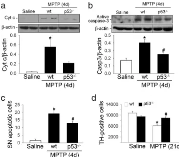
Fig. 3. Loss of p53 in gene-targeted mice attenuates MPTP-induced SNpc DA
apoptotic cell death. (a and b) At the peak of MPTP-induced mitochondria-dependent apoptotic cell death (4 days after the last MPTP injection), cyto-chrome c release and caspase-3 activation are reduced in ventral midbrain cytosolic fractions of p53-deficient mice. (c) The number of MPTP-induced SNpc apoptotic neurons is decreased in the ventral midbrain of p53-deficient mice compared with similarly treated wild-type mice. Morphological criteria to identify apoptotic cells included shrinkage of the cell body, chromatin condensation, and the presence of distinct, round, well defined chromatin clumps, demonstrated on thionin staining, as in ref. 8. (d) p53-deficient mice exhibit a significant protection against MPTP-induced SNpc DA cell death, as determined by assessing the number of SNpc DA neurons at 21 days after the last MPTP injection.*, P⬍ 0.05 compared with saline-injected mice; #, P ⬍ 0.05 compared with MPTP-injected wild-type mice.
operate in a cell type- and/or stimulus-specific manner (32, 33). In neurons, Bim is subject to the transcriptional control of JNK (17, 18), a pathogenic pathway that has been implicated in MPTP-induced SNpc DA neurodegeneration (4, 19). Moreover, evidence from in vitro studies indicates that JNK activation is required for Bax mitochondrial translocation and activation of the mitochondrial apoptotic pathway elicited by DNA damage (34, 35). We assessed whether MPTP-induced up-regulation of Bim depended on JNK. In particular, we examined the involve-ment of JNK3, a JNK isoform selectively expressed in the nervous system that has been shown to play a critical role in stress-induced neuronal apoptosis and to transcriptionally reg-ulate Bim in ischemic neuronal injury (36). Supporting a role for JNK3 in bim transcriptional induction caused by complex I inhibition, ablation of JNK3 in mutant mice prevented MPTP-induced bim mRNA up-regulation (Fig. 4b).
At the peak of MPTP-induced bim mRNA up-regulation (24 h after the last MPTP injection), levels of BimEL, the predominant
and physiologically relevant Bim isoform in neurons (17, 37), were markedly increased in the ventral midbrain of MPTP-intoxicated mice (Fig. 4c), but not in regions unaffected by MPTP (data not shown). BimELwas detected exclusively in the mitochondrial
frac-tion of ventral midbrain homogenates (Fig. 4c) in agreement with its known localization as an integral mitochondrial membrane protein in neurons (17). Overall, our results indicate that Bim is transcriptionally induced in a JNK-dependent manner after MPTP intoxication and that it is localized in mitochondrial fractions.
Loss of Bim Attenuates Bax Mitochondrial Translocation, Cytochrome c Release, and SNpc DA Apoptotic Cell Death Caused by Complex I Inhibition.Supporting a pathogenic role of Bim in SNpc DA cell death caused by complex I inhibition, loss of Bim in
gene-targeted mice significantly attenuated MPTP-induced Bax mi-tochondrial translocation (Fig. 4d), mimi-tochondrial release of cytochrome c (Fig. 4d), and apoptotic cell death (Fig. 4e). As a consequence, Bim-deficient mice exhibited significant protec-tion against MPTP-induced SNpc DA cell death (Fig. 4f ). This protective effect was not due to altered metabolism of MPTP in the absence of Bim because the striatal levels of MPP⫹did not differ between MPTP-intoxicated bim⫺/⫺ and wild-type mice (data not shown). In contrast to SNpc DA cell bodies, striatal levels of dopamine, as measured by HPLC, were decreased to a similar extent in MPTP-injected bim⫺/⫺and wild-type mice (SI Fig. 7). Overall, our results indicate that the BH3-only protein Bim participates in MPTP-induced Bax mitochondrial translo-cation and SNpc DA cell death.
Discussion
Mitochondrial oxidative damage and Bax have both been shown to play critical roles in neuronal degeneration caused by complex I deficiency (6, 8). In particular, free radical production, sec-ondary to complex I blockade, increases the “releasable” soluble pool of cytochrome c in the mitochondrial intermembrane space through peroxidation of the inner mitochondrial lipid cardio-lipin, whereas activated Bax triggers neuronal death by perme-abilizing the outer mitochondrial membrane, thereby releasing cytochrome c into the cytosol, where it promotes activation of the caspase cascade as part of the “apoptosome” (6). In agree-ment with this model, activation of Bax is required for mito-chondria-dependent SNpc DA apoptotic cell death caused by MPTP-induced complex I inhibition (6, 8). Of note, although the mechanism by which Bax induces cytochrome c release is still a matter of intense debate, our preliminary data indicate that in the context of complex I deficiency, it seems to be independent
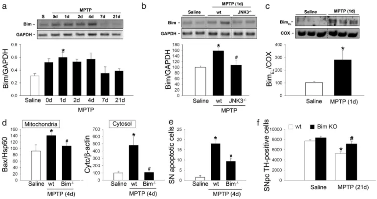
Fig. 4. Role of Bim in MPTP-induced SNpc DA neurodegeneration. (a) Ventral midbrain bim mRNA expression is induced in a time-dependent manner after
MPTP intoxication, peaking at 24 h post-MPTP. (b) At its peak, MPTP-induced bim mRNA up-regulation is prevented in JNK-3-deficient mice. (c) At the peak of MPTP-induced bim mRNA up-regulation, mitochondrial BimELprotein levels are markedly increased in the ventral midbrain. (d) Loss of Bim in gene-targeted mice
significantly attenuates MPTP-induced Bax mitochondrial translocation (Left) and cytochrome c release (Right), as assessed by Western blot analysis of mitochondrial and cytosolic fractions from ventral midbrain, respectively. (e and f ) Bim-deficient mice exhibit a significant reduction in the number of MPTP-induced SNpc apoptotic neurons (e) and a significant protection from MPTP-induced SNpc DA cell death ( f).*, P⬍ 0.05 compared with saline-injected mice; #, P⬍ 0.05 compared with MPTP-injected wild-type mice.
of the mitochondrial permeability transition pore because (i) Bax-induced cytochrome c release in MPP⫹-treated brain mi-tochondria is not responsive to the mimi-tochondrial pore blocker cyclosporin A (SI Fig. 8A), and (ii) mice deficient for cyclophilin D, a critical component of the mitochondrial pore, do not exhibit reduced susceptibility to MPTP (SI Fig. 8 B and C).
On the basis of our results, we propose a pathogenic scenario in which neuronal death caused by complex I deficiency results from a self-amplifying cascade of deleterious events, starting at the mitochondria by the alteration of the oxidative phosphorylation and finishing also at the mitochondria by the activation of the pro-grammed cell death machinery. In this scenario (Fig. 5), MPTP impairs mitochondrial respiration in SNpc DA neurons by inhibit-ing complex I of the electron transport chain. Inhibition of complex I blocks the flow of electrons along the mitochondrial electron transport chain, which results in increased production of reactive oxygen species (ROS). Mitochondrial ROS then increase the soluble pool of cytochrome c in the mitochondrial intermembrane space by a mechanism involving peroxidation of cardiolipin, whereas ROS outside the mitochondria, probably emanating also from sources other than complex I inhibition (38), damage different cellular elements, such as lipids, proteins, and DNA. DNA damage activates both p53 and JNK. p53 induces transcriptional up-regulation of Bax, whereas JNK participates in Bax mitochondrial translocation through transcriptional activation of the BH3-only protein Bim. Once localized to the mitochondrial outer membrane, Bax induces the release of cytochrome c into the cytosol and the ensuing caspase activation and cell death. Supporting the relevance of this scenario to SNpc DA neurodegeneration occurring in PD, several elements of this molecular cascade have been demonstrated in postmortem human brain samples from PD patients, including complex I deficiency (1–3); ROS production (39); oxidative damage to lipids (40, 41), proteins (42, 43), and DNA (44, 45); JNK activation (19, 46, 47); Bax activation (9, 10); and activation of caspase-9 (11) and caspase-3 (9, 48).
The complexity of this cascade, which probably includes other factors not depicted here, guarantees exquisite control over neu-ronal cell death and provides several targets of potential therapeutic significance. Approaches aimed at targeting different key elements of this cascade, especially those acting upstream of cytochrome c release, all result in a significant attenuation of MPTP-induced SNpc DA neurodegeneration (5). We have previously identified Bax as a potential therapeutic target for SNpc DA
neurodegen-eration linked to MPTP-induced complex I inhibition (6, 8). Here, we provide evidence that interfering upstream of Bax, by targeting molecular pathways involved in either Bax transcriptional or post-translation regulation, also provides significant neuroprotection in this model. However, probably because of the multiplicity of regulatory pathways converging on Bax activation, these strategies (at least singly) do not provide as much neuroprotection as target-ing Bax itself (8), most notably at the level of striatal DA terminals. Because the symptoms of PD are caused by the loss of DA terminals in the striatum, preventing the death of SNpc DA cell bodies without preventing the degeneration of their axons is unlikely to be a helpful therapeutic strategy. Neurons might have two self-destruction programs, one for the cell body and a second one for the axon (49). So, a combination of antiapoptotic and axon-protective strategies might be required to obtain optimal clinical benefit from such neuroprotective approaches.
Materials and Methods
Animals and Treatment.The following lines of mutant mice were used here: (i) p53-deficient mice (50), (ii) JNK3-deficient mice (47), (iii) Bim-deficient mice (51), and (iv) cyclophilin D-deficient mice (52). The different lines of mutant mice were maintained in a C57BL/6 genetic background. Eight- to 10-week-old wild-type or mutant male mice received one i.p. injection of MPTP-HCl per day (30 mg⫺1䡠kg⫺1day of free base; Sigma– Aldrich, St. Louis, MO) for 5 consecutive days and were killed at the indicated time points after the last MPTP injection; control mice received saline injections only. For all of the experiments, 3 to 10 mice per group were used.
Subcellular Fractionation.Protein extraction of mitochondrial and cytosolic fractions at the indicated time points was performed on fresh ventral midbrain tissue from saline- and MPTP-injected mice, as previously described (6).
PARP Activity andin Situ Histochemistry.Ventral midbrain, stria-tum, and cerebellum from mice killed at the indicated time points post-MPTP were homogenized. From each sample, 20g of the protein extract were used to determine PARP activity and assayed with a commercial kit (Trevigen, Gaithersburg, MD) using [32P]NAD (PerkinElmer, Boston, MA) following the
man-ufacturer’s instructions. For PARP histochemistry, fresh cryo-stat-cut sections (14m) were fixed in ethanol and permeabil-ized with Triton X-100. The assay was performed using biotinylated NAD⫹ as described previously (53).
Antibodies. The following primary antibodies were used for Western blot analyses: active caspase-3 (BD Biosciences, San Jose, CA), -actin (clone AC15; Sigma–Aldrich), Bax (B-9; Santa Cruz Biotechnology, Santa Cruz, CA), Bim (Bim/BOD, AAP-330; Stressgen Biotechnologies, San Diego, CA), COX-IV (Molecular Probes, Eugene, OR), cytochrome c (Pharmingen, San Diego, CA), DAT (Chemicon, Temecula, CA), and HSP60 (Santa Cruz Biotechnology).
RNA Extraction and RT-PCR. Total RNA was extracted at the indicated time points from midbrain, striatal, and cerebellar samples from saline and chronic MPTP-treated mice and used for RT-PCR analysis using the following primer sequences: Bax, 5⬘-CTGAGCTGACCTTGGAGC-3⬘ and 5⬘-GACTCCAGCCA-CAAAGATG-3⬘; p53, 5⬘-CACTGGAGTCTTCAAGTGTG-3⬘ and 5⬘-GTCTGGGACAGCCAAGTCTG-3⬘; Puma/Bbc3, 5⬘-TGCAGTCAGAGCAGCAACAAC-3⬘ and 5⬘-GGTCACCAT-GAGTCCTTCAGC-3⬘; Noxa, 5⬘-GGATCTAAGTCCCCTG-TACGC-3⬘ and 5⬘-TTTCCCTCTCATCACAGTCCA-3⬘; Bim, 5⬘-GGTAATCCCGACGGCGAAGGGAC-3⬘ and 5⬘-AA-GAGAAATACCCACTGGAGGACC-3⬘; -actin, 5⬘-CTTT-GATGTCACGCACGATTTC-3⬘ and
5⬘-GGGCCGCTCTAG-Fig. 5. Proposed pathogenic scenario induced by complex I deficiency with
MPTP in which DA neuronal death results from a self-amplifying cascade of deleterious events that start at the mitochondria with the alteration of oxidative phosphorylation and finish at the mitochondria with the activation of the programmed cell death machinery. In this scenario, p53 transcription-ally up-regulates Bax, whereas JNK promotes Bax mitochondrial translocation through transcriptional activation of the BH3-only protein Bim.
GCACCAA-3⬘; and GAPDH, 5⬘-GTTTCTTACTCCTTG-GAGGCCAT-3⬘ and 5⬘-TGATGACATCAAGAAGTGGT-GAA-3⬘.
Quantification of DA Neurodegeneration.The total number of ty-rosine hydroxylase (TH)-positive SNpc neurons was counted in the different groups of mice at 21 days after the last MPTP or saline injection by stereology using the optical fractionator method (Ste-reoInvestigator; MBF Bioscience, Williston, VT). Immunostaining was performed with a polyclonal antibody to TH (Calbiochem, EMD Biosciences, San Diego, CA). Quantification of the number of apoptotic neurons in the SN of MPTP- and saline-injected mice was assessed as previously described (8). Because apoptotic neurons may have lost their TH expression as a result of the dying process (54), counts of apoptotic profiles refer to all cells in the substantia nigra.
HPLC Measurements.Striatal levels of dopamine and metabolites were measured by HPLC with electrochemical detection, as previously described (8). Striatal MPP⫹levels were determined by HPLC with UV detection ( ⫽ 295 nm) 90 min after a single i.p. injection of MPTP (30 mg/kg).
Statistical Analysis.All values are expressed as the mean⫾ SEM. Differences among means were analyzed with one- or two-way
ANOVA with treatment or genotype as the independent factors. When ANOVA showed significant differences, pairwise compari-sons between means were tested by Student–Newman–Keuls post hoc testing. In all analyses, the null hypothesis was rejected at the 0.05 level.
We thank Dr. R. A. Flavell (Yale University, New Haven, CT) for providing the JNK-3-deficient mice and Drs. M. Forte (Oregon Health and Sciences University, Portland, OR) and P. Bernardi (University of Padova, Padova, Italy) for providing the cyclophilin D-deficient mice. This work was supported by the European Commission’s Marie Curie Excellence grant and Marie Curie International Reintegration grant (to M.V.), Fundacio´ la Caixa (to M.V.), National Institutes of Health (NIH)/National Institute on Aging (to S.P. and M.V.), U.S. Depart-ment of Defense (S.P. and M.V.), NIH/National Institute of Neuro-logical Disorders and Stroke (S.P.), NIH/National Institute on Envi-ronmental Health Sciences (S.P.), Parkinson’s Disease Foundation (S.P.), Muscular Dystrophy Association/Wings-Over-Wall Street (S.P.), and National Health and Medical Research Council (Australia) and Leukemia and Lymphoma Society of America (P.B. and A.S.). J.B.S. was supported by Deutsche Forschungsgemeinschaft grants and the Center for Molecular Physiology of the Brain (Go¨ttingen, Ger-many). M.V.’s group is part of the Centro de Investigacio´n Biome´dica en Red en Enfermedades Neurodegenerativas (CIBERNED, Instituto de Salud Carlos III, Spain).
1. Parker WD, Jr, Boyson SJ, Parks JK (1989) Ann Neurol 26:719–723. 2. Schapira AH, Cooper JM, Dexter D, Clark JB, Jenner P, Marsden CD (1990)
J Neurochem 54:823–827.
3. Keeney PM, Xie J, Capaldi RA, Bennett JP, Jr (2006) J Neurosci 26:5256–5264. 4. Dauer W, Przedborski S (2003) Neuron 39:889–909.
5. Vila M, Przedborski S (2003) Nat Rev Neurosci 4:365–375.
6. Perier C, Tieu K, Guegan C, Caspersen C, Jackson-Lewis V, Carelli V, Martinuzzi A, Hirano M, Przedborski S, Vila M (2005) Proc Natl Acad Sci USA 102:19126–19131. 7. Roucou X, Martinou JC (2001) Cell Death Differ 8:875–877.
8. Vila M, Jackson-Lewis V, Vukosavic S, Djaldetti R, Liberatore G, Offen D, Korsmeyer SJ, Przedborski S (2001) Proc Natl Acad Sci USA 98:2837–2842. 9. Tatton NA (2000) Exp Neurol 166:29–43.
10. Hartmann A, Michel PP, Troadec JD, Mouatt-Prigent A, Faucheux BA, Ruberg M, Agid Y, Hirsch EC (2001) J Neurochem 76:1785–1793.
11. Viswanath V, Wu Y, Boonplueang R, Chen S, Stevenson FF, Yantiri F, Yang L, Beal MF, Andersen JK (2001) J Neurosci 21:9519–9528.
12. Fannjiang Y, Kim CH, Huganir RL, Zou SF, Lindsten T, Thompson CB, Mito T, Traystman RJ, Larsen T, Griffin DE, et al. (2003) Dev Cell 4:575–585. 13. Duan W, Zhu X, Ladenheim B, Yu QS, Guo Z, Oyler J, Cutler RG, Cadet JL,
Greig NH, Mattson MP (2002) Ann Neurol 52:597–606.
14. Letai A, Bassik M, Walensky L, Sorcinelli M, Weiler S, Korsmeyer S (2002)
Cancer Cell 2:183.
15. Villunger A, Michalak EM, Coultas L, Mullauer F, Bock G, Ausserlechner MJ, Adams JM, Strasser A (2003) Science 302:1036–1038.
16. Jeffers JR, Parganas E, Lee Y, Yang C, Wang J, Brennan J, MacLean KH, Han J, Chittenden T, Ihle JN, et al. (2003) Cancer Cell 4:321–328.
17. Putcha GV, Moulder KL, Golden JP, Bouillet P, Adams JA, Strasser A, Johnson EM (2001) Neuron 29:615–628.
18. Whitfield J, Neame SJ, Paquet L, Bernard O, Ham J (2001) Neuron 29:629–643. 19. Xia XG, Harding T, Weller M, Bieneman A, Uney JB, Schulz JB (2001) Proc
Natl Acad Sci USA 98:10433–10438.
20. Mihara M, Erster S, Zaika A, Petrenko O, Chittenden T, Pancoska P, Moll UM (2003) Mol Cell 11:577–590.
21. Chipuk JE, Kuwana T, Bouchier-Hayes L, Droin NM, Newmeyer DD, Schuler M, Green DR (2004) Science 303:1010–1014.
22. Chipuk JE, Bouchier-Hayes L, Kuwana T, Newmeyer DD, Green DR (2005)
Science 309:1732–1735.
23. Endo H, Kamada H, Nito C, Nishi T, Chan PH (2006) J Neurosci 26:7974–7983. 24. Hong SJ, Dawson TM, Dawson VL (2004) Trends Pharmacol Sci 25:259–264. 25. Mandir AS, Przedborski S, Jackson-Lewis V, Wang ZQ, Simbulan-Rosenthal M, Smulson ME, Hoffman BE, Guastella DB, Dawson VL, Dawson TM (1999) Proc
Natl Acad Sci USA 96:5774–5779.
26. Miyashita T, Reed JC (1995) Cell 80:293–299.
27. Thornborrow EC, Patel S, Mastropietro AE, Schwartzfarb EM, Manfredi JJ (2002) Oncogene 21:990–999.
28. Willis SN, Adams JM (2005) Curr Opin Cell Biol 17:617–625.
29. Kuwana T, Bouchier-Hayes L, Chipuk JE, Bonzon C, Sullivan BA, Green DR, Newmeyer DD (2005) Mol Cell 17:525–535.
30. Chen L, Willis SN, Wei A, Smith BJ, Fletcher JI, Hinds MG, Colman PM, Day CL, Adams JM, Huang DC (2005) Mol Cell 17:393–403.
31. Willis SN, Fletcher JI, Kaufmann, T, van Delft MF, Chen L, Czabotar PE, Ierino H, Lee EF, Fairlie WD, Bouillet P, et al. (2007) Science 315:856–859. 32. Huang DC, Strasser A (2000) Cell 103:839–842.
33. Puthalakath H, Strasser A (2002) Cell Death Differ 9:505–512.
34. Tournier C, Hess P, Yang DD, Xu J, Turner TK, Nimnual A, Bar-Sagi D, Jones SN, Flavell RA, Davis RJ (2000) Science 288:870–874.
35. Lei K, Nimnual A, Zong WX, Kennedy NJ, Flavell RA, Thompson CB, Bar-Sagi D, Davis RJ (2002) Mol Cell Biol 22:4929–4942.
36. Kuan CY, Whitmarsh AJ, Yang DD, Liao G, Schloemer AJ, Dong C, Bao J, Banasiak KJ, Haddad GG, Flavell RA, et al. (2003) Proc Natl Acad Sci USA 100:15184–15189.
37. O’Reilly LA, Cullen L, Visvader J, Lindeman GJ, Print C, Bath ML, Huang DC, Strasser A (2000) Am J Pathol 157:449–461.
38. Lotharius J, O’Malley KL (2000) J Biol Chem 275:38581–38588.
39. Wu DC, Teismann P, Tieu K, Vila M, Jackson-Lewis V, Ischiropoulos H, Przedborski S (2003) Proc Natl Acad Sci USA 100:6145–6150.
40. Dexter DT, Carter CJ, Wells FR, Javoy-Agid F, Agid Y, Lees A, Jenner P, Marsden CD (1989) J Neurochem 52:381–389.
41. Yoritaka A, Hattori N, Uchida K, Tanaka M, Stadtman ER, Mizuno Y (1996)
Proc Natl Acad Sci USA 93:2696–2701.
42. Alam ZI, Daniel SE, Lees AJ, Marsden DC, Jenner P, Halliwell B (1997)
J Neurochem 69:1326–1329.
43. Giasson BI, Duda JE, Murray IV, Chen Q, Souza JM, Hurtig HI, Ischiropoulos H, Trojanowski JQ, Lee VM (2000) Science 290:985–989.
44. Alam ZI, Jenner A, Daniel SE, Lees AJ, Cairns N, Marsden CD, Jenner P, Halliwell B (1997) J Neurochem 69:1196–1203.
45. Zhang J, Perry G, Smith MA, Robertson D, Olson SJ, Graham DG, Montine TJ (1999) Am J Pathol 154:1423–1429.
46. Teismann P, Tieu K, Choi DK, Wu DC, Naini A, Hunot S, Vila M, Jackson-Lewis V, Przedborski S (2003) Proc Natl Acad Sci USA 100:5473–5478. 47. Hunot S, Vila M, Teismann P, Davis RJ, Hirsch EC, Przedborski S, Rakic P,
Flavell RA (2004) Proc Natl Acad Sci USA 101:665–670.
48. Hartmann A, Hunot S, Michel PP, Muriel MP, Vyas S, Faucheux BA, Mouatt-Prigent A, Turmel H, Srinivasan A, Ruberg M, et al. (2000) Proc Natl Acad Sci
USA 97:2875–2880.
49. Raff MC, Whitmore AV, Finn JT (2002) Science 296:868–871.
50. Donehower LA, Harvey M, Slagle BL, McArthur MJ, Montgomery CA, Jr, Butel JS, Bradley A (1992) Nature 356:215–221.
51. Bouillet P, Metcalf D, Huang DC, Tarlinton DM, Kay TW, Kontgen F, Adams JM, Strasser A (1999) Science 286:1735–1738.
52. Basso E, Fante L, Fowlkes J, Petronilli V, Forte MA, Bernardi P (2005) J Biol
Chem 280:18558–18561.
53. Bakondi E, Bai P, Szabo EE, Hunyadi J, Gergely P, Szabo C, Virag L (2002)
J Histochem Cytochem 50:91–98.
54. Jackson-Lewis V, Vila M, Djaldetti R, Gue´gan C, Liberatore G, Liu J, O’Malley KL, Burke RE, Przedborski S (2000) J Comp Neurol 424:476–488.