HAL Id: tel-01499845
https://tel.archives-ouvertes.fr/tel-01499845
Submitted on 1 Apr 2017
HAL is a multi-disciplinary open access archive for the deposit and dissemination of sci-entific research documents, whether they are pub-lished or not. The documents may come from teaching and research institutions in France or abroad, or from public or private research centers.
L’archive ouverte pluridisciplinaire HAL, est destinée au dépôt et à la diffusion de documents scientifiques de niveau recherche, publiés ou non, émanant des établissements d’enseignement et de recherche français ou étrangers, des laboratoires publics ou privés.
Pseudolysogeny and sequential mutations build
multiresistance to virulent bacteriophages in
Pseudomonas aeruginosa
Libera Latino
To cite this version:
Libera Latino. Pseudolysogeny and sequential mutations build multiresistance to virulent bacterio-phages in Pseudomonas aeruginosa. Bacteriology. Université Paris-Saclay, 2016. English. �NNT : 2016SACLS274�. �tel-01499845�
NNT : 2016SACLS274
T
HESE DE DOCTORAT
DE
L’U
NIVERSITE
P
ARIS
-S
ACLAY
PREPAREE A
L
’U
NIVERSITE
P
ARIS
-S
UD
INSTITUT DE BIOLOGIE INTEGRATIVE DE LA CELLULE
E
COLED
OCTORALE N° 577
Structure et dynamique des systèmes vivants
Spécialité de doctorat : Sciences de la Vie et de la Santé
Par
Mme Libera Latino
Pseudolysogeny and sequential mutations build multiresistance to virulent
bacteriophages in Pseudomonas aeruginosa
Thèse présentée et soutenue à Orsay, le 20 septembre 2016 : Composition du Jury :
M. Guyonvarch Armel Professeur Université Paris-Saclay, Orsay Président M. Bikard David Chargé de Recherche 1 Institut Pasteur, Paris Rapporteur Mme Brøndsted Lone Professeur associé University of Copenhagen, Copenhagen Rapporteur Mme Bidnenko Elena Chargé de Recherche 1 INRA, Jouy-en-Josas Examinatrice M. Hochberg Michael Directeur de Recherche Université Montpellier II, Montpellier Examinateur M. Vergnaud Gilles Directeur de Recherche Université Paris-Saclay, Orsay Directeur de thèse Mme Pourcel Christine Directeur de Recherche Université Paris-Saclay, Orsay Co-directeur de thèse
Acknowledgments
I would like to thank my thesis committee members, Dr Lone Brøndsted and Dr David Bikard for spending time reading my manuscript and suggesting improvements; Dr Elena Bidnenko, Professor Michael Hochberg and Professor Armel Guyonvarch for examining my work as well. I appreciate your precious suggestions.
Words will never be enough to express my gratitude to Dr Gilles Vergnaud and Dr Christine Pourcel for welcoming me in their laboratory dedicating precious time training and supervising me during the last four years. I would like to express my special appreciation and thanks to my advisor Christine, you have been a tremendous mentor for me. You have always encouraged my research, my ideas and allowed me to grow as a scientist. Your advices on research as well as on my career have been priceless and contributed to make the person I am today. Moreover, I would like to thank you for all the enjoyable moments spent during the different conferences we attended around the world and for giving me precious life advices. I also wish to thank to Professor Michaël Dubow, Dr Marie-Agnès Petit and Dr Emmanuelle Guillot-Combe for tutoring me during my entire thesis and providing me with helpful suggestions.
Thanks to all past and present members of the team, and particularly to Cédric Midoux, David Christiany and Yann Blouin for being so terribly patient with me asking bioinformatics (sometimes very weird) questions. To Yolande Hauck, for sharing a lot during the last year, her office and her time. You have been always so caring and tender to me and morally supported me (sometimes with the help of too much chocolate and ice cream).
A special thanks to Dr Agnès Delahodde and Raynald Cossard for kindly spending their time training me with C. elegans manipulation; Dr Barry Holland and Dr Simon Seror for the interesting discussions on bacteria motility. To all the people of the Institute that made it possible, from secretaries, Muriel Decraene and Catherine Drouet helping me with the administrative work, to people working on the second floor as Emeline Dubois always present for supporting me with great life advices and Philippe Bouloc and Chantal Bohn for interesting and sometimes very funny discussions during lunch times. In particular, a special thanks to Eric Espagne, an amazing friend always ready to help me or simply listen to me. I would like to thank also all my friends for making my stay in Paris full of great memories and last but not least my parents that have always believed in me and firmly supported my choices.
Thanks to all of you, with this thesis defense I close another wonderful chapter of my life and I am ready to start a new one, sure that I will always have your support.
I pictured myself as a virus…and tried to sense what it would be like.
TABLE OF CONTENT
INTRODUCTION ... 1
I BACTERIA AND BACTERIOPHAGES ... 1
I.I PSEUDOMONAS AERUGINOSA IS AN OPPORTUNISTIC PATHOGEN ... 1
I.I.I The genetic diversity of P. aeruginosa ... 2
I.I.II Emergence of multidrug resistant P. aeruginosa ... 4
I.I.III Biofilm formed by P. aeruginosa in patients ... 6
I.II BACTERIOPHAGES ... 8
I.II.I Discovery of phages and first phage therapy assays ... 9
I.II.II Phage life strategies ... 10
I.II.III The importance of phage research ... 14
I.II.IV Molecular interaction between virulent phages and bacteria during infection ... 16
I.III PHAGES EMPLOYED IN PHAGE THERAPY ... 18
I.III.I Phages of P. aeruginosa ... 18
The Myoviridae ... 18
The Siphoviridae ... 19
The Podoviridae ... 20
I.III.II Selection of phages for therapeutic use ... 21
I.III.III Phage prophylaxis and therapy to treat P. aeruginosa infections ... 24
II PHAGE-BACTERIA INTERACTIONS ... 27
II.I PSEUDOLYSOGENY ... 28
II.I.I History about the definition of pseudolysogeny ... 29
II.I.II Impact of pseudolysogeny on bacterial populations ... 31
II.II SUPERINFECTION EXCLUSION: A MECHANISM OF BACTERIAL IMMUNITY AGAINST PHAGES ... 33
II.III BACTERIAL RESISTANCE AGAINST PHAGES ... 37
II.III.I Mutations of phage receptors ... 37
The bacterial lipopolysaccharide ... 37
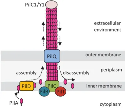
Bacterial appendages: type IV pili ... 40
P. aeruginosa mucoid phenotype ... 43
II.III.II Restriction modification system ... 44
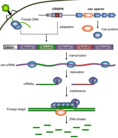
II.III.III The CRISPR-Cas system ... 45
GOAL OF THE WORK ... 49
RESULTS AND DISCUSSION ... 51
I SELECTIVE PRESSURE IMPOSED ON P. AERUGINOSA BY A TEMPERATE PHAGE .. 53
I.II BACTERIAL RESISTANCE AGAINST PHAGE AB31 ... 55
I.III AB31 IS A CHIMERIC PHAGE ... 59
CONCLUSION... 59
II TOLERANT VARIANTS SURVIVING INFECTION WITH VIRULENT PHAGES ... 61
II.I RATIONALE AND EXPERIMENTAL PROCEDURE ... 61
II.I.I Genetic diversity of phages used in this study ... 61
Phage Ab05 ... 62
Phage Ab09 ... 62
Phage Ab17 ... 63
Phage Ab27 ... 64
II.I.II Choices about the infection conditions ... 64
II.I.III Phages combinations ... 65
II.II PHENOTYPE OF PHAGE-TOLERANT VARIANTS ... 66
II.II.I Colony morphology of phage-tolerant variants on different medium ... 67
II.II.II Clustering of the thirty-two variants according to their phage-tolerance pattern ... 69
II.III A WIDE RANGE OF CHROMOSOMAL MUTATIONS IS SELECTED BY PHAGES ... 73
II.III.I Phase variation mutations are reversible ... 78
II.III.II Deletions ... 79
II.III.III Nucleotide substitutions ... 79
II.IV PERSISTENCE OF PHAGE DNA CONFERS IMMUNITY ... 80
II.IV.I Pseudolysogenic phages with tail fiber mutations ... 81
II.IV.II Stability of the pseudolysogenic stage ... 83
II.IV.III High frequency of double mutants is due to persistence of phage DNA ... 86
II.IV.IV Pseudolysogeny confers immunity to phages ... 91
II.V MUTATIONS AFFECT PHAGE RECEPTORS... 92
II.V.I Pilus type IV mutants are defective in twitching motility and biofilm formation ... 92
II.V.II The mucoid phenotype could be reverted through complementation ... 94
II.V.III Lipopolysaccharide is modified in Ab09-, Ab17- and Ab27-tolerant variants ... 95
The Wzy mutants ... 97
The Wzz2 mutant ... 99
The Pgi, WapH, DnpA and WbpL mutants ... 99
II.V.IV The lipopolysaccharide of pseudolysogens ... 102
GLOBAL DISCUSSION ... 105
I PSEUDOLYSOGENY ALLOWS SELECTION OF MUTANTS... 105
I.I CONDITIONS FAVORING THE APPEARANCE OF PSEUDOLYSOGENS ... 105
I.II PSEUDOLYSOGENY CONTRIBUTES TO THE SELECTION OF SINGLE/DOUBLE MUTANTS ... 106
I.III IMMUNITY PROVIDED BY THE PSEUDOLYSOGENIC PHAGE ... 107
II RED QUEEN DYNAMICS/ARM RACE CO-EVOLUTION ... 109
II.I FITNESS COST LINKED TO ACQUISITION OF PHAGE-RESISTANCE ... 109
II.II PSEUDOLYSOGENIC MUTANT PHAGES ... 111
CONCLUSIONS ... 113
PERSPECTIVES ... 117
NEW INSIGHTS INTO PSEUDOLYSOGENY ... 117
PHAGE-RESISTANCE IN ANOTHER P. AERUGINOSA STRAIN ... 118
PSEUDOLYSOGENY AND CRISPR-Cas SYSTEM ... 119
THE LIPOPOLYSACCHARIDE AS A PHAGE RECEPTOR ... 119
LINK BETWEEN PHAGE AND ANTIBIOTIC RESISTANCE ... 119
MATERIAL AND METHODS ... 121
I PHAGES ... 121
I.I Phage amplification ... 121
I.II Electron microscopy ... 122
II RESISTANT-VARIANT COLLECTION PREPARATION ... 122
II.I Isolation of phage-resistant variants ... 122
II.II Calculation of the frequency of resistance ... 123
II.III Phage susceptibility assay ... 124
III PHENOTYPIC CHARACTERIZATION ... 124
III.I Adsorption assay ... 124
III.II Twitching motility assay ... 125
III.III Planktonic growth rate and biofilm formation ... 125
III.IV Virucide assay ... 125
III.V Lipopolysaccharide analysis ... 126
IV PSEUDOLYSOGENS ANALYSIS ... 126
IV.I Colony transfer and hybridization ... 126
IV.II Stability of phage persistence in pseudolysogens ... 127
IV.III DNA extraction, PCR and sequencing ... 127
IV.IV Gene cloning and expression ... 129
IV.V Whole-genome sequencing ... 129
BIBLIOGRAPHY ... 131
ANNEX I ... 149
ANNEX II ... 177
ANNEX III ... 195
ABBREVIATIONS
A: adenine A600: absorbance at 600 nm bp: base pair C: cytosine CF: cystic fibrosisEOP: efficiency of plating g: grams
G: guanine
ICU: intensive care unit kbp: kilo base pair l: liter
LB: Luria Bertani broth LPS: lipopolysaccharide MOI: multiplicity of infection
PAGE: polyacrylamide gel electrophoresis PBS: phosphate saline buffer
PCR: polymerase chain reaction PEG: polyethylene glycol
SMG: saline magnesium gelatin buffer SNP: single nucleotide polymorphism T: thymine
1
INTRODUCTION
I
BACTERIA
AND
BACTERIOPHAGES
I.I PSEUDOMONASAERUGINOSA IS AN OPPORTUNISTIC PATHOGEN
Pseudomonas aeruginosa is a gram-negative bacterium that can be naturally found in soil and water. This bacterium is able to colonize biotic and abiotic surfaces and to naturally resist different classes of antimicrobial agents. P. aeruginosa is a member of the normal microbial flora in humans. Representative colonization rates for specific sites in humans are up to 2% for skin, 3.3% for the nasal mucosa, 6.6% for the throat, and from 2.6 up to 24% for fecal samples (Morrison and Wenzel 1984). Generally it does not cause disease in healthy people or in people without injury. For instance, when the bacteria enter the lungs of healthy hosts, they are cleared rapidly, without the initiation of an inflammatory response, by a variety of innate host defense strategies such as mucociliary clearance or by the resident macrophages. In contrast to its harmless nature in healthy people and, more generally, in the environment, P. aeruginosa is an opportunistic pathogen. It dangerously affects people whose immune system is weakened by a disease, such as people with acquired immune deficiency syndrome, or burned patients, people with ulcers or intubated in intensive care units, those with postoperative infections and patients affected by cystic fibrosis (Bodey, Bolivar et al. 1983). Indeed, patients with impaired immunity have higher risks of contracting P. aeruginosa nosocomial infection (Morrison and Wenzel 1984). Colonization rates may exceed 50% during hospitalization, especially among patients with trauma, cutaneous wounds or breach in mucosal barriers due to mechanical ventilation, tracheostomy, catheters, surgery, or severe burns (Ohara and Itoh 2003; Thuong, Arvaniti et al. 2003; Erol, Altoparlak et al. 2004). Nosocomial infections, contracted from the environment or staff of a healthcare facility (by
2 contaminated equipment, bed linens, or air droplets), can be spread in the hospital environment, nursing home environment, rehabilitation facility, clinic or other clinical settings. Disruption of the normal microbial flora as a result of antimicrobial therapy has also been shown to increase colonization by P. aeruginosa (Takesue, Yokoyama et al. 2002).
P. aeruginosa can establish two forms of interaction with its host: acute and chronic infection (Furukawa, Kuchma et al. 2006). Acute infections include acute pneumonia in hospitalized and, in particular, mechanically ventilated patients, skin infections and sepsis in patients with extensive burns, corneal infections in individuals wearing contact lenses, urinary tract infections in patients with catheters, bacteremia and sepsis in immunocompromised patients, particularly neutropenic patients receiving cytotoxic therapies, and post-surgical wound infections.
P. aeruginosa is the principal pathogen in the lungs of patients with CF. In some cases, P. aeruginosa breaks down host defenses in tissues such as the lungs and disseminates in the bloodstream, leading to death of a patient within hours or days. Through the type III secretion system, P. aeruginosa is able to secrete a variety of extracellular toxins facilitating systemic dissemination via the bloodstream (Vance, Rietsch et al. 2005), and acute infections such as in pneumonia (Barbieri and Sun 2004; Matsumoto 2004). In contrast, persistent or chronic infection is characterized by pulmonary tissue damage and respiratory failure which even long-term antibiotic therapy does not eradicate, leading to the patient death in some cases (Burns, Ramsey et al. 1993; Høiby 1993).
I.I.I THE GENETIC DIVERSITY OF P. AERUGINOSA
The success of P. aeruginosa as a worldwide-spread bacterium and opportunistic pathogen is based on its broad genetic repertoire.
3 The complete bacterial genome of a species, the pan-genome, includes the "core genome" containing genes present in all strains, and the "dispensable genome" also called “accessory genome” containing genes present in a subset of strains (Medini, Donati et al. 2005). The P. aeruginosa pan-genome consists of a core genome of about 5000 genes (Valot, Guyeux et al. 2015; van Belkum, Soriaga et al. 2015), and a dispensable genome of more than 40 000 genes. Seventy-five percent of the latter are present only in a few clones or strains (Hilker, Munder et al. 2015). Several studies have described the population structure as non-clonal,
panmictic with a few clones associated to diseases, environmental conditions or antibiotic
resistance (Kiewitz and Tümmler 2000; Curran, Jonas et al. 2004). Studies of the diversity of
strains present in CF patients in France showed that they are mostly non-clonal, apart from
clone C and clone PA14 (Vu-Thien, Corbineau et al. 2007; Llanes, Pourcel et al. 2013). This
suggested that there is continuous modification of bacterial genomes by genetic exchanges,
while clones emerge in particular contexts due to evolutionary pressures. In contrast, in
another study performed by Wiehlmann and colleagues, a representative strain collection of
diverse habitats and geographic origin was typed to describe the global population structure of
P. aeruginosa (Wiehlmann, Wagner et al. 2007). The majority of P. aeruginosa strains was
shown to belong to a few dominant clones widespread in diseases and environmental habitats
such as clone PA14 represented by the completely sequenced reference strain PA14 (Lee,
Urbach et al. 2006), clone C (Larbig, Christmann et al. 2002), clone K (Klockgether, Reva et
al. 2004), clone M (Römling, Wingender et al. 1994), clone TB (Tümmler, Koopmann et al.
1991), clone CHA (Dacheux, Attree et al. 1999), and clone LES (Midlands and Liverpool
4 I.I.II EMERGENCE OF MULTIDRUG RESISTANT P. AERUGINOSA
P. aeruginosa possess a high capability of developing resistance to antimicrobial agents, molecules able to kill or simply inhibit the growth of bacteria such as antibiotics and aseptics, commonly employed in hospitals. Thus, nosocomial infections due to an antimicrobial-resistant P. aeruginosa spread are often difficult to eradicate. Even more problematic is the development of resistance during the course of therapy, a complication which has been shown to double the length of hospitalization and overall cost of patient care (Lister, Wolter et al. 2009). P. aeruginosa can develop resistance to different classes of antibiotics such as carbapenems, aminoglycosides and fluoroquinolones (Ozer, Duran et al. 2012) either through the acquisition of resistance genes on mobile genetic elements such as plasmids, or through mutational processes that alter the expression and function of chromosomally encoded mechanisms (Lister, Wolter et al. 2009).
Carbapenem-resistance may be related to decreased bacterial outer membrane permeability due, for example, to a loss or modification of porins or to overexpression of efflux pumps, or to expression of carbapenemases (Mesaros, Nordmann et al. 2007; Rodríguez-Martínez, Poirel et al. 2009). Indeed, several studies performed on P. aeruginosa coming from different sources and different countries such as Europe, Asia and South America have shown that some P. aeruginosa isolates present a set of genes encoding antibiotic-inactivation enzymes such as carbapenemases (Nordmann and Poirel 2002; Scheffer, Gales et al. 2010; Vitkauskienė, Skrodenienė et al. 2011; Correa, Montealegre et al. 2012; Llanes, Pourcel et al. 2013). Many carbapenemase genes are carried by plasmids and are easily transferable.
Aminoglycoside resistance may be due to aminoglycoside-modifying enzymes. High level of resistance to multiple aminoglycosides can also be associated with the acquisition of a
5 methylase gene, able to methylate the 16S rRNA in P. aeruginosa (Yokoyama, Doi et al. 2003).
Fluoroquinolone resistance among P. aeruginosa isolates has been linked mostly to mutations in chromosomal genes, with alterations in the fluoroquinolone targets DNA gyrase and topoisomerase IV, or overexpression of multidrug efflux pumps (Lister, Wolter et al. 2009).
The simultaneous presence of aminoglycoside-modifying enzymes and chromosomal mutations in genes encoding the fluoroquinolone-target enzymes, leads to the emergence of multidrug-resistant P. aeruginosa (Carmeli, Troillet et al. 1999). Polymyxins are among the very few therapeutic options left against such strains (Levin, Barone et al. 1999). In the 1970s, polymyxin B agents, antibiotics produced by a strain of Bacillus polymyxa, were employed to treat infections caused by gram-negative bacteria, including P. aeruginosa, but they were soon abandoned because of reported nephrotoxicity and neurotoxicity, and replaced by other, less toxic, antibiotics (Falagas and Kasiakou 2006). Another antibiotic belonging to the polymyxin group, polymixin E or colistin, is used in clinical practice as the emergence of multidrug-resistant organisms has renewed the interest in this mildly toxic therapeutic option (Sabuda, Laupland et al. 2008).
The emergence and spread of multidrug-resistant P. aeruginosa strains has significantly increased during the past years and this can be attributed to the wide use of new generation antibiotics and to the action, at the genetic level, of horizontal transfer of plasmids carrying different combinations of antibiotic-resistance determinants (Poole 2005; Nordmann, Poirel et al. 2011). It constitutes a serious threat for future therapy (Barbier and Wolff 2010).
6 I.I.III BIOFILM FORMED BY P. AERUGINOSA IN PATIENTS
The failure of antibiotic treatment of some chronic P. aeruginosa infections has been hypothesized to depend from two main events: the formation of a biofilm known, in general, to protect bacteria from environmental stressful conditions (Costerton, Stewart et al. 1999), and the selection, through prolonged use of antibiotics, of antibiotic-resistant variants.
Biofilms constitute a protective mode of growth that allows survival in a hostile environment. The biofilms contain channels in which nutrients can circulate, and cells in different regions of a biofilm exhibit different patterns of gene expression (Davies, Chakrabarty et al. 1993). These sessile biofilm communities can give rise to non-sessile individuals, planktonic bacteria that can rapidly multiply and disperse (Figure 1) (Mizan, Jahid et al. 2015).
FIGURE 1. HYPOTHETICAL DEVELOPMENT OF A BIOFILM. The mature biofilm is produced through
different steps: 1) the planktonic bacterial cells attach on a surface; 2) the production of structures devoted to the attachment is arrested and the bacterial cells start to proliferate; 3) the bacterial cells produce an exopolysaccharide matrix and “communicate” with other bacterial cells through quorum sensing mechanism; 4) the mature biofilm is formed and 5) some cells disperse and colonize other free surfaces. Figure extracted and modified from (Mizan, Jahid et al. 2015).
7 Planktonic bacteria expose themselves to deleterious agents in their environment, such as phages or potent antimicrobial agents in a clinical setting. Thus, it is not surprising that chronic bacterial infections could involve bacterial biofilms, which are not easily eradicated by conventional antibiotic therapy.
Analysis of quorum sensing signals released by the bacteria in the sputum of CF patients chronically infected with P. aeruginosa suggested that this bacterium exist in the form of biofilms (Singh, Schaefer et al. 2000). The factors responsible for the induction of biofilm formation in lungs of CF patients are still not completely clear. In patients affected by this disease, the transmembrane regulator chloride channel located at the apical membranes of epithelial cells is defective, thus the fluid present in the airways of CF people is characterized by high salt concentration; the salt is believed to inhibit the activity of antimicrobial peptides and proteins of the innate immunity present in the airways, therefore the bacterium become free to colonize the epithelium in the form of a biofilm (Welsh and Smith 1993).
Antibiotic treatment during chronic P. aeruginosa infections of CF patients sometimes allows the disappearance of some symptoms usually associated with acute infections. This could be explained by the fact that planktonic cells released by the biofilm matrix and provoking the typical symptoms of acute infection, are susceptible to the antibiotic being used and can be killed. However, it is possible that the use of antibiotic could also select for planktonic cells resistant to antibiotics that can, in turn, colonize other parts of the epithelium giving rise to an antibiotic-resistant biofilm, determining the failure of the ongoing infection treatment (Costerton, Stewart et al. 1999; Singh, Schaefer et al. 2000; Drenkard and Ausubel 2002).
Current knowledge about bacterial biofilms suggests that effective eradication depends upon the use of therapeutic agents able to penetrate the exopolysaccharide matrix, and promote the detachment of bacterial cells, thus impairing the emergence of antibiotic-resistant variants.
8 I.II BACTERIOPHAGES
Bacteriophages (or phages) are the most abundant and most diversified microorganisms on Earth. They can be seen as obligate bacterial predators since they need bacteria for their multiplication, and can be found in all reservoirs populated by bacterial hosts including soil, aquatic environments (Srinivasiah, Bhavsar et al. 2008), human gut (Mills, Shanahan et al. 2013) etc.
Interactions between phages and their bacterial hosts are complex and play significant roles in
shaping the structure of environmental microbial communities. Phage survival depends on its
ability to infect the bacterial host. Adaptation to selective pressures such as host resistance,
determines its abundance and diversification. Co-evolution of the phage tail fibers and
bacterial receptors determines bacterial host ranges, mechanisms of phage entry, and other
infection parameters. Phage host ranges have been shown to be highly variable in terms of
specificity, ranging from phages with extremely narrow ranges of hosts within a single
species to those that can infect bacteria across genera (Weitz, Poisot et al. 2013).
In natural habitats, phages and bacteria are in a constant arms race that proceeds in continuous cycles of co-evolution. As soon as the bacteria develop mechanisms to prevent phage infection, for example, bacterial receptor modification and degradation of invading phage DNA (Labrie, Samson et al. 2010), phages can evolve mechanisms to target such resistant bacteria (Samson, Magadán et al. 2013). This arms race continues and become one of the major forces to both widen the genetic diversity and maintain the equilibrium within microbial communities.
9 I.II.I DISCOVERY OF PHAGES AND FIRST PHAGE THERAPY ASSAYS
The very first evidences of an antibacterial activity that could be attributed to bacteriophages were obtained by Ernest Hankin. In 1896 this British bacteriologist reported the presence of an antibacterial agent in the water of two Indian rivers. The agent was able to pass through a porcelain filter, was inactivated by heat, and could impede the spread of a cholera epidemic (Hankin 1896). Twenty years later (in 1915), Frederick Twort, a medically trained bacteriologist from England, suggested that the antibacterial activity previously mentioned, could be due, among other possibilities, to the presence of a virus. Two years later (in 1917), Félix d’Hérelle, a French-Canadian bacteriologist working at the Pasteur Institute in Paris, reported the discovery of bacteriophages (see (Duckworth 1976) for an analysis of the controversy of who should be credited for the discovery of bacteriophages) and subsequently developed their therapeutic use (phage therapy). In 1915 d’Hérelle had been assigned to conduct an investigation on the dysentery outbreak among the French troops stationed at Maisons-Laffitte (on the outskirts of Paris). He filtered some patient fecal samples and incubated them with a Shigella strain isolated from the same patients, then he inoculated the mixture in rabbits in order to develop a vaccine against dysentery, and spread it also on agar plates to look at the bacterial growth; he observed that the bacterial growth on the plate was not uniform due to the presence of small, clear areas that he first called taches and only later plaques (Summers 1991). This discovery opened the way to a new therapeutic approach of bacterial infections. D’Hérelle was the first to use phages to successfully treat, in 1919, at the Necker-Enfants-Malades Hospital in Paris, five children affected by dysentery (Dublanchet and Fruciano 2008). However, the results of the treatment were published only later and were preceded by the first publication on the subject made by Richard Bruynoghe and Joseph Maisin in 1921, who used phages to treat a staphylococcal skin disease. After that, different treatment employing phages were performed and all were shown to be quite efficient in
10 eradicating the infection, such as those performed by d’Hérelle in India, on thousands of people affected by cholera or bubonic plague (Sulakvelidze, Alavidze et al. 2001).
Despite its initial success, with the advent of the first antibiotics and, consequently, the discovery of several new molecules from 1940 to 1990, phage therapy was abandoned in Western Europe and the Americas, and re-considered only recently (Dublanchet and Fruciano 2008; Abedon, Kuhl et al. 2011). However, it became a major anti-bacterial therapy in the former USSR and is still in use in Russia and different countries in Eastern Europe (Abedon, Kuhl et al. 2011).
Phage therapy is also being considered to treat animal and plant infections. For instance, different strains of the bleeding canker bacterium, Pseudomonas syringae pv. actinidiae, devastating kiwi plantations in New Zealand, threatening to cause significant economic damage to the country. The group of Peter Fineran at the University of Otago published an investigation of more than 200 phages that show activity against this pathogen, laying the foundation for the design of a phage cocktail to deal with this problem (Frampton, Taylor et al. 2014). Further potential for the application of phages lies in aquaculture (Richards 2014), such as shrimp farming, where the current approach to fighting bacterial disease is an unsustainable release of antibiotics into the water. Moreover, phages can be used to treat farm animals carrying human pathogens as, for example, bacterial species of the genus Campylobacter, frequently responsible for human enteric disease with occasionally very serious outcomes (Adak, Meakins et al. 2005), therefore a lot of efforts are devoted to the isolation of bacteriophages for these bacteria (Sørensen, Gencay et al. 2015).
I.II.II PHAGE LIFE STRATEGIES
Phages are viruses composed of a genome made of a single- or double-strand DNA, or RNA molecule, packaged into a capsid and, in some cases possessing a tail and tail fibers. In this
11 manuscript I will discuss the largest group of bacteriophages, the Caudoviridae, which are constituted of a head containing the phage double-stranded DNA genome and a tail that, according to its characteristics allow the distinction of three phage families: the Myoviridae with long and contractile tail, the Siphoviridae with long non-contractile, flexible tail, and Podoviridae with short non-contractile tail (Figure 2) (Ackermann 2007).
These phages are generally separated into “lytic” (alias “virulent”) and “lysogenic” (alias “temperate”), on the basis of their capacity to integrate their genome into the bacterial chromosome (Hobbs and Abedon 2016). Whatever the characteristics of the considered phages, a number of capacities are required for a successful life cycle: adsorption to the bacterial surface, injection of the nucleic acid into the cell, replication and expression of proteins encoded by the phage genome, assembly of the virion structure, cell lysis, release of phage particles and further opportunities for transmission of infection to another host.
FIGURE 2. THREE TAILED PHAGE FAMILIES. Phages belonging to the Myoviridae family possess a
contractile tail with terminal tail fibers; those belonging to the Podoviridae family have a short tail with tail spikes and those belonging to the Siphoviridae family possess a long flexible non-contractile tail. Figure extracted and modified from (Elbreki, Ross et al. 2014).
12 Adsorption is the first step of physical interaction between a phage and its host: a reversible adsorption on the cell surface is followed by a second irreversible and stable interaction between phage tail structures, such as tail fibers and the phage receptor. Then, the cell wall is permeabilized by the action of enzymes present in the viral capsid or on the phage tail, and the phage nucleic acid is internalized in the bacterial cell. At this stage, the phage can enter into different cycles according to its nature (lytic, temperate or pseudolysogenic). However, all the phages go through similar phases during their replication and virion maturation. After phage adsorption, the so-called latent period starts and ends with the cell lysis. During the latent period the phage DNA is replicated and phage proteins are expressed, resulting in the assembling of functional phage particles (eclipse period) which are, then, released during the rise period, basically consisting in the bacterial cell lysis. The number of phage particles released by each infected cell defines the phage burst size, and can be different not only considering phages belonging to the same genus, but also when the same phage is cultured on different bacteria. Phages can use different mechanisms to lyse the bacterial cells. For some of the tailed phages, cell lysis is accomplished through a two steps process mediated by two phage-encoded proteins: the holin, an enzyme that damage the bacterial membrane allowing the endolysin to hydrolize the peptidoglycan and release of the virions (Ackermann 1998).
Lytic phages cannot integrate their genome into the bacterial chromosome and therefore can only perform a productive cycle after injection of their genome (Figure 3). In contrast, lysogenic phages have the possibility to enter a lysogenic cycle, during which the phage genome is integrated into the bacterial chromosome to become a prophage, and persist in a latent or dormant state that does not promote cell death or the production of phage particles (Figure 3). Some prophages persist as low copy number plasmids and do not integrate into the bacterial chromosome (for example, the coliphages P1 and N15) (Edlin, Lin et al. 1977; Ravin, Ravin et al. 2000). Prophages are replicated together with the bacterial host
13 chromosome, and this lysogenic state is maintained by the repression of the phage lytic genes. A switch to lytic production is initiated when stressful conditions (Little 1984) induce the excision of the phage genome, which is followed by the expression of lytic genes that promote DNA replication, phage particle assembly, DNA packaging and bacterial lysis.
FIGURE 3. PHAGE LIFE STRATEGIES. Lytic cycle: the phage adsorbs on the bacterial surface and injects
its DNA into the cell. The phage DNA is immediately replicated and the phage proteins are expressed in order to produce functional phage particles, then released at the end of the lytic cycle terminating with cell lysis.
Lysogenic cycle: after adsorption and DNA injection in the host cell, a temperate phage can integrate its genome
inside the bacterial chromosome becoming a prophage and entering a dormant state persisting until an external stress, inducing bacterial DNA damage, resume the phage lytic cycle. Pseudolysogeny: after adsorption and DNA injection in the host cell, the phage DNA remains in an episomal form, not integrating in the bacterial chromosome as a prophage nor entering in a productive lytic cycle. It has been frequently observed in starved cell and when the environmental conditions become again favorable for the host replication, the phage can enter a lytic cycle or lysogenic cycle according to the phage nature.
14 There exists a third, even if still poorly understood, phage life strategy called pseudolysogeny, represented by an unstable situation in which the virulent phage fails to finish the reproductive cycle and lyse the host or, in case of temperate phage, to become established as a prophage (Baess 1971). This phenomenon has been frequently observed under nutrient-deprived conditions, when bacterial cells cannot support DNA replication or protein synthesis. The phage genome is maintained, until the nutritional status is restored, at which point it enters either a lysogenic or a lytic life cycle (Fuhrman 1999) (Figure 3).
I.II.III THE IMPORTANCE OF PHAGE RESEARCH
As previously mentioned, phage research has seen many peaks over the past century, particularly in relation with their use as therapeutic agents. The vast majority of clinical trials involving the use of phages have been conducted in Eastern Europe, especially in Poland, Georgia, and Russia, where phage therapy has been employed for decades without interruptions (Sulakvelidze, Alavidze et al. 2001); in the rest of the World, after the initial successes, phage research dedicated to clinical use was abandoned due to the discovery of antibiotics. It has regained some attention as a therapeutic approach against the problematic rise in antibiotic-resistant pathogens mostly in the past decade.
Basic research on different aspects of bacteriophages life cycles and diversity has been abundant. The development of molecular biology and biotechnology techniques has contributed to, but also benefited from, the development of this research, as phages encode enzymes and promoters with peculiar characteristics that make them good tools for genetic manipulation (Haq, Chaudhry et al. 2012).
Most of the research was performed on phages targeting gram-negative bacteria, in particular on Escherichia coli, as one particular strain of this bacterium became the main model organism for microbiology. Consequently, coliphages such as T phages (Demerec and Fano
15 1945) and lambda (Lederberg and Lederberg 1953) served as model phages in the development of molecular tools and the fundamental understanding of phage-host interactions.
T-even phages (T2, T4 and T6) have been major model systems in the development of modern genetics and molecular biology since the 1940s. Coliphages T2 and T4 were fundamental for the recognition of nucleic acids as genetic material and for the definition of gene by mutational, recombinational, and functional analyses (Miller, Kutter et al. 2003). These phages were instrumental to demonstrate that the genetic code is constituted of triplets. The phages allowed the discovery of mRNA, of the importance of recombination in DNA replication, of light-dependent and light-independent DNA repair mechanisms, of restriction and modification of DNA, of self-splicing introns in prokaryotes, of translational bypassing etc (Brenner, Jacob et al. 1961; Crick, Barnett et al. 1961).
One of the characteristics that make the phage T4 a good tool for molecular studies is the total inhibition of the host gene expression mediated by the phage during the infection. Studies performed to elucidate the virion assembly mechanisms, phage DNA replication and recombination led to important insights into macromolecular interactions between phage and host proteins (Alberts 1987; Alberts and Miake-Lye 1992) that constitute nowadays a standard reference for similar studies performed on phages belonging to different genera.
T-even phages possess many proteins with redundant functions and this could explain their ability to exploit a broad range of potential hosts and to resist different sorts of antiviral mechanisms imposed by the host (Kutter, d'Acci et al. 1994; Abedon, Herschler et al. 2001). Phage T4 research led to the identification of several enzymes with widespread applications in genetic engineering, such as DNA and RNA ligase, polynucleotide kinase, and DNA polymerase.
16 T5 and T7 phages have been model phages for structural analyses and mechanisms of DNA injection into the bacteria, respectively. The overall structure of T5 has been revealed by cryo-electron microscopy and image reconstruction. The early events of T5 capsid assembly have also been, even if partly, deciphered; the initial prohead I is assembled from a precursor protein possessing an N-terminal scaffolding domain which is cleaved by the T5-encoded head protease, yielding the mature prohead II (Huet, Conway et al. 2010). Packaging of DNA into prohead II is accompanied by expansion of the capsid, which involves large structural rearrangements of the coat protein subunits and allows accommodation of the full-length genome (Preux, Durand et al. 2013). The mature capsid is then decorated with some proteins, which bind as monomers to the center of the hexamers (Effantin, Boulanger et al. 2006). T5 properties are generally applicable to the large Siphoviridae family of dsDNA tailed phages.
As previously mentioned, T7 has mostly been studied to clarify the molecular mechanisms acting during phage interaction with the bacterial host. Details about the molecular interaction between the phage and its host during infection are provided in the following paragraph.
I.II.IV MOLECULAR INTERACTION BETWEEN VIRULENT PHAGES AND BACTERIA DURING INFECTION
Phage host range is defined by looking at which bacterial genera, species and strains a given
phage is able to lyse (Kutter 2009). Bacteriophages generally target their hosts at the
strain-specific level. For this reason they have often been used as genotyping tools to classify
bacterial strains at the subspecies level.
In addition to the initial interaction between the tail and its receptor, bacteriophages rely on the action of bacterial proteins at different stages of their life cycle. For example, during a normal lytic infection cycle by phage T7, the viral genome is injected in two steps. In an initial phase after phage adsorption on the surface, only part of the viral genome enters the
17 bacterial cell (García and Molineux 1995). Transcription is started by the RNA polymerase of the host, recognizing strong promoters located at the very beginning of the phage DNA early region. After transcription and translation of the phage RNA polymerase, the remaining part of the phage genome is internalized (Figure 4) (García and Molineux 1999). The viral cycle proceeds using the phage-encoded RNA polymerase.
FIGURE 4. PHAGE T7 DNA INJECTION IN THE HOST CELL. Three phage proteins, Gp14, Gp15 and
Gp16, are ejected from the phage capsid and form a channel and a motor that allow the injection of ~ 1 kbp of phage DNA inside the host cell. The remaining phage DNA is pulled inside the cell after the action of the bacterial RNA polymerase (RNA pol). Figure extracted and modified from (Molineux and Panja 2013).
The mRNAs of phage early genes produced by the host RNA polymerase code for several phage proteins involved in the shut-off of host defenses. Among them are the Gp0.3 coding for an anti-restriction protein (Studier 1975) or Gp0.7, a protein kinase that phosphorylates the β’ subunit of host RNA polymerase and affects its termination properties (Severinova and Severinov 2006). Once the T7 RNA polymerase is synthetized, middle and late regions are transcribed including the Gp2 gene encoding a potent inhibitor of host RNA polymerase (Hesselbach and Nakada 1977).
More generally, phages use the bacterial machinery for their replication and gene synthesis and in some cases they might not find the perfect environment to perform a full productive cycle, thus initiating a pseudolysogenic stage. The pseudolysogenic life cycle will be described in more details in the paragraph “II.I PSEUDOLYSOGENY”.
18 I.III PHAGES EMPLOYED IN PHAGE THERAPY
I.III.I PHAGES OF P. AERUGINOSA
More than 97% of isolated phages active against P. aeruginosa strains belong to the Caudoviridae. The other phages that infect P. aeruginosa have single-stranded DNA genomes or RNA genomes and they will not be described in this manuscript.
Given the wide distribution of P. aeruginosa clones, it is possible to imagine that their phages
could be commonly found and distributed in few defined genera able to cover, when
combined, a large host spectrum. To date, the characterized P. aeruginosa phages have been
distributed in at least 7 genera of virulent phages (T7-like, ΦKMV-like, LUZ24-like,
LIT1-like, PB1-LIT1-like, ΦKZ-LIT1-like, JG004-like) and into a similar number of temperate genera
(Ceyssens and Lavigne 2010). Within each genus, phages can show a different host spectrum
according to the specificity of their tail receptor-recognizing proteins (Chaturongakul and
Ounjai 2014). Although several studies performed using P. aeruginosa strains have shown
that combining different phages allows to cover a large host range spectrum, almost 15% of
clinical strains were shown to be resistant to all phages tested (Essoh, Blouin et al. 2013).
THE MYOVIRIDAE
The фKZ-like viruses are myoviruses also defined as giant phages due to the big size of their genome: the representative of this genus, the фKZ phage, possesses a genome of ~280 kbp in length (Mesyanzhinov, Robben et al. 2002). Like phage фKZ, phage EL possesses a genome of ~211 kbp in length and a similar morphology (Hertveldt, Lavigne et al. 2005). Phages of this genus have a broad host range and high burst size.
19 Phages belonging to the PB1-like genus present high similarities at genomic (>96%) and morphological level (Ceyssens, Miroshnikov et al. 2009). They have a wide host-range and have been shown to target LPS as receptor.
The PAK-P1-like (Debarbieux, Leduc et al. 2010) and KPP10-like (Uchiyama, Rashel et al. 2012) phages are worldwide spread genera. Previously, these two groups of phages constituted the Felix O1-like genus since they share structural characteristics and genome organization similar to that of the Salmonella phage Felix O1 (Whichard, Weigt et al. 2010). Performing a phylogenetic analysis of the sequenced genomes of phages belonging to the Felix O1 group, Henry and colleagues proposed to distinguish two main genera: the Pakpunaviridae, and the Kpp10viridae (Henry, Bobay et al. 2015).
In contrast to the virulent behavior of the phages belonging to the four genera just described, phages belonging to the CTX genus are temperate myoviruses carrying the cholera-toxin (CTx) that provides the lysogen bacteria with new virulent characteristics (Hayashi, Baba et al. 1990).
THE SIPHOVIRIDAE
The siphoviruses of P. aeruginosa are mostly temperate phages. Phages D3112, B3, DMS3, PM105 and PA1Ø (genome sequences reported by (Wang, Chu et al. 2004), (Braid, Silhavy et al. 2004), (Budzik, Rosche et al. 2004), (Pourcel, Midoux et al. 2016) and (Kim, Rahman et al. 2012), respectively) are all Mu-like phages which replicate by transposition and are capable of transferring fragments of bacterial DNA from one strain to another (Harshey 2014). This ability to carry fragments of bacterial DNA from a donor cell (the phage-infected cell) to a recipient (the cell infected by a phage released by a primarily infected cell) is called transduction. With the exception of phage PA1Ø (lacking phage repressor), they are all temperate.
20 The D3-like phages including phage D3 (Kuzio and Kropinski 1983) and PAJU2 (Uchiyama, Rashel et al. 2009), are temperate phages with similar virion structure, able to change the bacterial serotype upon lysogenization.
The YuA-like phages carry anti-restriction genes enabling the phage DNA to be protected against the bacterial restriction endonucleases (Ceyssens, Mesyanzhinov et al. 2008).
Apart from phage PA1Ø, two lytic phages belonging to the Siphoviridae family have been isolated: phage Kakheti25 isolated in Georgia (Karumidze, Thomas et al. 2012) and phage KPP23 isolated in Japan (Yamaguchi, Miyata et al. 2014).
THE PODOVIRIDAE
Most podoviruses of P. aeruginosa are lytic phages. ф-KMV-like phages possess unique characteristics: their packaged DNA possesses single-strand interruptions whose biological function is still unknown (Kulakov, Ksenzenko et al. 2009). ф-KMV-like phages encode an alginate-degrading enzyme located at the C-terminal of the tail spike protein providing the phage with the ability of penetrating deeply in biofilms (Glonti, Chanishvili et al. 2010) and making them suitable for therapeutic cocktail preparations. Phages belonging to this genus target the type IV pilus as a receptor (Chibeu, Ceyssens et al. 2009). The genome of all the ф-KMV-like phages encodes an RNA polymerase responsible for the transcription of genes located in the middle and late region of their genome (Lavigne, Burkal'tseva et al. 2003).
Phages of the LUZ24 genus do not encode an RNA polymerase, thus their multiplication relies entirely on the bacterial transcription machinery (Ceyssens and Lavigne 2010). Similarly to ф-KMV-like phages, their packaged genome possesses single-stranded DNA breaks (Essoh, Latino et al. 2015).
21 The LIT1-like phages are morphologically similar to the coliphage N4 and constitute another genus of the Podoviridae family; they are known to carry a large virion-associated RNA polymerase that is injected with the phage DNA inside the host cell (Wittmann, Klumpp et al. 2015).
A number of phages not yet assigned to a specific genus also belong to the Podoviridae. F116, a temperate generalized transducing phage, is particularly interesting as it possesses an alginate-degrading activity that it uses to reach its receptor, the type IV pilus (Byrne and Kropinski 2005). Despite the presence of an integrase encoding gene in its genome, this phage is believed to be carried by the host strain in a plasmid form, the so-called carrier state (Miller, Pemberton et al. 1977) discussed in “II.I PSEUDOLYSOGENY”.
I.III.II SELECTION OF PHAGES FOR THERAPEUTIC USE
The first steps in implementing a phage therapy protocol is phage isolation and selection according to the characteristics of the pathogen to be targeted (Gill and Hyman 2010; Goodridge 2010). The phage choice will be oriented by the therapeutic objectives. Pyophage or Intestiphage are multi-species phage cocktails that cover a wide host spectrum. They are commercialized in some countries to allow the simultaneous elimination of different bacterial targets (genera, species and strains) (Kutter, De Vos et al. 2010). A second approach targets a specific bacterial species either with a cocktail of well-characterized phages (Merabishvili, Pirnay et al. 2009; Gill and Hyman 2010), or with single phages. In the latter situation, also called “sur mesure” (Pirnay, De Vos et al. 2011; Ravat, Jault et al. 2015), single phages could be successful in eliminating particular strains. However, several studies have demonstrated that the use of a phage cocktail is advantageous not only to provide wider host range target, but also to prevent or delay the emergence of mutations in the bacterial population that can
22 result in the selection of phage‐resistant clones and consequent therapeutic failure (Tanji, Shimada et al. 2004; Gu, Liu et al. 2012).
Three main characteristics define the suitability of a phage for therapeutic use: a strictly virulent behavior, the absence of encoded toxins, and the inability to perform generalized transduction. However, the virulent behavior of therapeutic phages does not ensure total safety of the treatment; indeed virulent phages could recombine with the host prophages forming chimeras with temperate properties. Chimera phages, such as the P. aeruginosa phage Ab31, isolated in Abidjan by our group (ANNEX I) (Essoh, Latino et al. 2015) do exist in nature although the frequency of such events has not been quantified. Phage Ab31 seems to derive from a recombination event between a phage similar to temperate P. aeruginosa phage PAJU2 and a phage similar to the lytic phage AF of P. putida (Latino, Essoh et al. 2014). In the ANNEX II, we show that phage Ab31 could select phage-resistant variants with genome rearrangements and phenotypic characteristic such as mucoidy that, in a phage therapy context, could give an unfavorable prognosis (Latino, Essoh et al. 2014).
Some phages, such as the Shiga toxin-converting phages of E. coli (Tozzoli, Grande et al. 2014), are known to encode toxins or other proteins that could somehow be acquired by the bacteria, producing a more virulent variant with a competitive advantage over other bacteria. Concerning this point, it is essential to sequence and annotate the phages that are going to be employed and try to assign functions to each of the genes. Once each phage has been sufficiently annotated and the absence of putative toxins or integrases has been confirmed, their behavior in vitro should be studied.
Other secondary characteristics should also be considered when selecting phages for therapeutic purposes. Those with a broad host range would be good candidates for phage therapy as it would increase the chances to target different bacterial strains. Phages belonging
23 to P. aeruginosa PB1 and ф-KMV genera possess a wide host spectrum, and are commonly employed for the constitution of phage cocktails. PB1-like phages have been recently used to treat infections caused by P. aeruginosa in burn patients (Merabishvili, Pirnay et al. 2009) and in laboratory simulated chronic pneumonia conditions (Garbe, Wesche et al. 2010). However, it has been reported that isolated clinical P. aeruginosa strains are frequently resistant to such phages (Pleteneva, Shaburova et al. 2008; Ceyssens, Miroshnikov et al. 2009). Phages belonging to the P. aeruginosa фKZ genus are also characterized by a broad lytic spectrum and have been commonly employed for the preparation of cocktails for therapeutic use (Krylov, Shaburova et al. 2013). Unfortunately they have been shown to be able to persist inside the bacterial host in a pseudolysogenic state and to perform generalized transduction (Krylov, Miroshnikov et al. 2010; Pleteneva, Burkal'tseva et al. 2011; Krylov, Kropinski et al. 2012). The persistence of phage DNA in a pseudolysogen host may favor its recombination with prophages carried by the infected host, producing a phage with undesirable characteristics. It may also induce a change in the bacterial phenotype: P. aeruginosa PAO1 pseudolysogenized by фKZ phages, converts to a mucoid phenotype, a characteristic very undesirable during the course of P. aeruginosa infection (Krylov, Miroshnikov et al. 2010). To safely use фKZ-like phages, it might be interesting to select mutants losing the ability to pseudolysogenize the host. Although фKZ-like phages and their variants seem to be good candidates for the constitution of phage cocktails due to their broad host spectrum, they should be avoided.
The adsorption properties of phages are also important when it comes to use them to constitute a cocktail because of their strict correlation with selection of phage-resistant mutants. Synergy between phages takes into account the adsorption characteristics of each single phage constituting the cocktail (Schmerer, Molineux et al. 2014). For instance, one phage can synergistically affect the infection of a second phage impacting three main phases
24 of its infection cycle: adsorption rate, burst size or latent period. The synergy impacting the phage adsorption on the bacterial surface has been frequently observed, especially for phages carrying depolymerizing activity that can degrade the bacterial capsule (Azeredo and Sutherland 2008) allowing other phages to better adsorb and infect the host (Bull, Vimr et al. 2010). At the same time, phages can also negatively interfere with each other; for example, during coinfection, one phage can strongly affect the burst size of a second phage. So, it is necessary when combining different phages, to carefully choose them according to their adsorption characteristics, such as the nature of their target receptor (LPS, type IV pilus, etc.), in order to lower the possibility of cross-resistant variant selection or phage interference.
Studies on P. aeruginosa suggest the potential utility of phages to reduce or eliminate biofilms produced by different strains, including PAO1 (Pires, Sillankorva et al. 2011), and to prevent biofilm formation on medical devices, such as catheters (Fu, Forster et al. 2010). In particular, phages belonging to PB1-like, LIT1-like and ф-KMV-like genera have been shown to be efficient in the in vitro dissociation of P. aeruginosa biofilm (Alves, Perez-Esteban et al. 2015).
I.III.III PHAGE PROPHYLAXIS AND THERAPY TO TREAT P. AERUGINOSA INFECTIONS
The advantages of using phages as a therapy to treat infections are numerous; indeed, phages are highly specific for the infecting bacterium, thus being harmless for the host; phages can be used to kill antibiotic resistant bacteria since resistance to phages and to antibiotics seems not to be mutually dependent; moreover it has been shown that the combination of both antimicrobials, phages and antibiotics, can have a synergistic effect on the eradication of infection (Torres-Barceló and Hochberg 2016); it is not necessary to administer high doses of phages, since phages multiply in the bacterial cells releasing new phage particles; this process continues until all the susceptible cells are completely destroyed.
25 Attempts to treat P. aeruginosa infections with phages have been shown to efficiently help to reduce the frequency of local nosocomial infections in clinical settings. Ahiwale and colleagues isolated the P. aeruginosa lytic phage BVPaP-3, able to efficiently disperse biofilm formed in vitro by a multidrug-resistant P. aeruginosa strain isolated in the hospital. Used at a multiplicity of infection (MOI) of 0.001, it behaved as a disinfectant to prevent biofilm formation on medical devices (Ahiwale, Tamboli et al. 2011). In a similar study, the frequency of hospital infections caused by P. aeruginosa was shown to drop from 40.8% to 8.93% when virulent phages adapted to local P. aeruginosa were employed (Aslanov, Iafaev et al. 2003).
Phages have been shown to be efficient not only as disinfectants, thus impeding the spread of P. aeruginosa nosocomial infections, but also as a therapeutic treatment. Different animal models have been developed to investigate the efficiency and safety of phage therapy. For instance, Wang and colleagues, showed that a virulent phage chosen among 29 phages isolated from hospital sewage, was able to reduce the mortality of mice infected intraperitoneally with an imipenem-resistant P. aeruginosa strain (Wang, Hu et al. 2006). KPP10-like and PAK-P1-like phages have been employed to conduct experiments in vivo, on mice model, to assess their efficacy in the treatment of induced murine gut-derived sepsis, or P. aeruginosa pulmonary infections, respectively (Watanabe, Matsumoto et al. 2007; Debarbieux, Leduc et al. 2010; Uchiyama, Rashel et al. 2012). Mice infected with P. aeruginosa strain PAK were shown to be cured when the lytic phage PAK-P1 was administrated a few hours after infection (Debarbieux, Leduc et al. 2010). In the course of the same study, different results were obtained when the phage was tested in vitro on CF P. aeruginosa strains derived from patients with primary colonization or chronic infection. Phage PAK-P1 effectively lysed 50% of the primary colonization strains, but it only moderately lysed 10% of the chronic ones. This could be related to the fact that phage
PAK-26 P1 was isolated using a planktonic culture, and was thus not adapted to kill bacteria deriving from biofilm-like structure typical of chronic infections (Debarbieux, Leduc et al. 2010). In this case it is worth to mention that, although phages could give positive results in the treatment of P. aeruginosa infections, the choice of the phage should be rationalized according to the stage of infection. In chronically infected patients, it would be recommended to use phages carrying a depolymerizing activity able to lyse bacteria embedded in a biofilm (Hanlon, Denyer et al. 2001).
Although phages seem to represent a promising tool to fight some bacterial pathogens, as previously outlined, the use of chemotherapy (antibiotics) should not be abandoned. Phages used in combination with antibiotics, have been shown to have a synergistic effect on the killing of the bacteria and in the lowering of the resistant variants emergence (Torres-Barceló and Hochberg 2016). Alone or in association with antibiotics, phages seem to constitute a promising treatment for pathogens, but there are only few large-scale clinical studies performed to evaluate their safety and efficacy, according to modern rules. Although during almost a century, large amount of treatments were performed in Easter Countries, they were not conducted following the legislation dictating the rules to perform clinical trials. Moreover, most of the reports about the results obtained from these treatments were written in Russian and only few have been translated in English (N. Chanishvili 2012).
In 2009 in the United Kingdom, a clinical trial to treat an antibiotic-resistant P. aeruginosa causing chronic otitis was conducted and reported to be effective and safe (Wright, Hawkins et al. 2009). Another clinical study, Phagoburn, evaluating the efficacy of the treatment of antibiotic-resistant infections of burn wounds, is ongoing in France, Belgium and Switzerland (Kingwell 2015).
27
II
PHAGE-BACTERIA
INTERACTIONS
The interactions most frequently observed between bacteria and their predators are beneficial (synergistic) or adverse (antagonistic) ones. The beneficial interactions, including mutualism, can be observed in nature mainly between temperate phages and their hosts. For example, phage conversion, described as a change in the bacterial phenotype when lysogenized by a temperate phage, may result in an increased fitness of the host. Examples of this advantageous condition have been reported for various phages infecting E. coli (Edlin, Lin et al. 1977). An increased fitness of the host should also result in higher replication of the phage genome, and this might be considered a mutualistic interaction. Moreover, the establishment of a lysogenic interaction always confers immunity to the lysogenic cell against superinfection with the same or related phage types (Ackermann and DuBow 1987). In contrast, virulent phages mainly establish an antagonistic interaction, better known as predator–prey interaction, with their host.
Overall, a description of phages as parasites of bacteria seems to be an oversimplification regarding the sophisticated and diverse interactions that they can have with their host. Interactions between bacteria and phages are complex and comprise a continuum from mutualistic to parasitic, even among the same set of actors at different stages of their life cycles (Dennehy 2014). In some cases, phages provide bacteria with critically important genes, including antibiotic resistance, metabolic and virulence genes (Chibani-Chennoufi, Bruttin et al. 2004; Comeau and Krisch 2005), and protect their host from infection by other viruses (Bondy-Denomy and Davidson 2014). Phages are also lethal parasites of bacteria, they modulate host populations and drive their diversification by selecting for resistant mutants in co-evolutionary arms race (Brockhurst, Buckling et al. 2005; Weitz, Hartman et al. 2005; Rodriguez-Valera, Martin-Cuadrado et al. 2009; Dennehy 2012).
28 II.I PSEUDOLYSOGENY
Much work has been devoted to characterizing classical phage-bacteria interactions, such as the lysis-lysogeny paradigm of temperate phages and the strictly lytic life cycle. However, it has long been known that other phage-bacteria interactions exist, most prominently pseudolysogeny, which has been described as an intermediate state between the lytic and lysogenic lifestyles (Ripp and Miller 1998; Abedon 2009). For historical and technical reasons, this strategy remains poorly understood. Given the significant ecological impacts of pseudolysogenic phages discussed below (Wommack and Colwell 2000; Paul 2008; Clokie, Millard et al. 2011; Łoś and Węgrzyn 2012; Maura and Debarbieux 2012), a better understanding of pseudolysogeny is sought.
Pseudolysogeny is believed to play an important role in the long-term survival of phages as it might prevent poor replication or even degradation of the phage chromosome in a host that is too starved to support further steps in lytic or lysogenic development. In addition, it provides a transient intracellular refuge for the phage chromosome in environments characterized by low host densities and short capsid half-lives (Ripp and Miller 1998). Despite its ecological importance (Łoś and Węgrzyn 2012), few formal molecular evidences currently exist for the regulation of such a state and its possible impact on the physiology of the cell (Cenens, Mebrhatu et al. 2013).
Until now, there is no literature about the molecular mechanism (phage-bacterial proteins interaction and regulation) that could constitute the basis for the establishment of pseudolysogeny with lytic phages. In contrast, for temperate phages, even if only in few cases, the regulation of carrier state, defined by Ackermann and DuBow (Ackermann and DuBow 1987) as lysogeny by a plasmid phage, has been reported. Cenens and colleagues identified in Salmonella Typhimurium phage P22 a locus, named pid, expressed only in what
29 they called “phage carrier cells” and able to specifically derepress the dgo-operon of the host involved in the galactonate metabolism (Cenens, Makumi et al. 2013). The reason why Pid would specifically target galactonate metabolism and whether or not this interaction is beneficial for the phage and/or the host so far remains unclear.
II.I.I HISTORY ABOUT THE DEFINITION OF PSEUDOLYSOGENY
The definition of pseudolysogeny has always been controversial and nowadays is still ambiguous. The first definition of pseudolysogeny, could be associated with the observations made by Delbrück in 1946 on “pseudolysogenesis”, during which the contaminating phage reproduced at the expense of phage-susceptible bacteria produced by mutation during growth of a phage-resistant culture (Delbrück 1946). These observations were in accordance with the hypothesis made by D’Herelle (1930) saying that in a “symbiose bactérie-bactériophage” there were bacteria covering a certain spectrum of susceptibility to phage action, while the phage population covered a considerable range of virulence.
However, the first historical discussion about the concept of pseudolysogeny, mentioned as “carrier strain” was presented by Lwoff (Lwoff 1953). Lwoff pointed out the distinction between “lysogenic strains”, in which all the bacteria are lysogenic and perpetuate hereditarily the power to produce phage that cannot be eliminated by cultivating the strain in anti-phage serum, and “carrier strains”, also called “pseudolysogenic strains”, constituted of a mixture of bacteriophages and bacteria in a more or less stable equilibrium. The majority of the bacteria are phage-resistant, some are sensitive variants that could be infected by extrinsic phages and allow phage multiplication. In contrast to “lysogenic strains”, the “carrier strains” could lose their phage-producing power if treated with phage-antiserum or simply purified through colony re-isolation (Lwoff 1953).