Circulating CXCR5[superscript +]CXCR3[superscript
+]PD-1[superscript lo] Tfh-like cells in HIV-1
controllers with neutralizing antibody breadth
The MIT Faculty has made this article openly available. Please share
how this access benefits you. Your story matters.
Citation Martin-Gayo, Enrique, Jacqueline Cronin, Taylor Hickman, Zhengyu
Ouyang, Madelene Lindqvist, Kellie E. Kolb, Julian Schulze zur Wiesch, et al. “Circulating CXCR5[superscript +]CXCR3[superscript +]PD-1[superscript lo] Tfh-like cells in HIV-1 controllers with neutralizing antibody breadth.” JCI Insight 2, 2 (January 2017)
As Published http://dx.doi.org/10.1172/jci.insight.89574
Publisher American Society for Clinical Investigation
Version Author's final manuscript
Citable link http://hdl.handle.net/1721.1/115126
Terms of Use Creative Commons Attribution-Noncommercial-Share Alike
Circulating CXCR5+CXCR3+PD-1Lo Tfh-like cells in HIV-1 controllers with
neutralizing antibody breadth
Enrique Martin-‐Gayo1, Jacqueline Cronin1, Taylor Hickman1, Zhengyu Ouyang1, Madelene Lindqvist1,2, Kellie E. Kolb1,3, Julian Schulze zur Wiesch4, Rafael Cubas5,
Filippos Porichis1, Alex K. Shalek1,3, Jan van Lunzen4, Elias K. Haddad6, Bruce D. Walker1,2,7, Daniel E. Kaufmann1,2,8, Mathias Lichterfeld1,9 and Xu G. Yu1.
1 Ragon Institute of MGH, MIT and Harvard, Cambridge, MA
2Center for HIV/AIDS Vaccine Immunology and Immunogen Discovery (CHAVI-‐ID) 3 MIT Institute for Medical Engineering & Science (IMES) and Chemistry, Cambridge,
MA
4 University Medical Center Hamburg, Hamburg, Germany 5 Vaccine & Gene Therapy Institute of Florida, Port St. Lucie, FL
6Drexel University, Division of Infectious Diseases and HIV Medicine, Philadelphia, PA
7 Howard Hughes Medical Institute, Chevy Chase, MD, USA
8 CHUM Research Center, University of Montreal, Montreal, Quebec, Canada 9 Infectious Disease Divisions, Brigham and Women's Hospital and Massachusetts
General Hospital, Boston, MA
Corresponding author: Xu G. Yu, M. D.
Associate Professor of Medicine Ragon Institute of MGH, MIT and Harvard
Massachusetts General Hospital 400 Technology Square Cambridge, MA 02139, USA
Phone: 857-‐268-‐7004 e-‐mail: [email protected]
Conflict of interest statement
The authors have declared that no conflict of interest exists.
Abstract
HIV-‐1-‐specific broadly-‐neutralizing antibodies (bnAbs) typically develop in individuals with continuous high-‐level viral replication and increased immune activation, conditions that cannot be reproduced during prophylactic immunization. Understanding mechanisms supporting bnAb development in the absence of high-‐ level viremia may be important for designing bnAb-‐inducing immunogens. Here, we show that the breadth of neutralizing antibody responses in HIV-‐1 controllers was associated with a relative enrichment of circulating CXCR5+ CXCR3+ PD-‐1Lo CD4 T cells. These CXCR3+ PD-‐1Lo Tfh-‐like cells were preferentially induced in vitro by functionally superior dendritic cells from controller neutralizers, and able to secrete IL-‐21 and support B cells. In addition, these CXCR3+ PD-‐1Lo Tfh-‐like cells contained higher proportions of stem cell-‐like memory T cells, and upon antigenic stimulation differentiated into PD-‐1Hi Tfh-‐like cells in a Notch-‐dependent manner. Together, these data suggest that CXCR5+ CXCR3+ PD-‐1Lo cells represent a dendritic cell-‐ primed precursor cell population for PD-‐1Hi Tfh-‐like cells that may contribute to the generation of bnAbs in the absence of high-‐level viremia.
Introduction
Antibodies with broad neutralizing activity against different strains of HIV-‐1 (bnAbs) (1,2), represent immune responses that in principle, could be reproduced in healthy individuals to prevent infection with HIV-‐1. However, mechanisms required to generate and maintain such bnAbs seem extremely complex, and remain poorly understood. Follicular CD4+ T helper cells (Tfh) are critical for priming of B cell responses within lymph node germinal centers that leads to the development of bnAbs (3,4). Tfh cells are phenotypically characterized by the expression of the surface receptor CXCR5, and their developmental program is regulated by the master transcription factor Bcl-‐6 (5,6). Functionally, Tfh cells enhance maturation, immunoglobulin class-‐switching and affinity maturation in B cells by secreting cytokines such as IL-‐21 and IL-‐4 (7,8), and through contact-‐dependent mechanisms (9,10). The molecular and cellular signals necessary for Tfh development represent an area of active investigation, but current data from experimental animal models suggest that antigen presentation by dendritic cells (DC) is necessary and sufficient to initiate a Tfh development program (11,12), while cognate interactions with activated B cells seem required to sustain dendritic cell-‐primed Tfh cells (13).
Tfh cells reside in lymphoid tissue (14), but a population of CXCR5+ PD-‐1+ CD4+ T lymphocytes circulating in the peripheral blood has been proposed to act as peripheral counterparts of Tfh cells (pTfh) (15,16). In comparison to germinal center Tfh, peripheral blood CXCR5+ CD4 T cells express reduced levels of ICOS, Bcl-‐ 6 and cellular activation markers such as CD69 and HLA-‐DR, but maintain the ability to stimulate Ab production and Ig class switching in B cells in vitro upon reactivation with cognate antigens (15,17), suggesting that they represent Tfh-‐ committed memory cells. pTfh cells have been further subdivided into distinct subsets based on expression of CXCR3 and CCR6 receptors, but the contribution of each subtype to the development of humoral immunity remains controversial (16-‐ 19). In HIV-‐1 infection, associations between circulating CXCR5+ CXCR3-‐ PD-‐1+ Tfh and the breadth of HIV-‐1-‐specific neutralizing antibodies were made in a cohort of
chronically infected individuals with continuously-‐ongoing high plasma viral loads and high immune activation (16). In contrast, following immunization with Influenza vaccines (19) or Human Papilloma-‐Virus (HPV) vaccines (20) (i. e. during more limited antigen exposure), humoral immune responses were correlated with CXCR3+ CXCR5+ PD-‐1+ CD4 T cells, and CXCR3+ CXCR5+ CD4 T cells were also observed in blood and lymph nodes in rhesus macaques immunized with an SIV vaccine (21). In addition, recent studies in non-‐human primate models also reported induction of CXCR3+ Tfh in chronic SIV infection (22). Therefore, the contribution of pTfh subsets to the development of protective Ab responses seems to be context-‐ dependent, and requires further investigation.
HIV-‐1 controllers are able to spontaneously maintain low or undetectable levels of viral replication, and arguably provide the most informative opportunity to study effective HIV-‐1 immune defense mechanisms. Most prior studies in these patients have focused on cellular mechanisms of antiviral immune control, and identified highly-‐functional HIV-‐1-‐specific memory CD4 and CD8 T cell responses as the predominant correlate of antiviral immune defense (23); this represents a sharp contrast to HIV-‐1 progressors in whom there is considerable evidence for a defective and functionally-‐exhausted memory cell response to HIV-‐1. Mechanisms of HIV-‐1-‐specific humoral immunity and memory pTfh cells in HIV-‐1 controllers remain largely uncertain, although prior studies noted that the development of HIV-‐ 1-‐specific antibodies with increased neutralizing breadth seems rare in these patients (24). In the present study, we show that relative enrichment of CXCR5+ CXCR3+ PD-‐1Lo CD4 T cells is associated with increased HIV-‐1 neutralizing antibody breadth in controllers. Importantly, CXCR3+ PD-‐1Lo Tfh-‐like cells were efficiently primed by myeloid DC (mDC) from HIV-‐1 controller neutralizers, were phenotypically enriched for immature, stem cell-‐like CD4 T cells, and were able to partially support B cell differentiation and secreted high levels of IL-‐21 upon antigen stimulation, suggesting they might contribute to humoral responses in these patients.
Results
Ratios of circulating PD-1Lo/PD-1Hi CXCR3+ Tfh-like cells correlate with neutralizing antibody breadth in controllers.
Circulating CXCR5+ CD4 T cells can be classified into CXCR3+ and CXCR3-‐ subsets (Supplemental Figure 1A). In untreated HIV-‐1 patients with progressive disease, evolution of broadly-‐neutralizing antibody responses has been associated with proportions of circulating CXCR5+ CXCR3-‐ PD-‐1+ Tfh-‐like cells, which seem to represent the peripheral counterpart of conventional Tfh located in lymphoid tissues and are therefore termed pTfh [16]. However, cellular immune responses supporting bnAbs evolution under conditions of limited antigen exposure are unknown. To address this, we selected a cohort of controllers who spontaneously maintained HIV-‐1 replication levels of less than 1500 copies/ml and further subdivided them into two groups based on the presence (neutralizers) or the absence (non-‐neutralizers) of antibodies with neutralizing activity against Tier-‐2 HIV-‐1 viruses in plasma, with no significant difference in viral loads, CD4 counts and HLA allele distributions (Supplemental Table 1 and 2). We found that in HIV-‐1 controller neutralizers, the proportions of CXCR5+ CXCR3-‐ PD-‐1+ pTfh were significantly lower than the proportions of CXCR5+ CXCR3+ PD-‐1+ CD4 T cells (Figure 1A). In addition, in contrast to prior studies in HIV-‐1 progressors (16), the proportions of CXCR5+ CXCR3-‐ PD-‐1+ pTfh were not associated with the breadth of neutralizing antibodies in controllers (p=0.49, R=0.14). Similarly, proportions of CXCR5+ CXCR3+ PD-‐1+ cells were also unrelated to neutralizing antibody breadth in this patient population (p=0.76, R=0.06). Based on the intensity of PD-‐1 surface expression, we further classified circulating CXCR5+ T cells into PD-‐1Hi and PD-‐1Lo cells (Supplemental Figure 1B), as previously suggested (16). Interestingly, we found that proportions of both CXCR3+ and CXCR3-‐ PD-‐1Lo cells in the peripheral blood were higher in controller neutralizers compared to controller non-‐ neutralizers, while CXCR3+ and CXCR3-‐ PD-‐1Hi cell subsets were not different between these two patient populations (Figure 1B). Yet, neither proportions of PD-‐ 1Lo nor PD-‐1Hi CXCR5+ CD4 T cells were associated with neutralizing antibody
breadth in controller neutralizers, independently of CXCR3 expression (data not shown). In contrast, the ratios between PD-‐1Lo and PD-‐1Hi cells within CXCR5+ CXCR3+ T cells were positively correlated with the breadth of the HIV-‐1-‐specific neutralizing antibody responses (Figure 1C, nominal p=0.02) in controller neutralizers. These associations seemed to be specific of the CXCR5+ CXCR3+ CD4 T cell compartment, since the ratio of PD-‐1Lo vs. PD-‐1Hi cells within the CXCR5+ CXCR3-‐ CD4 T cell populations was unrelated to neutralizing antibody breadth (Figure 1C). Together, our data indicate that enrichment of CXCR5+ CXCR3+ PD-‐1Lo relative to CXCR5+ CXCR3+ PD-‐1Hi Tfh-‐like cells in blood is associated with the breadth of neutralizing antibodies in the absence of high-‐level HIV-‐1 replication.
Primary myeloid dendritic cells from HIV-1 controller neutralizers preferentially prime naïve CD4 T cells into PD-1Lo Tfh-like cells.
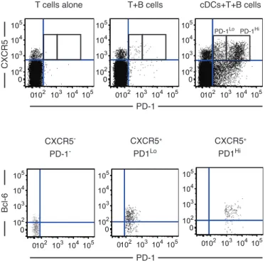
A number of experimental animal studies suggest that dendritic cells are indispensable for initiating a Tfh development program in naïve CD4 T cells (11,12). Therefore, we hypothesized that the relative enrichment of PD-‐1Lo vs. PD-‐1Hi CXCR3+ Tfh-‐like cells in the blood from controller neutralizers might be associated with differential priming by DCs. To evaluate the ability of human myeloid DCs (mDC) to polarize CD4 T cells towards Tfh lineage commitment in vitro, we established a co-‐ culture system with autologous naïve CD4 T cells and naïve B cells from HIV-‐1 negative individuals in the presence or absence of primary mDC isolated from the blood of an allogeneic healthy donor, without addition of exogenous antigens. After 7 days of co-‐culture, maturation of naïve CD4 T cells into cells with a Tfh-‐like phenotype was analyzed by flow cytometry. As shown in Figure 2A, and consistent with previous studies (25), CD4 T cells were unable to efficiently upregulate the expression of the Tfh markers, CXCR5 and PD-‐1, when cultured in media or in the presence of naïve B cells alone. However, when mDC were added to the co-‐culture, a significant proportion of CXCR5+ PD-‐1+ T cells was detected on day 7 of culture (Figure 2A), suggesting de novo differentiation of naïve CD4 T cells into Tfh-‐like cells
in vitro. Similarly to cells from peripheral blood, CXCR5+ T cells induced in this assay could also be subdivided into PD-‐1 Lo and PD-‐1 Hi subpopulations (Figure 2A). Further phenotypical analysis revealed that both PD-‐1 Lo and PD-‐1 Hi CXCR5+ T cells also expressed additional Tfh markers such as Bcl-‐6 and ICOS (26,27) (Figure 2B and Supplemental Figure 2A), while expression of FoxP3, a marker for follicular regulatory T cells (Tfr) (28), was low (Supplemental Figure 2A). Of note, most DC-‐ primed CXCR5+ PD-‐1+ T cells expressed the chemokine receptor CXCR3 (Supplemental Figure 2A). To examine whether the primed T cells generated in our co-‐culture system were more similar to Th1 or Tfh1 cells, we analyzed intracellular expression levels of the Th1 transcription factor Tbet (29). As shown in Supplemental Figure 2A-‐2B, Tbet was detected in CXCR5+ PD-‐1+ T cells, but levels of expression were much lower than in bona fide Th1 cells primed with LPS-‐pulsed mDC (Supplemental Figure 2B). Therefore, our data indicate that human primary mDC are required to efficiently prime naïve CD4 T cells into CXCR5+ PD-‐1+ Tfh1-‐like cells in the presence of B cells in vitro. Importantly, in vitro generation of CXCR3+ ICOS+ PD-‐1Lo and PD-‐1Hi Tfh1-‐like cells was also observed in the presence of primary mDC isolated from lymph node biopsies from HIV-‐1 negative donors (Supplemental Figure 2C).
Using this experimental system, we next determined the ability of mDC from different cohorts of HIV-‐1-‐infected individuals to prime Tfh-‐like cells in vitro. To this end, we evaluated the Tfh-‐priming potential of mDC from controller neutralizers (NT) and non-‐neutralizers (NN), Viremic (CP) and HAART-‐treated (H) chronically HIV-‐1-‐infected individuals. A cohort of HIV-‐1 negative persons (NG) was also included for comparative purposes. Subsequently, mDC were isolated from all study groups and their in vitro Tfh priming abilities were tested by co-‐culture assays using identical allogeneic naïve CD4 T and B cells from HIV-‐1 negative donors. As shown in Figure 2C, generation of PD-‐1Hi Tfh-‐like cells was less efficient in the presence of mDC from HIV-‐1 infected donors, compared to uninfected individuals, but mDC from controller neutralizers and non-‐neutralizers seemed equally effective in inducing cells with a PD-‐1Hi Tfh phenotype. However, mDC from controller neutralizers
induced higher proportions of PD-‐1Lo Tfh-‐like cells compared to controller non-‐ neutralizers and chronically infected, viremic or HAART-‐treated individuals (Figure 2C). Notably, the ratios of PD-‐1Lo vs. PD-‐1Hi Tfh-‐like cells generated in the presence of mDC from controller neutralizers were higher than in assays with mDC from all other study cohorts, and significantly exceeded the ratios of PD-‐1Lo vs. PD-‐1Hi cells generated in the presence of healthy individuals (Figure 2D). Notably, preferential induction of CXCR5+ PD-‐1Lo T cells by mDC from HIV-‐1 controller neutralizers was not associated with significantly higher levels of specific cytokines (Supplemental Figure 2D), despite a tendency for higher detection of IL-‐6, IL-‐10 and IL-‐21. Similarly, cultures performed with mDC from controller neutralizers did not show specific phenotypic alterations in co-‐cultured B cells (Supplemental Figure 3A-‐3B), or significant changes in the immunoglobulin (Ig) classes secreted by B cells (Supplemental Figure 3C). However, the contribution of class G Ig to the total amount of secreted Ig was highest in assays with mDC from controller neutralizers (Supplemental Figure 3C). Together, these data indicate that primary mDC from controller neutralizers have superior abilities to prime PD-‐1 Lo CXCR3+ CXCR5+ Tfh-‐ like cells in vitro, compared to mDC from controller non-‐neutralizers.
Distinct transcriptional signatures in mDC from controller neutralizers
To better understand molecular mechanisms underlying enhanced abilities of mDC from controller neutralizers to prime Tfh-‐like cells, we analyzed transcriptional profiles of mDC from 4 controller neutralizers compared to cells from 4 controller non-‐neutralizers. These analyses revealed significant differences (FDR-‐adjusted p<0.05) in the expression of 930 genes in mDC from controller neutralizers (Figure 3A, Supplemental Table 3), as determined by DESeq2 (30). Overall, pathways upregulated in controller neutralizers shared a high number of genes with interconnected functions in innate and adaptive immunity, consistent with the diverse role of DC in orchestrating antiviral immune defense (Figure 3B). Interestingly, we observed that pathways previously associated with Tfh priming, such as ICOSL-‐ICOS interactions (p=3.98x10-‐11) and CD40-‐dependent signaling (p=0.00079) (31) (Figure 3B), were enriched in transcriptional signatures of mDC
from controller neutralizers. In addition, genes encoding for signaling components of the IL-‐6 (p=0.001) and IL-‐1 (p=9.54x10-‐6) pathways, critical for functional development of Tfh cells and the inhibition of alternative T helper cell lineages (32,33), were also significantly upregulated in mDC from controller neutralizers (Figure 3B), as were transcripts involved in general DC maturation and activation, features of mDC previously involved in improved Tfh generation (34). Consistent with these observations, biocomputational analysis identified signaling through CD40 (p=1.05x10-‐23) or CD86 (p=5.05x10-‐10), secretion of multiple effector cytokines such as IFNα (p=3.98x10-‐26), TNFα(1.96 x 10-‐16), IL-‐1β (p=4.55 x 10-‐13), IL-‐6 (p=3.2x10-‐11), IL-‐10 (p=4.5x10-‐15) and IL-‐18 (p=2.89x10-‐17) (Figure 3C), and transcription factors and signal mediators such as NFKB1 (p=5.88x10-‐16), STAT3 (p=9.47x10-‐15), IRAK4 (p=7.45x10-‐13) and ID3 (p=3.97x10-‐21) as putative upstream regulators of transcriptional signatures in mDC from controller neutralizers (Figure 3C-‐D). In contrast, transcriptional pathways down-‐regulated in DC from controller neutralizers had no immediate connection to Tfh priming and DC function (Figure 3B). To further explore these observations, we performed functional in vitro co-‐ culture assays between DC and T/B cells using a trans-‐well experimental system. As previously described for CXCR5+ CXCR3-‐ PD-‐1+ cells (31,35), in vitro priming of CXCR3+ CXCR5+ PD-‐1Lo Tfh-‐like cells seemed to require direct contact with mDC (Supplemental Figure 4A). Flow cytometry analysis revealed that among all costimulatory molecules tested, ICOS-‐L and more significantly CD40 tended to be expressed at higher levels on mDC from controller neutralizers (Figure 3E, Supplemental Figure 4B), consistent with the described transcriptional profiling experiments. Importantly, inhibition of cellular communication via CD40/CD40L (Figure 3F) significantly abrogated the priming of Tfh-‐like cells by mDC, although such interactions alone did not explain the preferential induction of PD-‐1Lo by mDC from controller neutralizers. Collectively, these data indicate that mDC from controller neutralizers are characterized by transcriptional signatures associated with key pathways involved in Tfh priming.
CXCR5+ CXCR3+ PD-1Lo CD4 T cells are detectable in lymphoid tissues in vivo.
We next investigated the presence of CXCR5+ CXCR3+ PD-‐1Lo Tfh-‐like cells in peripheral blood and tissue samples isolated directly ex vivo in samples from HIV-‐1-‐ negative persons (Supplemental Figure 1A-‐1B). We found that similarly to peripheral blood, the proportions of CXCR5+ CXCR3+ CD4 T cells approximated those of CXCR5+ CXCR3-‐ CD4 T cells in the lymph node, and that the majority of CXCR5+ CD4 T cells in these tissues were PD-‐1Lo, independently of CXCR3 co-‐ expression (Supplemental Figure 1A-‐1B). In contrast, in tonsil tissue, PD-‐1Lo cells were mostly detected in CXCR5+ CXCR3+ cells, while CXCR5+ CXCR3-‐ CD4 T cells consisted almost entirely of PD-‐1Hi cells. Notably, CXCR3-‐ PD-‐1Hi cells were the dominant cell subset within CXCR5+ lymphocytes in tonsils, while CXCR3+ PD-‐1Lo cells represented the largest cell compartment within CXCR5+ CD4 T cells from blood and lymph nodes (Supplemental Figure 1B). Moreover, within lymph nodes and tonsils, Bcl-‐6 was upregulated in all CXCR5+ CD4 T cells, irrespective of their CXCR3 or PD-‐1 expression levels, as opposed to CXCR5+ CD4 T cells from peripheral blood, in which Bcl-‐6 levels were very low and indistinguishable from those of total CD4 T cells, consistent with prior results (3) (Supplemental Figure 5). ICOS was more strongly expressed in PD-‐1Hi Tfh-‐like cells compared to PD-‐1Lo cells across all tissues (Supplemental Figure 5). Together, our data indicate that CXCR5+ CXCR3+ PD-‐1Lo cells are present in vivo in lymphoid tissues.
CXCR3+ PD-1Lo Tfh-like cells support B cell activation and differentiation.
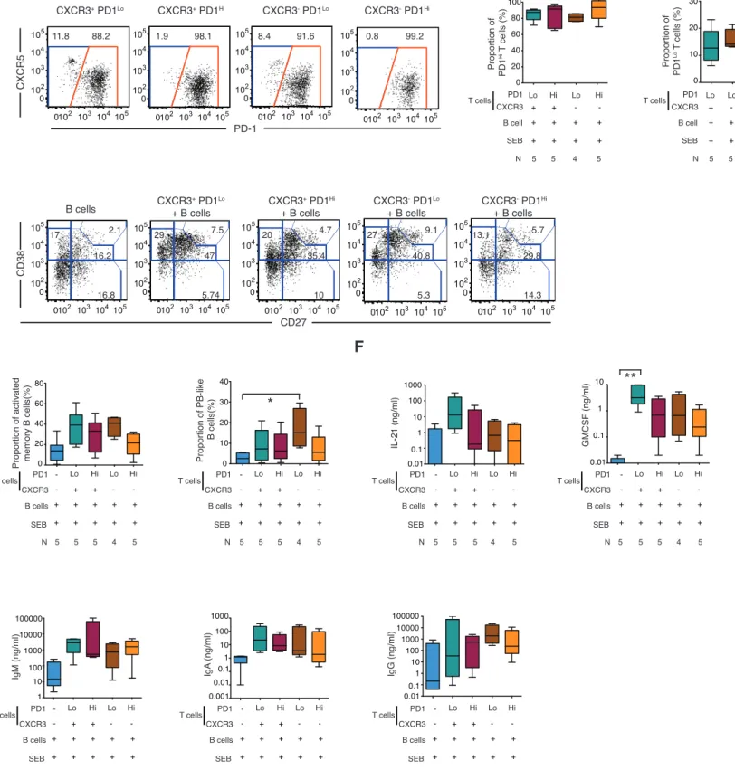
Previous studies defined CXCR3+ pTfh as poor supporters of B cell maturation based on their weak ability to stimulate class switching to IgG in B cells in vitro (15,16). However, our results suggest that the presence of CXCR3+ PD-‐1Lo Tfh-‐like cells might be beneficial for the development of neutralizing Abs against HIV-‐1 in controllers. To better understand the functional characteristics of CXCR3+ PD-‐1Lo Tfh-‐like cells, different pTfh cell subsets from the blood of HIV negative individuals (Supplemental Figure 1A-‐1B) were sorted and cultured with autologous total B cells in the presence of staphylococcal endotoxin B (SEB). After 6 days of culture, differentiation
of pTfh subsets, maturation of B cells, cytokine secretion profiles and levels of secreted immunoglobulins were analyzed. As shown in Figure 4A-‐4B, most CXCR3+ and CXCR3-‐ CXCR5+ PD-‐1Lo Tfh-‐like cells differentiated into PD-‐1Hi Tfh-‐like cells in the presence of antigens and autologous B cells; however, some of the CXCR3+ and CXCR3-‐ PD-‐1Lo Tfh-‐like cells seemed to be able to retain their original PD-‐1Lo phenotype during co-‐culture (Figure 4A, 4C). In addition, significant levels of proliferation were detected on all subsets of pTfh cells present on day 6 of culture (Supplemental Figure 8A). Co-‐culture of B cells with any of the pTfh cell subsets was accompanied by a tendency for decreased proportions of resting memory B cells (Figure 4D, Supplemental Figure 6A), and a marked upregulation of CD38 and CD27, leading to elevated frequencies of CD38Int CD27Int activated memory B cells, CD38Hi CD27Hi plasmablast-‐like B cells (Figure 4D-‐4E) and CD38Hi CD27-‐ transitional B cells (Supplemental Figure 6A). Notably, PD-‐1Lo pTfh tended to be more efficient than PD-‐ 1Hi cells in inducing activated memory B cells (Figure 4E). Overall, PD-‐1Lo and PD-‐1Hi CXCR3+ pTfh cells were able to induce comparable level of phenotypic maturation in B cells as CXCR3-‐ pTfh subsets. Interestingly, PD-‐1Lo CXCR3+ pTfh seemed to secrete higher levels of IL-‐21 and, more significantly, of GM-‐CSF, IL-‐6 and IL-‐10 in a six-‐day co-‐culture assay with total B cells and SEB (Figure 4F and Supplemental Figure 6B). In agreement with previous studies, only CXCR3-‐ pTfh were able to efficiently induce full class switching to IgG in B cells, dominated by IgG1 (Figure 4G, Supplemental Figure 6C) (16-‐18). In contrast, PD-‐1Lo and PD-‐1Hi CXCR3+ cells seemed capable of stimulating B cells to produce IgM and IgA, IgG3 but not IgG1, as previously reported (16-‐18), suggesting a more limited ability of these pTfh subsets to support Ig class switching to IgG in our six-‐day co-‐culture assays (Figure 4G). Together, these data indicate that both CXCR3+ and CXCR3-‐ PD-‐1Lo pTfh subsets mature into PD-‐1Hi cells upon Ag stimulation, and that CXCR3+ pTfh cells display distinct, but partially overlapping functional abilities to support B cells compared to conventional CXCR3-‐ pTfh. These findings suggest that both CXCR3+ and CXCR3-‐ pTfh might participate in the development of Ab breadth at different levels.
While most PD-‐1Lo Tfh-‐like cells differentiated into PD-‐1Hi upon antigenic stimulation, we observed a small proportion of PD-‐1Lo Tfh-‐like cells that were able to retain their original CXCR5+ PD-‐1Lo phenotype during co-‐culture with B cells and SEB, suggesting self-‐renewal or homeostatic proliferation. To test this possibility, we first analyzed the expression of memory cell markers (Figure 5A-‐5B) on different Tfh-‐like subsets from human blood and lymphoid tissues. As shown in Figure 5C, PD-‐1Lo and PD-‐1Hi cells from both CXCR3+ and CXCR3-‐ peripheral Tfh were dominated by CCR7+ CD45RO+ central-‐memory T cells; in contrast, total CD4 T cells were dominated by naïve cells. In tonsils and lymph nodes, CXCR5+ CXCR3+ PD-‐ 1Lo CD4 T cells also preferentially exhibited a central-‐memory phenotype, while PD-‐ 1Hi cells, specifically within the CXCR3-‐ T cell pools, included higher proportions of CCR7-‐ CD45RO+ effector-‐memory cells (Figure 5C). Importantly, we observed that PD-‐1Hi cells from all tissue compartments consisted mostly of CD45RO+ cells, while both CXCR3+ and CXCR3-‐ PD-‐1Lo Tfh populations contained larger and more distinct populations of CD45RO-‐ naïve (NA)-‐like cells (Figure 5B). Subsequent experiments demonstrated that many of these CD45RO-‐ Tfh expressed CD95, a memory cell marker that when expressed on otherwise naïve-‐appearing CD4 T cells defines a highly immature population of extremely long-‐lived memory CD4 T cells with stem cell-‐like properties (Figure 5B) (36). In fact, in contrast to total CD4 T cells (Figure 5D), a large proportion of NA-‐like cells present in the different CXCR5+ subsets from the analyzed tissue compartments exhibited a CD95+ T stem cell memory phenotype, and the relative proportions of these CD45RO-‐ CD95+ cells tended to be higher within the PD-‐1Lo populations (Figure 5D). Importantly, a similar memory subset distribution was observed between circulating Tfh-‐like cell populations from HIV-‐1 controller neutralizers and non-‐neutralizers (Supplemental Figure 7A). Interestingly, frequencies of stem cell-‐memory T cells within the CXCR3+ PD-‐1Lo cell populations appeared to be increased in HIV-‐1 controller neutralizers (Supplemental Figure 7B). Therefore, CXCR5+ PD-‐1Lo CD4 T cells seemed to contain higher proportions of more immature stem cell-‐memory T cells.
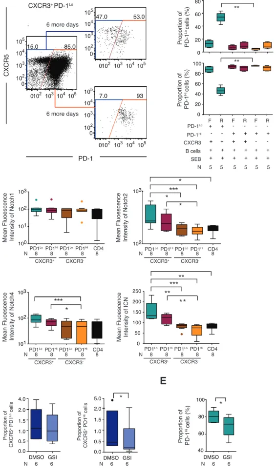
To investigate stem cell-‐like precursor properties of PD-‐1Lo Tfh-‐like cells, we analyzed the developmental fate of CXCR5+ CXCR3+ PD-‐1Lo in serial co-‐culture experiments. For this purpose, CXCR5+ CXCR3+ PD-‐1Lo cells that maintained their original phenotype during an initial six-‐day co-‐culture assay with B cells and SEB were sorted and exposed to six additional days of culture with B cells and SEB. Interestingly, a considerable proportion of sorted CXCR5+ CXCR3+ PD-‐1Lo CD4 T cells that remained PD-‐1Lo during the initial culture also retained their original phenotype (Figure 6A-‐6B) during secondary culture, although a proportion of these cells was able to differentiate into PD-‐1Hi cells (Figure 6A-‐6B). In contrast, the phenotype of sorted PD-‐1Hi pTfh remained unchanged after the primary and secondary cultures, independently of CXCR3 expression (Figure 6B). Importantly, cells capable of maintaining a PD-‐1Lo phenotype during primary (Supplemental Figure 8B-‐8C) and secondary cultures (Supplemental Figure 8D) showed signs of active proliferation. This suggests that even though the majority of PD-‐1Lo CXCR5+ cells ultimately transition to PD-‐1Hi cells, a proportion of these cells appear to maintain their original phenotype while proliferating, consistent with homeostatic self-‐renewal, a key component of a stem cell-‐like functional profile.
To investigate mechanisms that may dictate the ability of PD-‐1Lo pTfh to repopulate PD-‐1Hi pTfh cells, we focused on the Notch signaling cascade, which has previously been implicated in regulating the developmental fate of organ-‐specific stem cells (37,38), memory T cells (39-‐41) and T follicular helper cells (42). We observed that Notch receptors 2 and 4, as well as the Notch active intracellular domain, were upregulated on CXCR5+ CXCR3+ PD-‐1Lo cells and, to a lesser extent, on CXCR5+ CXCR3+ PD-‐1Hi CD4 T cells from peripheral blood when compared to total CD4 T cells (Figure 6C). Moreover, inhibition of Notch signaling by gamma secretase inhibitors (GSI) in naïve T cells co-‐cultured with autologous naïve B cells and allogenic mDC significantly inhibited the generation of PD-‐1Hi but not PD-‐1Lo Tfh-‐ like cells in vitro (Figure 6D). A similar inhibition in the generation of PD-‐1Hi cells was observed when Notch signaling was blocked in sorted CXCR3+ PD-‐1Lo pTfh co-‐ cultured with autologous B cells and SEB (Figure 6E), suggesting that Notch
signaling is critical in regulating differentiation of PD-‐1Lo CXCR5+ CD4 cells into PD-‐ 1Hi cells. Together, these data suggest that circulating CXCR5+ CXCR3+ PD-‐1Lo CD4 T cells represent a distinct population of progenitor cells able to repopulate the PD-‐1Hi effector pTfh cells in a Notch-‐dependent fashion.
Discussion
It is highly likely that the long-‐lasting induction of HIV-‐1-‐specific bnAbs by vaccines will require support from Tfh or Tfh-‐like cells. In previous studies, peripheral CXCR5+ CXCR3-‐ PD-‐1+ cells were found to correlate strongly with the development of HIV-‐1-‐specific bnAbs in patients with progressive diseases (16); however, such patients had continuous high-‐levels of HIV-‐1 replications and elevated levels of immune activation that hardly reflect conditions for immune induction reproducible in uninfected recipients of a prophylactic HIV-‐1 vaccine candidate. Here, we used HIV-‐1 controllers, a group of patients widely used for investigation of HIV-‐1 immune defense mechanisms in previous studies (43-‐45), as a model for studying HIV-‐1-‐ specific antibodies with increased neutralizing breadth in the absence of high-‐levels of viremia. Our observations demonstrate that relative ratios between CXCR5+ CXCR3+ PD-‐1Lo and CXCR3+ CXCR5+ PD-‐1Hi Tfh-‐like cells in the blood from HIV-‐1 controllers correlated with increased neutralizing breadth of HIV-‐1 antibodies in this particular patient population. In contrast, peripheral CXCR5+ CXCR3-‐ CD4 T cells were unrelated to HIV-‐1-‐specific antibodies with broader neutralizing breadth in these patients, irrespectively of PD-‐1 expression. Interestingly, CXCR5+ CXCR3+ PD-‐ 1Lo Tfh-‐like cells expressed comparable levels of Tfh markers such as ICOS and Bcl-‐6 in lymphoid tissue and peripheral blood as conventional CXCR5+ CXCR3-‐ PD-‐1Hi Tfh/pTfh. Moreover, CXCR5+ CXCR3+ PD-‐1Lo Tfh-‐like cells effectively produced cytokines for stimulation of B cell development and were able to differentiate into PD-‐1Hi Tfh-‐like cells, although they were inferior to CXCR3-‐ Tfh-‐like cells in inducing B cell Ig class switching to IgG1 in our co-‐culture assays. Together, these studies suggest that at least under conditions of low viral loads and immune activation, CXCR5+ CXCR3+ PD-‐1Lo cells may make important contributions to the induction and/or maintenance of HIV-‐1 antibody responses with broad neutralizing breadth. Dendritic cells in HIV-‐1 controllers seem to have a distinct functional and phenotypic profile that can contribute to the induction of highly functional HIV-‐1-‐ specific T cell responses. These abilities have been associated with an altered
expression profile of immunoregulatory receptors (46), and with enhanced abilities for HIV sensing and innate immune recognition (47). However, the impact of dendritic cells from these patients on Tfh development is still unclear, and investigations of dendritic cells on induction of Tfh have mostly been limited to animal studies (11,48,49) or experiments with in vitro generated monocyte-‐derived DC (MDDC) (25,33), rather than primary dendritic cells. To our knowledge, this is the first study to analyze the effects of primary dendritic cells on Tfh priming in a rare group of HIV-‐1 controllers who are able to generate broader neutralizing breadth in the absence of high-‐level viral replications. Our results demonstrate that when co-‐cultured with B cells, mDC from these patients have a preferential ability to induce Tfh-‐like cells with low expression of PD-‐1, and a chemokine receptor expression profile that includes co-‐expression of CXCR3 and CXCR5. Notably, we found that Tfh priming by mDC from controllers seems to be associated with increased transcriptional expression of genes involved in functional DC activity and in Tfh priming. Interestingly, secretion of specific cytokines, such as IL-‐6, a cytokine with a recognized role in Tfh priming by dendritic cells (33), tended to be higher in cultures with cells from neutralizers and was also activated in transcriptional signatures of mDC from these patients. Moreover, enhanced cell-‐contact dependent interactions mediated by CD40 and possibly ICOSL, which were expressed at higher levels in mDC from controller neutralizers (Figure 3E) and have previously been implicated in Tfh generation (50), may play an important role in this context. Notably, in our experimental in vitro system, primary mDC did not efficiently prime CXCR3-‐ CXCR5+ Tfh-‐like cells, suggesting that the priming of this subset is more independent of dendritic cells or might represent a different stage of T cell differentiation. Collectively, our findings highlight the importance of mDC as critical mediators of Tfh lineage commitment and suggest that manipulation of mDC might be an attractive strategy to improve Tfh differentiation in future HIV-‐1 vaccine studies.
Unexpectedly, this study showed that the emergence of HIV-‐1 antibodies with broader neutralizing breadth in HIV-‐1 controllers was associated with Tfh-‐like cells
expressing CXCR3, a surface marker denoting cells considered less efficient in supporting B cell immunoglobulin class-‐switching in previous in vitro studies (15-‐ 18). While our observations confirm that CXCR3+ Tfh-‐like cells are inferior to CXCR3-‐ Tfh-‐like cells in supporting IgG1 class switching, we did demonstrate that CXCR3+ Tfh-‐like cells have abilities to induce class switching to IgG3 and to alternative Ig subtypes, to secrete high levels of B cell-‐supporting cytokines and to induce phenotypical maturation of B cells at equivalent levels as CXCR3-‐ Tfh-‐like cells, at least when analyzed over a longer, six-‐day co-‐incubation period with total B cells and SEB stimulation, and not just in a short-‐term culture assay lasting for 48 hours (15). Together, these results suggest that CXCR5+ CXCR3+ Tfh-‐like cells can contribute to helping B cell responses, and that CXCR5+ CXCR3-‐ and CXCR5+ CXCR3+ pTfh might have distinct, but partially overlapping roles for supporting B cells. Consistent with a role as circulating memory cells, CXCR5+ CXCR3+ PD-‐1Locells in HIV-‐1 controllers may primarily contribute to maintaining long-‐term survival of B cells producing more broadly cross-‐reactive antibodies, and not to the initial priming of such immune responses, which is likely to preferentially occur in lymph node germinal centers and may largely be independent of circulating Tfh-‐like cells. A preferential role of CXCR3+ Tfh-‐like cells in maintaining or expanding pre-‐existing B cell responses, but not in the initial priming of new antigen-‐specific B cells, was also hypothesized in alternative contexts associated with limited antigen exposure, such as in recipients of vaccines against Influenza (19). In contrast, in HIV-‐1 progressors with continuously-‐ongoing high-‐level viremia, maintenance of HIV-‐1-‐ specific B cells may more significantly depend on the presence of elevated amounts of circulating viral antigens. Notably, our data do not exclude the possibility that CXCR3-‐ pTfh, which were also significantly elevated in controller neutralizers (Figure 1B), participate individually or in collaboration with CXCR3+ pTfh in generating or maintaining more broadly-‐neutralizing antibody-‐secreting B cells in these patients.
A large number of studies suggest that circulating CXCR5+ T cells are memory Tfh cells (3), but the dynamics of their development and long-‐term survival remain
poorly understood. Our data suggest that phenotypically-‐defined subsets of circulating CXCR5+ CD4 T cells may not only differ by effector functions, but also involve a developmental and maturational aspect. A linear developmental hierarchy from long-‐lasting immature central-‐memory and stem cell-‐memory to short-‐lived effector-‐memory cells is experimentally well validated within the CD8 T cell memory compartment (36), and has also been suggested to occur with CD4 T cells (51). The results presented here suggest that a similar developmental hierarchy also exists within the pools of circulating Tfh-‐like cells, and that PD-‐1Lo Tfh represent a long-‐lived precursor cell population that can replenish the more mature and short-‐ lived PD-‐1Hi effector Tfh cells. Interestingly, these PD-‐1Lo Tfh-‐like cells included a substantial proportion of CD45RO-‐ naive-‐like T cells, a cell population that was excluded in prior studies (15-‐17), but includes a fraction of highly-‐immature memory T cells with increased stem cell-‐like properties (36). The ability to maintain a larger proportion of PD-‐1Lo memory Tfh-‐like cells that are enriched for a T memory stem cell phenotype relative to PD-‐1Hi pTfh could represent a distinct aspect of HIV-‐1 controllers with more broadly neutralizing antibody responses, and appears to separate these patients from HIV-‐1 progressors in which high-‐level viremia may lead to more differentiated and possibly more exhausted PD-‐1Hi pTfh cell populations (52). Notably, an optimal balance between immature precursor cells and more committed effector cells has previously also been found to be critical for regulating CD8 T cell immunity (53). In addition, a higher proportion of total immature memory CD8 and CD4 T cells are associated with improved prognosis in chronic HIV-‐1 infection (54) and can be enriched in the absence of viremia and immune activation in treated HIV-‐1-‐positive individuals (55). Interestingly, we found evidence that Notch signaling, previously mostly shown to regulate hematopoietic and thymic precursor cell differentiation (37,38), is also involved in regulating the transition of long-‐lasting PD-‐1Lo into short-‐lived PD-‐1Hi Tfh-‐like cells. This suggest that stem cell pathways, previously recognized in the context of traditional organ-‐specific stem cells, may also contribute to regulation and fate decisions within the memory Tfh pools, and may offer molecular targets for
selectively influencing memory Tfh development. A role for Notch in Tfh memory evolution is also consistent with the recent observation that Notch signaling was necessary for Tfh differentiation in experimental animal models (42).
Taken together, these studies demonstrate that the CXCR5+ CXCR3+ PD-‐1Lo Tfh-‐like population serves as precursor cells for PD-‐1Hi cells, supports B cell development through cytokine secretion, is associated with maintenance of more broadly-‐ neutralizing HIV-‐1 antibody responses in the absence of high-‐level HIV-‐1 viremia and can be selectively primed by dendritic cells from HIV-‐1 controllers with neutralizing antibody responses. These studies may help to delineate mechanisms for inducing HIV-‐1 antibodies with higher levels of neutralizing breadth by prophylactic vaccines.
Methods
Study Participants
HIV-‐1 controllers who had maintained < 1500 copies/ml HIV-‐1 viral loads (VL) for a median of 5 years (range 2-‐14) in the absence of antiretroviral therapy, with (neutralizers, NT, n=25, median VL 123 copies/ml, range 20-‐1400 copies/ml; median CD4 counts 769.5 cells/ml, range 418-‐1545 cells/ml) or without (non-‐ neutralizers, NN, n=20; median VL 75 copies/ml, range 48-‐1470 copies/ml; median CD4 counts 844.5 cells/ml, range 407-‐2117 cells/ml) neutralizing antibodies against Tier-‐2/3 HIV-‐1 viruses, untreated chronic progressors (CP, n=14, median VL 28352.5 copies/ml, range 2368-‐90500 copies/ml; median CD4 T cell counts 313 cells/ml, range 88-‐955 cells/ml), ART-‐treated chronically HIV-‐1-‐infected patients with suppressed HIV-‐1 viremia (H, n=14, median VL <49 copies/ml; median CD4 T cell counts 490 cells/ml, range 183-‐964 cells/ml) and HIV-‐1 seronegative healthy persons (NG, n=14) were recruited for this study. Samples of mononuclear cells extracted from inguinal lymph nodes were obtained by surgical excision from HIV-‐1 negative study persons. Mononuclear samples from human tonsil tissue of HIV-‐1 negative individuals who underwent routine tonsillectomies were obtained as previously described (52).
Analysis of the neutralizing breadth of HIV-1-specific antibodies
As previously described (56), HIV-‐1 neutralization breadth was measured in a TZM-‐ bl cell-‐based pseudovirus neutralization assay against a panel of Env-‐pseudoviruses derived from 9 Clade B Tier 2 and two Tier 3 neutralization sensitivities: AC10.0.29*, RHPA4259.7*, THRO4156.18*, REJO4541.67*, WITO4160.33*, TRO.11*, SC422661.8*,QH0692.42*, CAAN5342.A2# and Tier 3: PVO.4* and TRJO4551.58*. The Clade B and C isolates are denoted by the superscripts * and # respectively. Neutralization was defined as at least 50% inhibition of infection at a 1:20 dilution. The neutralization breadth was defined as the percentage of the 11 isolates neutralized by each plasma sample. All samples were screened for non-‐HIV-‐1-‐ specific neutralization using murine leukemia virus-‐pseudotyped virions