HAL Id: inserm-00277129
https://www.hal.inserm.fr/inserm-00277129
Submitted on 6 Apr 2009
HAL is a multi-disciplinary open access archive for the deposit and dissemination of sci-entific research documents, whether they are pub-lished or not. The documents may come from teaching and research institutions in France or abroad, or from public or private research centers.
L’archive ouverte pluridisciplinaire HAL, est destinée au dépôt et à la diffusion de documents scientifiques de niveau recherche, publiés ou non, émanant des établissements d’enseignement et de recherche français ou étrangers, des laboratoires publics ou privés.
No clear link between VKORC1 genetic polymorphism
and the risk of venous thrombosis or peripheral arterial
disease.
David Smadja, Marie-Anne Loriot, Lucia Hindorff, Lucille Mellottee, Pascale
Gaussem, Joseph Emmerich
To cite this version:
David Smadja, Marie-Anne Loriot, Lucia Hindorff, Lucille Mellottee, Pascale Gaussem, et al.. No clear link between VKORC1 genetic polymorphism and the risk of venous thrombosis or peripheral arterial disease.. Thrombosis and Haemostasis, Schattauer, 2008, 99 (5), pp.970-972. �10.1160/TH07-12-0742�. �inserm-00277129�
No clear link between VKORC1 genetic polymorphism and the risk of venous thrombosis or
peripheral arterial disease
David M. Smadja1-3, Marie-Anne Loriot1,2,4, Lucia A. Hindorff5, Lucille Mellottee1,4, Pascale Gaussem1-,3,
Joseph Emmerich1-3
1Université Paris Descartes, Paris, France; 2AP-HP, Hôpital européen Georges Pompidou, Paris, France; 3INSERM Unité 765, Faculté
de Pharmacie, Paris, France; 4INSERM Unité 775, Paris, France; 5Department of Epidemiology, School of Public Health, University
of Washington, Seattle, Washington, USA
Correspondence to: David M. Smadja
Université Paris Descartes, Faculté de Pharmacie INSERM U765 Avenue de l’Observatoire 75006 Paris, France Tel.: +33 1 56 09 3933, Fax: +33 1 56 09 3913 E-mail: david.smadja@egp.aphp.fr Received December 17, 2007
Accepted after minor revision March 14, 2008 Prepublished online April 9, 2008
doi:10.1160/TH07-12-0742
Thromb Haemost 2008; 99: 970–972 Dear Sir,
Vitamin K, discovered in the 1930s, functions as a cofactor for the posttranslational carboxylation of glutamate residues (1). The vitamin K-epoxide reductase complex (VKOR) allows vit-amin K epoxide to be converted back into reduced vitvit-amin K. Its activity is the rate-limiting step of vitamin K-dependent protein gamma-carboxylation. Gamma-carboxylation is essential for the biological activity of clotting factors (factors II, VII, IX and X, and proteins C, S, and Z). Genetic and acquired disorders of some of these factors are linked to venous thromboembolism (VTE). Matrix Gla protein and bone Gla protein (osteocalcin), two vitamin K-dependent proteins involved in calcium home-ostasis, have been implicated in the pathogenesis of athero-sclerosis, myocardial infarction and stroke (2, 3). Moreover,
VKOR has been implicated in angiogenesis, a process with an
important role in cardiovascular disease (4). Studies of two families showing vitamin K-dependent clotting factor deficien-cy type 2 (VKCFD2) permit to identify the VKOR subunit 1 (VKORC1) gene. Another four patients resistant to vitamin K an-tagonists (VKA) point to VKORC1 as a candidate locus at which certain mutations or polymorphisms confer resistance to phar-macotherapy (5). VKORC1 as a target of coumarin-based drugs was confirmed in several single patients with coumarin resis-tance, showing heterozygous mutations in VKORC1 exhibiting impaired coumarin inhibition (5, 6). Common VKORC1 poly-morphisms have since been found to affect the VKA dose re-sponse and warfarin dose requirements (7–10). The frequency of these polymorphisms differs among ethnic populations (8).
Various nomenclature for VKORC1 haplotypes were pro-posed : VKORC1*2 haplotype, strictly corresponds to the haplo-type combination called “A” by Rieder’s study (7), while
VKORC1*1, *3 and *4 haplotype, correspond to the haplotype
combination called “B”. Two polymorphisms, the C1173T and
G-1639A, are in completelinkage disequilibrium and can be used to distinguish between the haplotype combinations A and B.
The simple genotypingof VKORC1 G-1639Aor C1173T with the CYP2C9*3 polymorphism could predicta high risk of overdose before initiation of anticoagulationwith acenocouma-rol (9–12). Indeed, muted allele A or T (G-1639Aor C1173T) ex-plainsabout one third of the variability of the pharmacologic re-sponse(37% of factor VII decrease and 30% of international nor-malised ratio [INR] change) (9). Here we studied the functional promoter polymorphism of VKORC1 G-1639A (rs9923231) to identify the major VKORC1 haplotype 2 (group A of Rieder’s classification) (7) and “non VKORC1*2” haplotype (group B). The G allele of the G-1639A SNP corresponds to the group B
VKORC1 haplotype and the A allele to the group A VKORC1
ha-plotype.
Two recent studies showed a link between VKORC1 haplo-types and the risk of cardiovascular disease. The first, a Chinese case-control study, suggested that VKORC1 B/B haplotype was a major risk factor for coronary heart disease (CHD), stroke, and aortic dissection, probably through the role of osteocalcin in vas-cular calcification (13). The second, a French case-control study, showed a protective effect of the VKORC1 A/A haplotype on VTE (14). However, several other studies have failed to show any association between the VKORC1 genotype and the risk of VTE, CHD or stroke (15, 16). To test the hypothesis that VKORC1- dependent effects on the coagulationcascade and vascular cal-cification would contribute to susceptibilityto vascular diseases, we investigated the previously unexplored association of
VKORC1 A and B haplotypes with peripheral arterial disease
(PAD). Conflicting findings prompted us to seek also a link be-tween VKORC1 A and B haplotypes and VTE.
We studied two matched case-control studies, namely the Pallas study (PAris Lower Limb Atherothrombosis Study) for PAD and the FARIVE study (FActeurs de RIsques et de récidives
de la maladie thromboembolique VEineuse) for first episodes of
VTE. All the participants gave their writteninformed consent, and the Paris-Cochin Ethics Committee approved the protocols. The PAD cases were consecutive Caucasian men under 70 years of age, who are described in detail elsewhere (17, 18). The FA-RIVE study included men and women with a first proximal ve-nous thrombosis and/or pulmonary embolism. For each case, age- and sex-matched controls were recruited among patients hospitalized during the same period with no history of arterial or venous thrombosis or cancer.
VKORC1 A/B status was determined with the Taqman allelic
discrimination assay (Applied Biosystems) targeting the dis-criminant SNP G-1639A. The primers and probes (available on
Letters to the Editor
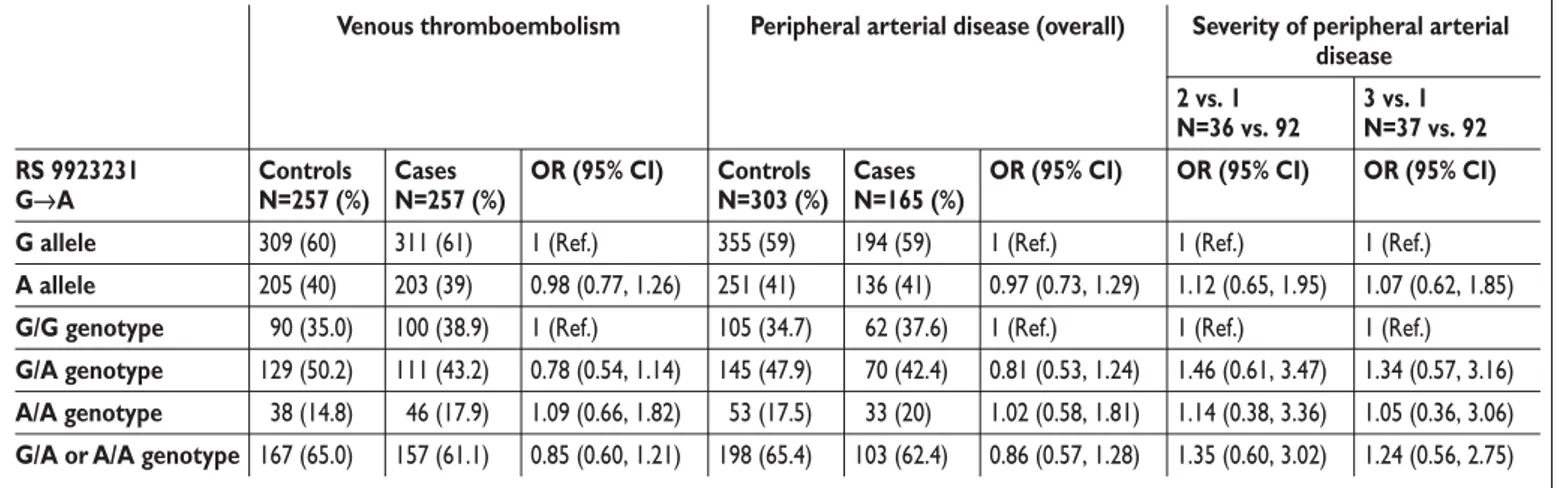
Table 1: Association of the VKORC1 genotype with venous thromboembolism (VTE) and peripheral arterial disease (PAD). The G
al-lele of the G-1639A SNP corresponds to the group B VKORC1 haplotype of Rieder’s classification and the A alal-lele to the group A VKORC1 haplotype.
Venous thromboembolism Severity of peripheral arterial disease 2 vs. 1 N=36 vs. 92 3 vs. 1 N=37 vs. 92 RS 9923231 G→A Controls N=257 (%) Cases N=257 (%)
OR (95% CI) OR (95% CI) OR (95% CI) G allele 309 (60) 311 (61) 1 (Ref.) 1 (Ref.) 1 (Ref.)
A allele 205 (40) 203 (39) 0.98 (0.77, 1.26) 1.12 (0.65, 1.95) 1.07 (0.62, 1.85)
G/G genotype 090 (35.0) 100 (38.9) 1 (Ref.) 1 (Ref.) 1 (Ref.)
G/A genotype 129 (50.2) 111 (43.2) 0.78 (0.54, 1.14) 1.46 (0.61, 3.47) 1.34 (0.57, 3.16)
A/A genotype 038 (14.8) 046 (17.9) 1.09 (0.66, 1.82) 1.14 (0.38, 3.36) 1.05 (0.36, 3.06)
G/A or A/A genotype 167 (65.0) 157 (61.1) 0.85 (0.60, 1.21) 1.35 (0.60, 3.02) 1.24 (0.56, 2.75)
Peripheral arterial disease (overall)
Controls N=303 (%) 355 (59) 251 (41) 105 (34.7) 145 (47.9) 053 (17.5) 198 (65.4) Cases N=165 (%) 194 (59) 136 (41) 062 (37.6) 070 (42.4) 033 (20) 103 (62.4) OR (95% CI) 1 (Ref.) 0.97 (0.73, 1.29) 1 (Ref.) 0.81 (0.53, 1.24) 1.02 (0.58, 1.81) 0.86 (0.57, 1.28)
request) were chosen with Primer Express Software (Applied Biosystems, Courtaboeuf, France). STATA 8 software was used for statistical analyses. Odds ratios (ORs) and 95% confidence intervals (CIs) were estimated by means of conditional logistic regression. The genotype was modelled by assuming either a dominant model (1 or 2 copies of the minor allele) or a model with heterozygotes and homozygotes modelled separately. All analyses adjusted for the matching variables of age and gender. We adjusted only for matching factors, since acquired risk fac-tors are not expected to confound genetic associations except through selection bias.
The VKORC1 genotype frequencies in the VTE and PAD pa-tients and controls are reported in Table 1. The VKORC1 genetic variants were in Hardy-Weinberg equilibrium. The observed fre-quencies of minor allele A in cases and controls of the two studies ranged from 0.39 to 0.41. VKORC1 A were carried by 61.1% of VTE cases and 65% of controls (OR=0.85; 95% CI: 0.6–1.21). The VKORC1 genotype was not associatedwith VTE: the ORs were 0.78 (95% CI: 0.54–1.14) and 1.09 (95% CI: 0.66–1.82), respectively, for VKORC1 A haplotype heterozygos-ity and homozygosheterozygos-ity. This confirmed that the VKORC1 geno-type does not influence the risk of VTE (16). VKORC1 A was car-ried by 62.4% of PAD cases and 65.4% of control subjects (OR=0.86, CI 0.57–1.28). The VKORC1 genotype was not as-sociatedwith PAD: the ORs were 0.81 (95% CI: 0.53–1.24) and 1.02 (95% CI: 0.58–1.28), respectively, for VKORC1 A haplo-type heterozygosity and homozygosity. We also studied the pos-sible influence of the VKORC1 genotype on the severity of PAD, categorized as follows: group 1: intermittent claudication (IC) with a walking distance above 100 m (N=92); group 2: IC with a walking distance below 100 m (N=36); and group 3: critical limb ischemia (N=37). Among cases, the genotype frequencies were not significantly different in group 2 compared with group 1 (OR 1.12, 95% CI 0.69–1.83) or in group 3 compared with group 1 (OR 0.99, 95% CI 0.61–1.62).
VKORC1 could play a role in VTE or PAD by affecting
gamma-carboxylation of vitamin K-dependent proteins, namely clotting factors in VTE and osteocalcin in PAD. In a Chinese study, the VKORC1 B/B haplotype was associated with 1.7– and 1.8-fold increased risks of stroke and CHD, while VKORC1 A/A was associated with protection from VTE in a French population (14). However, these results were not confirmed in other Cau-casian populations (15, 16). The VKORC1 A genotype frequen-cies in the two case-control studies analyzed here are similar to those previouslyreported in European Caucasians (14, 15), but slightly higher than in a recent US study (16). However, differ-ences in the geographic distribution of VKORC1 polymorphisms are unlikely to explain the lack of correlation with PAD and VTE observed here. Our study size was modest, and results were con-sistent with a wide range of possible associations, as evidenced by the wide CIs.
Our results suggest the absence of a clear association be-tween VKORC1 polymorphisms that determine warfarin sensi-tivity and the risk of VTE and PAD. Further investigations are needed to clarify the role of VKORC1 variants in the pathogen-esis of arterial and venous thrombosis, in particular with impli-cations of Gas6 or extra-hepatic Gla protein levels. Interactions with other candidate genes or local environmental factors might explain why VKORC1 gene variants are associated with arterial or venous thrombosis in certain populations.
Acknowledgements
We thank Veronique Remones, Fouad Dali-Ali, Patricia Verrier, Marjorie Starck and Isabelle Martinez for their technical assistance. This work was supported by research grants from Programme Hospitalier de Recherche Clinique FARIVE (PHRC AOR 02057), Fondation pour la Recherche Médicale, Fondation de France and the Leducq foundation (Leducq Trans-atlantic Network of Excellence on Atherothrombosis Research-LINAT). Letters to the Editor
References
1. Cranenburg EC, Schurgers LJ, Vermeer C. Vitamin
K: the coagulation vitamin that became omnipotent. Thromb Haemost 2007; 98: 120–125.
2. Luo G, Ducy P, McKee MD, et al. Spontaneous
cal-cification of arteries and cartilage in mice lacking ma-trix GLA protein. Nature 1997; 386: 78–81.
3. Herrmann SM, Whatling C, Brand E, et al.
Poly-morphisms of the human matrix gla protein (MGP) gene, vascular calcification, and myocardial infarction. Arterioscler Thromb Vasc Biol 2000; 20: 2386–2393.
4. Wang Y, Zhen Y, Shi Y, et al. Vitamin k epoxide
re-ductase: a protein involved in angiogenesis. Mol Cancer Res 2005; 3: 317–323.
5. Rost S, Fregin A, Ivaskevicius V, et al. Mutations in
VKORC1 cause warfarin resistance and multiple co-agulation factor deficiency type 2. Nature 2004; 427: 537–541.
6. Bodin L, Horellou MH, Flaujac C, et al. A vitamin
K epoxide reductase complex subunit-1 (VKORC1) mutation in a patient with vitamin K antagonist resis-tance. J Thromb Haemost 2005; 3: 1533–1535.
7. Rieder MJ, Reiner AP, Gage BF, et al. Effect of
VKORC1 haplotypes on transcriptional regulation and warfarin dose. N Engl J Med 2005; 352: 2285–2293.
8. Geisen C, Watzka M, Sittinger K, et al. VKORC1
haplotypes and their impact on the inter-individual and inter-ethnical variability of oral anticoagulation. Thromb Haemost 2005; 94: 773–779.
9. Bodin L, Verstuyft C, Tregouet DA, et al.
Cytoch-rome P450 2C9 (CYP2C9) and vitamin K epoxide re-ductase (VKORC1) genotypes as determinants of ace-nocoumarol sensitivity. Blood 2005; 106: 135–140.
10. Oldenburg J, Bevans CG, Fregin A, et al. Current
pharmacogenetic developments in oral anticoagulation therapy: the influence of variant VKORC1 and CYP2C9 alleles. Thromb Haemost 2007; 98: 570–578.
11. Siguret V. Impact of pharmacogenetics on
interindi-vidual variability in the response to vitamin K antagon-ist therapy. Pathol Biol (Paris) 2007; 55: 295–298.
12. Loriot MA, Beaune P. Vitamin K epoxide
reduc-tase: Fresh blood for oral anticoagulant therapies. Rev Med Interne 2006; 27: 979–982.
13. Wang Y, Zhang W, Zhang Y, et al. VKORC1
haplo-types are associated with arterial vascular diseases (stroke, coronary heart disease, and aortic dissection). Circulation 2006; 113: 1615–1621.
14. Lacut K, Larramendy-Gozalo C, Le Gal G, et al.
Vitamin K epoxide reductase genetic polymorphism is
associated with venous thromboembolism: results from the EDITH Study. J Thromb Haemost 2007; 5: 2020–2024.
15. Watzka M, Nebel A, El Mokhtari NE, et al.
Func-tional promoter polymorphism in the VKORC1 gene is no major genetic determinant for coronary heart dis-ease in Northern Germans. Thromb Haemost 2007; 97: 998–1002.
16. Hindorff LA, Heckbert SR, Smith N, et al.
Com-mon VKORC1 variants are not associated with arterial or venous thrombosis. J Thromb Haemost 2007; 5: 2025–2027.
17. Reny JL, Alhenc-Gelas M, Fontana P, et al. The
fac-tor II G20210A gene polymorphism, but not facfac-tor V Arg506Gln, is associated with peripheral arterial dis-ease: results of a case-control study. J Thromb Haemost 2004; 2: 1334–1340.
18. Fontana P, Gaussem P, Aiach M, et al. P2Y12 H2
haplotype is associated with peripheral arterial disease: a case-control study. Circulation 2003; 108: 2971–2973.
Letters to the Editor