ORIGINAL ARTICLE
13
N-ammonia myocardial perfusion imaging with a PET/CT
scanner: impact on clinical decision making
and cost-effectiveness
Patrick T. Siegrist&Lars Husmann&
Martina Knabenhans&Oliver Gaemperli&
Ines Valenta&Tobias Hoefflinghaus&Hans Scheffel&
Paul Stolzmann&Hatem Alkadhi&Philipp A. Kaufmann
Received: 22 August 2007 / Accepted: 4 November 2007 / Published online: 5 December 2007
# Springer-Verlag 2007
Abstract
Purpose The purpose of the study is to determine the impact of13N-ammonia positron emission tomography (PET) myo-cardial perfusion imaging (MPI) on clinical decision making and its cost-effectiveness.
Materials and methods One hundred consecutive patients (28 women, 72 men; mean age 60.9±12.0 years; range 24– 85 years) underwent13N-ammonia PET scanning (and com-puted tomography, used only for attenuation correction) to assess myocardial perfusion in patients with known (n=79) or suspected (n=8) coronary artery disease (CAD), or for suspected small-vessel disease (SVD; n=13). Before PET, the referring physician was asked to determine patient treatment if PET would not be available. Four weeks later, PET patient management was reassessed for each patient individually. Results Before PET management strategies would have been: diagnostic angiography (62 of 100 patients), diag-nostic angiography and percutaneous coronary intervention (PCI; 6 of 100), coronary artery bypass grafting (CABG;
3 of 100), transplantation (1 of 100), or conservative medical treatment (28 of 100). After PET scanning, treatment strat-egies were altered in 78 patients leading to: diagnostic angiography (0 of 100), PCI (20 of 100), CABG (3 of 100), transplantation (1 of 100), or conservative medical treatment (76 of 100). Patient management followed the recommen-dations of PET findings in 97% of the cases. Cost-effectiveness analysis revealed lower costs of €206/patient as a result of PET scanning.
Conclusion In a population with a high prevalence of known CAD, PET is cost-effective and has an important impact on patient management.
Keywords 13N-ammonia myocardial perfusion imaging . Impact on patient management . Clinical decision making . Cost-effectiveness . Positron emission tomography
Introduction
Clinical decisions regarding the need for angioplasty or bypass surgery are often based on qualitative angiographic evaluations of coronary lesions. However, it is well known that coronary anatomy does not accurately reflect the pathophysiological significance of lesions [1–3], as stenosis significance depends upon a complex relationship between coronary pressure, lesion length and shape, the number and size of branching arteries, and their relationship to the coronary lesion [1,4,5]. Single photon emission computed tomography (SPECT) and positron emission tomography (PET) are widely established methods for the evaluation of the physiological significance of coronary lesions [6–9]. Myocardial perfusion imaging (MPI) with 13N-ammonia P.T. Siegrist and L. Husmann contributed equally to this work.
P. T. Siegrist
:
L. Husmann:
M. Knabenhans:
O. Gaemperli:
I. Valenta:
T. Hoefflinghaus:
P. A. Kaufmann (*)Cardiovascular Center, University Hospital Zurich, Raemistrasse 100,
CH-8091 Zurich, Switzerland e-mail: [email protected]
H. Scheffel
:
P. Stolzmann:
H. AlkadhiInstitute of Diagnostic Radiology, University Hospital Zurich, Zurich, Switzerland
P. A. Kaufmann
Center for Integrative Human Physiology, University Zurich, Zurich, Switzerland
and PET is considered the clinical ‘gold standard’ for perfusion [10], and it has been suggested to have a higher accuracy for the assessment of coronary artery disease (CAD) compared to SPECT [11–14], which is primarily due to its higher spatial resolution. Furthermore,13N-ammonia MPI enables the quantification of perfusion and thus the determination of the coronary flow reserve (CFR), a parameter with high sensitivity and predictive value for CAD, and it is considered a prerequisite for the detection of small vessel disease (SVD; [15–17]).
Nonetheless, neither the impact of MPI using PET on decision making in the daily clinical routine, nor its cost-effectiveness, have so far been investigated prospectively. Thus, the objective of the present study was to evaluate the impact of MPI using PET with 13N-ammonia on clinical decision-making and to evaluate its cost-effectiveness.
Material and methods Patients
Between January 2004 and February 2005, 100 consecutive patients (28 female, 72 male; mean age 60.9±12.0 years; age range 24–85 years) were prospectively enrolled in the study and underwent13N-ammonia MPI on a PET scanner to rule out suspected CAD (n=8), for further evaluation of known CAD (n=79), and for diagnosis of suspected small vessel disease (n=13).
All patients were specifically referred to PET scanning for clinical indications as mentioned above. SPECT was consid-ered to be no alternative in suspected SVD (n=13), complex three-vessel disease (n=62), or patients with a history of two-vessel disease and suspected progression to multi-two-vessel- multi-vessel-disease (n=13). In the remaining 12 patients, PET was considered superior to SPECT (by the referring doctors) to rule out ischemia, due to its higher reported specificity [11].
Written informed consent was obtained from all patients, and approval was provided by the local ethics committee. Study design
Before PET scanning, the referring physician was asked to determine patient management if MPI would not be available. Choices were
a) diagnostic angiography,
b) diagnostic angiography and percutaneous coronary intervention (PCI),
c) coronary artery bypass grafting (CABG), d) transplantation, or
e) conservative medical treatment with or without anti-ischemic medication.
Four weeks after PET scanning, patient management was reevaluated using the same categories mentioned above. Treatment changes were assessed for each patient individ-ually. Furthermore, we evaluated whether the chosen strategy of patient management was “in line” with MPI findings and the resulting recommendations, assuming that ischemia would justify PCI, CABG, or antiischemic medication, while normal findings and scars would justify conservative management with or without medication or transplantation. If patient management was “not in line” with the resulting recommendations, management was reevaluated with respect to the clinical context.
PET
Imaging was performed on a Discovery LS PET-computed tomography (CT) scanner (GE Healthcare). This scanner integrates the Advance PET system, with a 7-mm recon-structed in-plane resolution, and a four-row helical CT scanner (LightSpeed Plus). First, a CT scout scan providing an anteroposterior and lateral view of the chest area was acquired. This scan was used to localize the field of view for the following emission and transmission scans. All patients received a 700- to 900-MBq injection of 13 N-ammonia into a peripheral vein before the start of serial transaxial tomographic imaging of the heart. Ammonia was injected during 10 s with a dynamic imaging sequence, which was previously designed to get a sampling rate sufficient to measure the tracer bolus and consisted of twelve 10-s, four 30-s, one 60-s, and two 300-s frames. Transmission data for photon attenuation correction were acquired in a nongated CT transmission scan of the chest area, using the following parameters: scan length, 15 cm; rotation time, 0.5 s; total scan time, 3.9 s; tube voltage, 140 kV; and slice thickness, 5 mm. Attenuation correction (AC) was performed using the standard reconstruction soft-ware of the Discovery LS. For CT-based AC, this softsoft-ware performs a bilinear conversion of the CT Hounsfield units into linear attenuation coefficients at the PET energy, as outlined previously in detail [18]. To achieve accurate image fusion, the same landmarks were used for repeated scans. MPI was performed at rest and during standard pharmacologically induced stress, that is, a 7-min infusion of adenosine, 0.14 mg/ min/kg of body weight [19].
Data analysis
Images were transferred to an external workstation (Advantage Workstation 4.03, GE Healthcare). On reformatted short-axis slices and polar plots, the left ventricle was subdivided into a total of six segments (anterior, inferior, posterior, septal, lateral, apex), as previously published [20]. 13N-ammonia uptake for each segment at rest and at pharmacologic stress
was visually graded as “normal” 13N-ammonia uptake, or “mild” and “severe” reduction of uptake.
Quantitative myocardial blood flow (MBF) and coronary flow reserve (CFR) were determined in all patients with suspected SVD using the PMOD software package (PMOD Technologies). Therefore, a spherical ROI was placed into the blood pool of the left ventricle. Myocardial and blood pool time–activity curves were generated from the dynamic frames and corrected for radioisotope decay. MBF was estimated by model fitting of the blood pool and myocar-dial time–activity curves [21]. Corrections for partial volume and spillover (both accounting for the resolution distortion) have been performed using the method described and validated by Hutchins et al. [6], as previously reported [18, 22]. CFR was calculated as the ratio of hyperemic to resting MBF.
Cost-effectiveness analysis
We performed a cost-effectiveness analysis to assess direct costs and benefits as a consequence of the additional appli-cation of PET to the standard patient management proce-dures. Cost analysis included all direct costs of standard MPI with PET and cost of CABG, PCI, and transplantation and were derived from our accounting department and reflect real cost for the year 2005, including physician fees. The costs for conservative medical treatment were not included into the analysis due to the limited follow-up at the time of analysis. Costs are expressed in euros.
Statistical Analysis
Quantitative variables were expressed as mean±SD and categorical variables as frequencies, median (25th, 75th percentiles), or percentages. All statistical analysis was con-ducted using SPSS software (SPSS 12.0.1, Chicago, IL, USA).
Results
PET was successfully performed in all patients, and no complications occurred. All images were of adequate image quality and suitable for analysis. Baseline characteristics of the study group are presented in Table1, displaying a high prevalence of known CAD.
MPI revealed no perfusion defect in 30 of 100 patients, while it showed fixed perfusion defects (scar) in 34 of 100 patients and ischemia (with or without scar) in 36 of 100 patients. Segment-based MPI findings are presented in detail in Table2.
Overall impact of PET on patient management
If MPI had not been available, the referring physicians would have chosen the following patient-management strategies: a) diagnostic angiography in 62 of 100,
b) diagnostic angiography and PCI in 6 of 100, Table 1 Patient demographics
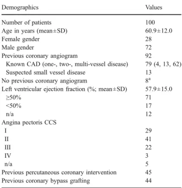
Demographics Values
Number of patients 100
Age in years (mean±SD) 60.9±12.0
Female gender 28
Male gender 72
Previous coronary angiogram 92
Known CAD (one-, two-, multi-vessel disease) 79 (4, 13, 62) Suspected small vessel disease 13
No previous coronary angiogram 8a Left ventricular ejection fraction (%; mean±SD) 57.9±15.0
≥50% 71 <50% 17 n/a 12 Angina pectoris CCS I 29 II 41 III 22 IV 3 n/a 5
Previous percutaneous coronary intervention 45 Previous coronary bypass grafting 44
aPatients had a low likelihood for coronary artery disease.
Table 2 PET findings in 100 patients
Segments Reduced uptake at rest Reduced uptake at stress Scar Ischemia Scar and ischemia (n) Mild (n) Severe (n) Mild (n) Severe (n) Mild (n) Severe (n) Mild (n) Severe (n)
Anterior 14 3 21 6 13 3 8 2 1 posterior 10 5 8 8 7 5 1 0 3 Inferior 13 4 15 9 10 4 5 2 3 Lateral 29 6 27 19 20 6 7 4 9 Septal 1 0 0 1 0 0 0 0 1 Apex 7 8 8 10 6 8 2 1 1
c) CABG in 3 of 100,
d) transplantation in 1 of 100, or
e) conservative medical management in 28 of 100 patients. In all patients with suspected SVD (n=13), antiischemic medication would have been continued.
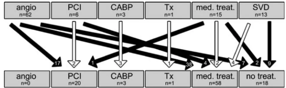
Patient management after the PET is shown in Fig.1. It was influenced in 78% of the study population:
a) diagnostic angiography in 0 of 100,
b) diagnostic angiography and PCI in 20 of 100, c) CABG in 3 of 100,
d) transplantation in 1 of 100, or
e) conservative medical management in 76 of 100 patients. Among the patients with suspected SVD, antiischemic medication was stopped in 9 of 13 patients.
Individual clinical decision-making after PET
Patients with a normal MPI got conservative medical man-agement (13 of 30; 43%), or conservative (cardiac) medical management was stopped (rule out CAD or SVD; 16 of 30; 53%), which was considered “in line” with PET results; whereas one patient (1 of 30; 3%) underwent CABG surgery despite normal MPI, which was considered“not in line” with PET results.
In case of a fixed perfusion defect (scar), 94% of the patients were subsequently treated conservatively (32/34), which was considered “in line” with PET results. Two patients with a fixed perfusion defect underwent PCI (2 of 34; 6%) because, compared to previous PET examinations, the scars were new; therefore, patient management was also considered“in line” with PET results.
Eighteen of 36 (50%) patients with ischemia underwent PCI and 2 of 36 (6%) underwent CABG, both of which was considered“in line” with PET results (Fig.2). One patient (1 of 36, 3%) with ischemia subsequently underwent trans-plantation, which was also considered “in line” with PET
results because ischemia was of mild extent, and therefore, CABG surgery was not considered as a treatment option. Fifteen of these 36 patients (42%) with ischemia received conservative medical treatment, which was also considered “in line” with PET results in 13 patients because (a) ischemia Fig. 1 Demonstration of changes in patient management after MPI
using PET. Squares at top row represent patient management if MPI would not have been available (angio Diagnostic angiography, PCI diagnostic angiography and percutaneous coronary intervention, CABG coronary artery bypass grafting, Tx transplantation, med. treat. medical treatment, SVD small vessel disease). White arrows indicate
no changes in patient management; black solid arrows indicate changes in patient management (numbers given in arrow heads). Squares at bottom row represent patient management after PET (no treat. No treatment). Scanning the patient changed management in 78 of 100 patients
Fig. 2 A 71-year-old patient who had had bypass surgery 8 years before PET was referred to PET for evaluation of myocardial perfusion because of increasing symptoms of angina pectoris. Because symptoms were mild, the referring physician would have treated the patient conservatively with antiischemic medication if MPI would not have been available. Three axial, one horizontal, and one coronal image (five images at the bottom) demonstrate decreased 13
N-ammonia uptake at rest in the inferior and apical myocardium, indicating a scar of mild extent. Five images at the top demonstrate a further decrease of13N-ammonia uptake at pharmacological stress in
the inferior and apical myocardium, indicating severe ischemia. Due to results of PET, patient management was altered, and the patient was referred to PCI
was of mild extent, and intervention was considered unnecessary (seven patients), (b) PCI was technically not possible and ischemia was considered to be minor for CABG (two patients), (c) patient rejected PCI (one patient), and (d) SVD was confirmed (three patients). By contrast, patient management was considered“not in line” in two patients, who were referred for preoperative evaluation of CAD and underwent noncardiac surgery despite severe ischemia.
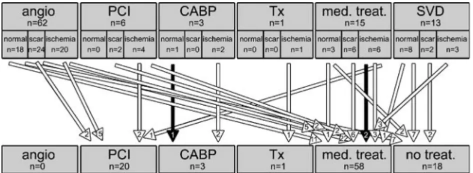
Redirection of patient management is illustrated in Fig.3. In summary, after PET scanning, the management of 3 of 100 patients remained discrepant (“not in line”) to the results of MPI.
Determination of the presence of small-vessel disease SVD was confirmed in 4 of 13 patients (31%) referred to PET. One of them had a visually normal MPI, but a decreased CFR. Three patients had a reduced global CFR and minor ischemia on MPI, attributed to localized pro-nunciation of SVD (n=2) or to the concomitant presence of macro-vessel disease (n=1) (Fig. 3).
In 9 of 13 patients (69%), SVD was ruled out, and anti-ischemic medication was stopped. Seven patients had normal MPI and normal CFR, while two patients had normal CFR, but a mild myocardial scar, not considered relevant (Fig.3). Cost-effectiveness analysis
All patients referred to PET to determine the presence of SVD were excluded from the cost-effectiveness analysis because long-term follow-up was not performed, precluding assessment of long-term costs and/or savings of medical treatment. For the remaining 87 patients, analysis revealed lower total savings of€17,910 (€206 per patient) because of the additional application of PET to the standard patient management procedures (Table3).
Discussion
MPI is a widely established method for the noninvasive evaluation of the pathophysiological significance of coro-nary lesions in patients with CAD. Our study adds to the previous knowledge that (a) in a population with a high prevalence of known CAD, MPI using 13N-ammonia and PET has an impact on patient management in 78% of the cases; (b) the additional application of PET is cost-effective; and (c) patient management followed the recom-mendations of PET findings in 97% of the cases.
Previous clinical studies [23, 24] have analyzed the in-fluence of 18F-fluorodeoxyglucose PET myocardial viabil-Fig. 3 Demonstration of changes in patient management with respect
to the results of MPI with PET. Squares at top row represent patient management if MPI would not have been available (angio Diagnostic angiography, PCI diagnostic angiography and percutaneous coronary intervention, CABG coronary artery bypass grafting, Tx transplanta-tion, med. treat. medical treatment, SVD small vessel disease). Squares
in the second row represent MPI findings for each subgroup. Patient management followed the recommendations of PET findings in 97 cases (white arrows) and did not follow the recommendations of PET in 3 cases (black arrows). Squares at bottom row represent patient management after MPI using PET (no treat. No treatment)
Table 3 Cost-effectiveness Method Treatment without PET (n) Treatment after PET results (n) Difference (n) Difference (€) Diagnostic angiographya 62 0 −62 −149,420 Diagnostic angiography and PCIb 6 20 14 48,860 CABG 3 3 0 0 transplantation 1 1 0 0 Medical treatmentc 15 63 48 0 n/ad 13 13 PETe 0 87 87 82,650 Total € −17,910 a
Costs of diagnostic angiography in our hospital including 1 day hospital stay:€2,410/patient
bCosts of diagnostic angiography percutaneous coronary intervention
(PCI) in our hospital including one day hospital stay:€3,490/patient
cCosts of conservative medical therapy were not included into the
cost-effectiveness analysis
dPatients referred for the evaluation of small-vessel disease were not
included into the cost-effectiveness analysis because PET is the accepted reference standard for diagnosis
e
ity examinations on patient management and found an impact in 57–68% of patients. However, search for viability must, by its nature, remain confined to a highly selected population of CAD patients with a history of myocardial infarction. By contrast, our study using PET for the as-sessment of myocardial perfusion in a consecutive, unse-lected patient population yielded an impact on patient management in 78% of patients.
Moreover, 97% of treatment strategies were in concor-dance with the results from PET scanning. This underlines the clinical value of MPI with PET. We found only 1% of patients who underwent CABG despite normal PET MPI, comparing favorably with the 3–4% reported in retrospec-tive studies with SPECT MPI in patients with unknown CAD [25,26]. Conversely, the proportion of patients in our study who underwent PCI or CABG after detection of ischemia was 56%, lying well within the range of 36–60% reported in previous studies [25,26].
In patients with unknown CAD, an additional MPI using SPECT followed by selective coronary angiography has been shown to be more cost-effective compared with a direct catheterization-first strategy [27]. In our study, we found that the additional application of PET to the standard patient management procedures is also cost-effective in a patient population with a high prevalence of known CAD because direct coronary angiographic strategies can frequently be avoided. This cost-effectiveness would be maintained even with an increase in PET costs by up to 20%. So far, the routine use of PET has not been accepted as a standard for MPI in many centers and health care systems despite its higher diagnostic accuracy compared to SPECT, as reported in the ACC/AHA/ ASNC guidelines of nuclear cardiology [11]. The latter par-ticularly document a superior specificity of PET. This most likely translates into a higher cost-effectiveness of PET com-pared to SPECT, as recently reported by Merighe et al. [28].
MPI using PET is the accepted reference standard for the diagnosis of SVD [29]. In our study population, SVD was ruled out in 69% of patients with suspected SVD, and antiischemic medical management was consecutively stopped, which most likely would further improve the long-term cost-effectiveness of the method, although this aspect was not incorporated into our analysis.
We acknowledge the following limitations to our study. We did not assess how many and which alternative noninvasive tests for ischemia would have been chosen if PET would not have been available, as PET was selected deliberately by the referring physician as the best choice. Therefore, we cannot comment on the performance of alternative options. We did not perform a long-term clinical patient follow-up to evaluate whether the impact of PET on patient management leads to a prognostic benefit. However, such benefit has been reported for MPI SPECT in many studies [11], and it appears reasonable to assume similar performance of PET MPI. As a
further potential limitation, we did not include in our cost-effectiveness calculation the fact that some patients planned for angiography may possibly have ended with PCI. This may have lead to an underestimation of the cost-effectiveness of PET, further strengthening our results.
Furthermore, costs for medical management were not included, due to the limited follow-up at the time of analysis. However, as standard medical treatment in CAD patients would not differ largely between different strategy groups, any change of strategy would only cause minor changes in costs for medication. Finally, we excluded 13 patients referred to PET for the evaluation of SVD because PET is the accepted reference standard for diagnosis, and additional patient management procedures cannot be expected, regardless of the results of PET.
Conclusions
In a population with a high prevalence of known CAD, PET is cost-effective and has an important impact on patient management, while patient management follows the recom-mendations of PET findings in 97% of the cases.
Acknowledgements This study was supported by a grant from the Swiss National Science Foundation (SNSF-professorship grant no. PP00A-114706) and by a grant of the National Center of Competence in Research, Computer Aided and Image Guided Medical Interven-tions (NCCR CO-ME) of the Swiss National Science Foundation.
References
1. Topol EJ, Nissen SE. Our preoccupation with coronary luminol-ogy. The dissociation between clinical and angiographic findings in ischemic heart disease. Circulation 1995;92:2333–42. 2. Schelbert HR, Wisenberg G, Phelps ME, Gould KL, Henze E,
Hoffman EJ, et al. Noninvasive assessment of coronary stenoses by myocardial imaging during pharmacologic coronary vasodila-tion. VI. Detection of coronary artery disease in human beings with intravenous N-13 ammonia and positron computed tomog-raphy. Am J Cardiol 1982;49:1197–207.
3. Gould KL, Kelley KO, Bolson EL. Experimental validation of quantitative coronary arteriography for determining pressure-flow characteristics of coronary stenosis. Circulation 1982;66:930–7. 4. Miller DD, Donohue TJ, Younis LT, Bach RG, Aguirre FV, Wittry
MD, et al. Correlation of pharmacological 99mTc-sestamibi myocardial perfusion imaging with poststenotic coronary flow reserve in patients with angiographically intermediate coronary artery stenoses. Circulation 1994;89:2150–60.
5. Zijlstra F, Fioretti P, Reiber JH, Serruys PW. Which cineangio-graphically assessed anatomic variable correlates best with functional measurements of stenosis severity? A comparison of quantitative analysis of the coronary cineangiogram with measured coronary flow reserve and exercise/redistribution thallium-201 scintigraphy. J Am Coll Cardiol 1988;12:686–91.
6. Hutchins GD, Schwaiger M, Rosenspire KC, Krivokapich J, Schelbert H, Kuhl DE. Noninvasive quantification of regional blood
flow in the human heart using N-13 ammonia and dynamic positron emission tomographic imaging. J Am Coll Cardiol 1990;15:1032–42. 7. Bergmann SR, Herrero P, Markham J, Weinheimer CJ, Walsh MN. Noninvasive quantitation of myocardial blood flow in human subjects with oxygen-15-labeled water and positron emission tomography. J Am Coll Cardiol 1989;14:639–52.
8. Araujo LI, Lammertsma AA, Rhodes CG, McFalls EO, Iida H, Rechavia E, et al. Noninvasive quantification of regional myocardial blood flow in coronary artery disease with oxygen-15-labeled carbon dioxide inhalation and positron emission tomography. Circulation 1991;83:875–85.
9. Hesse B, Tagil K, Cuocolo A, Anagnostopoulos C, Bardies M, Bax J, et al. EANM/ESC procedural guidelines for myocardial perfusion imaging in nuclear cardiology. Eur J Nucl Med Mol Imaging 2005;32:855–97.
10. Gould KL. Clinical cardiac positron emission tomography: State of the Art. Circulation 1991;84(Suppl I):I-22–I-36.
11. Klocke FJ, Baird MG, Lorell BH, Bateman TM, Messer JV, Berman DS, et al. ACC/AHA/ASNC guidelines for the clinical use of cardiac radionuclide imaging—executive summary: a report of the American College of Cardiology/American Heart Association Task Force on Practice Guidelines (ACC/AHA/ASNC Committee to Revise the 1995 Guidelines for the Clinical Use of Cardiac Radionuclide Imaging). J Am Coll Cardiol 2003;42:1318–33. 12. Stewart RE, Schwaiger M, Molina E, Popma J, Gacioch GM,
Kalus M, et al. Comparison of rubidium-82 positron emission tomography and thallium-201 SPECT imaging for detection of coronary artery disease. Am J Cardiol 1991;67:1303–10. 13. Tamaki N, Yonekura Y, Senda M, Yamashita K, Koide H, Saji H,
et al. Value and limitation of stress thallium-201 single photon emission computed tomography: comparison with nitrogen-13 ammonia positron tomography. J Nucl Med 1988;29:1181–8. 14. Go RT, Marwick TH, MacIntyre WJ, Saha GB, Neumann DR,
Underwood DA, et al. A prospective comparison of rubidium-82 PET and thallium-201 SPECT myocardial perfusion imaging utilizing a single dipyridamole stress in the diagnosis of coronary artery disease. J Nucl Med 1990;31:1899–905.
15. Camici PG, Marraccini P, Lorenzoni R, Buzzigoli G, Pecori N, Perissinotto A, et al. Coronary hemodynamics and myocardial metabolism in patients with syndrome X: response to pacing stress. J Am Coll Cardiol 1991;17:1461–70.
16. Rosen SD, Uren NG, Kaski JC, Tousoulis D, Davies GJ, Camici PG. Coronary vasodilator reserve, pain perception, and sex in patients with syndrome X. Circulation 1994;90:50–60.
17. Epstein SE, Cannon RO 3rd. Site of increased resistance to coronary flow in patients with angina pectoris and normal epicardial coronary arteries. J Am Coll Cardiol 1986;8:459–61. 18. Koepfli P, Wyss CA, Namdar M, Klainguti M, von Schulthess GK,
Luscher TF, et al. Beta-adrenergic blockade and myocardial
perfusion in coronary artery disease: differential effects in stenotic versus remote myocardial segments. J Nucl Med 2004;45:1626–31. 19. Cerqueira MD, Verani MS, Schwaiger M, Heo J, Iskandrian AS. Safety profile of adenosine stress perfusion imaging: results from the Adenoscan Multicenter Trial Registry. J Am Coll Cardiol 1994;23:384–9.
20. Margonato A, Chierchia SL, Xuereb RG, Xuereb M, Fragasso G, Cappelletti A, et al. Specificity and sensitivity of exercise-induced ST segment elevation for detection of residual viability: compar-ison with fluorodeoxyglucose and positron emission tomography. J Am Coll Cardiol 1995;25:1032–8.
21. Muzik O, Beanlands RS, Hutchins GD, Mangner TJ, Nguyen N, Schwaiger M. Validation of nitrogen-13-ammonia tracer kinetic model for quantification of myocardial blood flow using PET. J Nucl Med 1993;34:83–91.
22. Jorg-Ciopor M, Namdar M, Turina J, Jenni R, Schwitter J, Turina M, et al. Regional myocardial ischemia in hypertrophic cardio-myopathy: impact of myectomy. J Thorac Cardiovasc Surg 2004;128:163–9.
23. Beanlands RS, deKemp RA, Smith S, Johansen H, Ruddy TD. F-18-fluorodeoxyglucose PET imaging alters clinical decision making in patients with impaired ventricular function. Am J Cardiol 1997;79:1092–5.
24. Felix RC, Correa PL, de Azevedo JC, Dohmann HF, Mesquita ET, Mesquita CT. Clinical impact of positron emission tomography by coincidence system with 18F-FDG on therapeutic decision-making of patients with ischemic cardiomyopathy after myocar-dial infarction. Arq Bras Cardiol 2006;86:337–45.
25. Bateman TM, O’Keefe JH Jr., Dong VM, Barnhart C, Ligon RW. Coronary angiographic rates after stress single-photon emission computed tomographic scintigraphy. J Nucl Cardiol 1995;2:217–23. 26. Nallamothu N, Pancholy SB, Lee KR, Heo J, Iskandrian AS. Impact on exercise single-photon emission computed tomographic thallium imaging on patient management and outcome. J Nucl Cardiol 1995;2:334–8.
27. Shaw LJ, Hachamovitch R, Berman DS, Marwick TH, Lauer MS, Heller GV, et al. The economic consequences of available diagnostic and prognostic strategies for the evaluation of stable angina patients: an observational assessment of the value of precatherization ischemia. Economics of Noninvasive Diagnosis (END) Multicenter Study Group. J Am Coll Cardiol 1999;33: 661–9.
28. Merhige ME, Breen WJ, Shelton V, Houston T, D’Arcy BJ, Perna AF. Impact of myocardial perfusion imaging with PET and (82)Rb on downstream invasive procedure utilization, costs, and out-comes in coronary disease management. J Nucl Med 2007;48: 1069–76.
29. Camici PG, Crea F. Coronary microvascular dysfunction. N Engl J Med 2007;356:830–40.