Association of Retinal Nerve Fiber Layer Thickness With Brain
Alterations in the Visual and Limbic Networks in Elderly Adults
Without Dementia
Juan Luis Méndez-Gómez, PhD; Amandine Pelletier, PhD; Marie-Bénédicte Rougier, MD, PhD; Jean-François Korobelnik, MD; Cédric Schweitzer, MD, PhD;
Marie-Noëlle Delyfer, MD, PhD; Gwenaëlle Catheline, PhD; Solène Monfermé, MD; Jean-François Dartigues, MD, PhD; Cécile Delcourt, PhD; Catherine Helmer, MD, PhD
Abstract
IMPORTANCE The eye is a sensory organ that is easily accessible for imaging techniques, allowing the measurement of the retinal nerve fiber layer (RNFL) thickness. The eye is part of the central nervous system, and its neurons may be susceptible to degeneration; therefore, changes in the RNFL thickness may reflect microstructural and volume alterations in the brain.
OBJECTIVE To explore the association between the peripapillary RNFL thickness and brain alterations in the visual and limbic networks in elderly people without dementia.
DESIGN, SETTING, AND PARTICIPANTS Cross-sectional analysis of the Three-City/Antioxydants, Lipides Essentiels, Nutrition et Maladies Oculaires (Alienor) Study cohort (April 2009 to December 2010). The dates of analysis were July 2017 to August 2018. The setting was a population-based study in France. The brain volume analysis included 104 participants, and the diffusion tensor imaging analysis included 79 participants.
MAIN OUTCOMES AND MEASURES Global RNFL was assessed by spectral-domain optical coherence tomography. Brain volumes were assessed via T1-weighted magnetic resonance imaging by measurement of the global white and gray matter fractions and the hippocampal fraction. Brain microstructural alterations were assessed with diffusion tensor imaging at the level of the posterior thalamic radiations, the limbic system tracts (the fornix and cingulum bundles), and the posterior limb of the internal capsule (control region). Linear regression models adjusted for several confounders were performed.
RESULTS Among a total of 104 participants, the mean (SD) age was 80.8 (3.9) years, and the cohort was 56.7% women (n = 59). The mean (SD) global RNFL thickness was 89.3 (12.9) μm. A thicker RNFL was associated with a greater hippocampal fraction (quantity of increase β = 0.013; 95% CI, 0.001-0.025 per 10-μm increase in the RNFL thickness) and better diffusion tensor imaging variables in the global cingulum (mean diffusivity β = −0.007; 95% CI, −0.015 to −0.000) and the
hippocampal part of the cingulum (mean diffusivity β = −0.009; 95% CI, −0.016 to −0.002 and radial diffusivity β = −0.010; 95% CI, −0.018 to −0.002) and the posterior thalamic radiations (fractional anisotropy β = 0.008; 95% CI, 0.000-0.017). No significant associations were found with other magnetic resonance imaging volumes or with other diffusion tensor imaging variables. In particular, there was no significant association with the control region of interest.
CONCLUSIONS AND RELEVANCE Results of this study suggest that in elderly individuals without dementia, a thicker RNFL was associated with better magnetic resonance imaging variables both in a
(continued)
Key Points
Question Is the peripapillary retinal nerve fiber layer (RNFL) thickness correlated with brain alterations in regions that are vulnerable to neurodegenerative processes? Findings Among 104 elderly people with dementia in this population-based study, a thicker RNFL was associated with a higher hippocampal fraction and better diffusion tensor imaging variables in the global and hippocampal part of the cingulum, 2 regions closely associated with the neurodegenerative processes of Alzheimer disease.
Meaning The assessment of axonal thickness in the retina, which is a quick measurement to perform, may provide some elements of brain magnetic resonance imaging abnormalities at an early stage of neurodegeneration.
+
Supplemental contentAuthor affiliations and article information are listed at the end of this article.
Abstract (continued)
region that included the visual pathways and in regions particularly involved in the neurodegenerative processes of Alzheimer disease.
JAMA Network Open. 2018;1(7):e184406. doi:10.1001/jamanetworkopen.2018.4406
Introduction
Because of their common embryologic origin, the retinal nerve fiber layer (RNFL) is anatomically connected with the central nervous system; therefore, the eye is a sensory organ that is part of the central nervous system, with neurons that may be susceptible to direct or indirect degeneration.1
Using advanced imaging techniques, such as spectral-domain optical coherence tomography (SD-OCT), provides easy and quick access to accurately evaluate the RNFL, which may reflect neurodegenerative changes in the brain.2
Previous studies have reported RNFL thinning in patients with Alzheimer disease (AD) and mild cognitive impairment (MCI) vs healthy controls; these results were confirmed in 2 recent
meta-analyses,3,4
revealing significantly reduced mean RNFL thickness in AD and MCI. Moreover, within a large population, the Rotterdam Study5
recently found that a thinner RNFL was associated with a higher risk of developing dementia and AD in subsequent years, suggesting that a thinner RNFL may be a biomarker for dementia and AD. Nevertheless, beyond the clinical diagnosis of AD and MCI, the relationship between the RNFL and brain structures remains underexplored in the elderly population. Indeed, previous imaging studies have explored the connections between the RNFL and brain alterations observed in specific pathologies, such as multiple sclerosis6-10
and glaucoma.11
However, to our knowledge, only 2 previous studies have explored this association beyond these specific pathologies. The first, a population-based study12
that included 164 elderly people who were mainly cognitively impaired without dementia showed that a reduced RNFL thickness was associated with a reduced gray matter (GM) volume in the temporal lobe. The second study13
included 79 neurologically healthy older adults and found an association between RNFL thinning and smaller mediotemporal lobe volumes. However, these 2 studies analyzed only the brain volume. Yet, other brain imaging techniques, such as diffusion tensor imaging (DTI), can be used to assess the white matter (WM) microstructural integrity, revealing another aspect of the
neurodegenerative process. Moreover, several studies since 2006 indicate that DTI variable measurement is a sensitive and powerful method, showing global age-related modifications14
and fine modifications of specific networks, such as the limbic system.15,16
We hypothesized that changes in the RNFL thickness may reflect microstructural and volume alterations in the brain, not only in the visual pathways but also in regions that are particularly vulnerable to the neurodegenerative processes occurring in AD and/or MCI. Therefore, within a population-based study of elderly people in France, we aimed to explore the associations between the peripapillary RNFL thickness and brain alteration measures using anatomical images to assess the global and hippocampal volumes and using diffusion-weighted images to extract variables in specific WM bundles in the posterior thalamic radiations (including the optic radiations), the WM connecting tracts of the limbic system (ie, the fornix and cingulum bundles), and the posterior limb of the internal capsule as a control region.
Methods
Study Population
Our study population is from the Three-City Study,17
a prospective population-based cohort of French adults 65 years or older that aims to estimate the risk of dementia and cognitive impairment attributable to vascular factors in 3 cities in France (Bordeaux, Dijon, and Montpellier). Among the initial 9294 participants, our study focused on those from the Bordeaux site; 2104 participants were
included at baseline (1999-2001) and subsequently reexamined 2, 4, 7, 10, and 12 years later. Data regarding their sociodemographic characteristics, lifestyle, physical and mental health, medications, disabilities, and cognitive function were assessed at baseline and at each follow-up. At the 10-year follow-up, a brain imaging examination was proposed; 239 participants agreed to undergo this examination.
At the 7-year follow-up (in 2006-2007), 963 participants agreed to participate in the Antioxydants, Lipides Essentiels, Nutrition et Maladies Oculaires (Alienor) Study,18
consisting of an ophthalmological examination. These participants were then followed up every 2 years with eye examinations that were concurrent with the Three-City Study follow-ups. In 2009-2010, SD-OCT examination of the retina and optic nerve was included in the eye examination.19
Our study sample consisted of people with valid peripapillary RNFL measurements obtained in April 2009 to December 2010, and valid brain magnetic resonance imaging (MRI) (without any major cerebral pathologies) obtained at the same time (Figure). The dates of analysis were July 2017 to August 2018. People with prevalent dementia, which was systematically screened for and diagnosed on the Diagnostic and Statistical Manual of Mental Disorders (Fourth Edition) criteria20
using a 3-step standardized procedure previously described,17
were excluded. We also excluded people with a medical history of stroke, as well as those with eye diseases affecting the RNFL measurement (glaucoma, vitreomacular traction, myopic chorioretinopathy, peripapillary choroidal
neovascularization, or myelinated retinal nerve fibers). In addition, because the ocular axial length (AL) is associated with the RNFL thickness,21
we also excluded people with missing values for AL. Finally, our study sample included 104 participants for the brain volume analysis and 79 participants for the DTI analysis. Compared with the total Three-City Study population, our study sample was older, included more women, had a lower educational level, had more hypertension, and had a lower Mini-Mental State Examination score (eTable in theSupplement).
Figure. Flowchart of the Selected Participants
624 Alienor Study
508 Alienor Study participants with RNFL
177 MRI data among Alienor Study participants
177 Valid brain volumes dataa
104 Study sample for brain
volumes data 79 Study sample for DTI variables data 136 Valid DTI variables dataa 116 Excluded (no RNFL measurement
or valid measurement)
331 Excluded (no MRI)
73 Excluded
17 Eye disease affecting RNFL measurement 10 Prevalent dementia
4 Medical history of stroke 42 Missing axial length
57 Excluded
16 Eye disease affecting RNFL measurement 5 Prevalent dementia 2 Medical history of stroke 34 Missing axial length
Alienor indicates Antioxydants, Lipides Essentiels, Nutrition et Maladies Oculaires; DTI, diffusion tensor imaging; MRI, magnetic resonance imaging; and RNFL, retinal nerve fiber layer.
aValid MRI images without the presence of tumors or
major cerebrovascular pathologies and showing good-quality processes.
Ethics committee approvals were obtained from the ethical committee of Bordeaux for the Alienor Study and from the ethical committees of the university hospitals of Le Kremlin-Bicêtre and Sud-Méditerranée III for the Three-City Study. Each participant signed an informed consent form. We followed the Strengthening the Reporting of Observational Studies in Epidemiology (STROBE) reporting guideline for observational studies to guide the reporting of this study.
MRI Assessment of Brain Structure
MRI AcquisitionThe MRI examinations were performed using a 3-T imaging system (Achieva; Philips Medical Systems) equipped with an 8-channel head coil (SENSE; Philips Medical Systems). For each person, anatomical high-resolution MRI volumes were acquired in a transverse plane using a 3-dimensional (3-D) magnetization-prepared rapid gradient echo (MPRAGE) T1-weighted sequence, and diffusion-weighted images were acquired using a single-shot echo-planar sequence (21 noncollinear directions and b value of 1000 s/mm2
). MRI Processing
Brain Volumes|Global brain volumes were assessed using voxel-based morphometry22,23
implemented in the VBM8 toolbox (http://dbm.neuro.uni-jena.de/vbm/) within SPM8 software (https:// www.fil.ion.ucl.ac.uk/spm/software/spm8/). Regarding the hippocampal volume, segmentations and volume estimations were performed using FIRST,23
a component of Functional Magnetic Resonance Imaging of the Brain (FMRIB) Software Library. More details have been reported previously.24
The right and left hippocampal volumes were averaged for the statistical analyses. The hippocampal fraction (HF) (the hippocampal volume divided by the total intracranial volume [TIV]) was used as an index of hippocampal atrophy. Similarly, our analyses included the GM fraction (the GM volume divided by the TIV), the WM fraction (the WM volume divided by the TIV), and the brain parenchymal fraction (BPF) (GM plus WM volumes divided by the TIV).
DTI Variables |The integrity of WM microstructure was examined using DTI. The DTI variables evaluate both the directionality and the magnitude of water diffusion in brain tissue. Within WM, the diffusion of water is restricted along bundles.25
Fractional anisotropy (FA) represents the degree of the directionality of water diffusivity along the fibers; axial diffusivity (AxD) and radial diffusivity (RD) quantify the magnitude of diffusion along the principal and perpendicular directions of the fibers, respectively; and mean diffusivity (MD) represents a global measure of diffusion. Therefore, higher directionality, as indicated by FA, and lower magnitude of diffusion, as indicated by diffusivity variables (ie, AxD, RD, and MD), generally indicate preserved microstructure. We evaluated the DTI variables in the following areas: (1) the posterior thalamic radiations, including the optic tracts, and (2) the bundles of the limbic system, which are thought to be altered prematurely in AD processes (ie, the fornix and cingulum bundles). The latter 2 bundles were analyzed both globally and specifically in their part running along the hippocampus (as defined in the Johns Hopkins University atlas,26
the stria terminalis of the fornix and the hippocampal cingulum). We also analyzed a control region of interest, the posterior limb of the internal capsule (including the motor bundles), which was not specifically involved in either the visual pathway or AD.
SD-OCT Measures
Participants underwent a retinal SD-OCT examination without pupil dilation using a software program (SPECTRALIS, version 5.4.7.0; Heidelberg Engineering) and performed by the same experienced technician. The SD-OCT offers a detailed cross-sectional image of the retina.27
The RNFL thickness was acquired using the following conditions19
: high-speed resolution mode, circle diameter of 3.5 mm, size x of 768 pixels (10.9 mm), size z of 496 pixels (1.9 mm), scaling x of 14.14 lumens per pixel, scaling z of 3.87 lumens per pixel, and automatic real-time (ART) mode of 16 images. The minimum reliable value of the global thickness was retained when a reliable measurement was
obtained in both eyes; otherwise, only the measurement from the eye with a reliable value was retained.
Other Variables
The covariates considered were age, sex, educational level, APOE4 genotype, diabetes (antidiabetic treatment and/or a fasting glucose levelⱖ126 mg/dL [to convert to millimoles per liter, multiply by 0.555] or a nonfasting glucose levelⱖ198 mg/dL), hypertension (blood pressure >140/90 mm Hg or antihypertensive medication), tobacco consumption, cataract surgery, vertical diameter of the optic nerve disc (the mean of 2 measurements), and AL. Axial length was assessed for all participants via anatomical high-resolution MRI images in a transverse plane based on a 3-D MPRAGE T1-weighted sequence.28
Two operators (J.L.M.-G. and S.M.) separately measured the AL, and the mean of 2 measurements was retained. In case of a difference exceeding 2 mm between the 2 values, a third measurement was made, and the 2 closer measurements were retained for the mean. This procedure was validated in a subsample of 79 participants with available AL measurements obtained with noncontact partial coherence laser interferometry (IOL Master; Carl Zeiss Meditec AG); these latter correlated strongly with the AL measurements from the MRI images (Pearson product moment correlation r = 0.89, P < .001).
Statistical Analysis
Descriptive statistics were calculated for each MRI subsample (brain volume analysis and DTI analysis). Multiple linear regression models were performed to analyze the association between the RNFL and MRI variables. The β coefficients, which correspond to the quantity of increase in brain variables (either brain volumes or DTI variables) per 10-μm increase in the RNFL thickness, and their 95% CIs are provided. The models were adjusted for ocular variables, age, sex, educational level, APOE4 genotype, diabetes, hypertension, and AL. The distribution of the residuals in the models was examined graphically to test the assumption of normal distribution. All statistical analyses were performed with a software program (SAS, version 9.3; SAS Institute Inc), and P < .05 (2-sided) was considered significant.
Results
The population characteristics are listed in Table 1. Among a total of 104 participants, the mean (SD) age was 80.8 (3.9) years, and the cohort was 56.7% women (n = 59). Among the participants, 16.0% (16 of 100) were APOE4 carriers, 8.7% (9 of 104) had diabetes, 73.1% (76 of 104) had hypertension, 34.6% (36 of 104) were smokers, and 40.4% (42 of 104) had undergone cataract surgery. The mean (SD) global RNFL thickness was 89.3 (12.9) μm.
Regarding the brain volumes, the mean (SD) BPF was 76.1% (2.4%), and the mean (SD) HF was 0.49% (0.07%). Regarding the DTI variables, the mean (SD) global FA was 0.46 (0.02), and the mean (SD) global MD was 0.78 (0.03).
RNFL Thickness and Brain Volumes
The multivariable analysis identified a significant association between the RNFL thickness and the hippocampus: a thicker RNFL was associated with a greater HF (quantity of increase β = 0.013; 95% CI, 0.001-0.025 per 10-μm increase in the RNFL thickness; P = .03) (Table 2). No significant associations were found between the RNFL and other MRI volume measures (ie, BPF and global GM and WM fractions).
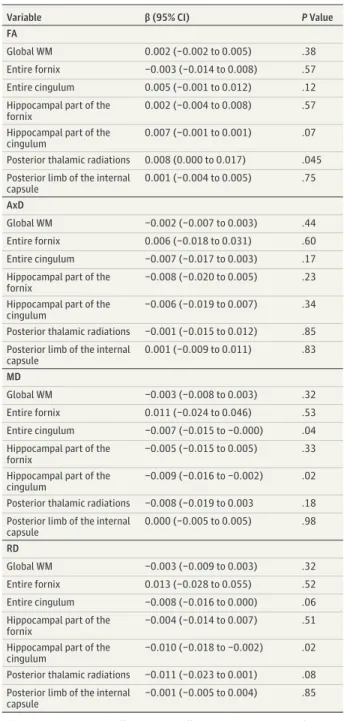
RNFL Thickness and DTI Variables
The multivariable analysis showed significant associations between the RNFL thickness and regions that include the visual pathway and regions implicated in AD-associated neurodegenerative processes (Table 3). A thicker RNFL was associated with a higher FA (reflecting better integrity of
Table 1. Characteristics of the Study Participants Variable Brain Volumes (n = 104)a DTI Variables (n = 79)b Age, mean (SD), y 80.8 (3.9) 80.4 (3.8) Female, No. (%) 59 (56.7) 45 (57.0)
Educational level, No. (%)
Elementary school 9 (8.7) 6 (7.6)
Short secondary school 40 (38.5) 34 (43.0)
Higher level 55 (52.9) 39 (49.4)
APOE4 genotype, No./total No. (%) 16/100 (16.0) 12/76 (15.8)
Diabetes, No. (%) 9 (8.7) 7 (8.9)
Hypertension, No. (%) 76 (73.1) 57 (72.2)
Smokers, No. (%) 36 (34.6) 25 (31.6)
Cataract surgery, No. (%) 42 (40.4) 32 (40.5) Vertical diameter of the optic nerve disc,
mean (SD), mm
2.0 (0.2) 2.1 (0.2) Axial length, mean (SD), mm 23.7 (1.1) 23.7 (1.2) Global RNFL thickness, mean (SD), μm 89.3 (12.9) 89.2 (13.2) MMSE score, mean (SD) 27.7 (2.2) 27.8 (2.1) Brain volumes, mean (SD), % of TIV
BPF 76.1 (2.4) NA
HF 0.49 (0.07) NA
WM fraction 35.7 (2.2) NA
GM fraction 40.3 (2.5) NA
DTI variables, mean (SD)
Global FA NA 0.46 (0.02)
Global AxD, 1000 s/mm2 NA 1.21 (0.03)
Global RD, 1000 s/mm2 NA 0.56 (0.03)
Global MD, 1000 s/mm2 NA 0.78 (0.03)
Abbreviations: AxD, axial diffusivity; BPF, brain parenchymal fraction; DTI, diffusion tensor imaging; FA, fractional anisotropy; GM, gray matter; HF, hippocampal fraction; MD, mean diffusivity; MMSE, Mini-Mental State Examination (score range, 0-30; higher scores indicate better cognition); NA, not applicable; RD, radial diffusivity; RNFL, retinal nerve fiber layer; TIV, total intracranial volume; WM, white matter.
a
Missing data for brain volumes: 4 for APOE genotype, 3 for vertical diameter of the optic nerve disc, 1 for MMSE score, and 7 for HF.
bMissing data for DTI variables: 3 for APOE genotype, 3 for vertical diameter of
the optic nerve disc, and 1 for MMSE score.
Table 2. Associations Between the Global RNFL Thickness and Brain Volumes Among 97 Participantsa
Variable, % of TIV β (95% CI) P Value
BPF 0.174 (−0.202 to 0.551) .36
HFb 0.013 (0.001 to 0.025) .03
WM fraction 0.051 (−0.324 to 0.425) .79 GM fraction 0.123 (−0.245 to 0.492) .51 Abbreviations: BPF, brain parenchymal fraction; GM, gray matter; HF, hippocampal fraction; RNFL, retinal nerve fiber layer; TIV, total intracranial volume; WM, white matter.
a
Seven brain volumes participants who had missing values for at least 1 covariate were excluded from the multivariable analysis. The multiple linear regression models were adjusted for age, sex, educational level, APOE genotype, diabetes, hypertension, tobacco consumption, cataract surgery, vertical diameter of the optic nerve disc, and axial length. The β coefficients are presented for a 10-μm increase in the RNFL thickness.
WM bundles) (β = 0.008; 95% CI, 0.000-0.017 per 10-μm increase in the RNFL thickness; P = .045) in the posterior thalamic radiations. A thicker RNFL was also associated with a lower MD in the global cingulum (β = −0.007; 95% CI, −0.015 to −0.000; P = .04), as well as a lower MD (β = −0.009; 95%
Table 3. Associations Between the Global RNFL Thickness and DTI Variables Among 73 Participantsa
Variable β (95% CI) P Value
FA
Global WM 0.002 (−0.002 to 0.005) .38
Entire fornix −0.003 (−0.014 to 0.008) .57 Entire cingulum 0.005 (−0.001 to 0.012) .12 Hippocampal part of the
fornix
0.002 (−0.004 to 0.008) .57 Hippocampal part of the
cingulum
0.007 (−0.001 to 0.001) .07 Posterior thalamic radiations 0.008 (0.000 to 0.017) .045 Posterior limb of the internal
capsule 0.001 (−0.004 to 0.005) .75 AxD Global WM −0.002 (−0.007 to 0.003) .44 Entire fornix 0.006 (−0.018 to 0.031) .60 Entire cingulum −0.007 (−0.017 to 0.003) .17 Hippocampal part of the
fornix
−0.008 (−0.020 to 0.005) .23 Hippocampal part of the
cingulum
−0.006 (−0.019 to 0.007) .34 Posterior thalamic radiations −0.001 (−0.015 to 0.012) .85 Posterior limb of the internal
capsule 0.001 (−0.009 to 0.011) .83 MD Global WM −0.003 (−0.008 to 0.003) .32 Entire fornix 0.011 (−0.024 to 0.046) .53 Entire cingulum −0.007 (−0.015 to −0.000) .04 Hippocampal part of the
fornix
−0.005 (−0.015 to 0.005) .33 Hippocampal part of the
cingulum
−0.009 (−0.016 to −0.002) .02 Posterior thalamic radiations −0.008 (−0.019 to 0.003 .18 Posterior limb of the internal
capsule 0.000 (−0.005 to 0.005) .98 RD Global WM −0.003 (−0.009 to 0.003) .32 Entire fornix 0.013 (−0.028 to 0.055) .52 Entire cingulum −0.008 (−0.016 to 0.000) .06 Hippocampal part of the
fornix
−0.004 (−0.014 to 0.007) .51 Hippocampal part of the
cingulum
−0.010 (−0.018 to −0.002) .02 Posterior thalamic radiations −0.011 (−0.023 to 0.001) .08 Posterior limb of the internal
capsule
−0.001 (−0.005 to 0.004) .85
Abbreviations: AxD, axial diffusivity; DTI, diffusion tensor imaging; FA, fractional anisotropy; MD, mean diffusivity; RD, radial diffusivity; RNFL, retinal nerve fiber layer; WM, white matter.
aSix DTI variables participants who had missing values for at least 1 covariate
were excluded from the multivariable analysis. Multiple linear regression models were adjusted for age, sex, educational level, APOE genotype, diabetes, hypertension, tobacco consumption, cataract surgery, vertical diameter of the optic nerve disc, and axial length. The β coefficients are presented for a 10-μm increase in the RNFL thickness. The DTI variables are expressed in 1000 s/mm2.
CI, −0.016 to −0.002; P = .02) and a lower RD (β = −0.010; 95% CI, −0.018 to −0.002; P = .02) in the hippocampal part of the cingulum. There were no significant associations with other DTI variables in the regions explored. In particular, there was no significant association with the control region of interest (the posterior limb of the internal capsule).
Discussion
In an elderly population without dementia, we found that a thicker global peripapillary RNFL was associated with better DTI variables in the posterior thalamic radiations, which include the visual pathway. In addition, a thicker RNFL was also associated with a higher HF and better DTI variables in the global and hippocampal part of the cingulum, a region closely associated with the
neurodegenerative processes of AD. In contrast, no significant associations were found between the RNFL and the DTI variables in the control region located outside the visual pathway and the regions involved in AD. No significant associations were found with global MRI variables.
Previous studies reported the associations between a thinner RNFL and lesions in the visual pathway in patients with multiple sclerosis,6
visual dysfunction,28
and glaucoma.29-31
Our results add complementary knowledge; we found an association between the RNFL thickness and the DTI variables in the posterior thalamic radiations, which include the visual pathway, not specifically in patients with diseases that directly influence the RNFL thickness but in a general elderly population. Therefore, even beyond specific pathologies inducing RNFL lesions, the RNFL may reflect the brain’s visual pathway. To our knowledge, only 2 previous studies12,13
have explored the association between the RNFL and brain MRI beyond the context of specific pathologies. The study by Ong et al12
included a population of 164 elderly participants and showed no association between the RNFL and the occipital lobe, which includes the visual cortex. However, only the occipital volume and not the DTI variables were studied. Moreover, that population differed from ours, with 75% of the participants being cognitively impaired; therefore, it was far from a general elderly population. The study by Casaletto et al,13
including 79 neurologically healthy older adults without global cognitive impairment (median Mini-Mental State Examination score, 29.5), focused on the mediotemporal lobe and did not analyze the association with the visual pathway.
In addition to the expected association with the visual pathway, our study showed that the RNFL thickness was associated with the HF and with the DTI variables in the global and hippocampal part of the cingulum, 2 brain structures that are involved in the neurodegenerative processes of AD. In contrast, no significant associations were found between the RNFL thickness and the DTI variables in the control region, which was located outside the visual pathway and the regions involved in AD. The hippocampus is well known to be involved early in the neurodegenerative processes, as shown in patients with MCI32
and even in cognitively intact elderly people before they developed AD.29
In previous results from the Three-City Study,30
it was found that people who later developed AD were initially seen with mediotemporal lesions up to 5 years before the diagnosis. The DTI variables in the WM structures have also been associated with current or future clinical neurodegenerative states. Compared with healthy controls, significant alterations in FA, RD, and MD have been shown in the parahippocampal cingulum of patients with AD and patients with MCI who convert to AD.31
Compared with stable healthy people, FA alterations were also detected in the parahippocampal cingulum and in the fornix in cognitively healthy people who developed amnestic MCI 2 years later.33
An association between glaucoma and AD has previously been found,34-36
although some work failed to confirm it.37
Our present results, which show that even in people without glaucoma, abnormal RNFL thinning is associated with brain abnormalities in regions involved in the neurodegenerative processes of AD, are in favor of common neurodegenerative processes occurring at the retinal and brain level, leading to both glaucoma and AD.
In our elderly population, which included people who were not specifically selected based on cognitive capacity, the associations identified between the RNFL and brain structures outside the visual pathway suggest a potential use for the RNFL to reflect brain alterations in structures that are
particularly vulnerable in cognitive decline and AD. Concordant results were reported by Ong et al12
in their population with cognitive impairment and showed an association between the RNFL and the GM volume in the temporal lobe. In addition, Casaletto et al13
showed a specific association with the mediotemporal lobe.
We found no significant associations between the RNFL and global brain MRI variables. One explanation may be that our population was composed of generally well-functioning people; therefore, few participants had global alterations in the brain. In a previous MRI study38
of a small group of young people without multiple sclerosis (controls for patients with multiple sclerosis), the RNFL thickness was also not associated with BPF or GM and WM volumes. Moreover, Ong et al12
also found no association with the total brain or GM or WM volumes in their population with cognitive impairment; even in that population, an association was found only with the GM in the temporal lobe and not globally. However, before the moderate stage of dementia, global brain structures are preserved.
Therefore, associations with the RNFL seem to be specific to brain structures that are
particularly vulnerable in the elderly population and are involved in the neurodegenerative processes of cognitive decline and AD. Regardless of the MRI variables, our results were consistent: a thicker RNFL was associated with both better DTI variables in the global and hippocampal part of the cingulum and a higher HF. A thicker RNFL was associated with lower RD and MD in the hippocampal part of the cingulum but was not associated with AxD. The RD may better reflect the dysmyelination of neurons, whereas AxD may better reflect axonal integrity.39
Previous studies40,41
have consistently found decreased microstructural integrity in WM tracts of patients with AD or at risk of AD, particularly in parahippocampal regions, with alterations in RD variables, suggesting that demyelination may be a contributing mechanism. Moreover, only the cingulum bundle and not the fornix was found to be associated with RNFL. Although the finding needs to be confirmed, this association only with the main afferent and not the efferent pathway of the hippocampus may correspond to an early stage of alteration. Therefore, our results may reflect an “early, silent” form of brain damage that may subsequently progress to a clinical stage. Casaletto et al13
previously found that the RNFL was associated with the mediotemporal lobe but not with memory performance and concluded that the RNFL may represent an early marker of AD processes. Within the Alienor Study, it was also previously found that a thinner RNFL was associated with lower delayed memory scores but not with other cognitive scores (including the Mini-Mental State Examination score), concordant with early stages of the neurodegenerative process.42
Strengths and Limitations
This study has several strengths. It included a large population-based sample of elderly people who underwent exhaustive eye examinations, which allowed the exclusion of ocular pathologies that would likely have influenced the retinal measurements. The SD-OCT assessments were performed on the same SD-OCT machine by the same experienced technician according to a standardized protocol. Moreover, the spectral-domain technique provides the best quality and reproducibility currently available. The brain MRI measurements were also performed on the same machine and according to a standardized procedure. Two different types of MRI variables were analyzed (brain volumes and DTI variables) in different regions of the brain, allowing a better understanding of the global processes. Finally, our analyses accounted for numerous potential confounding factors.
Our study has some limitations. First, our population was composed of well-functioning people (who were well educated and had a conserved cognitive state) who accepted imaging and
ophthalmological examinations; these participants differed from those without these examinations. These participants may have had fewer WM microstructural lesions, which may have prevented the detection of early MRI changes associated with the RNFL. However, to our knowledge, this is the first study to explore this association in a well-functioning population without dementia. Second, our cross-sectional design prevented the evaluation of any temporal association between retinal and brain degeneration; this would require a future longitudinal approach to determine if one of these
degenerative processes precedes the other or if they are concurrent. Third, the analyses were explanatory and did not deal with multiple comparisons. Fourth, because our study was restricted to participants with both brain MRIs and ophthalmological examinations, we cannot exclude a possible lack of power.
Conclusions
In elderly individuals without dementia, the RNFL thickness was associated with MRI variables in both regions that included the visual pathways and regions involved in AD-related
neurodegeneration. Therefore, SD-OCT assessment of retinal axonal thickness may offer a way to explore brain MRI abnormalities in these regions. However, more studies are needed to demonstrate the potential utility of the RNFL thickness for detecting early brain neurodegeneration in
presymptomatic elderly adults.
ARTICLE INFORMATION
Accepted for Publication: September 13, 2018.
Published: November 9, 2018. doi:10.1001/jamanetworkopen.2018.4406
Open Access: This is an open access article distributed under the terms of theCC-BY License. © 2018 Méndez-Gómez JL et al. JAMA Network Open.
Corresponding Author: Catherine Helmer, MD, PhD, University Bordeaux, Institut National de la Santé et de la Recherche Médicale (INSERM), Bordeaux Population Health Research Center, Unité Mixte de Recherche (UMR) 1219, 146 Rue Léo Saignat, CS61292, F-33076 Bordeaux CEDEX, France (catherine.helmer@u-bordeaux.fr). Author Affiliations: University Bordeaux, Institut National de la Santé et de la Recherche Médicale (INSERM), Bordeaux Population Health Research Center, Unité Mixte de Recherche (UMR) 1219, Bordeaux, France (Méndez-Gómez, Pelletier, Rougier, Korobelnik, Schweitzer, Delyfer, Dartigues, Delcourt, Helmer); Centre National de la Recherche Scientifique (CNRS), Institut de Neurosciences Cognitives et Intégratives d’Aquitaine (INCIA), UMR 5287, Bordeaux, France (Pelletier, Catheline); Ophthalmology, University Hospital, Bordeaux, France (Rougier, Korobelnik, Schweitzer, Delyfer, Monfermé); École Pratique des Hautes Études (EPHE), Paris Sciences et Lettres (PSL) Research University, Bordeaux, France (Catheline); Memory Consultation, Centre Mémoire de Ressource et de Recherche (CMRR), University Hospital, Bordeaux, France (Dartigues); Clinical Epidemiology Unit, INSERM, Centre d’Investigation Clinique (CIC) 1401, Bordeaux, France (Helmer).
Author Contributions: Dr Helmer had full access to all of the data in the study and takes responsibility for the integrity of the data and the accuracy of the data analysis.
Concept and design: Mendez-Gomez, Korobelnik, Schweitzer, Delyfer, Dartigues, Delcourt, Helmer. Acquisition, analysis, or interpretation of data: Mendez-Gomez, Pelletier, Rougier, Korobelnik, Catheline,
Monferme, Delcourt, Helmer.
Drafting of the manuscript: Mendez-Gomez, Pelletier, Helmer.
Critical revision of the manuscript for important intellectual content: Rougier, Korobelnik, Schweitzer, Delyfer,
Catheline, Monferme, Dartigues, Delcourt, Helmer.
Statistical analysis: Mendez-Gomez, Pelletier, Helmer. Obtained funding: Korobelnik, Dartigues, Delcourt, Helmer.
Administrative, technical, or material support: Korobelnik, Schweitzer, Delyfer, Catheline. Supervision: Korobelnik, Delyfer, Dartigues, Helmer.
Conflict of Interest Disclosures: Dr Rougier reported being a consultant for Allergan and Bausch & Lomb; reported receiving funding for conferences from Bayer, Carl Zeiss Meditec, AbbVie, Laboratoires Théa, and Novartis; and reported receiving personal fees from Bayer, AbbVie, Allergan, Novartis, and Carl Zeiss Meditec. Dr Korobelnik reported being a consultant for Alcon, Allergan, Bayer, Carl Zeiss Meditec, Novartis, and Laboratoires Théa and reported receiving research grants from Roche and Alimera Sciences. Dr Schweitzer reported receiving personal fees from Laboratoires Théa, Alcon, Novartis, and Allergan. Dr Delyfer reported receiving advisory board or travel funding from Théa Laboratories, Bayer, and Novartis; reported receiving grants from Laboratoires Théa; and reported receiving personal fees from Allergan, Bayer, Novartis, and Bausch & Lomb. Dr Dartigues reported receiving research support and grants from Roche. Dr Delcourt reported receiving grants from Laboratoires Théa,
Fondation Voir et Entendre, Association Retina France, and Conseil Regional d’Aquitaine; reported receiving personal fees from Allergan, Bausch & Lomb, Laboratoires Théa, Novartis, and Roche; and reported being a consultant for Allergan, Bausch & Lomb, Novartis, Roche, and Laboratoires Théa. Dr Helmer reported receiving grants from France Alzheimer (a charitable foundation) and reported receiving grants from Roche. All funding received by the authors was outside of the present work. No other disclosures were reported.
Funding/Support: The Antioxydants, Lipides Essentiels, Nutrition et Maladies Oculaires (Alienor) Study is supported by Laboratoires Théa (which participated in the design of that study) and by Fondation Voir et Entendre, Association Retina France, Conseil Régional d’Aquitaine, and Fondation Pour la Recherche Médicale. The present study was supported by France Alzheimer (a charitable foundation). All funding received by the authors was outside of the present work.
Role of the Funder/Sponsor: Laboratoires Théa participated in the design of the Antioxydants, Lipides Essentiels, Nutrition et Maladies Oculaires (Alienor) Study but had no role in the conduct of the study; collection,
management, analysis, and interpretation of the data; preparation, review, or approval of the manuscript; and decision to submit the manuscript for publication. The other funding sources had no role in the design and conduct of the study; collection, management, analysis, and interpretation of the data; preparation, review, or approval of the manuscript; and decision to submit the manuscript for publication. No author was the recipient of the funding sources of the present work.
Additional Information: The Three-City Study is conducted under a partnership agreement between the Institut National de la Santé et de la Recherche Médicale (INSERM), the University Bordeaux 2 Victor Segalen, and Sanofi-Aventis. Fondation Pour la Recherche Médicale funded the preparation and initiation of the study. The Three-City Study is also supported by the Caisse Nationale Maladie des Travailleurs Salariés, Direction Générale de la Santé, Mutuelle Générale de l’Education Nationale (MGEN), Institut de la Longévité, Conseil Régional d’Aquitaine, Conseil Régional de Bourgogne, Fondation de France, Ministry of Research–INSERM Programme “Cohortes et Collections de Données Biologiques,” Agence Nationale de la Recherche (ANR) Programme National de Recherches en Alimentation et Nutrition Humaine (PNRA) 2006 and LongVie 2007, the “Fondation Plan Alzheimer” (Fondation de Coopération Scientifique [FCS] 2009-2012), and the Caisse Nationale de Solidarité pour l’Autonomie (CNSA).
REFERENCES
1. London A, Benhar I, Schwartz M. The retina as a window to the brain: from eye research to CNS disorders. Nat
Rev Neurol. 2013;9(1):44-53. doi:10.1038/nrneurol.2012.227
2. Jindahra P, Hedges TR, Mendoza-Santiesteban CE, Plant GT. Optical coherence tomography of the retina: applications in neurology. Curr Opin Neurol. 2010;23(1):16-23. doi:10.1097/WCO.0b013e328334e99b
3. den Haan J, Verbraak FD, Visser PJ, Bouwman FH. Retinal thickness in Alzheimer’s disease: a systematic review and meta-analysis.Alzheimers Dement (Amst). 2017;6:162-170.
4. Thomson KL, Yeo JM, Waddell B, Cameron JR, Pal S. A systematic review and meta-analysis of retinal nerve fiber layer change in dementia, using optical coherence tomography.Alzheimers Dement (Amst). 2015;1(2):
136-143.
5. Mutlu U, Colijn JM, Ikram MA, et al. Association of retinal neurodegeneration on optical coherence tomography with dementia: a population-based study [published online June 25, 2018].JAMA Neurol.
6. Gabilondo I, Martínez-Lapiscina EH, Martínez-Heras E, et al. Trans-synaptic axonal degeneration in the visual pathway in multiple sclerosis. Ann Neurol. 2014;75(1):98-107. doi:10.1002/ana.24030
7. Klistorner A, Sriram P, Vootakuru N, et al. Axonal loss of retinal neurons in multiple sclerosis associated with optic radiation lesions. Neurology. 2014;82(24):2165-2172. doi:10.1212/WNL.0000000000000522 8. Sabry HM, Ibrahim MA. Optical coherence tomography of retinal nerve fibre layer in multiple sclerosis and its correlation with the disease evolution. Egypt J Neurol Psychiat Neurosurg. 2010;47(1):21-27.
9. Scheel M, Finke C, Oberwahrenbrock T, et al. Retinal nerve fibre layer thickness correlates with brain white matter damage in multiple sclerosis: a combined optical coherence tomography and diffusion tensor imaging study. Mult Scler. 2014;20(14):1904-1907. doi:10.1177/1352458514535128
10. Siger M, Dziegielewski K, Jasek L, et al. Optical coherence tomography in multiple sclerosis: thickness of the retinal nerve fiber layer as a potential measure of axonal loss and brain atrophy. J Neurol. 2008;255(10): 1555-1560. doi:10.1007/s00415-008-0985-5
11. Murphy MC, Conner IP, Teng CY, et al. Retinal structures and visual cortex activity are impaired prior to clinical vision loss in glaucoma. Sci Rep. 2016;6:31464. doi:10.1038/srep31464
12. Ong YT, Hilal S, Cheung CY, et al. Retinal neurodegeneration on optical coherence tomography and cerebral atrophy. Neurosci Lett. 2015;584:12-16. doi:10.1016/j.neulet.2014.10.010
13. Casaletto KB, Ward ME, Baker NS, et al. Retinal thinning is uniquely associated with medial temporal lobe atrophy in neurologically normal older adults. Neurobiol Aging. 2017;51:141-147. doi:10.1016/j.neurobiolaging.2016. 12.011
14. Sullivan EV, Pfefferbaum A. Diffusion tensor imaging and aging. Neurosci Biobehav Rev. 2006;30(6):749-761. doi:10.1016/j.neubiorev.2006.06.002
15. Stoub TR, Barnes CA, Shah RC, Stebbins GT, Ferrari C, deToledo-Morrell L. Age-related changes in the mesial temporal lobe: the parahippocampal white matter region. Neurobiol Aging. 2012;33(7):1168-1176. doi:10.1016/j. neurobiolaging.2011.02.010
16. Pelletier A, Periot O, Dilharreguy B, et al. Age-related modifications of diffusion tensor imaging parameters and white matter hyperintensities as inter-dependent processes. Front Aging Neurosci. 2016;7:255. doi:10.3389/fnagi. 2015.00255
17. 3C Study Group. Vascular factors and risk of dementia: design of the Three-City Study and baseline characteristics of the study population. Neuroepidemiology. 2003;22(6):316-325. doi:10.1159/000072920 18. Delcourt C, Korobelnik JF, Barberger-Gateau P, et al. Nutrition and age-related eye diseases: the Alienor (Antioxydants, Lipides Essentiels, Nutrition et Maladies Oculaires) Study. J Nutr Health Aging. 2010;14(10): 854-861. doi:10.1007/s12603-010-0131-9
19. Rougier MB, Korobelnik JF, Malet F, et al. Retinal nerve fibre layer thickness measured with SD-OCT in a population-based study of French elderly subjects: the Alienor Study. Acta Ophthalmol. 2015;93(6):539-545. doi:
10.1111/aos.12658
20. American Psychiatric Association. Diagnostic and Statistical Manual of Mental Disorders. 4th ed. Washington, DC: American Psychiatric Association; 1994.
21. Savini G, Barboni P, Parisi V, Carbonelli M. The influence of axial length on retinal nerve fibre layer thickness and optic-disc size measurements by spectral-domain OCT. Br J Ophthalmol. 2012;96(1):57-61. doi:10.1136/bjo.2010. 196782
22. Ashburner J, Friston KJ. Voxel-based morphometry: the methods. Neuroimage. 2000;11(6, pt 1):805-821. doi: 10.1006/nimg.2000.0582
23. Patenaude B, Smith SM, Kennedy DN, Jenkinson M. A Bayesian model of shape and appearance for subcortical brain segmentation. Neuroimage. 2011;56(3):907-922. doi:10.1016/j.neuroimage.2011.02.046
24. Pelletier A, Periot O, Dilharreguy B, et al. Structural hippocampal network alterations during healthy aging: a multi-modal MRI study. Front Aging Neurosci. 2013;5:84. doi:10.3389/fnagi.2013.00084
25. Alexander AL, Hurley SA, Samsonov AA, et al. Characterization of cerebral white matter properties using quantitative magnetic resonance imaging stains. Brain Connect. 2011;1(6):423-446. doi:10.1089/brain.2011.0071 26. Hua K, Zhang J, Wakana S, et al. Tract probability maps in stereotaxic spaces: analyses of white matter anatomy and tract-specific quantification. Neuroimage. 2008;39(1):336-347. Medline: doi:10.1016/j.neuroimage. 2007.07.053
27. Gabriele ML, Wollstein G, Ishikawa H, et al. Three dimensional optical coherence tomography imaging: advantages and advances. Prog Retin Eye Res. 2010;29(6):556-579. doi:10.1016/j.preteyeres.2010.05.005 28. Akduman EI, Nacke RE, Leiva PM, Akduman L. Accuracy of ocular axial length measurement with MRI.
Ophthalmologica. 2008;222(6):397-399. doi:10.1159/000153419
29. den Heijer T, Geerlings MI, Hoebeek FE, Hofman A, Koudstaal PJ, Breteler MM. Use of hippocampal and amygdalar volumes on magnetic resonance imaging to predict dementia in cognitively intact elderly people. Arch
Gen Psychiatry. 2006;63(1):57-62. doi:10.1001/archpsyc.63.1.57
30. Bernard C, Helmer C, Dilharreguy B, et al. Time course of brain volume changes in the preclinical phase of Alzheimer’s disease. Alzheimers Dement. 2014;10(2):143-151.e1. doi:10.1016/j.jalz.2013.08.279
31. Ito K, Sasaki M, Takahashi J, et al. Detection of early changes in the parahippocampal and posterior cingulum bundles during mild cognitive impairment by using high-resolution multi-parametric diffusion tensor imaging.
Psychiatry Res. 2015;231(3):346-352. doi:10.1016/j.pscychresns.2015.01.020
32. Tabatabaei-Jafari H, Shaw ME, Cherbuin N. Cerebral atrophy in mild cognitive impairment: a systematic review with meta-analysis.Alzheimers Dement (Amst). 2015;1(4):487-504.
33. Zhuang L, Sachdev PS, Trollor JN, et al. Microstructural white matter changes in cognitively normal individuals at risk of amnestic MCI. Neurology. 2012;79(8):748-754. doi:10.1212/WNL.0b013e3182661f4d
34. Helmer C, Malet F, Rougier MB, et al. Is there a link between open-angle glaucoma and dementia? the Three-City–Alienor cohort.Ann Neurol. 2013;74(2):171-179.
35. Bayer AU, Ferrari F, Erb C. High occurrence rate of glaucoma among patients with Alzheimer’s disease. Eur
Neurol. 2002;47(3):165-168. doi:10.1159/000047976
36. Tamura H, Kawakami H, Kanamoto T, et al. High frequency of open-angle glaucoma in Japanese patients with Alzheimer’s disease. J Neurol Sci. 2006;246(1-2):79-83. doi:10.1016/j.jns.2006.02.009
37. Tsilis AG, Tsilidis KK, Pelidou SH, Kitsos G. Systematic review of the association between Alzheimer’s disease and chronic glaucoma. Clin Ophthalmol. 2014;8:2095-2104. doi:10.2147/OPTH.S69534
38. Gordon-Lipkin E, Chodkowski B, Reich DS, et al. Retinal nerve fiber layer is associated with brain atrophy in multiple sclerosis. Neurology. 2007;69(16):1603-1609. doi:10.1212/01.wnl.0000295995.46586.ae
39. Song SK, Sun SW, Ramsbottom MJ, Chang C, Russell J, Cross AH. Dysmyelination revealed through MRI as increased radial (but unchanged axial) diffusion of water. Neuroimage. 2002;17(3):1429-1436. doi:10.1006/nimg. 2002.1267
40. Gold BT, Powell DK, Andersen AH, Smith CD. Alterations in multiple measures of white matter integrity in normal women at high risk for Alzheimer’s disease. Neuroimage. 2010;52(4):1487-1494. doi:10.1016/j. neuroimage.2010.05.036
41. Salat DH, Tuch DS, van der Kouwe AJ, et al. White matter pathology isolates the hippocampal formation in Alzheimer’s disease. Neurobiol Aging. 2010;31(2):244-256. doi:10.1016/j.neurobiolaging.2008.03.013 42. Méndez-Gómez JL, Rougier MB, Tellouck L, et al. Peripapillary retinal nerve fiber layer thickness and the evolution of cognitive performance in an elderly population. Front Neurol. 2017;8:93. doi:10.3389/fneur. 2017.00093
SUPPLEMENT.