Diverse Roles for the Mitogen-Activated Protein Kinase
ERK2 Revealed by High-throughput Target Identification
Scott M. Carlson
B.S. Chemistry Stanford University, 2005 M.Phil. Computational Biology
Cambridge University, 2006
S3AR IES
ARCHIVES
SUBMITTED TO THE DEPARTMENT OF BIOLOGICAL ENGINEERING IN PARTIAL FULFILLMENT OF THE REQUIREMENTS FOR THE DEGREE OF
DOCTOR OF PHILOSOPHY IN BIOLOGICAL ENGINEERING AT THE
MASSACHUSETTS INSTITUTE OF TECHNOLOGY FEBRUARY 2012
© 2011 Massachusetts Institute of Technology. All rights reserved.
Signature of Author:_
- -. Af z '0' ,/ Certified by:
Department of Biological Engineering December 16,2011
Forest M. White Associate Professor of Biological Engineering Thesis Supervisor Accepted by:
K. Dane Wittrup Mares Professor of Chemical Engineering and Biological Engineering Co-Chair, Graduate Program Committee
Members of the Thesis Committee voting in favor of the defense: Chair, K. Dane Wittrup
Mares Professor of Chemical Engineering and Biological Engineering Chair, Graduate Program Committee
Thesis Supervisor, Forest M. White
Associate Professor of Biological Engineering Ernest Fraenkel
Associate Professor of Biological Engineering Roger J. Davis
Professor of Molecular Medicine, University of Massachusetts Howard Hughes Medical Institute
Biographical Note and Acknowledgement
The author attended Leland Stanford Junior University from 2001 to 2005 and received a Bachelor's of Science in Chemistry with a minor in Biology, and was awarded Distinction for academic performance, Honors for advanced coursework and research, a Firestone Medal for outstanding thesis in the natural sciences and the Merck Index Award for outstanding
undergraduate in Chemistry.
Following the B.S. the author was awarded a Gates Cambridge Scholarship and attended
Cambridge University from 2005-06 where he received a Master of Philosophy in Computational Biology.
The author began as a Ph.D. student in Biological Engineering at MIT in fall of 2006 and has been supervised by Associate Professor Forest M. White. He has been supported during this time by a Graduate Research Fellowship from the National Science Foundation, a Momenta
Presidential Fellowship from MIT, a Graduate Fellowship from the David H. Koch Institute for Integrative Cancer Research, a Whitaker Health Science Fellowship, and a Siebel Scholarship. Other financial support for the work described here has come from AstraZeneca, Pfizer Inc., and NIH grants R01DK42816, RO1CA 118705 and U54CAl 12967.
The research described here has been in collaboration with a large number of people and research groups. Individual contributions are noted in the text. Major collaborators include: Professor Roger Davis, University of Massachusetts Medical School
Assistant Professor Norman Kennedy, University of Massachusetts Medical School Institute Professor Phillip Sharp, MIT
Dr. Paul Boutz, Sharp Lab, MIT Mohini Jangi, Sharp Lab, MIT
Associate Professor Ernest Fraenkel, MIT Candace Coudinard, Fraenkel Lab, MIT Adam Labadorf, Fraenkel Lab, MIT Chris Ng, Fraenkel Lab, MIT
Additional technical assistance has been provided by Kevan Shokat, UCSF, and his graduate student Nicholas Hertz, as well as the Swanson Biotechnology Core Facilities at the Koch Institute.
Note on prior publication
Elements of this thesis were published in ACS Chemical Biology in January 2011 (ACS Chem. Biol., 2011, 6 (1), pp 75-85), and have been published in Science Signaling (Sci. Signaling, 2011 Oct 25; 4(196): rsl 1). They are reused here under copyright agreements that allow reuse the material in a thesis or dissertation.
Diverse Roles for the Mitogen-Activated Protein Kinase ERK2
Revealed by High-throughput Target Identification
By
Scott M. Carlson
Submitted to the Department of Biological Engineering on December 16th 2011, in
Partial Fulfillment of the Requirements for the Degree of Doctor of Philosophy in Biological Engineering
ABSTRACT
Many major human oncogenes contribute to cancer in large part by activating the mitogen-activated protein kinases (MAPK) ERKI and ERK2 (ERK). These kinases are critical in normal physiological processes from development to memory, and their activation in cancer drives growth and metastatic invasion. Understanding the effects of ERK signaling is especially important since vemurafenib, the first pharmaceutical directly targeting the MAPK pathway, has recently been approved to treat advanced melanoma. ERK controls cellular phenotypes by phosphorylating over two hundred known substrate proteins, however new ERK targets are reported frequently. We have used a chemical genetics approach to identify over one hundred novel substrates of ERK2. This approach utilizes an ERK2 kinase with "gatekeeper" mutation that allows it to bind bulky ATP analogs (AS-ERK2). AS-ERK2 can be used to label its direct substrates with thiophosphate in an in vitro kinase reaction. To achieve sufficient sensitivity we have improved on existing protocols for identification of thiophosphorylated peptides. Our improved protocol identified over one hundred novel ERK2 substrates in 3T3-L1 fibroblasts and in colon carcinoma cell lines. We investigated one novel ERK2 substrate, the transcriptional repressor ETV3, in detail and found that phosphorylation abrogates binding to DNA by ETV3, and that mutation of key phosphorylated residues to alanine blocks this effect. We also
identified several thousand ETV3 targets across the genome. The wide range of genes targeted by ETV3 suggests that it may act as a key regulator of cell cycle and metabolism in some cell types. We have also identified ERK2 substrates in the DLD1 colon carcinoma cell line, including several mRNA splicing factors and members of the MLL family of histone 3
methyltransferases. We are using high-throughput sequencing and biochemical experiments to determine whether these phosphorylation sites control the function of MLL proteins. Taken together these investigations greatly expand our knowledge of the ERK signaling pathway and have revealed greater connectivity among biological processes than had been appreciated. Supervisor: Forest M. White
Table of Contents
Cover page 1
Members of the Thesis Committee 2
Biographical Information and Acknowledgments 3
Note on prior publication 4
Abstract 5
Table of Contents 6
Chapter 1: Introduction 7
Chapter 2: Protocol optimization and discovery of ERK2 substrates 20 Chapter 3: Phosphorylation by ERK regulates the transcriptional repressor ETV3 45
Chapter 4: Quantitative rewiring of the ERK1/2 pathway in colon cancer driven 58 by activated K-Ras
Conclusions and future directions: 77
References: 80
Appendix 1: ERK2 substrates identified in 3T3-Ll cells 90 Appendix 2: ERK2 phosphorylation sites on IRS2, ETV3 and CDC42EP1 93
Appendix 3: MAPK motif immunoprecipitation 94
1.
Introduction
Virtually all cellular responses to environmental perturbations are controlled in part by tightly regulated protein phosphorylation signaling networks. Aberrant kinase activation or loss of phosphatase activity underlies many major human diseases including cancer, autoimmunity, and diabetes'. The early discovery of the transforming capability of constitutively active tyrosine kinases has led to extensive research on how signaling is deregulated in cancer4. As a result,
increased expression or activation of kinases has been found to play a causative role in many cancers, including the BCR-ABL fusion protein in chronic myeloid leukemia (CML), B-Raf in melanoma, the receptor tyrosine kinase (RTK) EGFR in lung cancer and glioblastoma, and the related RTK HER2 in breast cancer ,6. Because kinase-activated signaling networks are associated with proliferative, migratory, and invasive phenotypes, small molecule kinase
7
inhibitors are a major area of pharmaceutical research and development .
Clinical results and experiments in cell culture have shown that many cancer cells experience "oncogene addiction" such that inhibition or removal of the transforming "driver" mutation blocks malignant phenotypes and leads to apoptosis . Although this effect has been observed in a wide range of cancers our understanding of the process is very limited. This was recently highlighted by Dr. Harold Varmus, Director of the National Institutes of Health, in a request for applications to study twenty-four "Provocative Questions" in cancer biology and treatment ("While this cell death is an encouraging outcome for therapeutic approaches, we have little knowledge of why these cells become so strongly dependent on the continued expression of an active mutated oncogene"). These questions will serve throughout this dissertation as a framework linking experiments in molecular biology to issues of long-term clinical significance.
The phenomenon of oncogene addiction suggests that many cancers may have a
significant therapeutic window where targeted inhibition of an oncogene will destroy tumor cells or inhibit growth while having a minimal effect on normal tissues. Unfortunately clinical trials with targeted kinase inhibitors as mono-therapies for cancer treatment have been largely
disappointing due to confounding issues including lack of specificity, primary resistance because
7
of compensating or redundant signals, rapid development of acquired resistance, and toxicity7. Understanding why kinase inhibitors fail in the clinic and how to combine them with other chemotherapies or targeted treatments will require a much deeper knowledge of the signaling networks that they affect. The aim of this research has been to examine one of those pathways in detail by identifying downstream targeted by the mitogen-activated protein kinase (MAPK) ERK2. ERK2, along with the closely related ERKI, is a key signaling molecule involved in cell cycle control, differentiation and metabolism, among other processes. ERKI and ERK2 are activated by the same upstream signals and share many of their targets; I will refer to their combined activity as the ERK pathway. ERK is downstream of several major oncogenes including over-expression or mutation in EGFR, mutation in KRAS, NRAS, and BRAF. In this dissertation I will present an optimized approach for labeling and identifying substrates of ERK2, discovery of over one hundred novel substrates in a variety of biological systems, a quantitative analysis of how ERK2 substrates are affected by the presence of an oncogenic signal, evidence that ERK2 controls ETV3, a global regulator of gene transcription, and initial work using high-throughput sequencing to define novel roles for ERK signaling in histone methylation and
Targeting kinases in oncology
There is tremendous interest in using small molecule kinase inhibitors as therapeutics for cancer. Imatinib (marketed as Gleevec) was the first clinically effective kinase inhibitor to be designed on the basis of a known disruption in kinase signaling. Approximately 95% of CML carries a reciprocal translocation of chromosomes 9 and 22, leading to fusion of the genes for ABL tyrosine kinase and BCR. The BCR-ABL protein is constitutively active and its activity remains necessary for survival and growth of CML through the entire course of the disease. Motivated by the critical role of BCR-ABL in CML, imatinib was developed by in vitro rational design for inhibitors of tyrosine kinases. Imatinib was approved for clinical use in 2001 and often sends the disease into nearly complete remission. Unfortunately the cancer is not eliminated completely and will recur if treatment is withdrawn. In some cases CML can also recur by acquiring secondary mutations that block the activity of imatinib.
Following the success of imatinib there was excitement about an entire generation of rationally-designed kinase inhibitors for treating cancer. Over the past decade this vision has proven extremely difficult to realize'0. Starting from the 1980s one of the most promising kinase targets was over-expression and activating mutations in Epidermal Growth Factor Receptor
(EGFR), which occurs frequently in non-small cell lung cancers, pancreatic cancers, and glioblastoma, as well as other cancer types" 2. Efforts to target EGFR led to development of
gefitinib and then erlotinib, both of which are potent inhibitors of EGFR. Treatment of advanced cancers over-expressing EGFR with either inhibitor has been shown to extend life by an average of 3 to 6 months3. Balanced with potentially serious side-effects and a cost of tens of thousands of dollars it is still debated whether these treatments are cost-effective or even provide a net clinical benefit.
More recently rationally-targeted kinase inhibitors have been developed against BRAF, and specifically the constitutively active form carrying a V600E mutation4. BRAF is a kinase upstream in the MAPK pathway and it drives cell cycle and proliferation in large part by activating ERK. Activated BRAF is a critical mutation in approximately 60% of melanomas, and occurs less frequently in colon carcinoma and other cancers. Although BRAF mutation is an early event in development of melanoma (it is present in a large fraction of precancerous
dysplastic nevi 5), clinical trials have focused on treatment of late-stage disease for ethical
reasons. The first clinical BRAF inhibitor PLX4032 (Vemurafenib) was very recently approved (August 17, 2011) for treatment of late-stage melanoma with BRAF V600E. Although this is one of the most promising targeted therapies in recent years the progression-free survival is only about 6 months. Known mechanisms of acquired resistance include activation of MEKI (the immediate downstream target of BRAF), over-expression of the tyrosine receptor kinase
PDGFR@, and activating mutations in NRAS, although these mechanisms cover only about half of cases of acquired resistance 16,. Ongoing efforts are examining the effect of treatment with direct inhibitors of MEK, and co-treatment with inhibitors of the AKT pathway (especially targeting P13K, mTOR, and mTORC).
Elucidating signaling networks
Understanding how to use targeted inhibitors more effectively will require a detailed knowledge of the signaling networks that they affect, and how normal and diseased tissues respond at the molecular and cellular levels. Even very basic questions, like why particular oncogenes occur frequently in specific types of cancer, are essentially unanswered. Another of the NIH Provocative Questions recently highlighted this uncertainty ("Cancer-causing mutations
arise under different selection pressures during tumor development. It has been recognized for
some time that the
frequency
or timing of various cancer mutations differs widely among tissues,but we have little mechanistic understanding about why this occurs", PQ-22).
A range of chemical biology techniques have been developed for systemic analysis of signaling networks, including multiplexed or high-throughput assays for activity of key signaling nodes1 8-2 0 and mass spectrometry-based approaches that probe signaling networks on a global
scale2 1
,2 2. These approaches are providing new insight into signaling networks, including their function, topology, and response to chemical perturbations. With these tools systemic effects of kinase inhibitors can be quantified with site-specific resolution across the signaling network (reviewed in 23).
Global phosphoproteomic analysis uses affinity enrichment of phosphorylated peptides to provide a broad and unbiased view of the signaling network, including phosphorylation of serine, threonine, and tyrosine residues2 4 2 7. In these experiments cell lysates are digested to peptides and phosphorylated peptides are enriched using approaches such as immobilized metal affinity chromatography or binding to a titanium dioxide surface. From there high-resolution mass spectrometry and analysis of peptide fragmentation patterns allow specific phosphorylated peptides to be identified by statistical comparison to peptides from in silico digest of the proteome2 8. These experiments routinely identify hundreds to thousands of phosphorylated
peptides.
Unfortunately, this breadth tends to come at the cost of limited depth. Abundant proteins are more likely to be detected by global analysis, while many important signaling proteins are missed because they are present at much lower levels. Even so, this approach has been used to interrogate the effects of kinase inhibitors, as demonstrated by Pan et al.29 In this study, global
phosphoproteomics was performed to analyze the effects of U0126, a MEK1/2 inhibitor, and SB202190, a p38a inhibitor, on EGFR signaling in HeLa cells, and to quantify the effects of dasatinib, an inhibitor of ABL and SRC family kinases, on the BCR-ABL signaling in a leukemia cell line. Of the several thousand sites identified in this study, approximately 500 increased after stimulation with EGF and 200-300 decreased with inhibitor co-treatment.
Relative to global analysis, targeted phosphoproteomics techniques enable more in-depth characterization of a subset of the phosphoproteome, allowing effects of small molecule
inhibitors to be measured on low-abundance phosphorylation sites. This approach has been critical for the analysis of tyrosine kinase inhibitors, as tyrosine phosphorylation represents a small part (<1%) of total phosphorylation in mammalian cells3 0. The most common method for
targeted phosphoproteomics uses pan-specific antibodies targeting a subset of the
phosphoproteome, such as proteins or peptides containing phosphotyrosine or phosphorylation in the context of specific amino acid motifs31,32
In a recent example of targeted chemical phosphoproteomics, Moritz et al. developed antibodies recognizing phosphorylation in the context of the RXRXXS/T motif common to basophilic kinases including Akt, RSK and p70 S6K33. They used peptide immunoprecipitation (IP) followed by mass spectrometry analysis optimized for basic peptides to identify hundreds of basophilic phosphorylation sites affected by treatment with gefitinib, SU1 1274 and imatinib (inhibitors of activated EGFR, c-Met, and PDGFR respectively), as well as UO 126, wortmannin, and rapamycin (inhibitors of MEK1/2, P13K and mTOR). This analysis identified
phosphorylation sites downstream of activated RTKs in several cancer cell lines, and signaling nodes where tyrosine and basophilic phosphorylation are likely to interact. Importantly, the authors note that their basophilic phosphorylation sites are not necessarily direct substrates of
any particular basophilic kinase, even when a site responds to a particular targeted inhibitor. Demonstration of a direct kinase substrate relationship requires both a direct physical interaction, often shown biochemically, as well as an in vivo functional relationship.
Although phosphoproteomic analysis can reveal unexpected downstream effects it provides very little information about the underlying interactions and molecular mechanisms. For example, up- or down-regulation of a particular phosphorylation site does not indicate which upstream kinases, phosphatases or signaling pathways are responsible. Without more specific mechanistic insight it is often difficult to generate and prioritize testable hypotheses. In some
cases the upstream pathways can be inferred from literature, predicted by computational analysis looking at interaction network and influence diagrams, or guessed based on kinase sequence specificities. However, since our knowledge of kinase and phosphatase targets is so limited, it remains difficult to infer enzyme-substrate interactions among signaling molecules. The problem is made more difficult because many substrates are extremely low abundance, interactions are often transient, phosphorylation stoichiometry may be low, and particular residues are often phosphorylated by several kinases. Even using perturbation approaches like genetic knockdown or chemical inhibition may lead to pleiotropic effects due to complex feedback and cross-talk within signaling networks.
Chemical genetics
An alternative to global or targeted profiling of phosphorylation sites is to selectively tag and enrich direct kinase substrates, which allows exquisitely sensitive detection of novel
substrates by almost eliminating background from other phosphorylated peptides. In particular, chemical genetics is an approach in which a kinase or substrate of interest is mutated to introduce
interactions with a bio-orthogonal small molecule. For instance, Blair et al. engineered EGFR and c-Src to be irreversibly inhibited by 6-acrylamido-4-anilinoquinazoline and used a
fluorescent form of the inhibitor to study the fraction of active signaling molecules required to initiate downstream signals34, while Maly et al. replaced a particular serine/threonine
phosphorylation site with cysteine, then treated with an ATP analog to covalently crosslink the substrate to its kinase . Perhaps the most powerful of these chemical genetics approaches was developed by the Shokat lab in 199736. In this approach, structural analysis was used to design a mutation in the ATP-binding site of v-Src, thereby altering the structure to accommodate a bio-orthogonal ATP analog (for example, N6-benzyl adenosine triphosphosate). To differentiate substrates of the mutated, analog sensitive (AS) kinase from substrates of other kinases, a radiolabeled phosphate was included in the terminal position of the ATP analog, thereby resulting in the transfer of radiolabeled phosphate to the kinase substrate. In addition to radio-labeling of direct substrates, the altered ATP-binding pocket of the AS-kinase rendered it
sensitive to inhibition by a variety of bio-orthogonal ATP analog inhibitors which have minimal affinity for endogenous ATP-binding pockets7'38.
The AS-kinase approach has several advantages over other chemical methods: it allows direct kinase substrates to be definitively tagged, it bypasses the need to design and validate a specific inhibitor for every kinase, and it ensures a limited and consistent set of off-target effects since the same inhibitor can be used for different AS-kinases. Engineering inhibitor sensitivity into specific kinases can be particularly useful to differentiate roles of related kinases, such as the various Src-family kinases. AS alleles have been used to parse out the transcriptional and
apoptotic roles of JNKl and JNK2 following stimulation by tumor necrosis factor alpha and in p53-induced senescence39,40. The AS-kinase approach was also used to identify a role for
polo-like kinase 1 (PLKl) in positioning RhoA during cytokinesis, by replacing endogenous PLK1 with an analog-sensitive allele for targeted inhibition41 .
Discovering network topology by chemical genetics
In addition to studying the phenotypic effects of targeted inhibition of a selected kinase, the AS-kinase approach has been also been used to identify substrates for multiple different kinases, including v-Src42, JNK243, PKA4 4, CDK145, and ERK24 6. All of these studies were
relatively low-throughput, as they relied on radiolabeled ATP-analogs to label substrates, which were then identified through MS analysis of bands cut from 1 D or 2D electrophoresis gels.
Without the ability to selectively enrich radiolabeled substrates in a complex background of phosphoproteins and non-phosphoproteins, analysis of many of the bands resulted in
identification of multiple different proteins, from which the true substrate had to be determined. Although over 40 AS-kinases have been developed, successful substrate identification using these kinases was limited by inability to selectively enrich substrates, and by challenges associated with designing and validating AS-kinases that retain enough activity to function in
47 their biological context
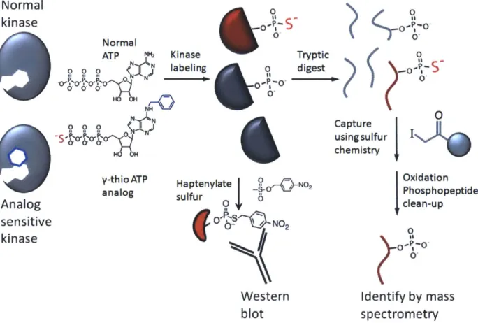
Radioactive tagging on y-phosphate of the ATP analog was originally used for selective labeling and detection of AS-kinase substrates, but purification and identification remained problematic. Recently, two procedures have been reported that use y-thiol-phosphate ATP analogs to thiophosphorylate substrates of cyclin/CDK complexes in whole cell lysate48'4. In
both cases, thiophosphate serves as a nucleophilic "handle" to purify peptides from tryptic digests so that substrates can be identified by tandem mass spectrometry (Figure 1). These experiments rely on the unique ability of the AS-kinase to bind and utilize the ATP analog; any
non-specific utilization of the ATP analog by other kinases in the cell will increase the background and may lead to false-positive substrate identifications.
In each case dozens of substrates of cyclin-dependent kinases were identified in yeast and human cells. A remarkable result from these AS-kinase based substrate identification
experiments is that only a small fraction (~10-20%) of the substrates have been previously reported. Given that dozens of substrates have been already been identified for many of these kinases by other methods, including classical biochemistry experiments, the AS-kinase results can be extrapolated to suggest that each of these kinases may have hundreds of substrates. In fact, compared to systems-level phosphoproteomic profiling experiments, the AS-kinase
experiments interrogate a much smaller part of the signaling network but reveal a similar level of
Normal
kinase
-P-5
S
.o
I}
I-Normal
ATP M Kinase Tryptic 0
0 0
:
0 N labeling o0- digest ~ .o~H~~ N N
0 NCapture
j
I
-S o- o 0usingsulfur i
chemistryW
y-thioATP Haptenylate 0jNO 2 Oxidation
analog sulfur - Phosphopeptide
Analog slu 0clean-up
sensitive
o _s
NO2kinase
s.
Western
Identify by mass
blot
spectrometry
Figure 1: an overview of how AS-kinases are used to label and identify substrates by Western blotting and by covalent capture of thiophosphorylated peptides.
complexity. These experiments suggest that signaling networks are much more highly connected than had been previously recognized and that a tremendous amount of mechanistic detail remains to be discovered at the level of direct interactions among signaling molecules, regulators and effector proteins.
Systems biology of the ERK1/2 mitogen-activated protein kinases
Having introduced the methods used in this dissertation, I would like to introduce the biological system in more detail. The extracellular-signal regulated kinases 1 and 2 (ERK1/2) are ubiquitously expressed in mammalian cells and phosphorylate proteins involved in
development5 0, glucose homeostasis5 1, immune function52 and memory53. Deregulation of
ERK1/2 activity is common in cancer and leads to proliferation, migration, resistance to 54
apoptosis, and loss of differentiated phenotypes
ERK1/2 regulates these various biological processes by phosphorylating hundreds of substrate proteins. Almost all of these substrates were identified by traditional biochemical approaches such as in vitro kinase assays, or by tracking phosphorylation in cells treated with activators or inhibitors of the ERK1/2 pathway. Substrate recognition is mediated by protein-protein docking sites, while the substrate binding cleft allows for serine or threonine
phosphorylation within an amino acid motif with proline strongly preferred at +1 and common at -2 56. ERK activity and substrate specificity is further regulated by scaffolds and adaptors that assemble the Raf/MEK/ERK activation cascade and direct sub-cellular localization5 7. Even
though many ERK substrates have been identified, incomplete knowledge of ERK targets remains a major hurdle to understanding the myriad biological consequences of ERK activity.
Despite the limitations of global phosphoproteomic analysis, several groups have used proteomic approaches to characterize ERK1/2 signaling by identifying phosphorylation sites that respond to MEK inhibition2 9' ,-60. Most of these studies only examined phosphorylation sites that are up- or down-regulated in the presence of the inhibitor. Among these groups, Kosako et al. used a combination of affinity chromatography and 2D gel electrophoresis to identify MEK-dependent phosphorylation sites, including 24 potential novel ERK targets, many of them members of the nuclear pore, and validated several of them as direct ERK1/2 substrates. In follow-up experiments the same group showed that phosphorylation of the nuclear pore by ERK1/2 regulates nuclear transport by reducing affinity of the pore for members of the
importin-p
proteins.All these studies of ERK1/2 signaling suffer from the weaknesses of global
phosphoproteomic analysis that were discussed earlier. To address these concerns we set out to identify additional downstream targets and pathways by using the chemical genetics approach developed by the Shokat lab.
ERK2 with Q103G substitution (AS-ERK2) uses ATP and ATP analogs efficiently and interacts normally with known substrates46 . AS-ERK2 was originally used to identify ubiquitin ligase EDD and nucleoporin TPR as direct substrates but discovery of additional targets was limited by their low abundance. To address this issue, we have optimized the solid-phase capture of thiophosphorylated peptides and added an additional phosphopeptide enrichment to reduce background, thereby enabling low-level substrate identification. Additionally, we have
incorporated quantitative mass spectrometry using stable isotope labeling in cell culture (SILAC)6 to establish statistical thresholds for identification of substrates over background phosphorylation, thereby decreasing false-positive substrate identification. Taken together these
constitute a major technical advance in the application of analog-sensitive kinases. Our approach is generally applicable to all AS-kinases and we hope that it will allow a much wider range of kinases and biological systems to be examined.
The second chapter of this dissertation describes protocol optimization and identification of novel ERK2 substrates in NIH 3T3-L1 fibroblasts. One of those substrates, a poorly studied the transcriptional repressor ETV3, is especially strongly phosphorylated by ERK1/2. The third chapter discusses the role of ERK activity in regulating ETV3, and presents a genome-wide
analysis of ETV3 transcriptional targets. The fourth chapter extends substrate capture and identification to a colon carcinoma system and analyzes the effect of an oncogenic mutation in KRAS on downstream targets of the ERK pathway. This analysis also reveals new targets involved in mRNA processing and histone methylation that may be important in the global rewiring that occurs during carcinogenesis.
2.
Protocol optimization and discovery of ERK2 substrates
To identify direct substrates of AS-kinases I began with the method originally developed by Allen et al.62 and Blethrow et al.48. Proteins in cell lysate are labeled with thiophosphate in a
reaction with AS-ERK2 and an N6-substituted y-thio ATP analog. The lysate is digested to peptides and thiophosphorylated peptides are captured on a solid-phase agarose support
functionalized with iodo-acetyl groups (SulfoLink beads), then released by oxidative hydrolysis to yield phosphorylated peptides. Phosphorylated peptides can then be identified by LC-MS/MS tandem mass spectrometry.
The first step in this project was to optimize a protocol to capture and identify
thiophosphate-labeled peptides from a complex mixture. I approached this by starting with a simple set of recombinant proteins and progressively increasing the amount of non-labeled protein. My initial studies used recombinant c-Jun protein thiophosphorylated by JNK1 in a reaction with ATPyS. First 10 picomole of labeled c-Jun was digested directly and reacted with SulfoLink beads. Mild washing followed by 5 minute treatment with dilute potassium
peroxomonosulfate recovered the labeled peptide with very little non-specific background. The same experiment with a 100-fold excess of bovine serum albumin added to the digest gave a similar result. Next I added 10 picomole of labeled c-Jun to 1 mg of cell lysate in 8M urea, a standard cell lysis buffer for mass spectrometry (the ratio of labeled to non-labeled peptide is approximately 1:100,000 by molar ratio). When I digested the mixture with trypsin and reacted with SulfoLink beads there was no detectable recovery of labeled peptide. I hypothesized that high salt concentration from the digest buffer may interfere with capture of the
hydrophobic cartridge (a standard procedure for White Lab phosphoproteomics experiments). With the additional clean-up step I was able to detect a significant amount of labeled c-Jun peptide, although the apparent yield was greatly reduced by approximately 90% relative to the less complex mixtures.
Since the eventual goal was detection of ERK2 substrates labeled in an in vitro kinase reaction I next added thiophosphorylated c-Jun into 1 mg cell lysate collected in a mild buffer suitable for kinase activity. Under this condition there was no detectable recovery of labeled c-Jun. I suspected that detergent from the buffer might interfere with peptide binding to the hydrophobic cartridge. Protein precipitation with either ammonium sulfate or
methanol/chloroform before digesting with trypsin resulted in a barely detectable recovery of labeled peptide, along with a dominant signal from non-phosphorylated background peptides. The signal for labeled c-Jun peptide was approximately 1/10,000 of the signal relative to
analyzing the same amount of c-Jun by itself. This was encouraging but would not be sufficient to discover novel ERK2 substrates.
Next I considered how to reduce level of background from non-specific binding, and whether sample might be lost by adsorption on surfaces throughout the procedure. Dilute phosphorylated peptides can be rapidly lost by absorption on glass, metal and plastic surfaces. To ensure that this would not interfere with my experiment I assembled a system using
microcapillary tubes so that labeled peptides were eluted directly onto a hydrophobic HPLC column. This also ensures that there are no losses from pipetting or transferring the sample between tubes.
Non-phosphorylated background can interfere with detection of phosphorylated peptides in two ways. First is that it takes up to a second for the mass spectrometer to isolate, fragment
and analyze a peptide, so a large number of peptides can overwhelm the ability of the instrument to collect fragmentation spectra. Second is that non-phosphorylated peptides take positive charge more readily than phosphorylated peptides, and can therefore suppress ionization of phosphorylated peptides.
To address the large non-phosphorylated background I first tried to adjust the wash stringency and elution conditions. Washing the SulfoLink beads in 50% acetonitrile, 5M sodium chloride, and 5% formic acid had only a minimal effect on the level of background peptides. This suggested that background peptides were not released from the agarose beads until
treatment with oxidizing agent. I varied the concentration of the oxidizing buffer from 1 mg/mL to 10 mg/mL of potassium monopersulfate, and elution times from a few seconds to ten minutes. More intense oxidizing treatments resulted in higher background, but reducing the treatment also limited recovery of labeled peptides. To ensure complete recovery I settled on 10 minute
treatment with 2 mg/mL oxidizing agent, even though it results in background signal that would be about 10 times too high to identify labeled ERK2 substrates.
To reduce the level of the non-phosphorylated background I included an additional phosphopeptide enrichment using immobilized metal affinity chromatography (IMAC) , which is a well-developed protocol in the White Lab2 4. This reduced the level of background by
approximately 90%, an acceptable level for identifying novel phosphorylated peptides. At this stage the apparent recovery of labeled c-Jun peptide was in the range of 1-3%, and I attempted to identify ERK2 substrates in cell lysate labeled by an in vitro kinase reaction. This experiment identified two known ERK substrates, stathmin and TPR, both well-known ERK targets and extraordinarily abundant structural proteins, but the experiment did not identify promising novel targets.
WT-ERK2 cells Sulfur-containing oxidize affinity Phosphorylated
-- --.. lodoacetyl peptides (elutethiophosphate) chromatography peptides
LgtAgLs-Beads -4HPLC-MS/MS
Light Arg/Lys Lyse Kinase Digest (H5) AS-ERK2 cells reaction
- - Discard Discard beads Non-phosphorylated Heavy Arg/Lys supernatant (cysteine-containing peptides
peptides)
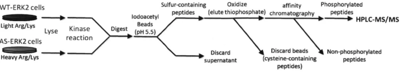
Figure 2: SILAC-labeled cells expressing AS- and wild-type ERK2 are lysed and substrates thiophosphorylated in vitro. After protein precipitation and tryptic digestion the labeled peptides are captured on iodoacetyl-agarose. The peptides are recovered by treatment with an oxidization agent and phosphorylated peptides are further enriched by IMAC prior to analysis by LC-MS/MS.
To further enhance recovery of labeled peptides I considered the binding chemistry of thiophosphate to iodoacetyl. Thiophosphate competes with cysteine to react with immobilized iodoacetyl. Because the reaction is greatly accelerated when the thiol group is negatively
charged I decided use the difference in pKa between thiophosphoric acid and cysteine to enhance the selectivity of the reaction. Blethrow et al. originally used a binding reaction at pH 7.0, corresponding to about 9% deprotonation of cysteine thiol (pKa depends on the local
environment but is in the range of 8.0 to 8.3). Since cysteine is at enormous excess relative to thiophosphate, this level of deprotonation could cause thiophosphate to lose out in competition to bind to the surface. Adjusting the pH of the binding buffer to 5.5 caused a several-fold increase
in recovery of thiophosphate, with the final apparent yield in the range of 10-20%. The final protocol is represented in Fig. 2 (including SILAC labels as discussed below).
There are several areas where this protocol could be optimized further, especially the final phosphopeptide enrichment step. The microcapillary-based IMAC system is sensitive to handling and requires considerable attention and manipulation. Other phosphopeptide
enrichment formats are available, notably batch-mode IMAC that requires much less handling, and titanium dioxide surfaces. Either of these approaches may improve the yield and specificity of the final enrichment and I hope that they will be tested in the future.
ERK2: WT AS WT AS Figure 3: Activated WT and AS-ERK2
AT P: + + -were immunoprecipitated from 3T3-LI
Ph~tTP7S + + cells and incubated with ELKi and either
PhEtATPyS:
- - + + ATP or ATP analog. Both kinases usepERK
normal ATP but only AS-ERK21thiophosphorylates ELKI with the ATP
analog. Thiophosphate is not recognized
SELK
1
by the phospho-ELK1 antibody as well as(Ser383)
normal phosphate.Selective labeling of AS-ERK2 substrates
Dr. Norman Kennedy at University of Massachusetts Medical School provided the AS-ERK2 gene (mouse AS-ERK2 with Q103G mutation). We produced a wild-type AS-ERK2 (WT-AS-ERK2) gene by site-directed mutagenesis and cloned both genes in pBabe retroviral vectors containing puromycin resistance. For an initial system we stably expressed HA-tagged WT and AS-ERK2 in 3T3-L1 fibroblast pre-adipocytes and verified by Western blot and ELISA that that both strains had similar expression of total ERK1/2. These cells were used in all the substrate tagging and identification experiments throughout this chapter. The 3T3-L1 cells were chosen as part of a collaboration involving Pfizer and several members of the White lab to study signaling down-stream of the insulin receptor.
Since the AS-kinase approach relies on selective affinity of the AS-kinase for bio-orthogonal ATP analogs, we confirmed selective utilization of PhEt-ATPyS by AS-ERK2 relative to wild-type ERK2 (WT-ERK2) by an in vitro kinase reactions using normal ATP or PhEt-ATPyS (Fig. 3). In this experiment the cells were serum-starved for 3 hours, treated for five minutes with EGF at 100 ng/mL, and lysed in cold kinase reaction buffer. We
immunoprecipitated WT or AS-ERK2 using anti-HA antibody immobilized on agarose beads, and then washed the beads and combined them directly with the recombinant ELK protein and ATP or ATP analog as indicated. The reactions were treated with p-nitrobenzylmesylate
(PNBM) at 2.5 mM for two hours and then analyzed by Western blot for alkylated thiophosphate.
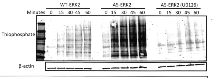
The next step was to determine the specificity of substrate labeling by AS-ERK2 in the context of a cell lysate. We serum-starved the AS-ERK2 and WT-ERK2 fibroblasts as before and treated them with PMA for two minutes, with or without 30 minute pretreatment with the MEK inhibitor UG 126 at 10 ptM. The cells were lysed as before, ATP analog was spiked directly
into the lysates, and they were incubated at 300 C for in vitro labeling reactions. As expected, labeling was strongest in the presence of AS-ERK2, and was dramatically decreased by pre-treatment with U0126. However, there was considerable labeling in the control cells expressing
WT-ERK2, indicating utilization of PhEt-ATPyS by wild-type kinases (Fig. 4). Interestingly, AS-ERK2 expressing cells treated with UO 126 + PMA had a lower level of labeling than WT-ERK2 cells treated with PMA alone. This suggests that much of the non-specific labeling is coming from normal ERK (especially since it is expressed at a high level), or else from kinases activated downstream of the ERK signal.
WT-ERK2 AS-ERK2 AS-ERK2 (U0126)
Minutes 0 15 30 45 60 0 15 30 45 60 0 15 30 45 60
Thiophosphate
*1. -in
Figure 4: Substrate labeling by WT or AS-ERK2 in cell lysate. Labeling in the WT
condition represents non-specific labeling by endogenous kinases. Much stronger labeling is observed with AS-ERK2, and labeling is almost completely abrogated by pretreatment with the MEK inhibitor U0126.
Total labeling intensity
1.2
1.0 AS-ERK2
Fluorescence 0.8 Figure 5: Total intensity for each of
(arbitrary 0.6 the conditions in Figure 4.
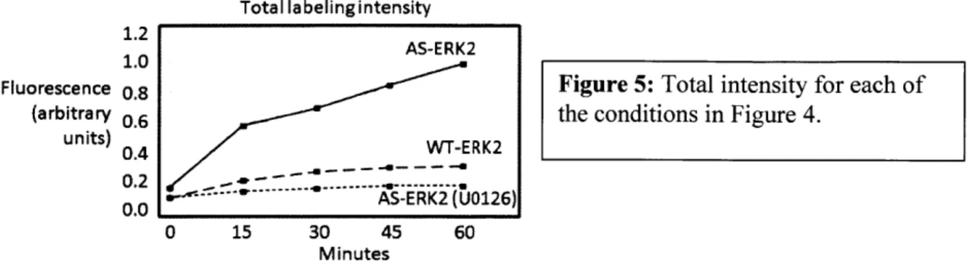
units) 0.4 WT-ERK2 0.2 --- 0... ---- 1 0.0 - AS-ERK2(U0126) 0 15 30 45 60 Minutes
The cumulative non-specific labeling by endogenous kinases indicated that a quantitative approach to measuring non-specific labeling was necessary to resolve low abundance substrates from background signals.
Identification of AS-ERK2 substrates
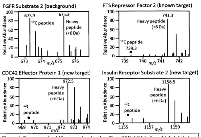
To distinguish ERK2 substrates from non-specific background we used SILAC for quantitative comparison between ERK2 and WT-ERK2 cell lysates. Cells expressing AS-ERK2 were grown in "heavy" L-arginine and L-lysine labeled with "C or "C and "N, while WT-ERK2 cells were grown in L-arginine and L-lysine containing entirely 12C and 14N. Samples were combined after in vitro kinase reactions and prior to tryptic digestion. The resulting peptides have distinctive mass signatures: background peptides have light and heavy ion signals with similar intensity, while ERK2 substrate peptides are enriched for heavy ions. Fig. 6 shows example MS spectra for one non-specific signal (FGFR substrate 2), one previously reported ERK substrate (ERF), and two novel substrates (CDC42EP 1 and IRS2).
This SILAC quantitative approach was combined with the solid-phase capture protocol to identify thiophosphorylated peptides from six independent labeling reactions with lysates of EGF-stimulated 3T3-Ll cells expressing WT or AS-ERK2. We used MASCOT 63 to match MS/MS spectra to phosphorylated peptides and examined the results for peptides matching the
FGFR Substrate 2 (background) 100 673.3 675.3 80- 12C peptide Heavy d peptide (+6 Da) 40A20 -673 674 m/z 675 676
CDC42 Effector Protein 1 (new target)
100 972.5 8 80 Heavy peptide S60 ~ (+6 Da) -o
<
40-
12C
u 20 -peptide -ii 20- e W 0 969 970 971 972 973 974 m/zETS Repressor Factor 2 (known target)
Insulin Receptor Substrate 2 (new target)
100 1158.5 80 - H eavy 60 - peptide (+6 Da) <40 - 2C peptide
20
-LL
1155 1157 m/z 1159minimal ERK2 substrate motif: pSer or pThr followed by proline. The MS/MS spectra for all potential ERK2 substrates were manually inspected to ensure proper placement of the
phosphorylated residue and validation with synthetic peptides was used to confirm any low-confidence assignment. SILAC values for phosphorylated peptides were normalized to values from non-phosphorylated peptides from within the same run.
Phosphorylated peptides from thirteen previously reported substrates were detected: Cx43, DYNC 112, DYNC1LIl, EGFR, ERF, KSR, LMNA, MAPKAP2, NUP153, TPR, STMN1, RPS3, and SORBS355,60. To differentiate AS-ERK2 substrates from background
phosphorylation we used these thirteen reported substrates to set a threshold for the quantitative difference in phosphorylation between the AS-ERK2 and WT-ERK2 expressing cells.
Figure 6: Representative MS spectra showing the effect of SILAC labeling. Light labeled peptides represent the negative control and heavy peptides the AS-ERK2 labeled lysate. The non-specific peptide has equal signals (top left), while the ERK2 substrates are dominated by signal from the heavy channel.
Previously reported substrates had background signals ranging from 6% to 33% of the signal from AS-ERK2 (Appendix 1), while non-specific peptides had ratios around 100% with a standard deviation (S.D.) of 19%. Since heavy-label incorporation into AS-ERK2 cells was greater than 98% (i.e. the SILAC ratio of protein from AS-ERK2 cells alone was ratio of less than 2%), we concluded that ERK2 substrate sites were subject to varying degrees of non-specific labeling. This result highlights the importance of quantitatively analyzing the negative control to distinguish background from bona fide substrates. To evaluate approximately 200 potential ERK2 substrates we used a threshold of 3.5 standard deviations below 100% in log-space, corresponding to a light:heavy ratio of 47% and a Bonferroni-corrected familywise error rate of 0.05 assuming that ratios are log-normal distributed under the null-hypothesis (e.g. less than 5% chance of one false positive).
A total of 98 peptides on 80 proteins met the criteria for in vitro ERK2 substrates, including 67 previously uncharacterized ERK2 substrate proteins. Three phosphopeptides matched the full motif but had light to heavy ratios close to 100% (ATF7, HIVEPI, G3BP-1). In agreement with our data, ATF7 phosphorylation does not respond to MEK inhibition in HeLa cells despite being homologous to ATF2 Thr53 (71 in human), a well-known ERK2 substrate
64
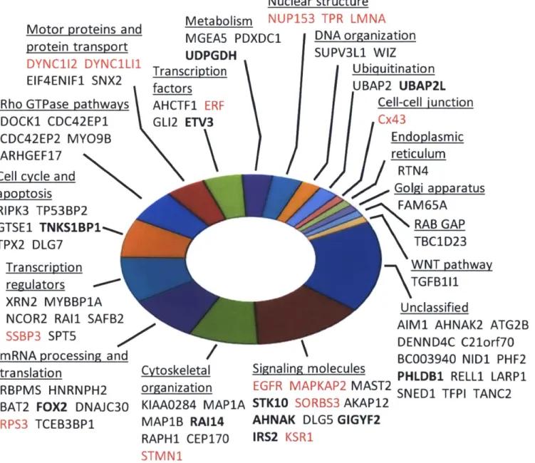
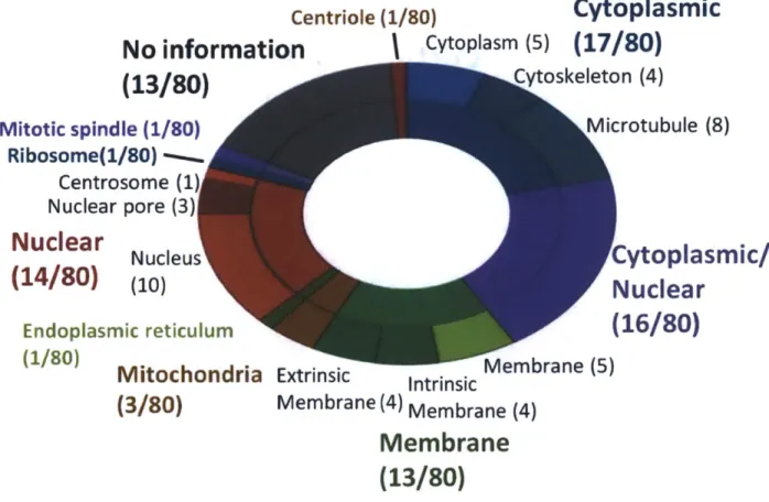
site . Without quantitative data these peptides could have been mistaken as ERK2 substrates. As expected based on the complex circuitry of the ERK2 network, substrates identified here span a wide range of both upstream and downstream-signaling pathways across many biological processes (Fig. 7). Consistent with dynamic ERK1/2 localization, substrates were associated with multiple cellular compartments, including cytoplasmic, nuclear, and membrane, among other compartments and organelles, and many substrates were further localized to structural components (cytoskeleton, microtubules, nuclear pore, plasma membrane) (Fig. 8).
Motor proteins and protein transport DYNC112 DYNC1LI1 EIF4ENIF1 SNX2 Rho GTPase pathways DOCK1 CDC42EP1 CDC42EP2 MYO9B
ARHGEF17
N
Cell cycle and
apoptosis RIPK3 TP53BP2 GTSE1 TNKSBP1', TPX2 DLG7 Transcription regulators XRN2 MYBBP1A NCOR2 RA11 SAFB2
SSBP3 SPT5
mRNA processing and translation
RBPMS HNRNPH2 BAT2 FOX2 DNAJC30
RPS3 TCEB3BP1
Nuclear structure
Metabolism NUP153 TPR LMNA
MGEA5 PDXDC1 UDPGDH Transcription factors AHCTF1 ERF GL12 ETV3 Cytoskeletal organization KIAA0284 MAP1A MAP1B RA114 RAPH1 CEP170 STMN1 DNA organization SUPV3L1 WIZ Ubiquitination UBAP2 UBAP2L Cell-cell iunction Cx43 Endoplasmic reticulum
RTN4
Golgi apparatus FAM65A RAB GAP TBC1D23 WNT pathwayTGFB111
UnclassifiedAIM1 AHNAK2 ATG2B DENND4C C21orf7O BC003940 NID1 PHF2 PHLDB1 RELL1 LARP1 SNEDI TFPI TANC2 Signaling molecules
EGFR MAPKAP2 MAST2 STK10 SORBS3 AKAP12
AHNAK DLG5 GIGYF2 IRS2 KSR1
Figure 7: The 80 proteins identified as in vitro substrates of ERK2 in 3T3-L1 fibroblasts. Proteins in red are previously reported ERK2 targets. Proteins in bold have ERK-dependent phosphorylation in intact cells, either in experiments described here (FOX2 and ETV3) or in phosphoproteomic experiments by other groups. Functional categories were derived by
Centriole (1/80)
Cytoplasmic
No information
I
Cytoplasm
(5)
(17/80)
(13/80)
C
toskeleton (4),Microtubule
(8)
Mitotic spindle (1/80)
Ribosome(1/80)
Centrosome (1
Nuclear pore (3)1
Nuclear
(14/80)
Nucleus
(10)
Endoplasmic reticulum
'
(1/80)
Mitochondria
(3/80)
'Cytoplasmic/
Nuclear
(16/80)
Extrinsin
Extrnsic
Intrinsic
Membrane (5)Membrane (4) Membrane (4)
Membrane
(13/80)
Figure 8: Subcellular location from GO annotations for each of the ERK2 substrates identified in 3T3-L1 cells.
Validating AS-ERK2 substrate specificity
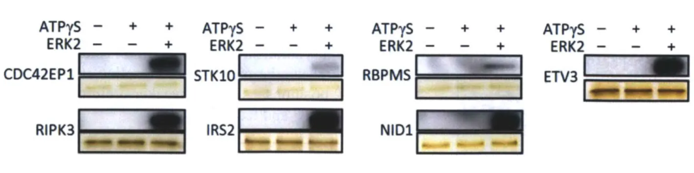
Although the ERK2 Q103G mutation has previously been shown to have no effect on substrate specificity, we tested several of the substrates identified in this study for their ability to be phosphorylated by WT-ERK2. We generated seven recombinant substrate proteins and all seven were phosphorylated in vitro by ERK2 to varying degrees (Fig. 9). We also mapped phosphorylation of recombinant CDC42EP 1, IRS2 and ETV3 by mass spectrometry following in vitro kinase reactions using wild-type ERK2 and identified additional ERK2-dependent
phosphorylation sites on each protein (Appendix 2).
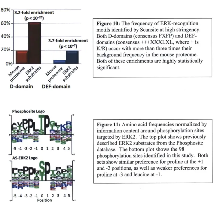
To determine whether the substrates identified in this study were consistent with known ERK1/2 binding motifs we used ScanSite to search substrates for ERK binding sequences: D-domains (positively charged residues three to five residues ahead of a hydrophobic sequence) and DEF-domains (FXFP, where X is any amino acid and Y may substitute for F)6 5. Both motifs were strongly enriched in the AS-ERK2 substrates compared to the SwissProt mouse proteome (Fig. 10). We also compared phosphorylation site motifs for AS-ERK2 substrates with known ERK2 substrates according to PhosphoSite (www.phosphosite.org) and found that major features of the two motifs were identical (Fig. 11).
ATPyS - + + ATPyS - + + ATPyS - + ATPyS - + ERK2 - + ERK2- - + ERK2- - + ERK2- - +
CDC42EP1 ST10 RBPMS ETV3
RIPK3 IRS2 NID1
Figure 9: Recombinant ERK2 substrates are thiophosphorylated by wild-type ERK2 in in vitro kinase reactions. This indicates that AS-ERK2 substrates are not artifacts of the Q103G mutation.
Although most detected substrates match the minimal ERKl/2 consensus motif SP/TP, and many match the full PXSP/PXTP motif, these motifs are neither necessary nor sufficient to identify ERK2 substrates. In fact, one phosphorylation site on the transcriptional repressor ETV3 was found that lacks the adjacent proline in the +1 position (discussed in the next chapter).
80%
J
-3.2-fold enrichment
(p <10-0)
60%.- Fi r.10: TheA Fre ue onn f ERK-rec- nitin
3.7-fold enrichment (p< 10-7) 0%i D A, D-domain DEF-domain Phosphosite Logo
motifs identified by Scansite at high stringency. Both D-domains (consensus FXFP) and DEF-domains (consensus +++XXXLXL, where + is K/R) occur with more than three times their background frequency in the mouse proteome. Both of these enrichments are highly statistically significant.
Figure 11: Amino acid frequencies normalized by information content around phosphorylation sites targeted by ERK2. The top plot shows previously described ERK2 substrates from the Phosphosite database. The bottom plot shows the 98
phosphorylation sites identified in this study. Both sets show similar preference for proline at the +1 and -2 positions, as well as weaker preferences for proline at -3 and leucine at -1.
40%-
20%--In vivo MEK-dependence of AS-ERK2 substrates
Several of the AS-ERK2 substrates identified here have previously been show to have altered phosphorylation following MEK inhibition. In particular, Pan et al. found reduced phosphorylation of AHNAK, ETV3, GIGYF2, PHLDB 1, RAI 14, TNKS 1 BP 1 and UBAP2L following treatment of EGF-stimulated HeLa cells with UO 12629. Kosako et al. found MEK-dependent phosphorylation of UDPGDH in 3T3-L1 fibroblasts6 0, Old et al. found
MEK-dependent phosphorylation of STK10 in the WM 115 melanoma cell line59, and Fritsche et al. found ERK-dependent phosphorylation of IRS266. The combination of in vitro interaction and cell-based assays supports these proteins as bona fide ERK2 substrates.
To identify additional phosphorylation sites affected by MEK inhibition in a global manner, we treated SILAC-labeled 3T3-L1 fibroblasts with EGF or PMA with or without U0126 pre-treatment. Lysates were digested with trypsin and antibodies recognizing the phosphorylated motifs PXpSP and PXpTP were used to immunoprecipitate peptides containing potential
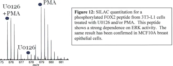
ERK1/2 substrate sites. These peptide IPs were processed by IMAC phosphopeptide enrichment and analyzed by LC-MS/MS. Only a couple dozen phosphopeptides were identified, probably because of restricted antibody specificity, but a number of known ERK2 substrates were identified, including ERF and the recently reported substrate NUP 153, as well as FOX2, a splicing factor and the novel substrate identified by AS-ERK26 7. Phosphorylation of both NUP 153 and FOX2 was strongly dependent on activity of MEK, indicating that these substrates are direct ERK2 targets in their biological context (Fig. 12).
In collaboration with Dr. Paul Boutz in the laboratory of Professor Phillip Sharp we have also looked at phosphorylation of FOX2 in MCF 1 OA cells. We have observed that FOX2 migrates more slowly by SDS-PAGE when ERK is activated. The effect is eliminated by
* PMA
875 876 877 878 879 880 881
m/z
treatment with phosphatase, or by expressing a form of FOX2 that does not include its second exon, which codes for the phosphorylation site. We have also performed detailed mapping for phosphorylation, methylation and acetylation across the FOX2 protein. We have observed only
one phosphorylation site that appears to be targeted by ERK, as well as methylation of arginines
361 and 445 and acetylation of lysine 257. Interestingly, there is only one sequence in FOX2
that matches a known ERK-binding motif, and that sequence is itself phosphorylated by an unknown kinase. The ERK-binding motif and ERK-target site both fall within an alternatively spliced cassette that is predominantly expressed in well-differentiated breast epithelial and carcinoma cell lines (personal communication, Paul Boutz). This leads us to believe that FOX2 contains an ERK-responsive cassette that can be regulated by alternative splicing, and possibly by phosphorylation of the ERK-binding motif. We used SILAC-labeled cells for quantitative co-IP to look for changes in protein binding when FOX2 is phosphorylated by ERK. Although we identified a number of heterogeneous nuclear ribonucleoproteins that associate with FOX2, none of them seem to do so in a phosphorylation-dependent manner. Additional experiments with phosphorylation site mutants are ongoing.
A complete list of the peptides identified in the motif IP experiment is contained in Appendix 3, along with a similar experiment in 3T3-L1 adipocytes which also found
MEK-Figure 12: SILAC quantitation for a
phosphorylated FOX2 peptide from 3T3-L1 cells treated with UO 126 and/or PMA. This peptide
shows a strong dependence on ERK activity. The same result has been confirmed in MCF 1 OA breast epithelial cells.
dependent phosphorylation of FOX2 following stimulation with insulin (adipocyte data was contributed by Dr. Katrin Schmelzle, a former postdoctoral associate in the White Lab).
Alternative methods and cellular systems
Comprehensive identification of kinase targets will require analysis of a wide range of cellular systems. A simple strategy would be to prepare activated AS-ERK2 from a strongly-expressing cell line, immunoprecipitate the kinase, and combine it with lysates from a range of cell types. We tested this by using thiophosphate Western blot to compare lysate labeled using immunoprecipitated AS-ERK2 with lysate from cells that had expressed AS-ERK2 directly. There was a stronger signal and more bands visible when AS-ERK2 was expressed in the target cells. We suspect that substrate recognition is enhanced by formation of protein complexes, possibly including scaffold and substrate proteins, prior to the lysis.
Based on this result we decided to explore expressing AS-ERK2 in additional cell culture systems. First we differentiated the 3T3-L 1 fibroblasts into adipocytes using an established protocol68. It proved difficult to get strong substrate labeling in adipocyte lysates, perhaps because of the abundant lipids. We also expressed AS-ERK2 in MCF 1 OA breast epithelial cells and in HepG2 hepatocellular carcinoma cells. MCF 1 OA cells normally have tightly-packed sheet-like morphology but can be induced to undergo epithelial-mesenchymal transition (EMT) by treatment with TGFp. When we conducted in vitro labeling reactions with these cells they showed an amount of labeling similar to the 3T3-Ll cells. Unfortunately when AS-ERK2 was expressed in MCF 1 OA they changed their morphology in a similar manner to TGFp treatment. The same did not occur when we expressed WT-ERK2 in the MCF 1 OA cells, perhaps suggesting that a difference in basal activity resulting from the Q103G mutation. We determined by
Western blotting that exogenous ERK2 expression was similar in each case, but the phenotypic difference may have resulted from a small difference in expression. We abandoned this system since it is not clear how to interpret the biological relevance of ERK substrates in MCF 1 OA cells following the apparent EMT.
Substrate labeling would be more biologically relevant if the reaction could occur in intact cells. We experimented with permeabilizing the 3T3-L1 fibroblasts and HepG2 cells. Treatment with the cholesterol-binding detergent digitonin did permeabilize both 3T3 and HepG2 cells to small molecules, but it also resulted in rapid detachment of 3T3 cells from the surface. HepG2 cells were more robust but we were unable to achieve a degree of substrate labeling even remotely close to that observed in in vitro reactions. There are several major challenges with this approach, especially competition with normal ATP (present at millimolar concentrations in most cells) and maintaining biological functions long enough to complete the reaction. Detecting substrates from in vitro labeling already stretches the sensitivity of our mass spectrometers, so we decided that identifying substrates from weaker labeling in vivo was unlikely to reveal new ERK2 targets. Our experience is consistent with work from the Shokat group. The only substrate identified when they conducted in-cell labeling reactions with ERK2 was TPR 62. TPR represents the strongest signal that we have seen in our own experiments and seems to be two to three orders of magnitude more abundant than most other substrates.
An alternative approach to identify labeled substrates could be using the thiophosphate antibody for protein IP. The Shokat group has identified a small number of kinase substrates by this approach and I have conducted a number of similar experiments. In my experiments the antibody demonstrated very poor specificity, always pulling down a large excess of non-thiophosphorylated proteins. A major disadvantage of protein IP is that proteins can often be
detected without discovering the thiophosphorylated residue. Given this weakness we did not choose to spend time optimizing the IP.
Discussion
Through an improved solid-phase capture chemical genetics strategy we identified a large number of known and novel substrates of the AS-ERK2 kinase. These novel substrates reveal a wide range of new connections between ERK2 and other signaling pathways, including adaptors and signaling molecules, regulation and effector proteins of the Rho GTPases, transcription factors, splicing regulators, and structural proteins. Validation using wild-type ERK2 shows that these substrates are not artifacts of the Q103G mutation, and cell-based assays indicate that a
significant fraction display MEK-dependence, and are therefore likely to be relevant in vivo. It is interesting that only 13 of the 80 substrates have been previously reported, and that these
represent a small fraction of the approximately 200 reported substrates. The limited overlap suggests that ERK2 may phosphorylate a much larger number of proteins than previously
recognized. We may not have identified a larger fraction of all the previously reported substrates because this cell line only expresses a restricted set of substrates. It may also be that some targets are not efficiently phosphorylated in the context of an in vitro reaction, either because they depend on particular localization or because they involve bridging interactions through scaffolds. Comprehensive identification of these targets will require a variety of complementary technologies and detailed examination of a wide range of cell types.
Novel substrates expand on the already considerable cross-talk between ERK and other pathways. For example, regulation of cell shape and migration is mediated in part by Rho GTPases, but very few ERK substrates have been reported in Rho signaling pathways69.
Identification of ERK2 substrate sites on RhoGEFs DOCK1 and ARHGEF 17, the RhoGAP MYO9B and the effector proteins CDC42EP1 and CDC42EP2 suggest that ERK regulates these pathways both upstream and downstream from the Rho GTPases. The ERK phosphorylation sites on these proteins may provide novel connections between MAPK signaling and regulation of cell morphology and migration. Additional investigation is needed to determine whether these phosphorylation sites function in normal physiological regulation of cytoskeletal structure and cell shape, or in pathological processes such as cancer metastasis.
Phosphorylation of adaptor proteins may represent feedback loops by which ERK regulates PI3K/Akt activity. Homology between the IRS2 target residues and a well-characterized sites on IRS 1 suggests that phosphorylation by ERK may reduce the effect of insulin signaling by competing with an overlapping P13K p8 5 binding site70. Serine 907 on
66
mouse IRS2 was recently identified as ERK-responsive in hepatoma cells and liver tissue Identification of additional ERK phosphorylation sites raises the possibility that the kinase regulates IRS2 through several modes of negative feedback. This mechanism is likely to be part of normal feedback regulation associated with insulin and other growth factor signaling, and could connect pathological ERK activation to insulin resistance caused by inflammation. Phosphorylation of GIGYF2A, GRB 10-associated adaptor protein, is another potential interaction with insulin signaling71, although this protein is not as well characterized as IRS2.
Another interesting point of cross-talk between ERK and other pathways is represented by phosphorylation of the GLI2 transcription factor. Although the ERK phosphorylation site has not previously been reported, it is proximal to residues near the C-terminus that are
phosphorylated by PKA and GSK3 to regulate GLI2 stability, and may therefore be critical for MEK-dependent cross-talk between Hedgehog and EGFR signaling7 2