HAL Id: hal-00567258
https://hal.archives-ouvertes.fr/hal-00567258
Submitted on 24 Oct 2018
HAL is a multi-disciplinary open access
archive for the deposit and dissemination of
sci-entific research documents, whether they are
pub-lished or not. The documents may come from
teaching and research institutions in France or
abroad, or from public or private research centers.
L’archive ouverte pluridisciplinaire HAL, est
destinée au dépôt et à la diffusion de documents
scientifiques de niveau recherche, publiés ou non,
émanant des établissements d’enseignement et de
recherche français ou étrangers, des laboratoires
publics ou privés.
Magneto-Chemotherapy
Charles Sanson, Odile Diou, Julie Thevenot, Emmanuel Ibarboure, Alain
Soum, Annie Brûlet, Sylvain Miraux, Eric Thiaudière, Sisareuth Tan, Alain
Brisson, et al.
To cite this version:
Charles Sanson, Odile Diou, Julie Thevenot, Emmanuel Ibarboure, Alain Soum, et al..
Dox-orubicin Loaded Magnetic Polymersomes:
Theranostic Nanocarriers for MR Imaging and
Magneto-Chemotherapy.
ACS Nano, American Chemical Society, 2011, 5 (2), pp.1122-1140.
�10.1021/nn102762f�. �hal-00567258�
Doxorubicin Loaded Magnetic Polymersomes: Theranostic
Nanocarriers for MR Imaging and Magneto-Chemotherapy
Charles Sanson,a,b Odile Diou,a,b Julie Thévenot,a,b Emmanuel Ibarboure,a,b Alain Soum,a,b Annie Brûlet,c,d Sylvain Miraux,e,f EricThiaudière,e,f Sisareuth Tan,g,h Alain Brisson,g,h Vincent Dupuis,i,j Olivier Sandre,a,b,i,j* and Sébastien Lecommandouxa,b*
a
Université de Bordeaux/IPB, ENSCBP, 16 avenue Pey Berland, 33607 Pessac Cedex, France
b CNRS, Laboratoire de Chimie des Polymères Organiques (UMR5629), Pessac, France c
CEA Saclay, LLB, 91191 Gif sur Yvette, France
d
CNRS, Laboratoire Léon Brillouin (UMR12), Gif sur Yvette, France
e
Université de Bordeaux, RMSB, 146 rue Léo Saignat, 33676 Bordeaux, France
f
CNRS, Résonance Magnétique des Systèmes Biologiques (UMR5536), Bordeaux, France
g
Université de Bordeaux, IECB, 33402 Talence Cedex, France
h
CNRS, Chimie et Biologie des Membranes et des Nanoobjets (UMR5248), Talence, France
i
UPMC Université Paris 6, 4 place Jussieu, 75005 Paris, France
j
CNRS, Physicochimie des Electrolytes, Colloïdes et Sciences Analytiques (UMR7195) Paris, France
Received for review October 14, 2010 and accepted December 24, 2010. Published online January 10, 2011, doi 10.1021/nn102762f
*
CORRESPONDING AUTHORS: Email: olivier.sandre@ipb.fr Tel: +33 (0)5-4000-3695, Fax: +33 (0)5-4000-8487,
Email: lecommandoux@enscbp.fr, Tel: +33 (0)5-4000-2241, Fax: +33 (0)5-4000-8487
ABSTRACT: Hydrophobically modified maghemite (γ-Fe2O3)
nanoparticles were encapsulated within the membrane of poly(trimethylene carbonate)-b-poly(L-glutamic acid)
(PTMC-b-PGA) block copolymer vesicles using a nanoprecipitation
process. This formation method gives a simple access to highly magnetic nanoparticles (MNPs) (loaded up to 70 wt %) together with a good control over the vesicles size (100 to 400 nm). The simultaneous loading of maghemite nanoparticles and doxorubicin was also achieved by nanoprecipitation. The deformation of the vesicle membrane under an applied magnetic field has been evidenced by small angle neutron scattering. These superparamagnetic hybrid self-assemblies display enhanced contrast properties that open potential applications for Magnetic Resonance Imaging. They can also be guided in a magnetic field gradient. The feasibility of controlled drug release by radio-frequency magnetic hyperthermia was demonstrated in the case of encapsulated doxorubicin molecules, showing the viability of the concept of magneto-chemotherapy. These magnetic polymersomes can be used as efficient multifunctional nano-carriers for combined therapy and imaging.
KEYWORDS: block copolymer vesicles, polymersome,
nanoprecipitation, superparamagnetic iron oxide nanoparticles, maghemite, magnetic hypethermia, magneto-chemotherapy, multifunctional, MRI contrast agent, doxorubicin, theranostics
Over the past decades, nanopolymeric therapeutics has proven to improve the effectiveness of cancer treatments in animal experiments.1-4 During this period, progresses in modern polymer (physico)-chemistry have enabled the design of polymeric carriers with ever higher levels of complexity
featuring addressable chemically reactive functions, defined chain architectures, and controlled morphologies and sizes. When applied to the field of drug delivery, these features allow achieving and combining several desirable properties such as high drug loading content, controlled release, increased circulation half-life and targeting of pathological areas or specific cell receptors. Polymer nanomedicines have the potential to increase the shelf life of chemotherapeutics before administration and to improve their efficacy after administration.5-7 A direct consequence of the latter is the reduction in the dosing concentration and frequency of administration of the drug, hence the minimization of toxic side effects on healthy tissues, which are currently a major problem in chemotherapy. Among the different classes of polymeric nanomedicines, block copolymer vesicles also termed polymersomes offer an attractive structure for drug delivery applications.8-12 This block copolymer self-assembly in a closed bilayer has fostered a considerable attention since both hydrophilic and hydrophobic drugs can be loaded either in the aqueous lumen or in the membrane core respectively,13, 14 thanks to a thick membrane that imparts long-term stability to the object. After drug loading, vesicle disruption inducing drug release can be either triggered by an environmental stimulus, such as pH, temperature, light, or oxidation,15-17 or can be the consequence of polymer hydrolytic or enzymatic degradation.18, 19 Even though a wide variety of polymer nanocarriers for drug delivery has shown efficient entrapment and controlled release of drugs in vitro, the evaluation of their biodistribution in vivo has become possible by non invasive methods. To address this issue, one strategy consists in incorporating imaging probes together with the drugs into the polymer nanoparticles. These dual polymer nanocarriers for simultaneous cancer imaging and treatment open the field to “theranostic nanomedicines”, combining diagnostic and
therapeutic components in an all-in-one nanoparticle.20, 21 Imaging probes to be loaded can belong to different families such as visible22, 23 and NIR fluorochromes,24, radiotracers25-27 or inorganic nanoparticles such as quantum dots,28-30 gold nanoparticles,31-35 or magnetic nanoparticles (MNPs).36, 37 Among the different MNPs, the so called “ultra-small superparamagnetic iron oxide” (USPIO) particles are synthetic γ-Fe2O3 or Fe3O4 nanometric grains in a perfectly dispersed
state (i.e. unclustered individual nanoparticles).38 As described in numerous review articles39-43, iron oxide MNPs also called USPIOs are commonly used as negative contrast-enhancing agents in MRI, enabling high spatial resolution acquisition, three-dimensional, non-invasive imaging of the human body. Hydrophilic “stealth” polymers are often employed to enhance the stability and biocompatibility of the MNPs in vivo by hindering their further aggregation and by an anti-fouling effect against proteins of the immune system called opsonins.42, 44 Besides the MRI contrast enhancement properties attributed to their ability to distort strongly the magnetic field lines,45 USPIOs can be used to kill cancer cells by their heating effect in radio-frequency magnetic fields. Hyperthermia (or thermal ablation) is identified as a promising approach in cancer therapy, particularly in combination with chemo- or radio-therapy.41 A promising hyperthermia route for treating deep tumors consists in concentrating MNPs around and inside the tumor site and increasing the temperature locally through conversion into heat of the energy from an external alternating magnetic field in the range of radio frequencies 100 kHz – 1 MHz. This magnetic hyperthermia led to an intense research activity both on the optimization of the conditions of treatment (power, concentration, geometrical parameters…)46-48 and on the characteristics of the USPIOs themselves (chemical nature, distribution of sizes…).39, 49-57
For this purpose of obtaining multifunctional drug vectors, hydrophilic USPIOs have been loaded at first in the aqueous compartment of liposomes.58, 59 Under a permanent magnetic field, magnetic liposomes deform into elongated ellipsoids, as it was evidenced for giant unilamellar vesicles.60-62 Interesting studies dealt with much smaller magneto-liposomes analogous in sizes to the pegylated lipid vectors of the DOXIL™ formulation of the anticancer drug doxorubicin hydrochloride (DOX). To combine magnetism and thermo-sensitivity, DOX was encapsulated into magnetic vesicles with a lipid membrane initially in the gel state and becoming fluid at a temperature reachable by magnetic hyperthermia.63, 64 The application of a RF magnetic field led to massive release of encapsulated DOX since the magnetic hyperthermia was sufficient to reach locally the main chain phase transition temperature of the bilayer, thereby increasing the membrane permeability.65 Recently, an analogous study with small hydrophobic USPIOs embedded in the membrane of liposomes evidenced the release triggered by a RF magnetic field of a fluorescent dye used as a model of hydrophilic drug.66 The possibility to target a solid tumor by using magneto-liposomes and an extracorporeal magnet to guide them has also been reported.64, 67, 68 Despite the tremendous results obtained with liposomes, the morphology of lipid/MNP systems strongly varies with MNP and lipid concentrations.69 They also suffer from the classical issue of instability associated with lipid bilayers,70 which incite to use of polymersomes as an alternative to liposomes. We have shown for the first time that hydrophobic USPIOs can be loaded into polymersome membranes of PB-b-PGA and that a reversible variation of the membrane thickness can be induced by the application of a magnetic field.71-73 Later, Förster et al. induced the bridging of adjacent bilayers and formed multi-lamellar hybrid polymersomes by incorporating hydrophobic USPIOs into PI-b-PEO bilayers at a feed weight ratio up to 20 % sufficiently large to guide the vesicles by a magnetic field gradient.74
In the present article, we describe a convenient procedure to prepare well-defined magnetic polymersomes featuring a hydrophobic internal membrane core made of the biodegradable block poly(trimethylene carbonate) (PTMC) and a polypeptide biocompatible corona of poly(L-glutamic acid) (PGA). Having synthesized USPIOs with the appropriate characteristics (size and hydrophobic coating), those were embedded together with the efficient antitumor drug doxorubicin hydrochloride (DOX) into the membrane of dual-loaded vesicles by one-step nanoprecipitation. This process allowed reaching quantitative loading contents and controlling the final sizes with low polydispersity. The two-dimensional confinement of USPIOs inside the vesicular membrane was evidenced by small angle neutron and light scattering techniques and observed by atomic force and transmission electron microscopy. The magnetic membrane of the PTMC-b-PGA polymersomes was shown to be reversibly deformable under a permanent magnetic field. The release of DOX under local hyperthermia conditions induced by an oscillating RF magnetic field was also evidenced as a proof of concept of magneto-chemotherapy with magnetic polymersomes.
RESULTS AND DISCUSSION Characteristics of the USPIOs
After synthesis75, size fractionation76 and surfactant coating77, the radius of gyration and the hydrodynamic radius
of the USPIO nanoparticles used in this work were
RGUSPIO=3.05±0.06 nm and RHUSPIO=4.7±0.07 nm as measured
respectively by SANS and DLS. The ratio
RGUSPIO/RHUSPIO=0.65 is not far from the theoretical value 0.775
for dense spherical particles,78, 79 the gap being reasonably ascribed to the contribution of the surfactant layer to RHUSPIO
only. Those sizes are in good agreement with the distribution measured by VSM (Supporting Information, S-d),80, 81 described by a Log-normal law of median diameter
DmagUSPIO=6.3 nm, width σ=0.22 and weight averaged diameter
Dw=7.5 nm. Concerning the magnetic hyperthermia capability,
a specific loss power (SLP) of 14 W/g was reported for USPIOs synthesized by the same route and of analogous distribution of diameters (DmagUSPIO=6.7 nm, σ=0.20, Dw=7.7
nm) but at larger frequency (fRF=700 kHz) and much higher
field intensity H0 (24.8 kA/m).54 Using its expected variation
with these parameters (~fRFH0 2
), we estimate a SLP value of 0.07 W/g in the conditions of biocompatible RF field used in this work (fRF=500 kHz and H0=2.12 kA/m).
Characteristics, structure and stability of USPIO-loaded vesicles
In a previous study, the conditions of nanoprecipitation with the PTMC-b-PGA block copolymer were varied: choice of THF or DMSO as good solvent of the blocks, order (solvent into water or reverse) and duration of the addition...82 The influence of each experimental parameter was rationalized in order to finely tune the sizes and PDI of the vesicles. In addition, the PTMC block was shown to be semi-crystalline with an apparent melting temperature in vesicles near 34°C (lower that value at 37°C in the bulk) that has a strong influence on the size of the vesicles and on their interactions.83 In the present study, we checked that incorporating inorganic nanoparticles at the nanoprecipitation step did not affect the self-assembly process of the diblock copolymer and that vesicular morphologies were still obtained. The conditions were selected according to the low PDI obtained, the compatibility of the organic solvent and of the obtained vesicles’ sizes with in vivo applications. The copolymer was first dissolved in DMSO with or without γ-Fe2O3 USPIOs.
Then water was added (up to 90 % of the final volume) at a controlled flow rate to trigger self-assembly. As the flow rate strongly influences the final size of the vesicles,82 we
considered two sets of conditions: an almost instantaneous addition (5 seconds) leading to small vesicles (RH=45-67 nm)
denoted WDi and a 15 minutes-addition leading to larger ones (RH=187-202 nm) denoted WD15. The characteristics of
nanoparticles’ dispersions prepared by either one or the other of these conditions at increasing USPIO feed weight ratios (FWR) are shown in Table 1.
Table 1. Size and polydispersity index (PDI) of nanoparticles’
dispersions prepared with increasing feed weight ratios of USPIO relatively to copolymer. Vesicles were prepared by nanoprecipitation in DMSO by adding water either in 5 s (WDi) or in 15 min (WD15). The image is a macroscopic view of samples.
Sample code FWR (%) RH (nm) PDI
WDi-0 0 67 0.07 WDi-20 20 50 0.14 WDi-35 35 45 0.16 WDi-50 50 47 0.16 WDi-70 70 52 0.18 WD15-0 0 202 0.05 WD15-20 20 196 0.09 WD15-35 35 195 0.20 WD15-50 50 187 0.22
The vesicles were found homogeneous in sizes, as observed by small PDI values in DLS. The loading of vesicles by USPIOs progressively increases the PDI (while remaining low) and slightly decreases RH. This hydrodynamic size decrease
(more pronounced for WDi than for WD15) and slight broadening of the sizes’ distribution (for both nanoprecipitation speeds) are ascribed to a larger hydrophobic effect when the copolymer is combined with USPIOs coated by surfactants, and thus to a larger driving force for a faster self-assembling process. No aggregation occurred below a critical USPIO FWR. Beyond this threshold value, the hydrophobic USPIOs began to aggregate during nanoprecipitation forming ill-defined
macroscopic clusters that rapidly migrated to the vial walls when approaching a magnet. This maximum FWR was respectively 50 wt % for WD15 and 70 wt % for WDi. The larger threshold FWR with WDi vesicles compared to WD15 ones can be ascribed to a much faster kinetics of formation, thereby minimizing the probability of USPIOs’ clustering before the completion of co-assembly with the copolymer. The maximum loading content of USPIOs in the membrane of the WDi-70 vesicles corresponds to a local volume fraction
% 1 . 12 mb USPIO=
Φ
. Interestingly, this is close to the reportedvalue of 11% for the insertion of USPIOs in bilayers of polystyrene-b-polyacrylate, whereas larger volume fractions
e.g. 21% lead to a morphological transition into micelles via
nanoparticles’ clustering.84 Fully dispersed and stable suspensions were observed below and up to these maximum USPIO FWR values. In these conditions, the shelf life is longer than several months at room temperature. Static light scattering (SLS) measurements conducted on the WDi-50 sample strongly suggested a vesicular morphology. By drawing the Berry plot85 over a scattering angular range from 50° to 150° and a concentration range from 0.2 to 1 mg/mL, we obtained the z-averaged radii of gyration (RG,z) allowing to calculate the ratio
ρ= RG/RH. While vesicles are characterized by ρ values close to
1, ρ values around 0.775 are expected for spherical micelles.78,
79
WDi-50 suspensions had a ρ value of 1.02 in good agreement indeed with vesicular self-assemblies.
A further insight to the exact morphology of USPIO-loaded PTMC24-b-PGA19 particles in either WDi or WD15 conditions
was brought by small angle neutron scattering (SANS) experiments. Figure 1 represents the intra-aggregate structure factor Sintra(q) of the USPIOs measured by SANS in a solvent
mixture matching the neutron scattering length density of the copolymer. The shape of Sintra(q) reflects both the interactions
between the USPIOs inside the object and the overall shape of their aggregates in the attractive regime.86, 87 In the small q-regime, Sintra(q) followed a power law with a slope
approximately -2 typical of flat samples, supporting a vesicle-type morphology. In this q-region (Kratky-Porod regime), the thickness of the USPIO layer in the vesicle membrane can be calculated from the slope of Ln[q2×Sintra(q)] plotted vs. q2
which is -δ2/12.88 From the experimental data, we obtained respectively δ=13 nm and δ=10 nm for samples WD15-50 and WDi-70. The vesicular membrane thus contains no more than one or two layers of magnetic colloids. More precisely, the data were properly fitted using a hollow sphere form factor with respectively an internal radius R=130 nm and shell thickness
δ=12 nm for WD15-50, and R=45 nm lumen radius with
membrane thickness δ=9 nm for WDi-70. These radii deduced from SANS fits agree pretty well with the hydrodynamic radii on Table 1 measured by DLS. At large wave-vectors, the q-4 scaling law is typical of the Porod’s regime expected for nanoparticles with a smooth interface. Moreover, Sintra(q)
presents a correlation peak around 8×10⁻² Å-1 (see Figure 1),
associated to a most probable USPIO inter-particle distance
dmax =2π/qmax= 7.8 nm. Considering their weight-average
diameter Dw=7.5 nm, we deduce that the USPIOs are closely
packed inside the vesicular membrane for both WD15-50 and WDi-70 samples. The SANS curve of WDi-50 vesicles in D2O
where the neutron scattering contrast of the USPIOs is almost matched also exhibits this correlation peak (Supporting Information S-c), as explained by the close-packed structure of holes in the copolymer membrane confining the USPIOs at a high local volume fraction.
A B
Figure 1. SANS curves of PTMC24-b-PGA19 vesicles WD15-50 (A) and WDi-70 (B), centrifuged then dispersed at 10 mg/mL in a
mixture H2O:D2O (65.6/34.4 v/v). Experimental intra-aggregate structure factors Sintra(q) of USPIOs are plotted as open circles. The solid
lines represent the simulated form factors respectively for hollow spheres of mean radius R=130 nm (PDI=0.17) with membrane thickness δ=12 nm (PDI=0.3) for WD15-50 (A), R=45 nm (PDI=0.35) with δ=9 nm (PDI=0.3) for WDi-70 (B).
Figure 2. TEM images of USPIO-loaded vesicles prepared by nanoprecipitation. (A) Low magnification picture of WD15-50 vesicles
(scale bar 1 µm); (B) Close-up view of a WD15-50 vesicle containing ~1500 USPIOs as measured by image analysis (scale bar 300 nm); (C) WDi-70 vesicles spreading on the substrate, which enables counting ~190 USPIOs on the left and ~220 USPIOs on the right (scale bar 100 nm); (D) Image of negatively stained WDi-50 vesicles, showing a group of vesicles laying intact on the carbon substrate (scale bar 50 nm); (E) Cryo-TEM image showing homogeneously dispersed WDi-50 vesicles (scale bar 200 nm). Inset: close-up view of two vesicles showing a mantle of respectively ~80 and ~110 close-packed USPIOs with some uncovered areas (scale bar 50 nm).
To summarize our SANS results, we found membrane thicknesses equal to 12.5±0.5 nm and 9.5±0.5 nm (either by scaling law or by form factor fitting) for WD15-50 and WDi-70 vesicles respectively. Due to the chosen H2O/D2O solvent
matching the copolymer scattering, these values represent the thickness of the USPIOs’ layer only. In D2O solvent were the
neutron scattering signal originates both from the magnetic contrast of iron oxide and the nuclear contrast of the copolymer, we measured a total membrane thickness 29.1±0.6 nm from Kratky-Porod’s plots of the data reported in the ESI file (figure S-c) for WDi vesicles independently of their iron oxide content (from 0 to 50% FWR), in accordance with the value 30±2 nm reported for the total membrane thickness of non magnetic vesicles made of PTMC24-b-PGA12 with a similar
PTMC block of molar mass Mn=2750 g/mol.
82
The measurement by SANS of the hydrophobic bilayer thickness for WDi vesicles well compares to the value δ=9.6 nm measured by cryo-TEM for polymersomes made of poly(ethylene oxide)-b-polybutadiene (noted EO26-BD46 or
OB2) with a total molar mass of 3600 g/mol and a hydrophilic fraction of 28%, thus a hydrophobic block mass of 2600 g/mol close to the one of PTMC here.70 The 25% increase of hydrophobic thickness for WD15 vesicles is ascribed to the swelling of vesicles’ membranes by the incorporation of USPIOs, which was presumably not possible for WDi ones due to their much higher curvature.
The WD15-50 and WDi-70 samples were further observed by TEM (Figure 2) and AFM (Figure 3) to confirm the vesicular morphology. TEM images mainly show the arrangement of the USPIOs because of the low electron scattering density of the copolymer compared to iron oxide. For both nanoprecipitation conditions (WD15 and WDi), hollow structures made of a close-packed arrangement of USPIOs were observed. The diameters measured on the TEM images 2B and 2C are around 750 nm and 150 nm respectively for WD15-50 and WDi-70 vesicles, which is larger than two times their hydrodynamic radii reported on Table 1 (374 nm and 104 nm respectively). This apparent discrepancy is ascribed to the total spreading of the vesicles onto the carbon substrate. The drying step during sample preparation and the strong wetting on substrates presumably induced the rupture of membranes, which explains the presence of fragments as well as not entirely closed structures.71-73 Unlike images A, B and C of Figure 2 that were obtained by spraying the samples onto the grids, image D originates from a more gentle protocol combined with staining (see Experimental) that led to vesicles sitting intact on the substrate. Both images D and E (cryoTEM) show apparent diameters much closer to the light scattering results, undoubtedly confirming the proposed structure. However we chose to show images A, B and C in spite of the spreading effect, because the flattening of the membrane onto the substrate enables to count the USPIOs per vesicle much easily than with the projection of intact spherical vesicles (D and E).
Figure 3. Tapping Mode™ AFM phase and height images of 1×1 μm surfaces of PTMC24-b-PGA19 vesicles prepared by
nanoprecipitation WDi without magnetic nanoparticles (upper panel) and with 50 wt % USPIO WDi-50 (lower panel). The average heights are measured on the right by cross-sections.
The WDi vesicles were also observed by AFM with and without the presence of 50 wt % USPIO. AFM phase images of empty vesicles (WDi-0) showed spherical vesicles, which aqueous interior leaked out due to drying and strong adsorption onto the freshly cleaved mica surface. When USPIOs were incorporated into the membrane (WDi-50), those presented multiple bright spots. The contrast of phase AFM pictures being proportional to the surface toughness,89, 90 we identify those bright spots with the hard inorganic USPIOs embedded
within the soft polymer matrix and spatially distributed over the vesicular surface as large patches. The average thicknesses of membranes spread on mica were analyzed on the AFM height images. These profiles revealed that the presence of USPIOs increase the thickness from 8 nm to 15 nm, the difference being very close to the weight average inorganic diameter Dw=7.5
nm. If the vesicles were adhering intact on the mica substrate, simply deflated by soft drying conditions, one would expect to measure an inorganic thickness equivalent to two layers of
USPIOs (respectively from the top and the bottom of the closed membrane). But the AFM images correspond to a single membrane thickness instead, which indicates that the vesicles had burst and spread onto the substrate, as depicted schematically in Supporting Information S-h.
To conclude on the SANS, (cryo)TEM and AFM results, these measurements show that due to their hydrophobic coating, the USPIOs are confined in 2 dimensions within the membranes, between the two leaflets of the copolymer bilayer.
Magnetization, migration and deformation under magnetic field of USPIO-loaded vesicles
As for the magnetization properties, the magnetization curves of both individual USPIOs and USPIO-loaded vesicles were fitted according to Langevin’s law of paramagnetism, each USPIO being a giant magnetic monodomain with an average magnetic dipole of 8200 µB, which is also the
approximate number of Fe3+ ions per USPIO (see Supporting Information S-d). On figure S-5, the VSM curve of WD15-50 vesicles loaded with 50 wt % USPIO exhibits a plateau value
MS/ΦmS allowing to determine a concentration 0.43 g/L of
USPIOs very close to the expected one (0.429 g/L, taking into account the USPIO FWR and the dilution effect during dialysis). Like previously stated, the vesicular dispersions were homogeneous, as attested by the detection of neither aggregates by DLS nor clusters of bare USPIOs moving when approaching a permanent magnet. Altogether, these results suggested a 100 % loading efficiency for the USPIOs at feed weight ratios lower than 50 % for WD15 and 70 % for WDi vesicles.91 Thus one can use mass conservation to deduce the values of the volume fraction Φ of USPIOs inside the membranes, on the basis of their initial weight ratio (FWR) relatively to the copolymer and their respective mass densities dUSPIO=5.1 g/cm3
and dcopo=1 g/cm3. An estimate of the mean number of USPIOs
per vesicle of radius RH and membrane thickness δ can be
calculated according to:
V
Φ
V
N
USPIO mb USPIO mb USPIO /vesicle × = (1)where the volume of membrane is calculated by Vmb = 4π
RH2 δ using geometrical values obtained by DLS and SANS
respectively and VUSPIO is averaged over the size distribution
law of diameters (πDw3/6). These calculations for each set of
experiments gathered in Table 2 lead to estimated numbers of USPIOs in pretty good accordance with the values observed on the TEM pictures of Figure 2.
Table 2. Characteristics of USPIO-loaded vesicles: feed weight
ratio, hydrodynamic radius RH, local volume fraction Φ of
USPIOs in membrane and their number N per vesicle calculated by equation (1) with respectively δ=13 nm for WD15 samples and δ=10 nm for WDi ones (SANS values).
The migration of the WD15-50 vesicles under a controlled magnetic field gradient was also used to estimate differently the number of confined USPIOs averaged on a large population of vesicles. The assessment of the ability of magnetic polymersomes to be attracted and concentrated at a specific location in vivo is also particularly relevant. Compared to magnetophoresis experiments with objects of sizes around 10 µm such as giant liposomes or biological cells,62, 92-95 a supplemental difficulty arose from the low value of the Peclet’s hydrodynamic number, which means that the magnetophoretic motion of the vesicles was not significantly larger than their Brownian motion (see the videos supplied as ESI). Usually, magnetophoretic measurements with such colloidal particles prone to thermal agitation are done by measuring light absorption profiles as a function of time and space.96-99 In the present work, we chose alternatively a statistical method to infer the average drift velocity Vdrift and the diffusion constant
Dvesicle by following a large number of individual trajectories,
as once described in a study of Brownian colloids in a liquid crystal.100
A
B
Figure 4. Video microscopy snapshot (inverted bright field
image) superposed with the trajectories (A) during 10s of WD15 PTMC24-b-PGA19 vesicles loaded with 50 wt % of
USPIOs. The vesicles were discriminated from the background by image analysis with the ParticleTracker plugin for ImageJ. The magnetophoretic mobility of the vesicles is visualized by the shifts between the histograms (B) of displacements in the x (red markers) and the y (blue markers) directions for durations respectively of τ=1/24=0.042 s (circles) and 2τ (diamonds). The arrow indicates the direction of magnetic field gradient of intensity dB/dz=18.5 T/m. The scale bar represents 20 µm. Sample code USPIO FWR (%)
Φ
mb USPIO (%) RH (nm)N
USPIO /vesicle WD15-20 20 3.8 196 1085 WD15-35 35 6.4 195 1830 WD15-50 50 8.9 187 2340 WDi-20 20 3.8 50 55 WDi-35 35 6.4 45 75 WDi-50 50 8.9 47 110 WDi-70 70 12.1 52 190The Brownian motion appears isotropic with a unique translation diffusion constant Dves=1.11 µm
2
/s. The Stokes-Einstein’s formula gives a hydrodynamic radius deduced by video-microscopy RH
video
=196 nm comparable to the value obtained by DLS. Due to an imperfect alignment of the magnetic field gradient with the x axis, we had to extract both coordinates of the magnetophoretic motion to calculate the total drift velocity:
µm/s
09
.
1
2 2+
=
=
x y driftV
V
V
(2)This experimental value is compared to the theoretical estimate obtained by balancing the forces acting on a spherical magnetic vesicle at steady state in an external magnetic field gradient, which are the magnetophoretic force FB, and the
viscous drag, Fv, acting against it. The two forces are given by:
dz dB m
FB = (3)
and Fv =6πηRHVdrift (4)
where m is the magnetic moment of the vesicle,101 dB/dz is
the gradient of the magnetic field, η is the viscosity of the solvent, RH is the hydrodynamic radius of the vesicle, and Vdrift
is the velocity of the particle. From the exact balance of the magnetic and the viscous forces, we calculate a theoretical magnetophoretic velocity Vdrift≈0.5 μm/s for the WD15-50
vesicles under a field gradient dB/dz=18.5 T/m. The factor around one-half between the expected drift velocity and the value Vdrift=1.09 μm/s measured experimentally cannot be
ascribed to the statistical noise because the uncertainties of the average displacements were estimated at 0.4% and 1.6% for the histograms at τ and 2τ time steps containing respectively 64519 and 3920 data points. As a tentative explanation, we know from a reported work on giant magnetic liposomes that the drag coefficient is enhanced compared to Stokes’ formula (4) if the vesicles were deformed by the field into high aspect ratio ellipsoids during their migration.102 Another correction compared to the drag coefficient of a solid sphere originates also from the viscous dissipation inside the fluid magnetic membrane, for instance if it was subjected to a “caterpillar” or a “crawling” motion.103 In addition to these pure hydrodynamic effects, the measured Vdrift higher than its expected value might
be explained by an underestimate of the numbers of USPIOs per vesicle appearing in Table 2. This would occur for example in the case of a non negligible amount of “blank vesicles” that were undetected but increased the average LC of iron oxide inside the magnetic vesicles above the USPIO/copolymer ratio (FWR) used for nanoprecipitation.
Apart from estimating the magnetic payload of the vesicles, the magnetophoresis experiment is also relevant to estimate their efficiency for magnetic guiding both in vivo and in vitro. Their magnetophoretic mobility in the vicinity of a strong NdFeB magnet is indeed of the same order of magnitude than
values ≈1 µm/s reported by studies that evidenced the enhanced uptake of magnetic nanocarriers by cell cultures under field gradients104, 105. For in vivo experiments, it was hypothesized that the accumulation of magnetic colloids injected in the main blood stream at a specific region under magnetic field requires that their migration is faster than the blood velocity in the smallest vessels alimenting the tumor.98 However, the guiding of magnetic stealth liposomes injected in the caudal vein of mice by a strong permanent magnet applied directly on a solid tumor was evidenced, even though their drift velocity was 10 µm/s only.68 A model experiment consisting in attracting clusters of MNPs of sub-micron diameters (330 nm) by a permanent magnet while they were circulating in a flow loop showed that they were efficiently deposited at the surface of the capillary near the magnet even with a stream velocity as high as 1 cm/s.106 Therefore we believe that the USPIO loaded vesicles WD15-35 or WD15-50 are good candidates for such magnetic targeting applications, whereas the WDi vesicles might be too small and contain an insufficient number of USPIOs.
The magnetic response of PTMC24-b-PGA19 vesicles with
their membrane filled by 50 wt % (WD15-50) or 70 wt % USPIO (WDi-70) was also studied by anisotropic SANS under an applied magnetic field. Vesicles were dispersed in light water (H2O) in order to work in almost pure nuclear contrast
conditions under field. The magnetic contrast of the γ-Fe2O3
USPIOs in H2O being much lower than the nuclear contrast, the
anisotropy of the SANS signal was not simply due to magnetization but reflects the spatial organization of the USPIOs and their possible rearrangement under magnetic field. The SANS patterns of WD15-50 vesicles are shown on Figure 5 at increasing field intensities up to 1 T.
The scattering patterns became clearly anisotropic when a magnetic field was applied to the sample. The lines of iso-intensity in the 3×10⁻³ – 3×10⁻² Å-1 q-range were elliptical,
elongated perpendicularly to the field direction. One possible scenario compatible with this asymmetry consists in the deformation of the hollow spheres formed by the USPIOs into either oblate or prolate ellipsoids symmetric by rotation along the field direction. However, one should keep in mind that the observed q-range corresponds to the length scale of the membrane thickness rather than to the whole size and shape of the vesicles.71-73
In order to study this shape anisotropy more quantitatively, the scattering patterns were averaged in angular sectors around two directions parallel (//) and perpendicular (┴) to the magnetic field. Examples of the resulting intensity curves are plotted on Figure S-2 (Supporting Information S-b). By comparing the difference q⊥ - q// in these two directions relatively to the wave vector q0 obtained by an isotropic averaging at the same intensity value, an anisotropy factor could be calculated for each sample and each magnetic field intensity (Table 3).
Figure 5. Anisotropic SANS patterns of WD15-50 in H2O in the q range 3×10 -3
– 3×10⁻² Å-1 under a magnetic field (horizontal) of intensity B=0 T (left), B=0.1 T (middle) and B=1 T (right). Each color corresponds to an iso-intensity range.
Table 3. Anisotropy factor of WD15-50 and WDi-70 vesicles
calculated from anisotropic averaging of their SANS patterns at 6 cm-1 iso-intensity under increasing magnetic field intensities.
The calculated anisotropy factors confirmed the increase of membrane anisotropy with the applied magnetic field already visible on the SANS patterns. The anisotropy factor increased mainly between 0.1 and 0.6 T for both vesicular dispersions and remained almost constant up to 1 T. The plateau value reached at 0.6 T is ascribed to the saturation of the magnetic moment of a vesicle above B ≈ 0.7 T as observed on the magnetization curve (Figure S-5). It is worth noticing that the anisotropy parameter is smaller for WDi-70 than for WD15-50 vesicles, as explained by the number of USPIOs per vesicle (12 times smaller for WDi-70 than for WD15-50) and by the vesicle size (4 times smaller). Presumably due to their smaller size associated with a higher membrane curvature, WDi-70 vesicles are less prone to magnetic deformation than the much larger WD15-50 ones.
USPIO-loaded vesicles as contrast agents for MRI
The efficiency of MRI contrast agents based on USPIO is usually assessed by measuring the T1 (longitudinal) and T2
(transverse) relaxation times of the proton spins relaxations. Then the relaxation rates 1/T1 and 1/T2 are plotted versus total
iron concentration in mM and the resulting slopes (s-1mM-1) called respectively r1 and r2 relaxivities can be used to compare
different samples of USPIO differing by their size, dispersity, local concentration, aggregation state or any other parameter like the confinement in either a hydrophilic or a hydrophobic environment. In particular the encapsulation of USPIOs in hydrophobic polymers hampers the diffusion of water protons in the vicinity of USPIOs, which results in poor T1 contrast
enhancement,107 so that we can infer the same effect in our systems where the USPIOs are buried within a hydrophobic membrane. In addition, r1 decreases rapidly as a function of the
applied magnetic field (i.e. the Larmor’s resonance frequency) while r2 reaches a plateau value due to the so-called “secular
term” in its theoretical expression.45, 108 The T1 and T2
relaxation times were measured on a 4.7 T (200Mz) research MRI system. Their inverse values 1/T1 and 1/T2 were plotted as
a function of iron molar concentration for PTMC-b-PGA vesicles prepared in WDi condition with USPIOs’ FWR ranging from 20 to 70 wt %. The r1 and r2 relaxivities deduced
from the slopes are reported on Table 4. To assess the experimental sensitivity, the solutions were diluted by a factor from 20 to 500: no vesicle size variation was observed by DLS under such dilutions. While r1 values are weak and almost
constant (~3 s-1mM-1), r2 values are of the same order of
magnitude (~100 s-1mM-1) and even larger than for commercial
T2 contrast agents: these are either individual (AMI-227,
Ferumoxtran/Sinerem)109 or clustered (AMI-25,
Feridex/Endorem)110, 111 USPIOs coated with a Dextran polymer, the latter being denominated SPIOs because of their larger size. The clustering of USPIOs inside objects at a high
loading density is indeed known to enhance the negative contrast for T2-sequence IRM images compared to individual
USPIOs, as reported for hydrophobic USPIOs in micelles107, 112,
113
or hydrophilic ones in liposomes114 or electrostatic coacervates with charged polymers.115 However, the articles that link the experimental r2 values to theory are rather scarce:
they dealt with SPIOs made of magnetite cores aggregated by Dextran of varying sizes116 and more recently with individual
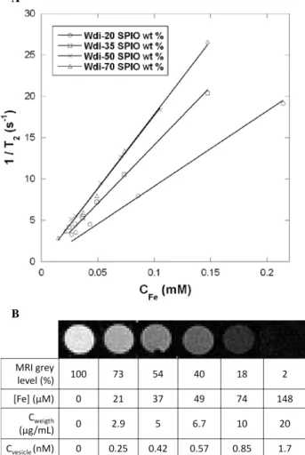
USPIOs coated by a controlled thickness of silica.117 In the present work, we note a steeper slope on Figure 6-A for WDi-35 (r2=134±2 s-1mM-1) as compared to WDi-20 (r2= 81±1 s -1
mM-1). These relaxivities are both larger than the value
r2=39±2 s -1
mM-1 reported for USPIOs individually dispersed in water synthesized by the same aqueous route and of the same size distribution.115 For 50 wt % USPIOs in the vesicles, the transverse relaxivity reached r2= 173±7 s-1mM-1, but then it
saturates near this value at r2= 182±4 s -1
mM-1 for the highest loading content 70 wt %. On the theoretical point, A. Roch et
al. predicted such a plateau of r2 when increasing the size of
clusters of USPIOs.116
A
B
Figure 6. (A) Transverse relaxation rates (1/T2, s-1) as a
function of iron concentration (mM) for PTMC24-b-PGA19
vesicles (WDi) loaded with 20, 35, 50 and 70 USPIO wt %. The slopes give the r2 value, respectively 81±1, 134±2, 173±7
and 182±4 s-1mM-1; (B) T2-weighted MRI images extracted
from T2 measurements experiment (4.7 T; multiple spin-echo
2D imaging sequence; TR=10000 ms; inter echo-time, 5 ms;
number of echo images, 256; FOV, 50×50 mm; matrix, 128×128; slice thickness, 1 mm) of WDi-70 vesicles at different dilution factors. The table gives the molar concentrations of iron ions, the total weight concentrations and the equivalent molar concentrations of vesicles.
To evidence the effect of USPIO-loaded vesicles on T2
-weighted MR images, Figure 6-B shows MR images of wells
B (T) (q⊥ - q//) / q0 (%) WD15-50 WDi-70 0.1 11.3 6.8 0.6 24.4 12.7 1 26.8 12.0
containing increasing concentrations of WDi-70 vesicles. A remarkable darkening (i.e. negative contrast enhancement) appeared even at low vesicle concentration. The MRI detection limit, defined as the copolymer concentration at which the MRI signal intensity decreases to 50 % of that of pure water, 107 was measured at 6.7 μg/mL for WDi-70 vesicles. Since the molar mass of the vesicles measured by SLS is 1.182×10⁷ g/mol (Supporting Information S-i), the above sensitivity limit corresponds to a vesicle concentration of approximately 0.57 nM, which is one order of magnitude lower than the 5 nM reported for magnetic micelles107 and, to our knowledge, the lowest value ever reported. For applications such as the evaluation of the bio-distribution or the targeting efficiency of a drug conveyed in nano-carriers, the concentration of ferric ions may not be the most relevant parameter for the radiologists. Therefore the r1 and r2 relaxivities are also expressed in Table 4
according to the concentration of vesicles in nM to facilitate the comparison with other nano-particular contrast agents. Unlike the relaxivities per ferric ion which saturate, their values per
WDi vesicle increase monotonously with the magnetic FWR inside the membrane, at an almost constant hydrodynamic size.
Table 4. Longitudinal (r1) and transverse (r2) relaxivities of
USPIO-loaded WDi vesicles when used as contrast agents for MRI at 4.7 T, deduced from the linear fits of the relaxation rates 1/T1 and 1/T2 versus molar concentrations both of ferric
ions (in mM) or of vesicles (in nM). The number of Fe3+ per vesicle is the product of the number of USPIOs per vesicle
(Table 2) by 8200 Fe3+ per USPIO on average.
Doxorubicin loading and release by macroscopic heating
To determine the feasibility of magnetically controlled drug release, a dual loading of USPIOs and of doxorubicin was carried out. The nanoprecipitation was performed at pH 10.5 in order to deprotonate the DOX (pKa~8.3), thus maximizing the
loading content (LC=34% without size variation at 50% FWR) and extending the release duration as described in a previous work.118 For each vesicular dispersion, the USPIO feed weight
ratio (FWR) was fixed at a value lower than the maximum USPIO loading (namely 50 wt % for WDi and 35 wt % for WD15) so that space was left in the membrane for DOX entrapment. The DOX FWR in the nanoprecipitation mixture was then progressively increased. A DOX FWR of 20% was selected for both vesicular types since a drug loading at this level did not alter the self-assembly of the vesicles significantly: Table 5 shows indeed a moderate variation of their hydrodynamic size (RH decreases by 16% for WD15 and
increases by 8% for WDi) and an unchanged surface charge. A larger 30% DOX FWR can be sustained by WD-15 vesicles without any size change, but for the smaller WDi-50 vesicles it leads to a two-fold size increase, presumably due to their larger curvature energy already invoked to explained their lower
deformability under static magnetic field. Table 5. Doxorubicin feed weight ratio (FWR), hydrodynamic
size, polydispersity index and ζ potential of WD15-35 and WDi-50 vesicles. The pictures show the corresponding sample tubes. Sample code RH (nm)
N
+ 3 Fe /vesicle ion Fe 1 3+ r (s-1mM-1) vesicle 1 r (s-1nM-1) ion Fe 2 3+ r (s-1mM-1) vesicle 2 r (s-1nM-1) WDi-20 50 4.5×10⁵ 2.8±0.02 1.3±0.01 81±1 37±0.4 WDi-35 45 6.2×10⁵ 3.6±0.08 2.2±0.05 134±2 83±1.2 WDi-50 47 9.0×10⁵ 3.6±0.2 3.3±0.2 173±7 156±6 WDi-70 52 1.6×10⁶ 3.5±0.1 5.5±0.2 182±4 283±7*not determined
After nanoprecipitation with dual-loading in DOX and USPIOs, an extensive dialysis against a large volume (4 L) of Tris buffer (pH 7.4, 30° C, ionic strength 150 mM) during 4h allowed to reduce the pH back to 7.4 and to completely remove the unbound drug and DMSO. As for the colloidal stability of these dual loaded vesicles, their ζ potential remained strongly negative (~ -40 mV). Therefore the corona of PGA chains was unaffected, which excludes the precipitation of the USPIOs and of the drug onto the hydrophilic chains and proves their embedment deeply inside the vesicular PTMC-b-PGA membrane. The loading content (LC) and loading efficiency (LE) of DOX were determined by spectrophotometry. Values obtained for both vesicular dispersions with or without USPIOs are gathered in Table 6. A DOX LC around 10 wt% was found in all cases, independently of the presence of USPIOs in the membrane. Finally, the colloidal stability of the WDi vesicles was tested in MEM cell culture medium with fetal bovine serum (10% v/v FBS), and no change in size was observed for 24 hours.
Table 6. Influence of USPIO feed weight ratio on the DOX
loading content and efficiency into WDi and WD15 vesicles. The FWR is in wt % relatively to copolymer in DMSO before nanoprecipitation. The LC is measured by spectrophotometry after nanoprecipitation and dialysis. The LE is the yield LC/FWR. Sample code USPIO FWR (%) DOX FWR (%) DOX LC (%) DOX LE (%) WD15 0 20 12.5 74 35 20 9 52 WDi 0 20 9.5 47.5 50 20 12 70
Comparing precisely the DOX loading efficiency between dual loaded vesicles and non magnetic ones, we observe that LE decreases by 22% for WD15 vesicles, whereas it increases by 22.5 % for WDi ones. As a result, the insertion of USPIOs and DOX appears competitive in the case of the larger WD15 vesicles, certainly due to a lack of space in the membrane (the difference between
Φ
USPIOmb and its maximal value being only 2.5 %, see Table 2). On the opposite, the incorporation seems cooperative for the smaller WDi vesicles. Such synergetic effect of dual loading has already been mentioned forcopolymer micelles, for which the LC of DOX could be enhanced from 3 to 12 wt % by the co-encapsulation with hydrophobic USPIOs.36
Subsequently, in vitro release studies from the several prepared vesicular dispersions were monitored in various conditions by comparing the absorbance at λmax=485 nm with
the DOX absorbance calibration curve (after background correction). The release kinetics in vitro at 37 °C of WDi-50 and WD15-35 vesicles fed with 20 wt % DOX appear almost similar. As seen on Figure 7 indeed, a plateau at around 50 wt % of released DOX was reached in both cases after one day. As stated in a previous work on the physicochemical conditions to optimize the loading and release of DOX with PTMC-b-PGA vesicles (but for a DOX LC of 34 wt % 3 times larger than in the present work and without USPIOs),118 temperature has a strong influence on the kinetics as well as on the amount of drug released: the plateau values at 5°C, 20°C, 37°C and 45°C were found respectively equal to 5%, 30%, 60% and 85% of the initial DOX load in the vesicles. This temperature sensitivity is presumably due to the semi-crystalline nature of the PTMC blocks inside membranes evidenced once by microcalorimetry83 and in this work by birefringence measurement (see Supporting Information S-e).
On Figure 7, only 15 % of DOX was released after 6 hours at 23°C compared to 45 % released after the same time at 37 °C thus above the melting temperature of PTMC in the membrane of vesicles.
Figure 7. In vitro release kinetic profiles of USPIO/DOX dual
loaded vesicles with initial 20% DOX FWR obtained for WDi-50 (○) and WD15-35 (□) vesicles at 37 °C and for WDi-50 vesicles at 23 °C (●). In all cases, the release medium was Tris 10 mM (pH 7.4, ionic strength 150 mM).
Doxorubicin release by magnetic hyperthermia
Having in mind this thermo-sensitivity of the release rate of DOX in vitro from dual-loaded PTMC-b-PGA vesicles, we studied the effect of an excitation by an oscillating magnetic field of the USPIOs confined in the membranes. Submitted to a strong radio-frequency field, USPIOs are known to dissipate heat originating from friction losses of their magnetic dipoles according to two different relaxation modes: Néel’s relaxation consisting in the flips of each dipole between the “easy axes” of the crystalline structure and the Brownian rotational diffusion of the USPIO grains in the solvent of viscosity η. According to a commonly accepted model,50 the specific loss power under a Sample Code DOX FWR (%) RH(nm) PDI ζ (mV) WD15-35 0 152 0.15 -39.6 20 124 0.23 -39.3 30 128 0.16 nd* WDi-50 0 56.5 0.22 -40.8 20 61 0.15 -42.0 30 137 0.20 nd*
field of frequency fRF and mean intensity H0 expressed in W/g writes:
(
)
2 0 RF RF 0 RF, ''(f )H d f H f SLP =π χ with( )
(
)
2 eff RF eff RF USPIO 2 S 0 1 3 '' τ τ µ χ f f T k V m f B + = (5)Here χ’’(fRF) is the loss term of the dynamic susceptibility of
an USPIO with specific magnetization mS, mass density d and
volume VUSPIO. The effective relaxation time τeff corresponds to
the fastest mode between the two mechanisms participating to thermal dissipation. Both of them can be expressed as functions of the particle volume:
(
KaVUSPIO kBT)
0 Néel =τ ×exp
τ (6)
with τ0≈10−9s and Ka ≈104J/m3
and τBrown=3ηVUSPIO kBT (7)
Although it does not take into account the possible variation of relaxation times with the magnetic field intensity,119 this
model correctly describes the strong dependence of the SLP with the size distribution of a suspension of USPIOs53 and gives an optimal diameter about 14-15 nm. When USPIOs are confined in a viscous environment as in lipid compartments inside biological cells (endosomes), membrane, the Brownian relaxation mode can be neglected.54, 120
Figure 8. Influence of a RF oscillating magnetic field on the in vitro release kinetics of WDi-50 vesicles at constant bath
temperature (23 °C); ●: B=0 T; ■: AC magnetic field (fRF=500
kHz, B0=2.65 mT).
Figure 8 displays the kinetic profiles at constant temperature 23 °C with and without the application of an oscillating magnetic field of frequency fRF=500 kHz and mean field
intensity H0=2.12 kAm-1. Although these field conditions might
appear weak, they were already over passing by more than a factor two the upper dose of RF irradiation fRF×H0<4.85×10
8
Am-1s-1 recommended for a human being.121 After 7 hours, the DOX release content is multiplied by a factor 2 under RF field compared to the same vesicles with USPIOs embedded in the membrane but kept away from the coil. The heat produced via Néel’s relaxation is believed to increase the fluidity of the semi-crystalline polymeric membrane, increasing dramatically the diffusion of the encapsulated DOX out of the membrane. It should be stressed that we have not observed any macroscopic heating of the vesicular dispersion. 122 Even if the global temperature of the suspension remained almost unchanged, we
infer that a local temperature raise around 7°C took place in the close vicinity of the membrane (i.e. at the nanometric scale). The approximately two-fold enhancement of DOX release rate under the RF magnetic field (Figure 8) was indeed of the same type than the three-fold enhancement observed when the vesicular suspension was heated macroscopically by 14°C thus above the melting temperature of the PTMC blocks (Figure 7). A control experiment performed by placing the WDi-50 vesicles during several hours under a constant magnetic field of intensity B0=0.4 T showed that a static deformation of the
vesicles had no impact on the release rate of DOX, thereby confirming the necessity to excite the USPIOs at a frequency in the RF-range close to their Néel’s relaxation in order to detect an effect on the membrane permeability of the vesicles.
CONCLUSIONS
In the present study, the formation of new hybrid vesicular self-assemblies from the biodegradable PTMC-b-PGA copolymer and hydrophobically coated γ-Fe2O3 nanoparticles
has been investigated. Hybrid vesicles have been obtained by one-step nanoprecipitation, leading to high loading content of magnetic nanoparticles (up to 70 wt %) in the membrane together with a good control over vesicles’ size and dispersity. The vesicular morphology was elucidated by combining light and neutron scattering techniques together with electronic and atomic force microscopy. These magnetic vesicles exhibited a long-term colloidal stability and showed suitable properties for biomedical applications: being guided by an external magnetic field gradient created by a small permanent magnet, they also showed an important contrast enhancement in Magnetic Resonance Imaging with a particularly low (sub-nanomolar) detection limit. Dual encapsulation of magnetic nanoparticles with doxorubicin in the biodegradable vesicular matrix is very promising as a versatile method to prepare multifunctional drug nanocarriers. The drug release rate could indeed be enhanced twice under the application of a RF oscillating magnetic field producing a local hyperthermia at the scale of the membranes.
The well-known hyperthermia effect of USPIOs was utilized here in a softer and gentler manner of action on the polymersomes’ membrane permeability than by thermoablation, which is based on the melting temperature of a semi-crystalline polycarbonate block. In future studies, we will enhance the RF-triggered release effect by using USPIOs with larger diameters (e.g. by a factor 2), which are known to exhibit much higher specific loss powers (~100 W/g or more). Apart from a higher thermal dissipation acting on the membrane fluidity and hence on the diffusion constants, those larger USPIOs will be partially ferrimagnetic, i.e. with a magnetic anisotropy energy Ea>kBT. This might introduce another
mechanism of membrane permeation, by direct rotation of the grains at the frequency of the oscillating magnetic field. Such a mechanism would be reminiscent of the “molecular drill” effect123-125 predicted long ago for lipid bilayers under mechanical stress by adsorption onto a corrugated surface.
To summarize, by exhibiting biocompatibility of the polymeric matrix, ease of preparation, contrast enhancement in MRI and triggered release under RF oscillating field, those hybrid vesicles are good candidates for the magneto-chemotherapeutic treatment of cancer. This work evidenced for the first time the concept of multi-functional polymersomes to combine imaging and therapy, opening new avenues to improve cancer treatments and to understand their mechanisms. The impact of such theranostic systems on tumor regression is currently under investigation.
EXPERIMENTAL DETAILS Materials and syntheses
Polymer, drug and buffers. PTMC24-b-PGA19 diblock
copolymer was synthesized by ring-opening polymerization (ROP) of γ-Benzyl-L-glutamate N-carboxyanhydride initiated by an amino functionalized PTMC macroinitiator upon a previously published method.82 All the experiments were
conducted on a PTMC24-b-PGA19 (Mn = 4900 g/mol) block
copolymer which presents a hydrophilic weight fraction of 50 wt % and a molar mass dispersity of 1.15. The solvent for nanoprecipitation (DMSO) was used without prior purification. Doxorubicin hydrochloride (CAS: 25316-40-9) was supplied by Discovery Fine Chemicals (Wimborne, UK). DOX was reconstituted in DMSO, stored at 5 °C and used within one month. Sodium chloride, Tris-HCl and Tris base were provided by Sigma.
Iron oxide nanoparticles. Superparamagnetic nanoparticles of maghemite (γ-Fe2O3), also called USPIOs, were synthesized
by alkaline coprecipitation of iron(II) and iron(III) salts75 and sorted according to their size by fractionated phase separations.76 Briefly, the ionic strength was increased to screen the electrostatic interactions between the nanoparticles and obtain successive fractions of narrower size distribution, as measured all along the sorting process by vibrating sample magnetometry (VSM) and on the final sample by scattering techniques (SLS, DLS and SANS). For dispersion in CH2Cl2,
the surface of the nanoparticles was grafted by the anionic surfactant Beycostat NB09 (CECA, Arkema group, France) used to disperse inorganic pigments in aromatic and chlorinated oils (but insoluble in aliphatic solvents), which is a mixture of mono- and diesters of phosphoric acid. The grafting procedure (30 min at 60°C, 20 mol% relatively to iron) was previously described.77
Preparation of empty, USPIO loaded and DOX/USPIO dual-loaded vesicles
Carbonate buffer (pH 10.5, 50 mM, 4.5 mL) was added onto PTMC24-b-PGA19 (5 mg) dissolved in DMSO (0.5 mL) under
magnetic stirring (1000 rpm) in a plastic tube (1.5 cm diameter), leading to a homogeneous dispersion of vesicles. A syringe pump controlled the water flow rate during injection. Two addition durations (5 seconds and 15 minutes respectively) of water solution into DMSO were used in order to tune the final average vesicle size. The resulting samples were respectively called WDi (for “instantaneous”) and WD15. The organic solvent was then removed by extensive dialysis against 4 L Tris buffer replaced at least twice (10 mM Tris, pH 7.4, 25 °C ionic strength 150 mM).
USPIO loading was performed at different feed weight ratios (FWR) (wt USPIO/wt copolymer) using the same nanoprecipitation
method. A negligible volume of USPIO suspension in CH2Cl2
(e.g. VCH2Cl2/Vwater=0.55% for wtUSPIO/wtpolymer=50 %) was
added into the DMSO/copolymer solution prior to the addition of water. For DOX/USPIO dual-loaded vesicles, doxorubicin hydrochloride was at first solubilized in the DMSO/copolymer solution at 2 mg/mL before mixing with the USPIOs. After water addition, organic solvent and free DOX were removed by dialysis for 4h with a membrane of 3500 g/mol MWCO against 4 L Tris buffer (10 mM Tris, pH 7.4; 30 °C, ionic strength 150 mM). The doxorubicin loading content (LC) was determined after vesicle rupture using sonication in a mixture containing 80 % volume of DMSO. This solvent mixture induced the aggregation of USPIOs that were then separated by centrifugation (1h, 10000 rpm). Then the titration of DOX was performed from the UV absorbance at λmax =485 nm using the
known value for doxorubicin in a DMSO/Tris buffer (80/20 v/v) mixture as calibration (see Supporting Information S-g).
Experimental Methods
Dynamic light scattering (DLS) and static light scattering (SLS) were performed using an ALV Laser goniometer, which consisted of a 35 mW HeNe linear polarized laser with a wavelength of 632.8 nm and an ALV-5000/EPP Multiple Tau Digital correlator with 125 ns initial sampling time. The samples were kept at constant temperature (25 °C) during all the experiments. The accessible scattering angle range ranged from 30° up to 150°. However, most of the dynamic measurements were carried out at 90°. Aliquots of the samples (1 mL in a 10 mm diameter cylindrical glass cell) were immersed in a filtered toluene bath. The data acquisition was done with the ALV-Correlator Control software and the counting time for DLS was fixed for each sample at 30 s. To perform light scattering in static mode, the differential refractive index increment dn/dc of PTMC24-b-PGA12 vesicles
in buffer was measured over a concentration range from 0.2 to 1 mg/mL by means of a differential refractometer (Wyatt Optilab rEX) operating at a wavelength of 658 nm and at 25°C. A dn/dc value of 0.3454 ± 6 10-4 mL/g was obtained for WDi vesicles loaded with 50 wt % USPIOs, which is larger than the value dn/dc=dn/dΦ/dUSPIO=1.08/5.1=0.21 mL/g reported for pure USPIOs coated with the same Beycostat surfactant.126 The mean hydrodynamic radii and polydispersity indexes (PDI) were determined using the 2nd order cumulant analysis.
Isotropic Small Angle Neutron Scattering (SANS) measurements were performed on the PAXY spectrometer of the Laboratoire Léon Brillouin (CEA-Saclay, France) equipped with a two dimension detector made of 128×128 cells. We used two configurations: the first one with a sample-to-detector distance of D=6.7 m and a neutron wavelength of λ=10 Å to cover a q range of 2.5×10⁻³ – 2.5×10⁻²Å-1; the second one with
D=2 m and λ=6 Å to cover a q range of 2×10⁻² – 0.2 Å-1. Full angular averaging of the detector cells at constant q was realized for the scattering patterns with the PASINET software available at www-llb.cea.fr.
The samples were prepared by nanoprecipitation, centrifuged and redispersed in the desired mixture of hydrogenated and deuterated solvents at a final concentration of 10 mg/mL. Three solvents were used in order to match the scattering length densities of the various components of the loaded magnetic polymersomes and to focus the contrast on selected features (see Supporting Information S-a). The magnetic scattering length density of the USPIOs estimated from the magnetization at saturation MS and the volume of the
nanoparticles was ρmagUSPIO ≈1010cm-2. One solvent was pure
H2O, which allowed observing mainly the nuclear scattering of
USPIO but also in a reduced way the copolymer signal. Pure D2O almost matched the nuclear signal of the USPIOs: this
scattering intensity revealed the fluctuation of the polymeric membrane together with the magnetic scattering of the USPIOs. Finally, the use of a H2O/D2O (65.6/34.4 v/v) mixture
matching the copolymer scattering length density enabled to focus on the nuclear scattering of the USPIOs only. The calculated contrast of neutrons scattering-length densities between γ-Fe2O3 and this H2O/D2O mixture was Δρ =5×10¹⁰
cm-2. SANS measurements were done in 5 mm thick quartz cuvettes for D2O or 1 mm thick ones for H2O and H2O/D2O
solvents to minimize the incoherent scattering. All the scattered intensity curves were corrected from the incoherent background of their proper solvents. They have been also normalized by the incoherent signal delivered by a 1 mm gap water sample in order to account for the efficiency of the detector cells. Absolute values of the scattering intensity, I(q) in cm-1, were obtained from the direct determination of the number of neutrons in the incident beam and the detector cell solid angle.127, 128
Here we mainly discuss the SANS signal obtained with USPIO loaded polymersomes’ suspensions in the H2O/D2O
mixture, which matches the copolymer. Following a method used for other kinds of nanocomposites made from colloids or


![[PDF] Formation MS Word 2007 pour débutant | Cours informatique](data:image/gif;base64,R0lGODlhAQABAIAAAP///wAAACH5BAEAAAAALAAAAAABAAEAAAICRAEAOw==)