HAL Id: hal-01487773
https://hal.univ-grenoble-alpes.fr/hal-01487773
Submitted on 17 May 2019
HAL is a multi-disciplinary open access
archive for the deposit and dissemination of
sci-entific research documents, whether they are
pub-lished or not. The documents may come from
teaching and research institutions in France or
abroad, or from public or private research centers.
L’archive ouverte pluridisciplinaire HAL, est
destinée au dépôt et à la diffusion de documents
scientifiques de niveau recherche, publiés ou non,
émanant des établissements d’enseignement et de
recherche français ou étrangers, des laboratoires
publics ou privés.
The cytotoxic Staphylococcus aureus PSMα3 reveals a
cross-α amyloid-like fibril
Einav Tayeb-Fligelman, Orly Tabachnikov, Asher Moshe, Orit
Goldshmidt-Tran, Michael R Sawaya, Nicolas Coquelle, Jacques-Philippe
Colletier, Meytal Landau
To cite this version:
Einav Tayeb-Fligelman, Orly Tabachnikov, Asher Moshe, Orit Goldshmidt-Tran, Michael R Sawaya,
et al.. The cytotoxic Staphylococcus aureus PSMα3 reveals a cross-α amyloid-like fibril. Science,
American Association for the Advancement of Science, 2017, 355 (6327), pp.831-833.
�10.1126/sci-ence.aaf4901�. �hal-01487773�
The cytotoxic Staphylococcus aureus PSM
α
3 reveals a cross-
α
amyloid-like fibril
Einav Tayeb-Fligelman
#1, Orly Tabachnikov
#1, Asher Moshe
#1, Orit Goldshmidt-Tran
1,
Michael R. Sawaya
2, Nicolas Coquelle
3, Jacques-Philippe Colletier
3, and Meytal Landau
1,†1
Department of Biology, Technion-Israel Institute of Technology, Haifa 3200003, Israel
2
Department of Biological Chemistry, Department of Chemistry and Biochemistry, and Howard
Hughes Medical Institute, University of California at Los Angeles, Los Angeles, California CA
9009590095, USA.
3
Université Grenoble Alpes, CNRS and CEA, and IBS, Grenoble 38044, France
#
These authors contributed equally to this work.
Abstract
Amyloids are ordered protein aggregates, found in all kingdoms of life, and are involved in
aggregation diseases as well as in physiological activities. In microbes, functional amyloids are
often key virulence determinants, yet the structural basis for their activity remains elusive. We
determined the fibril structure and function of the highly toxic, 22-residue phenol-soluble modulin
α3 (PSMα3) peptide secreted by Staphylococcus aureus. PSMα3 formed elongated fibrils that
shared the morphological and tinctorial characteristics of canonical cross-
β eukaryotic amyloids.
However, the crystal structure of full-length PSM
α3, solved de novo at 1.45 angstrom resolution,
revealed a distinctive “cross-
α” amyloid-like architecture, in which amphipathic α-helices stacked
perpendicular to the fibril axis into tight self-associating sheets. The cross-
α fibrillation of PSMα3
facilitated cytotoxicity, suggesting that this assembly mode underlies function in Staphylococcus
aureus.
One Sentence Summary
Fibrillation-dependent cytotoxicity of PSM
α3 functional amyloid is encoded by a cross-α
architecture.
Amyloids are structured protein aggregates that encompass a variety of structures, ranging
from small soluble oligomers to plaques of insoluble fibrils. Amyloids are most notorious
for their involvement in human neurodegenerative diseases (e.g., Alzheimer’s and
Parkinson’s diseases) (1). Insights into amyloid structures were long challenged by their
polymorphic and partially disordered nature (2, 3), but advances in x-ray and electron
micro-crystallography [e.g., (3–6)], cryo-electron microscopy [e.g., (7, 8)] and solid-state nuclear
magnetic resonance (NMR) spectroscopy [e.g., (2, 9–12)] have substantially furthered the
understanding of eukaryotic disease-associated amyloid properties and notable stability.
HHS Public Access
Author manuscript
Science
. Author manuscript; available in PMC 2019 February 13.
Published in final edited form as:
Science. 2017 February 24; 355(6327): 831–833. doi:10.1126/science.aaf4901.
A
uthor Man
uscr
ipt
A
uthor Man
uscr
ipt
A
uthor Man
uscr
ipt
A
uthor Man
uscr
ipt
Eukaryotic amyloids share a common structural feature, namely, the cross-
β spine, in which
individual
β-strands run perpendicular to the fibril axis (13).
In contrast to disease-associated amyloids, functional amyloids, evident mostly in microbes,
participate in diverse activities that benefit the producing organism (14–16). Thus far,
structural knowledge of microbial amyloids has been lacking, as have been the possible
differences between functional and disease-associated amyloids (17, 18). Functional
amyloids were recently suggested to play a role in the pathogenicity of Staphylococcus
aureus, a prominent cause of aggressive infections and an emerging public-health concern
(19, 20). These amyloids are formed by several members of a family of secreted virulent
peptides called phenol-soluble modulins (PSMs). PSMs stimulate inflammatory responses,
lyse human cells, and contribute to biofilm structuring (20, 21). High expression of PSM
αs
is linked to the virulence potential of methicillin-resistant S. aureus (MRSA) (22). Amyloid
fibrillation of some PSMs promote biofilm stability (20), yet the role of the amyloid state in
other PSM activities is unclear.
The 22-residue peptide PSM
α3 is the most cytotoxic and lytic member of the PSM family
(21, 23). PSM
α3 forms amphipathic helices (21, 23), as shown by solution NMR (24). Yet
the helix alone is not sufficient to achieve biological activities (21). We found that PSM
α3
formed elongated and un-branched fibrils (Fig. 1A), which bound the amyloid-indicator dye
Thioflavin T, generating high levels of fluorescence emission and a characteristic
amyloid-fibrillation curve (Fig. 1B and fig. S1). Whereas previously characterized amyloid proteins
convert into
β-pleated structures during fibril formation (1), we found that PSMα3
maintained its
α-helical conformation, both in solution and in the fibrils (fig. S2 and table
S1). The x-ray diffraction pattern of PSM
α3 indicated that the fibrils were indeed built from
the stacking of
α-helices (fig. S3 and supplementary methods).
To understand the atomic basis of these
α-helical fibrils, we solved the
micro-crystallographic fibril structure of full-length PSM
α3 at 1.45 Å resolution (Fig. 1 and table
S2), using de novo phasing methods (25). The structure revealed amphipathic
α-helices
positioned perpendicular to the fibril axis, which stacked into sheets that ran parallel to the
fibril axis and mated through the hydrophobic faces of the helix (Fig. 1, D and E, and figs.
S4 and S5). This “cross-
α” amyloid-like fibril is has not been observed previously in
structures of eukaryotic amyloids solved to date. The structural characteristics of PSM
α3
fibrils were nevertheless reminiscent of those displayed by cross-
β fibrils, which also feature
in-register stacking of a structural element into sheets, that mate through a dry interface (Fig.
2 and fig. S6). The chemical properties governing cross-
α fibril stability, i.e., buried surface
area and shape complementarity between sheets, resembled those of cross-
β structures (figs.
S4 to S7 and table S3). These structural characteristics suggested that the binding of the
amyloid-indicator dye Thioflavin T to PSM
α3 fibrils (Fig 1B and figs. S1 and S8) probably
occurs via cavities running parallel to the fibril axis. These cavities bear the characteristics
of repeating structures that exist mainly in
β-rich amyloid fibrils, but also within some
α-helical rich environments (26, 27). Thioflavin T binding to these cavities is often mediated
by aromatic side chains (26), which were indeed abundant in the PSM
α3 sequence (Fig.
1C). Overall, PSM
α3 fibrils not only shared the morphological and tinctorial properties of
amyloid fibrils, but also exhibited a cross-
α architecture reminiscent of cross-β amyloids,
A
uthor Man
uscr
ipt
A
uthor Man
uscr
ipt
A
uthor Man
uscr
ipt
A
uthor Man
uscr
ipt
notwithstanding the fundamental difference that the fibrils were formed of
α-helices rather
than
β-strands (Fig. 2).
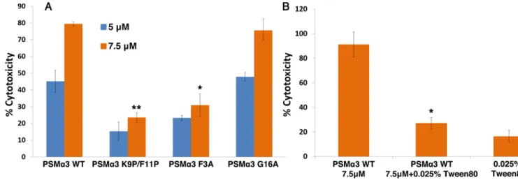
To explore whether fibrillation plays a role in PSM
α3 cytotoxicity, we performed
mutagenesis analysis to identify PSM
α3 mutants that do not fibrillate, and discovered F3A
and the K9P/F11P double mutant (A, Ala; F, Phe; K, Lys; P, Pro) (figs. S8 and S9). The two
mutants displayed much lower T-cell cytotoxicity compared to wild-type PSM
α3 (Fig. 3A).
In contrast, the G16A mutant (G, Gly), which forms fibrils recognized by Thioflavin T, thus
serving as control, was highly cytotoxic (Fig. 3A and figs. S8 and S9). Whereas the K9P/
F11P double mutant was mostly unstructured in solution, both G16A and F3A mutants
maintained
α-helical conformation (fig. S8), reinforcing the notion that helical conformation
alone is not sufficient for cytotoxicity. Furthermore, the addition of a biocompatible
surfactant maintained
α-helicity, but diminished fibrillation and abrogated PSMα3 toxicity
(Fig. 3B and figs. S8 and S10). The same pattern of fibrillation-dependent cytotoxicity was
recorded also against human embryonic kidney 293 (HEK293) cells (fig. S11), suggesting
that the lytic activity of PSM
α3 fibrils is not cell-specific. It is possible that this cytotoxicity
stems from self-assembly of helices that form large “carpets” of amphipathic sheets (fig. S6)
on the membrane surface, triggering its deformation (28). The exact conformation that
contributes to amyloid toxicity is still under debate. In some human disease-associated
amyloids, the toxic entity has been attributed to a prefibrillar conformation, whereas the
mature
β-rich fibrils detoxify the amyloid (29). Several eukaryotic amyloid proteins contain
α-helices in their monomeric or prefibrillar intermediate states [e.g., (30)], or even in the
fibril state [e.g., (27)], suggesting a link to the cytotoxicity induced by the fibrillation of
PSM
α3 into purely helical species.
In this work, we have demonstrated, at atomic resolution, that cross-
α fibrillation of PSMα3
into amyloid-like fibrils is required for cytotoxicity and suggest a key role for cross-
α fibrils
in S. aureus pathogenicity. PSM
α3 is thus a functional amyloid, displaying architecture and
properties similar to those of eukaryotic cross-
β fibrils, but differs in its secondary structure
elements. Among the large variety of super-helical assemblies found in nature,
α-helices
that stack perpendicular to the fibril axis are rare; the few examples include de
novo-designed amphiphilic peptides (28, 31, 32) and ultra-stable proteins of multiple tandem
copies of a helix-loop-helix unit (33) that bear no sequence relationship to PSM
α3. We thus
conclude that the cross-
α architecture is robust and compatible with divergent sequences. It
remains to be seen whether PSM
α3 is a unique example of a natural cross-α fibril. The
crystal structure of the PSM
α3 should contribute to research on protein aggregation,
biomaterial design, and antibacterial therapeutics.
Supplementary Material
Refer to Web version on PubMed Central for supplementary material.
Acknowledgments
We are grateful to D. Eisenberg for discussions and for his insight. We acknowledge S. Fleishman, R. Diskin, and N. Ben-Tal for helpful comments. M.L. thanks the U.S.-Israel Binational Science Foundation (BSF) (grant no. 2013254); Alon Fellowships for Outstanding Young Researchers by the Israeli Council for Higher Education;
A
uthor Man
uscr
ipt
A
uthor Man
uscr
ipt
A
uthor Man
uscr
ipt
A
uthor Man
uscr
ipt
David and Inez Mayers Career Advancement Chair in Life Sciences; the J. and A. Taub Biological Research Fund; the I-CORE Program of the Planning and Budgeting Committee and the Israel Science Foundation, Center of Excellence in Integrated Structural Cell Biology (grant no. 1775/12); the Support for Training and Career Development of Researchers (Marie Curie Career Integration Grants), Seventh Framework Program (FP7) of the European Commission, Single Beneficiary (grant no. 334260), DFG: Deutsch-IsraelischeProjektkooperation (DIP) (grant no. LA 3655/1-1), and BioStruct-X funded by FP7. J.-P.C. gratefully acknowledges the support of the France Alzheimer Foundation (FA-AAP-2013-65-101349) and the Agence Nationale de la Recherche
(ANR-12-BS07-0008-03). M.R.S. acknowledges support from the Howard Hughes Medical Institute. We acknowledge Y. Pazy-Benhar, H. Dvir, and D. Hiya (Technion Center for Structural Biology). We thank M. Baskin (Technion) for support with CD experiments. This work is based on research conducted at the ID13 and ID23-EH2 beamlines at the European Synchrotron Radiation Facility (ESRF), and at the Northeastern Collaborative Access Team beamlines funded by the National Institute of General Medical Sciences from the National Institutes of Health (P41 GM103403). The Pilatus 6M detector on 24-ID-C beamline is funded by a NIH-ORIP HEI grant (S10 RR029205). This research used resources of the Advanced Photon Source, a U.S. Department of Energy (DOE) Office of Science User Facility operated for the DOE Office of Science by Argonne National Laboratory under Contract no. DE-AC02-06CH11357. We appreciatively acknowledge ESRF and APS for beamtime and assistance during data collection. Coordinates and structure factors for the x-ray crystal structures of PSMα3 have been deposited in the Protein Data Bank (PDB) with accession code 5I55.
References and Notes:
1. Eisenberg D, Jucker M, The amyloid state of proteins in human diseases. Cell 148, 1188–1203 (2012). [PubMed: 22424229]
2. Paravastu AK, Leapman RD, Yau WM, Tycko R, Molecular structural basis for polymorphism in Alzheimer’s β-amyloid fibrils. Proc. Natl. Acad. Sci. U. S. A 105, 18349–18354 (2008). [PubMed: 19015532]
3. Colletier J-P, Laganowsky A, Landau M, Zhao M, Soriaga AB, Goldschmidt L, Flot D, Cascio D, Sawaya MR, Eisenberg D, Molecular basis for amyloid-beta polymorphism. Proc. Natl. Acad. Sci. U.S.A 108, 16938–16943 (2011). [PubMed: 21949245]
4. Nelson R, Sawaya MR, Balbirnie M, Madsen AO, Riekel C, Grothe R, Eisenberg D, Structure of the cross-beta spine of amyloid-like fibrils. Nature 435, 773–778 (2005). [PubMed: 15944695] 5. Sawaya MR, Sambashivan S, Nelson R, Ivanova MI, Sievers SA, Apostol MI, Thompson MJ,
Balbirnie M, Wiltzius JJ, McFarlane HT, Madsen AO, Riekel C, Eisenberg D, Atomic structures of amyloid cross-beta spines reveal varied steric zippers. Nature 447, 453–457 (2007). [PubMed: 17468747]
6. Rodriguez JA, Ivanova MI, Sawaya MR, Cascio D, Reyes FE, Shi D, Sangwan S, Guenther EL, Johnson LM, Zhang M, Jiang L, Arbing MA, Nannenga BL, Hattne J, Whitelegge J, Brewster AS, Messerschmidt M, Boutet S, Sauter NK, Gonen T, Eisenberg DS, Structure of the toxic core of alpha-synuclein from invisible crystals. Nature 525, 486–490 (2015). [PubMed: 26352473] 7. Chen SW, Drakulic S, Deas E, Ouberai M, Aprile FA, Arranz R, Ness S, Roodveldt C, Guilliams T,
De-Genst EJ, Klenerman D, Wood NW, Knowles TP, Alfonso C, Rivas G, Abramov AY, Valpuesta JM, Dobson CM, Cremades N, Structural characterization of toxic oligomers that are kinetically trapped during alpha-synuclein fibril formation. Proc. Natl. Acad. Sci. U.S.A 112, E1994–2003 (2015). [PubMed: 25855634]
8. Schmidt M, Rohou A, Lasker K, Yadav JK, Schiene-Fischer C, Fandrich M, Grigorieff N, Peptide dimer structure in an Abeta(1–42) fibril visualized with cryo-EM. Proc. Natl. Acad. Sci. U.S.A 112, 11858–11863 (2015). [PubMed: 26351699]
9. Xiao Y, Ma B, McElheny D, Parthasarathy S, Long F, Hoshi M, Nussinov R, Ishii Y, Abeta(1–42) fibril structure illuminates self-recognition and replication of amyloid in Alzheimer’s disease. Nat. Struct. Mol. Biol 22, 499–505 (2015). [PubMed: 25938662]
10. Van Melckebeke H, Wasmer C, Lange A, Ab E, Loquet A, Bockmann A, Meier BH, Atomic-resolution three-dimensional structure of HET-s(218–289) amyloid fibrils by solid-state NMR spectroscopy. J. Am. Chem. Soc 132, 13765–13775 (2010). [PubMed: 20828131]
11. Colvin MT, Silvers R, Ni QZ, Can TV, Sergeyev I, Rosay M, Donovan KJ, Michael B, Wall J, Linse S, Griffin RG, Atomic Resolution Structure of Monomorphic Abeta42 Amyloid Fibrils. J. Am. Chem. Soc 138, 9663–9674 (2016). [PubMed: 27355699]
A
uthor Man
uscr
ipt
A
uthor Man
uscr
ipt
A
uthor Man
uscr
ipt
A
uthor Man
uscr
ipt
12. Walti MA, Ravotti F, Arai H, Glabe CG, Wall JS, Bockmann A, Guntert P, Meier BH, Riek R, Atomic-resolution structure of a disease-relevant Abeta(1–42) amyloid fibril. Proc. Natl. Acad. Sci. U.S.A 113, E4976–4984 (2016). [PubMed: 27469165]
13. Sunde M, Serpell LC, Bartlam M, Fraser PE, Pepys MB, Blake CC, Common core structure of amyloid fibrils by synchrotron X-ray diffraction. J. Mol. Biol 273, 729–739 (1997). [PubMed: 9356260]
14. Maji SK, Perrin MH, Sawaya MR, Jessberger S, Vadodaria K, Rissman RA, Singru PS, Nilsson KP, Simon R, Schubert D, Eisenberg D, Rivier J, Sawchenko P, Vale W, Riek R, Functional amyloids as natural storage of peptide hormones in pituitary secretory granules. Science 325, 328– 332 (2009). [PubMed: 19541956]
15. Chapman MR, Robinson LS, Pinkner JS, Roth R, Heuser J, Hammar M, Normark S, Hultgren SJ, Role of Escherichia coli curli operons in directing amyloid fiber formation. Science 295, 851–855 (2002). [PubMed: 11823641]
16. DePas WH, Chapman MR, Microbial manipulation of the amyloid fold. Res. Microbiol 163, 592– 606 (2012). [PubMed: 23108148]
17. Schubeis T, Yuan P, Ahmed M, Nagaraj M, van Rossum BJ, Ritter C, Untangling a Repetitive Amyloid Sequence: Correlating Biofilm-Derived and Segmentally Labeled Curli Fimbriae by Solid-State NMR Spectroscopy. Angew. Chem. Int. Ed. Engl 54, 14669–14672 (2015). [PubMed: 26474178]
18. Shewmaker F, McGlinchey RP, Thurber KR, McPhie P, Dyda F, Tycko R, Wickner RB, The functional curli amyloid is not based on in-register parallel beta-sheet structure. J. Biol. Chem 284, 25065–25076 (2009). [PubMed: 19574225]
19. Schwartz K, Ganesan M, Payne DE, Solomon MJ, Boles BR, Extracellular DNA facilitates the formation of functional amyloids in Staphylococcus aureus biofilms. Mol. Microbiol 99, 123–134 (2016). [PubMed: 26365835]
20. Schwartz K, Syed AK, Stephenson RE, Rickard AH, Boles BR, Functional amyloids composed of phenol soluble modulins stabilize Staphylococcus aureus biofilms. PLoS Pathog 8, e1002744 (2012). [PubMed: 22685403]
21. Cheung GY, Kretschmer D, Queck SY, Joo HS, Wang R, Duong AC, Nguyen TH, Bach TH, Porter AR, DeLeo FR, Peschel A, Otto M, Insight into structure-function relationship in phenol-soluble modulins using an alanine screen of the phenol-soluble modulin (PSM) alpha3 peptide. FASEB J 28, 153–161 (2014). [PubMed: 24008753]
22. Wang R, Braughton KR, Kretschmer D, Bach TH, Queck SY, Li M, Kennedy AD, Dorward DW, Klebanoff SJ, Peschel A, DeLeo FR, Otto M, Identification of novel cytolytic peptides as key virulence determinants for community-associated MRSA. Nat. Med 13, 1510–1514 (2007). [PubMed: 17994102]
23. Laabei M, Jamieson WD, Yang Y, van den Elsen J, Jenkins AT, Investigating the lytic activity and structural properties of Staphylococcus aureus phenol soluble modulin (PSM) peptide toxins. Biochim. Biophys. Acta 1838, 3153–3161 (2014). [PubMed: 25194683]
24. Towle KM, Lohans CT, Miskolzie M, Acedo JZ, van Belkum MJ, Vederas JC, Solution Structures of Phenol-Soluble Modulins alpha1, alpha3, and beta2, Virulence Factors from Staphylococcus aureus. Biochemistry 55, 4798–4806 (2016). [PubMed: 27525453]
25. Rodriguez DD, Grosse C, Himmel S, Gonzalez C, de Ilarduya IM, Becker S, Sheldrick GM, Uson I, Crystallographic ab initio protein structure solution below atomic resolution. Nat. Methods 6, 651–653 (2009). [PubMed: 19684596]
26. Groenning M, Olsen L, van de Weert M, Flink JM, Frokjaer S, Jorgensen FS, Study on the binding of Thioflavin T to beta-sheet-rich and non-beta-sheet cavities. J. Struct. Biol 158, 358–369 (2007). [PubMed: 17289401]
27. Bousset L, Thomson NH, Radford SE, Melki R, The yeast prion Ure2p retains its native alpha-helical conformation upon assembly into protein fibrils in vitro. EMBO J 21, 2903–2911 (2002). [PubMed: 12065404]
28. Taylor KS, Lou MZ, Chin TM, Yang NC, Garavito RM, A novel, multilayer structure of a helical peptide. Protein Sci 5, 414–421 (1996). [PubMed: 8868477]
A
uthor Man
uscr
ipt
A
uthor Man
uscr
ipt
A
uthor Man
uscr
ipt
A
uthor Man
uscr
ipt
29. Stefani M, Structural features and cytotoxicity of amyloid oligomers: implications in Alzheimer’s disease and other diseases with amyloid deposits. Prog. Neurobiol 99, 226–245 (2012). [PubMed: 22450705]
30. Ghosh D, Singh PK, Sahay S, Jha NN, Jacob RS, Sen S, Kumar A, Riek R, Maji SK, Structure based aggregation studies reveal the presence of helix-rich intermediate during alpha-Synuclein aggregation. Sci. Rep 5, 9228 (2015). [PubMed: 25784353]
31. Prive GG, Anderson DH, Wesson L, Cascio D, Eisenberg D, Packed protein bilayers in the 0.90 A resolution structure of a designed alpha helical bundle. Protein Sci 8, 1400–1409 (1999). [PubMed: 10422828]
32. Mondal S, Adler-Abramovich L, Lampel A, Bram Y, Lipstman S, Gazit E, Formation of functional super-helical assemblies by constrained single heptad repeat. Nat. Commun 6, 8615 (2015). [PubMed: 26468599]
33. Brunette TJ, Parmeggiani F, Huang PS, Bhabha G, Ekiert DC, Tsutakawa SE, Hura GL, Tainer JA, Baker D, Exploring the repeat protein universe through computational protein design. Nature 528, 580–584 (2015). [PubMed: 26675729]
34. Rogers DR, Screening for the amyloid with the thioflavin-T fluorescent method. Am. J. Clin. Pathol 44, 59–61 (1965). [PubMed: 14314221]
35. Anthis NJ, Clore GM, Sequence-specific determination of protein and peptide concentrations by absorbance at 205 nm. Protein Sci 22, 851–858 (2013). [PubMed: 23526461]
36. Bohm G, Muhr R, Jaenicke R, Quantitative analysis of protein far UV circular dichroism spectra by neural networks. Protein Eng 5, 191–195 (1992). [PubMed: 1409538]
37. Micsonai A, Wien F, Kernya L, Lee YH, Goto Y, Refregiers M, Kardos J, Accurate secondary structure prediction and fold recognition for circular dichroism spectroscopy. Proc. Natl. Acad. Sci. U.S.A 112, E3095–3103 (2015). [PubMed: 26038575]
38. Hammersley AP, Svensson SO, Hanfland M, Fitch AN, Häusermann D, Two-Dimensional Detector Software: From Real Detector to Idealised Image or Two-Theta Scan. High Pressure Res 14, 235– 248 (1996).
39. Favre-Nicolin V, Cerny R, FOX, ‘free objects for crystallography’: a modular approach to ab initio structure determination from powder diffraction. J. Appl. Cryst 35, (2002).
40. Bortolotti M, Lonardelli I, ReX.Cell: a user-friendly program for powder diffraction indexing. J. Appl. Cryst 46, 259–261 (2013).
41. Boultif A, Louër D, Powder pattern indexing with the dichotomy method. J. Appl. Cryst 37, 724– 731 (2004).
42. Le Bail A, Monte Carlo indexing with McMaille. Powder Diffr 19, 249–254 (2004).
43. Kabsch W, XDS. Acta Crystallogr. D Biol. Crystallogr 66, 125–132 (2010). [PubMed: 20124692] 44. Holton J, XANES measurements of the rate of radiation damage to selenomethionine side chains.
J. Synchrotron Radiat 14, 51–72 (2007). [PubMed: 17211072]
45. Weiss M, Global indicators of X-ray data quality. J. Appl. Cryst 34, 130–135 (2001).
46. Adams PD, Afonine PV, Bunkoczi G, Chen VB, Davis IW, Echols N, Headd JJ, Hung LW, Kapral GJ, Grosse-Kunstleve RW, McCoy AJ, Moriarty NW, Oeffner R, Read RJ, Richardson DC, Richardson JS, Terwilliger TC, Zwart PH, PHENIX: a comprehensive Python-based system for macromolecular structure solution. Acta Crystallogr. D Biol. Crystallogr 66, 213–221 (2010). [PubMed: 20124702]
47. Terwilliger TC, Grosse-Kunstleve RW, Afonine PV, Moriarty NW, Zwart PH, Hung LW, Read RJ, Adams PD, Iterative model building, structure refinement and density modification with the PHENIX AutoBuild wizard. Acta Crystallogr. D Biol. Crystallogr 64, 61–69 (2008). [PubMed: 18094468]
48. Murshudov GN, Vagin AA, Dodson EJ, Refinement of macromolecular structures by the
maximum-likelihood method. Acta Crystallogr. D Biol. Crystallogr 53, 240–255 (1997). [PubMed: 15299926]
49. Emsley P, Cowtan K, Coot: model-building tools for molecular graphics. Acta Crystallogr. D Biol. Crystallogr 60, 2126–2132 (2004). [PubMed: 15572765]
A
uthor Man
uscr
ipt
A
uthor Man
uscr
ipt
A
uthor Man
uscr
ipt
A
uthor Man
uscr
ipt
50. Pettersen EF, Goddard TD, Huang CC, Couch GS, Greenblatt DM, Meng EC, Ferrin TE, UCSF Chimera--a visualization system for exploratory research and analysis. J. Comput. Chem 25, 1605–1612 (2004). [PubMed: 15264254]
51. Kyte J, Doolittle RF, A simple method for displaying the hydropathic character of a protein. J. Mol. Biol 157, 105–132 (1982). [PubMed: 7108955]
52. Petkova AT, Yau WM, Tycko R, Experimental constraints on quaternary structure in Alzheimer’s beta-amyloid fibrils. Biochemistry 45, 498–512 (2006). [PubMed: 16401079]
53. Schrodinger LLC, The PyMOL Molecular Graphics System, Version 1.8 (2015).
54. Lawrence MC, Colman PM, Shape complementarity at protein/protein interfaces. J. Mol. Biol 234, 946–950 (1993). [PubMed: 8263940]
55. Lee B, Richards FM, The interpretation of protein structures: estimation of static accessibility. J. Mol. Biol 55, 379–400 (1971). [PubMed: 5551392]
56. Saff EB, Kuijlaars ABJ, Distributing many points on a sphere. Math. Intelligencer 19, 5–11 (1997). 57. Winn MD, Ballard CC, Cowtan KD, Dodson EJ, Emsley P, Evans PR, Keegan RM, Krissinel EB,
Leslie AG, McCoy A, McNicholas SJ, Murshudov GN, Pannu NS, Potterton EA, Powell HR, Read RJ, Vagin A, Wilson KS, Overview of the CCP4 suite and current developments. Acta Crystallogr. D Biol. Crystallogr 67, 235–242 (2011). [PubMed: 21460441]
58. Altschul SF, Gish W, Miller W, Myers EW, Lipman DJ, Basic local alignment search tool. J. Mol. Biol 215, 403–410 (1990). [PubMed: 2231712]
59. Remmert M, Biegert A, Hauser A, Soding J, HHblits: lightning-fast iterative protein sequence searching by HMM-HMM alignment. Nat. Methods 9, 173–175 (2012).
60. Holm L, Rosenstrom P, Dali server: conservation mapping in 3D. Nucleic Acids Res 38, W545– 549 (2010). [PubMed: 20457744]
61. Patterson WR, Anderson DH, DeGrado WF, Cascio D, Eisenberg D, Centrosymmetric bilayers in the 0.75 A resolution structure of a designed alpha-helical peptide, D,L-Alpha-1. Protein Sci 8, 1410–1422 (1999). [PubMed: 10422829]
62. Hayouka Z, Thomas NC, Mortenson DE, Satyshur KA, Weisblum B, Forest KT, Gellman SH, Quasiracemate Crystal Structures of Magainin 2 Derivatives Support the Functional Significance of the Phenylalanine Zipper Motif. J. Am. Chem. Soc 137, 11884–11887 (2015). [PubMed: 26369301]
63. Miyazawa T, Blout ER, The Infrared Spectra of Polypeptides in Various Conformations: Amide I and II Bands1. J. Am. Chem. Soc 83, 712–719 (1961).
64. Yang H, Yang S, Kong J, Dong A, Yu S, Obtaining information about protein secondary structures in aqueous solution using Fourier transform IR spectroscopy. Nat. Protocols 10, 382–396 (2015). [PubMed: 25654756]
65. Haris PI, Chapman D, The conformational analysis of peptides using Fourier transform IR spectroscopy. Biopolymers 37, 251–263 (1995). [PubMed: 7540054]
66. Cabiaux V, Brasseur R, Wattiez R, Falmagne P, Ruysschaert JM, Goormaghtigh E, Secondary structure of diphtheria toxin and its fragments interacting with acidic liposomes studied by polarized infrared spectroscopy. J. Biol. Chem 264, 4928–4938 (1989). [PubMed: 2925676] 67. Chen VB, Arendall WB, 3rd, Headd JJ, Keedy DA, Immormino RM, Kapral GJ, Murray LW,
Richardson JS, Richardson DC, MolProbity: all-atom structure validation for macromolecular crystallography. Acta Crystallogr. D Biol. Crystallogr 66, 12–21 (2010). [PubMed: 20057044] 68. Diederichs K, Karplus PA, Improved R-factors for diffraction data analysis in macromolecular
crystallography. Nat. Struct. Biol 4, 269–275 (1997). [PubMed: 9095194]
69. Karplus PA, Diederichs K, Linking crystallographic model and data quality. Science 336, 1030– 1033 (2012). [PubMed: 22628654]
70. Brunger AT, Free R value: a novel statistical quantity for assessing the accuracy of crystal structures. Nature 355, 472–475 (1992). [PubMed: 18481394]
A
uthor Man
uscr
ipt
A
uthor Man
uscr
ipt
A
uthor Man
uscr
ipt
A
uthor Man
uscr
ipt
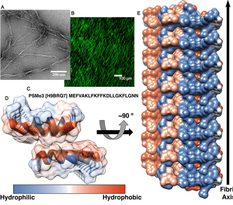
Fig. 1. The cross-α amyloid-like fibril of the full-length PSMα3.
(A) An electron micrograph of PSM
α3 fibrils. (B) Fluorescence microscopy images of
Thioflavin T stained PSM
α3 fibrils. (C) The sequence of S. aureus PSMα3 (UniProt
accession number is indicated in brackets). (D and E) The crystal structure of PSM
α3 at
1.45 Å resolution, colored according to hydrophobicity (a colored scale bar is shown). (D) A
view down the fibril axis. PSM
α3 forms parallel α-helical stacks, viewed as ribbons along
with a semitransparent surface representation. Facing helical sheets are oriented head to tail.
(E) A view perpendicular to the fibril axis. The helices, shown in surface representation, run
horizontally. Eight layers of
α-helices forming the cross-α structure are depicted.
Theoretically, fibrils can contain tens of thousands of layers. The
α-helical sheets interact
via their hydrophobic face, creating a tight interface. The higher order packing of the crystal
structure shows continuous rows of sheets that generate alternating hydrophobic and
hydrophilic interfaces (fig. S6A). Single-letter abbreviations for the amino acid residues are
A
uthor Man
uscr
ipt
A
uthor Man
uscr
ipt
A
uthor Man
uscr
ipt
A
uthor Man
uscr
ipt
as follows: A, Ala; D, Asp; E, Glu; F, Phe; G, Gly; K, Lys; L, Leu; M, Met; N, Asn; and V,
Val.
A
uthor Man
uscr
ipt
A
uthor Man
uscr
ipt
A
uthor Man
uscr
ipt
A
uthor Man
uscr
ipt
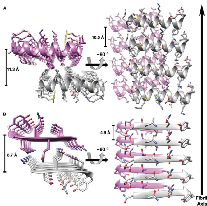
Fig. 2. PSMα3 cross-α fibril is reminiscent of amyloid cross-β structure.
(A) The crystal structure of PSM
α3. Two mating α-helical sheets are shown. (B) The steric
zipper structure of the NNQQNY N, Asn; Q, Gln; Y,Tyr) segment from yeast prion Sup35
(4) (PDB code 1YJO) forms the cross-
β spine of amyloid-like fibrils. The two mating
β-sheets are composed of parallel
β-strands. In both PSMα3 (A) and NNQQNY (B) structures,
side-chains protruding from the two sheets intermesh to form a dry, tightly
self-complementing interface. The two sheets, in purple and gray, are shown as ribbons, with
side chain as sticks. Heteroatoms are colored by atom type (nitrogen in blue, oxygen in red,
and sulfur in yellow). In the left panels, the view looks down the fibril axis, and in the right
panels, the view is roughly perpendicular to the fibril axis. The
α-helices (A) and β-strands
(B) run horizontally. Distances between mating sheets and between strands along the sheet
are displayed (table S3).
A
uthor Man
uscr
ipt
A
uthor Man
uscr
ipt
A
uthor Man
uscr
ipt
A
uthor Man
uscr
ipt
Fig. 3. PSMα3 toxicity against human T-cells is dependent on its ability to form fibrils.