HAL Id: hal-01935342
https://hal.archives-ouvertes.fr/hal-01935342
Submitted on 14 Dec 2018
HAL is a multi-disciplinary open access archive for the deposit and dissemination of sci-entific research documents, whether they are pub-lished or not. The documents may come from teaching and research institutions in France or abroad, or from public or private research centers.
L’archive ouverte pluridisciplinaire HAL, est destinée au dépôt et à la diffusion de documents scientifiques de niveau recherche, publiés ou non, émanant des établissements d’enseignement et de recherche français ou étrangers, des laboratoires publics ou privés.
Ureaplasma and/or Mycoplasma spp. to prevent
preterm birth: A randomized trial
Gilles Kayem, Alexandra Doloy, Thomas Schmitz, Yvon Chitrit, Philippe
Bouhanna, Bruno Carbonne, Jean Marie Jouannic, Laurent Mandelbrot,
Alexandra Benachi, Elie Azria, et al.
To cite this version:
Gilles Kayem, Alexandra Doloy, Thomas Schmitz, Yvon Chitrit, Philippe Bouhanna, et al.. Antibi-otics for amniotic-fluid colonization by Ureaplasma and/or Mycoplasma spp. to prevent preterm birth: A randomized trial. PLoS ONE, Public Library of Science, 2018, 13 (11), pp.e0206290. �10.1371/jour-nal.pone.0206290�. �hal-01935342�
Antibiotics for amniotic-fluid colonization
by Ureaplasma and/or Mycoplasma spp. to
prevent preterm birth: A randomized trial
Gilles KayemID1,2,3*, Alexandra Doloy4, Thomas Schmitz5,6, Yvon Chitrit7, Philippe Bouhanna8, Bruno Carbonne2,9, Jean Marie Jouannic2,10,
Laurent Mandelbrot5,7,11, Alexandra Benachi12, Elie Azria1,13, Francoise Maillard1, Florence Fenollar14, Claire Poyart4,15, Ce´cile Bebear16,17, Franc¸ois Goffinet1,15,18
1 Inserm UMR 1153, Obstetrical, Perinatal and Pediatric Epidemiology Research Team (Epope´), Center for Epidemiology and Statistics Sorbonne Paris Cite´, DHU Risks in pregnancy, Paris Descartes University, Paris, France, 2 Sorbonne University, Universite´ Pierre et Marie Curie, Paris, France, 3 Department of Obstetrics and Gynecology, CHI Cre´teil Hospital, Cre´teil, France, 4 Department of Microbiology, Cochin, Broca, Hoˆtel Dieu Hospital, AP-HP, Paris, France, 5 Paris Diderot-Paris VII University, Paris, France, 6 Department of Gynecology and Obstetrics, Robert Debre´ Hospital, APHP, Paris, France, 7 Department of Gynecology and Obstetrics, Louis Mourier Hospital, APHP, Paris, France, 8 Department of Gynecology and Obstetrics, CHI Poissy, Poissy, France, 9 Department of Obstetrics and Gynecology, Saint Antoine Hospital, APHP, Paris, France, 10 Department of Obstetrics and Gynecology, Trousseau Hospital, APHP, Paris, France, 11 Inserm IAME-UMR1137, Paris, France, 12 Department of Obstetrics and Gynecology, Antoine Beclère Hospital, Clamart, APHP, Paris, France, 13 Department of Gynecology and Obstetrics, Bichat Hospital, APHP, Paris, France, 14 Unite´ de Recherche sur les Maladies Infectieuses et Tropicales Emergentes CNRS UMR 6236 IRD 198, Marseille, France, 15 Paris Descartes University, Paris, France, 16 INRA, USC EA 3671, Mycoplasmal and Chlamydial Infections in Humans, Bordeaux, France, 17 University of Bordeaux, USC EA 3671, Mycoplasmal and Chlamydial Infections in Humans, Bordeaux, France, 18 Department of Obstetrics and Gynecology, Cochin, Broca, Hoˆtel Dieu Hospital, AP-HP, Paris, France
Abstract
Objective
To assess whether antibiotics used for treatment in asymptomatic second-trimester women positive for Mycoplasma or Ureaplasma spp. detected by amniotic-fluid PCR prevents pre-term delivery.
Design
A randomized, double-blind, placebo-controlled trial.
Setting
10 maternal fetal medicine centers in France.
Population
Women with a singleton pregnancy who underwent amniocentesis between 16 and 20 weeks’ gestation (weeks) for Down syndrome screening. A sample of 238 women with PCR-positive findings per treatment group was needed to show a 50% reduction in the pre-term delivery rate.
a1111111111 a1111111111 a1111111111 a1111111111 a1111111111 OPEN ACCESS
Citation: Kayem G, Doloy A, Schmitz T, Chitrit Y,
Bouhanna P, Carbonne B, et al. (2018) Antibiotics for amniotic-fluid colonization by Ureaplasma and/ or Mycoplasma spp. to prevent preterm birth: A randomized trial. PLoS ONE 13(11): e0206290.
https://doi.org/10.1371/journal.pone.0206290
Editor: Matthew Payne, University of Western
Australia, AUSTRALIA
Received: January 2, 2018 Accepted: October 1, 2018 Published: November 7, 2018
Copyright:© 2018 Kayem et al. This is an open access article distributed under the terms of the
Creative Commons Attribution License, which permits unrestricted use, distribution, and reproduction in any medium, provided the original author and source are credited.
Data Availability Statement: Data are available
from the Assistance Publique Hopitaux de Paris (APHP) Institutional Data Access / Ethics Committee for researchers who meet the criteria for access to confidential data. We confirm that interested researchers may access the data in the same manner as the authors. The contact for this access is: Shohreh AZIMI, Chef de projets, De´le´gationà la Recherche Clinique, et à l’innnovation (DRCI), Carre´ historique de l’Hoˆpital Saint-Louis, porte 23, 1 avenue Claude Vellefaux— 75475 Paris cedex 10, Tel +33 (0)1 44 84 17 79
Methods
Amniotic fluid was tested. Women with positive findings on real-time PCR of amniotic fluid for Mycoplasma hominis, Mycoplasma genitalium, Ureaplasma urealyticum and
Urea-plasma parvum were randomized to receive josamycin or placebo. Amniotic fluid was also
tested for 16S PCR.
Main outcome measures
The primary outcome was delivery before 37 weeks.
Results
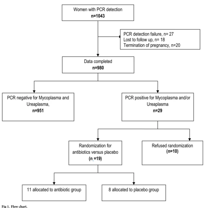
In total, 1043 women underwent amniotic-fluid screening with specific PCR detection between July 2008 and July 2011: PCR detection failed in 27 (2.6%), and 20 (1.9%) under-went termination of pregnancy. Among the 1016 women with PCR results, 980 had available data for the primary outcome (delivery before 37 weeks) and 29 (3.0%) were positive for
Ureaplasma and/or Mycoplasma spp. Because of the low rate of women with PCR-positive
findings, the trial was stopped prematurely. In total, 19 women were randomized to receive placebo (n = 8) or josamycin (n = 11) and their characteristics were comparable, as was the rate of preterm delivery and secondary outcomes. In comparing all PCR-positive and -nega-tive women regardless of treatment, PCR positivity for Ureaplasma and/or Mycoplasma spp. was not associated with any adverse pregnancy or neonatal outcome. Amniotic-fluid screening by 16S PCR showed no other bacterial colonization associated with preterm birth.
Conclusions
Because of a low amniotic fluid colonization rate, the trial was interrupted. Maternal amni-otic-fluid colonization by Mycoplasma and/or Ureaplasma spp. at 16–20 weeks in asymp-tomatic women is rare and not associated with adverse pregnancy outcomes.
Trial registration
ClinicalTrials.govNCT00718705
Introduction
Preterm delivery represents 5.5% to 12% of deliveries in high-income countries and is a major cause of infant morbidity and mortality [1]. Neonatal mortality occurs in 60% of cases in chil-dren born before 30 weeks’ gestation (weeks), and half the neurological, cognitive and respira-tory effects are observed before 32 weeks [2]. Infection could account for up to 50% of spontaneous preterm deliveries [3].
In women with spontaneous preterm labor and intact membranes, the most commonly identified bacteria in amniotic fluid areUreaplasma spp., Mycoplasma hominis, Gardnerella vaginalis, peptostreptococci, and Bacteroides species—all vaginal organisms of relatively low
virulence and all associated with increased rate of preterm delivery and chorioamnionitis [4]. The leading hypothesis is that these vaginal organisms may ascend first into the choriodecidual
Fax +33 (0)1 44 84 17 01, mail to:shohreh. [email protected].
Funding: PREMYC was funded by the French
ministry of public health (grant no.: AOM06046). The funders had no role in study design, data collection and analysis, decision to publish, or preparation of the manuscript.
Competing interests: Bayer has provided the
treatment (josamycin) only. Bayer did not provide any financial support for the study or to any investigator. This does not alter our adherence to PLOS ONE policies on sharing data and materials.
space early in pregnancy, remain undetected for months and finally result in the production of pro-inflammatory cytokines, chemokines and prostaglandins that cause cervical ripening, uterine contractions, rupture of membranes and preterm delivery [3]. Mycoplasmas and urea-plasmas have been detected in 6% and 11% of cases by PCR in amniotic fluid obtained for rou-tine chromosomal analysis during the second trimester in asymptomatic women. Their presence in amniotic fluid was found associated with preterm delivery [5,6,7].
Etiologic treatments to prevent preterm birth are lacking, and the potential benefit of the treatment forMycoplasma or Ureaplasma infection in second-trimester amniotic fluid has
never been evaluated [8]. We performed a randomized, prospective, multicenter, double-blind, placebo-controlled trial to assess the effect of antibiotics on reducing the risk of preterm delivery in second-trimester asymptomatic women who were PCR-positive for mycoplasmas, includingUreaplasma spp. (U. parvum and U. urealyticum) and M. hominis and M. genita-lium. Josamycin was the antibiotic used because at the time of the study, it was the sole
antibi-otic authorized for pregnant women in France and active against the studiedMycoplasma and Ureplasma spp. [9].
Materials and methods
The trial was approved by the national data protection authority (Commission Nationale de l’Informatique et des Liberte´s, CNIL) and by the committee for the protection of people partic-ipating in biomedical research (Comite´ de Protection des Personnes [CPP], CPP Poissy Saint Germain, no. 07051). This trial was registered at ClinicalTrials.gov (no. NCT00718705).
Participants
Women were recruited between July 2008 and July 2011 from 10 hospitals in the Paris region that had a prenatal diagnosis center. Women with a singleton pregnancy who underwent amniocentesis from 16 to 20 weeks for Down syndrome screening were eligible, and amniotic fluid was tested forU. urealyticum, U. parvum, M. hominis and M. genitalium by PCR.
Exclu-sion criteria were age < 18 years, known allergy to macrolides or lactose (included in placebo), known major structural or chromosomal fetal abnormality, and not speaking or understand-ing French. All participants gave written informed consent at 2 stages: before amniocentesis to agree to PCR analysis and, for those with PCR-positive results, before randomization.
Intervention
Women PCR-positive forMycoplasma or Ureaplasma spp. were randomized to receive
josa-mycin (Bayer) (1 g/day for 10 days) or placebo tablets. Each participant was given 20 tablets in 2 blister packs of 10 tablets each in one sealed box.
Josamycin was used because at the time of the study, it was the sole antibiotic authorized for pregnant women in France and it is active against the studiedMycoplasma and Ureplasma
spp.Ureaplasma spp. are susceptible to macrolides but not lincosamides [9]. Conversely,M. hominis is resistant to 14- and 15-member macrolides such as erythromycin but is sensitive to
16-member macrolides such as josamycin and lincosamides.M. genitalium is intrinsically
sus-ceptible to all macrolides; however, its increasing resistance rate to azithromycin is a concern [10]. Josamycin shows minimal inhibitory concentrations of <1 and <2 mg/l forM. hominis
andUreaplasma spp., respectively [11].
Randomization and masking
Staff at the Clinical Research Unit, Carre Saint Louis, Paris, France, created a randomization sequence with a 1:1 ratio. Boxes of josamycin and placebo were packed and labeled by the
National Agency of Drug Safety (Paris) according to this randomization sequence and shipped to each participant hospital. The randomization sequence, consisting of batches of 18 boxes with permuted blocks of randomly mixed sizes (2, 4 or 6) stratified by center, was created by using an interactive internet randomization system. All participants were blinded to treatment assignment for the duration of the trial, and the randomization code was not broken before all data had been collected, including the pregnancy and neonate follow-up until hospital dis-charge. Only the statistician and the independent Data Monitoring and Safety Committee had access to unblinded data during the study period. None of these people had any contact with participants in the study.
Procedure
Women were included between 15+0 and 20+6 weeks when amniocentesis was performed. An amount of 1 ml amniotic fluid was sent for real-time PCR in the microbiological center unit of Cochin hospital. If the PCR test was positive, a medical visit was scheduled and women were asked if they would agree to randomized treatment.
Gestational age was based on crown–rump length measured during the first trimester sonography. Treatment started between 20+0 and 23+6 weeks and was self-administered daily by participants for 10 days.
Demographic and obstetric characteristics were collected. Participants were interviewed by telephone after the end of the treatment to assess side effects and compliance.
Real-time PCR for
Ureaplasma and Mycoplasma spp.
One ml of amniotic fluid, to which 10μL of viral DNA used as control (Dia-IC/DNA(YD)-050, now renamed DICD-YD-L100, Diagenode), was extracted by using the NucliSENS easyMAG system (bioMe´rieux, France) according to the manufacturer’s instructions. Three distinct real-time PCR tests (U. urealyticum and U. parvum, M. genitalium, and M. hominis) were per-formed with LightCycler 2.0 (Roche). A sample was considered positive when any of the three tests reached a positive signal, given that the internal control (viral DNA) was positive.
U. urealyticum and U. parvum real-time PCR (targeting 90 bp of the urease gene for U.
urealyticum [12]) involved the primers UU-1524R
(5’-TTCCTGTTGCCCCTCAGTCT-3’) and UU-1623F (5’-AAGGTCAAGGTATGGAAGATCCAA-(5’-TTCCTGTTGCCCCTCAGTCT-3’) and Taqman probes UU-parvo (5’-FAM-TCCACAAGCTCCAGCAGCAATTTG-TAMRA-3’) and UU-T960 (5’-Yakima Yellow-ACCACAAGCACCTGCTACGATTTGTTC-TAMRA-3’).M. geni-talium PCR targeting 78 bp of the MgPa adhesin gene involved the primers MG-Pa-432R
GTTAATATCATATAAAGCTCTACCGTTGTTATC-3’) and MG-Pa-355F GAGAAAT-ACCTTGATGGTCAGCAA-3’), and the Taqman probe MG-Pa380 (5’-FAM-ACTTTGCAATCAGAAGGT-NFQMGB-3’).[13]M. hominis real-time PCR targeting 94
bp of theyidC gene involved the primers MHyidCfwd (5’-TCACTAAACCGGGTATTTTC
TAACAA-3’) and MHyidCrev (5’- TTGGCATATATTGCGATAGTGCTT-3’) and the Taqman probe MHyidC (5’-FAM- CTACCAATAATTTTAATATCTGTCGGTATG-BHQ-3’) [14]. A 20-μl PCR reaction mixture containing 5 μM each primer (final concentra-tion 0.5μM), 2.5 μM each probe (final concentration 0.25 μM), LightCycler FastStart DNA Master Hybprobe (Roche), and 5μl sample extract was amplified for 10 min at 95˚C, followed by 50 cycles of 15 s at 95˚C and 1 min at 60˚C.
16S rDNA PCR detection
Molecular analyses with broad-spectrum PCR and sequencing were performed to target 16S rDNA, as described [15,16]. All primers used for PCR and sequencing of 16S rDNA are in
S1 Table[15,16]. PCR products were purified by using the PCR kit Nucleofast 96 (Macherey-Nagel, Hoerdt, France), and sequencing involved use of the Big Dye Terminator v1.1 sequenc-ing kit (Applied Biosystems, Foster City, CA, USA). Products of the sequencsequenc-ing reaction were purified, and sequences were analyzed on an ABI PRISM 3130X Genetic Analyzer (Applied Biosystems, Foster City, CA, USA) [15]. The sequences were assembled and amended by using CodonCode Aligner v4.1.1 (CodonCode Corp., USA). Then, a correct consensus sequence was saved and compared with the GenBank database by using BLAST software (http://blast.ncbi. nlm.nih.gov/Blast.cgi). An isolate was correctly identified when it yielded > 98.7% sequence identity for the 16S rRNA sequence with the closest bacterial species sequence in GenBank [15–17].
Outcomes
The primary outcome was delivery before 37 weeks. Pre-specified secondary outcomes were 1) delivery before 22, 28 and 32 weeks; 2) number of liveborn infants; 3) hospital admission for preterm labor or preterm premature rupture of membranes (PPROM); 4) birth weight; and 5) selected neonatal complications including neonatal mortality, neonatal infection, respiratory distress syndrome, oxygen requirement at 36 weeks, intraventricular hemorrhage grade 1–4 according to Papile criteria [18], and periventricular leucomalacia and necrotizing enterocoli-tis according to the Bell classification [19]. Early-onset neonatal infection was defined by posi-tive bacteriology in blood or cerebrospinal fluid.
Statistical analysis
A sample size of 3200 was needed to detect 476 women (15%) who were PCR-positive for
Ureaplasma or Mycoplasma spp., 238 women per treatment group, to show a 50% reduction in
the preterm delivery rate from 17% to 8.5% with use of antibiotics, with a two-sided test (α error 5%, power 80%). The PCR-positive rate and 17% preterm delivery rate for PCR-positive women were estimated according to what was previously reported for second-trimester asymptomatic women at the time of the conception of the study [5,6,7].
All analyses were performed according to the intent-to-treat principle. All tests of signifi-cance were two-tailed and p < 0.05 was considered statistically significant. Comparisons between quantitative variables involved Kruskall Wallis test. For categorical variables, we used chi-square or Fisher exact test, as appropriate. All data management and analysis involved use of Stata 13.0 (Statacorp, College Station, TX, USA).
Results
Over 3 years, 1043 women underwent amniotic-fluid screening for Down syndrome and spe-cific PCR detection; for 27 (2.6%), PCR detection failed, and 20 (1.9%) underwent termination of pregnancy for medical reasons (2 were PCR-positive). In total, 18 women (1.7%) were lost to follow-up (all PCR-negative, except 2 with PCR detection failure) (Fig 1). The global rate of PCR positivity was 31/1016 (3%, 95% confidence interval 2–4%). Among the 1016 women with PCR results, 980 had available data for the primary outcome (delivery before 37 weeks) and 29 (3.0%) were positive forUreaplasma and/or Mycoplasma spp.: 13 (1.3%) for M. genita-lium, 10 (1%) for U. urealyticum, 7 (0.7%) for M. hominis and 2 (0.2%) for U. parvum. One
woman was positive forU. urealyticum, M. hominis and M. genitalium and one for U. urealyti-cum and U. parvum.
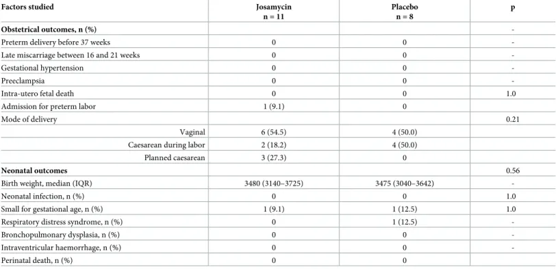
In all, 19 women agreed to randomized treatment with placebo (n = 8) or josamycin (n = 11). Characteristics of the 2 groups were comparable (Table 1) and the rate of preterm delivery and secondary outcomes was also similar (Table 2).
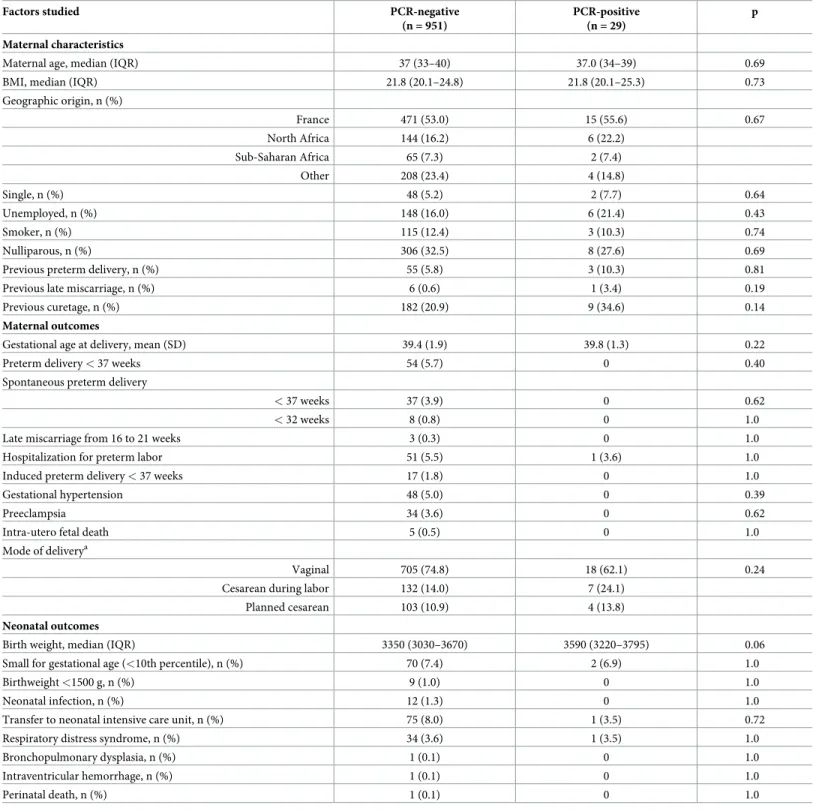
In comparing all PCR-positive and -negative women, PCR positivity was not associated with any adverse pregnancy or neonatal outcome (Table 3). Among women PCR negative with previous spontaneous preterm birth, 6/33 (18%) had again a spontaneous preterm birth. Among the two women PCR positive with spontaneous previous preterm birth, one was treated by a placebo and the other one treated by josamycin. None had a preterm birth.
Finally, all amniotic-fluid samples were screened again by using broad-spectrum PCR and sequencing targeting 16S ribosomal RNA to detect other bacteria. Five women were positive forSphingomonas sanxanigenes, Acinetobacter baumannii, Bacillus niabensis, Acinetobacter johnsonii and Streptococcus oralis, all considered probable contaminants. None of these
women delivered before 37 weeks.
Fig 1. Flow chart.
Discussion
Although we were unable to complete our trial because of a low recruitment, our main find-ings are that bacterial colonization in second-trimester amniotic fluid is rare. Indeed, our use of highly sensitive and specific techniques revealed a rate of 3% colonization of amniotic fluid
byMycoplasma and/or Ureaplasma spp. in second-trimester asymptomatic women. However,
this amniotic fluid colonization was not associated with preterm birth or other adverse mater-nal or neonatal outcomes.
Strengths and weaknesses
Our study’s main weakness is that we failed to recruit enough cases to test our primary hypoth-esis—that the use of antibiotics to reduce PCR-positiveUreaplasma or Mycoplasma spp.
infec-tion in amniotic fluid would reduce the incidence of preterm birth. Indeed, we found a marked decrease in number of inclusions because of generalized first-trimester screening for Down syndrome that led to a strong reduction in rate of second-trimester amniocentesis [20]. Moreover, the number of PCR-positive women was much lower than the 17% expected—3% [5,6]. Therefore, we stopped the study prematurely. As well, we included only women screened for Down syndrome who were therefore not representative of the overall population of women giving birth. However, the rate of preterm delivery (spontaneous or induced) agreed with French national rates, and we do not think that this study feature limits the scope of our results.
Our study has many strengths. This is the largest study to assess the association of amni-otic-fluid colonization byMycoplasma and/or Ureaplasma spp. and preterm delivery in
sec-ond-trimester asymptomatic women and to explore the bacterial colonization of
asymptomatic second-trimester women. Moreover, its multicentric and prospective design
Table 1. Characteristics of women with PCR positivity forUreaplasma and/or Mycoplasma spp. by treatment.
Factors studied Josamycin n = 11
Placebo n = 8
p
Maternal age, median (IQR) 38.0 (34.0–40.0) 36.5 (34.5–39.0) 0.59 BMI, median (IQR) 21.1 (20.7–27.3) 21.2 (20.1–25.3) 0.56 Geographic origin, n (%) France 4 (36.4) 4 (57.1) 0.44 North Africa 4 (36.4) 1 (14.3) Sub-Saharan Africa 0 1 (14.3) Others 3 (27.3) 1 (14.3) Single, n (%) 0 0 -Unemployed, n (%) 2 (18.2) 1 (14.3) 1.0 Smoker, n (%) 1 (9.1) 0 1.0 Nulliparous 8 (72.7) 6 (75.0) 1.0 Pre-existing diabetes, n (%) 0 0 -Pre-existing hypertension, n (%) 0 0 -Previous preterm delivery, n (%) 2 (18.2) 1 (12.5) 1.0
Previous fetal loss, n (%) 0 0
-Previous curetage, n (%) 4 (44.4) 3 (42.9) 1.0 Percentages are of the total available data; Comparisons between quantitative variables Kruskall Wallis test. For categorical variables, chi-square or Fisher exact test was used as appropriate; BMI, body mass index; IQR, interquartile range
reinforces the strengths of our results. However, DNA but not live cells of mycoplasmas and ureaplasmas was detected in our study, and we cannot exclude that the detected mycoplasmal DNA in the amniotic fluid could come from maternal blood across the placenta.
The real-time PCR used to detectUreaplasma spp., M. hominis and M. genitalium is highly
sensitive and specific. Indeed, real-time PCR is much more sensitive than culture for detecting these microorganisms and the 3 PCR assays were published and showed very good sensitivity and specificity; the limit of detection was < 5 to 7 genome copies depending on the assay [12– 14]. Furthermore, an extraction and amplification control was used for the 3 assays to confirm the quality of DNA extracts. These 3 PCR assays are routinely used in the French expert center for detecting human mycoplasmas. Thus, the low rate of amniotic-fluid positivity on PCR should not be related to the sensitivity of the detection methods used.
Finally, and to investigate whether women who delivered prematurely were positive for other bacteria during the second trimester of pregnancy, we also tested all amniotic-fluid sam-ples with a 16S rDNA PCR, and none of the women who delivered prematurely was positive on 16S rDNA PCR.
Interpretation
During 2003 to 2006, 3 studies reported that amniotic fluid in second-trimester asymptomatic women may be colonized byM. hominis and Ureaplasma spp. and that colonized women had
an increased rate of preterm delivery [5,6,7]. The prevalence of these bacteria, detected by a specific end-point PCR or by 16S rDNA PCR [6,21] in amniotic fluid, was 11% for Urea-plasma spp. and 6% for M. hominis between 15 and 20 weeks. One of those studies focused on
Table 2. Pregnancy outcomes for women with PCR positivity forUreaplasma and/or Mycoplasma spp. by treatment.
Factors studied Josamycin n = 11
Placebo n = 8
p
Obstetrical outcomes, n (%)
-Preterm delivery before 37 weeks 0 0
-Late miscarriage between 16 and 21 weeks 0 0
-Gestational hypertension 0 0
-Preeclampsia 0 0
-Intra-utero fetal death 0 0 1.0
Admission for preterm labor 1 (9.1) 0
Mode of delivery 0.21
Vaginal 6 (54.5) 4 (50.0) Caesarean during labor 2 (18.2) 4 (50.0)
Planned caesarean 3 (27.3) 0
Neonatal outcomes 0.56
Birth weight, median (IQR) 3480 (3140–3725) 3475 (3040–3642)
-Neonatal infection, n (%) 0 0 1.0
Small for gestational age, n (%) 1 (9.1) 1 (12.5) 1.0
Respiratory distress syndrome, n (%) 0 1 (12.5)
-Bronchopulmonary dysplasia, n (%) 0 0
-Intraventricular haemorrhage, n (%) 0 0
-Perinatal death, n (%) 0 0
IQR, interquartile range; Comparisons between quantitative variables involved Kruskall Wallis test. For categorical variables, chi-square or Fisher exact test was used as appropriate.
Table 3. Maternal characteristics and pregnancy outcomes by PCR positivity or negativity forUreaplasma or Mycoplasma spp.
Factors studied PCR-negative (n = 951)
PCR-positive (n = 29)
p Maternal characteristics
Maternal age, median (IQR) 37 (33–40) 37.0 (34–39) 0.69 BMI, median (IQR) 21.8 (20.1–24.8) 21.8 (20.1–25.3) 0.73 Geographic origin, n (%) France 471 (53.0) 15 (55.6) 0.67 North Africa 144 (16.2) 6 (22.2) Sub-Saharan Africa 65 (7.3) 2 (7.4) Other 208 (23.4) 4 (14.8) Single, n (%) 48 (5.2) 2 (7.7) 0.64 Unemployed, n (%) 148 (16.0) 6 (21.4) 0.43 Smoker, n (%) 115 (12.4) 3 (10.3) 0.74 Nulliparous, n (%) 306 (32.5) 8 (27.6) 0.69
Previous preterm delivery, n (%) 55 (5.8) 3 (10.3) 0.81 Previous late miscarriage, n (%) 6 (0.6) 1 (3.4) 0.19
Previous curetage, n (%) 182 (20.9) 9 (34.6) 0.14
Maternal outcomes
Gestational age at delivery, mean (SD) 39.4 (1.9) 39.8 (1.3) 0.22
Preterm delivery < 37 weeks 54 (5.7) 0 0.40
Spontaneous preterm delivery
< 37 weeks 37 (3.9) 0 0.62
< 32 weeks 8 (0.8) 0 1.0
Late miscarriage from 16 to 21 weeks 3 (0.3) 0 1.0
Hospitalization for preterm labor 51 (5.5) 1 (3.6) 1.0 Induced preterm delivery < 37 weeks 17 (1.8) 0 1.0
Gestational hypertension 48 (5.0) 0 0.39
Preeclampsia 34 (3.6) 0 0.62
Intra-utero fetal death 5 (0.5) 0 1.0
Mode of deliverya
Vaginal 705 (74.8) 18 (62.1) 0.24 Cesarean during labor 132 (14.0) 7 (24.1)
Planned cesarean 103 (10.9) 4 (13.8)
Neonatal outcomes
Birth weight, median (IQR) 3350 (3030–3670) 3590 (3220–3795) 0.06 Small for gestational age (<10th percentile), n (%) 70 (7.4) 2 (6.9) 1.0
Birthweight <1500 g, n (%) 9 (1.0) 0 1.0
Neonatal infection, n (%) 12 (1.3) 0 1.0
Transfer to neonatal intensive care unit, n (%) 75 (8.0) 1 (3.5) 0.72 Respiratory distress syndrome, n (%) 34 (3.6) 1 (3.5) 1.0
Bronchopulmonary dysplasia, n (%) 1 (0.1) 0 1.0
Intraventricular hemorrhage, n (%) 1 (0.1) 0 1.0
Perinatal death, n (%) 1 (0.1) 0 1.0
Percentages are for total available data (missing data < 10% only for sociodemographic and previous medical history data);
a
For this line and above, n = 940 (exclusion of cases of intra-utero fetal death and delivery from 16 to 21 weeks.);
BMI, body mass index; IQR, interquartile range; Comparisons between quantitative variables involved Kruskall Wallis test. For categorical variables, chi-square or Fisher exact test was used as appropriate.
Ureaplasma spp. in 254 asymptomatic women with amniotic fluid collected by amniocentesis
performed for chromosomal analysis [5]. Overall, 24% of PCR-positive women gave birth before 37 weeks versus 0.4% of PCR-negative women (p <0.001). The same analysis was car-ried out forM. hominis [6]. The prevalence ofM. hominis in the latter study was 6.4% among
456 patients. In total, 10.4% of the PCR-positive women gave birth prematurely versus 1.9% for PCR-negative women (p = 0.02). The main flaws of these studies were that they were retro-spective and conducted by the same team, therefore needing external confirmation by others.
Recently, new data contradicted these findings [22]. Payne et al. tested second-trimester amniotic-fluid cytokine levels andUreaplasma spp. colonisation in 480 Chinese and 492
Aus-tralian women.Ureaplasma spp. was detected in only 2 Chinese women who delivered
pre-term [23]. Rowland et al. prospectively investigated amniotic fluid from 344 asymptomatic women recruited in mid-pregnancy for detecting microbial DNA at the time of amniocentesis.
U. urealyticum, U. parvum, M. hominis and M. genitalium were not detected in amniotic fluid.
Three women delivered at 30, 31 and 33 weeks in this population. Our study agrees with these results and shows that amniotic-fluid bacterial colonization byMycoplasma or Ureaplasma
spp. or other bacteria is rare in asymptomatic women.
Several hypotheses can explain these differences with previous studies reporting high rates ofMycoplasma or Ureaplasma spp. colonization in amniotic fluid of second-trimester women.
Indeed, the design of the previous studies was retrospective, they were based on a much smaller number of women, and amniotic-fluid samples were stored after amniocentesis and were not from a prospective study with specific care (especially to avoid any bacterial contami-nation) [5,6,7]. The PCR results from previous studies may have included many false-positive results, specifically from contamination after amniocentesis.
One surprising finding is thatMycoplasma or Ureaplasma spp. colonization was not
associ-ated with preterm birth. This finding is not explained by the fact that 11 women received josa-mycin. Indeed, 18 women did not receive treatment, and none of the 29 women who were PCR positive had a preterm delivery. The other hypothesis might be that the techniques used are very sensitive and that the very low concentration detected is not able to induce local inflammation and a subsequent preterm delivery. Higher concentrations of bacteria occurring later in the pregnancy or in cases of preterm labor or preterm rupture of membranes may be a cofactor, increasing local inflammation and therefore the risk of preterm birth.
However, intra-amniotic infection by mycoplasmas and ureaplasmas may not be the main factor leading to preterm birth. Subclinical infection has been widely considered a significant etiological factor in the pathogenesis of inflammatory preterm birth, but the source of infec-tion and the gestainfec-tional window when it is acquired remain unclear. Colonizainfec-tion with low-virulence microorganisms, such asUreaplasma spp. (U. parvum and U. urealyticum) and M. hominis, ascending from the lower genital tract may be the precursor to intra-amniotic
sub-clinical infection, provoking, via metalloprotease and cytokine production, cervical changes in uterine contraction and rupture of membranes leading to preterm birth [3,24–27]. Genital mycoplasmas commonly isolated in amniotic fluid in cases of preterm labor or PPROM have been implicated in this process. However, whether the microorganisms present are themselves pathogenic is unclear: they may be susceptibility markers or have an adjunct role in the inflam-matory preterm birth cascade. Indeed,Mycoplasma or Ureaplasma spp. are part of the normal
vaginal flora in pregnant women [28,29]. The amniotic-fluid colonization in cases of preterm labor or PPROM may be attributed to cervical dilation or membrane rupture rather than cause preterm labor or PPROM. Although the presence of inflammation has been considered a consequence of microbial presence, the host inflammatory response may, by its effect on cer-vical dilation or rupture of membranes, favor microbial colonization and invasion. Some recent data favor this hypothesis. Indeed, recent studies using mass spectrometry of both
amniotic-fluid culture and broad-range PCR have found that sterile intra-amniotic inflamma-tion is as common as microbial-associated intra-amniotic inflammainflamma-tion in women with pre-term labor or PPROM, which suggests that sterile inflammation is more common and more closely associated with preterm birth than microbial-associated inflammation [30,31]. More-over, Gervasi et al. reported an increase in markers of inflammation in mid-trimester samples that in some cases was associated with spontaneous preterm birth, although samples did not contain microorganisms detectable with culture techniques [32]. Of note, sterile inflammation ignores any inflammation that may be present due to a viral agonist. Indeed, viruses generally go unnoticed in a diagnosis of infection because they are usually not screened. In addition, the presence of microbial DNA (without live organisms) may invoke an inflammatory response.
Conclusion
Although low amniotic-fluid colonization rate caused a premature interruption of the trial, our study shows that bacterial colonization of second-trimester amniotic fluid is rare. Coloni-zation byMycoplasma or Ureaplasma spp. occurred in 3% of cases and was not associated with
risk of preterm birth in this population.
Supporting information
S1 Table. Broad-spectrum PCR and sequencing.
(DOCX)
S1 Checklist. Consort check list.
(DOC)
S1 File. English translation of the study protocol.
(DOCX)
S2 File. Study protocol.
(PDF)
S3 File. Instruction for use of the Kit Ref. No.: DICD-XX-L100.
(PDF)
Acknowledgments
We thank Robert Cohen and Frederic Batteux who participated in the design of the study.
Author Contributions
Conceptualization: Gilles Kayem, Claire Poyart, Ce´cile Bebear, Franc¸ois Goffinet. Data curation: Francoise Maillard.
Formal analysis: Gilles Kayem, Francoise Maillard, Florence Fenollar. Funding acquisition: Gilles Kayem.
Investigation: Gilles Kayem, Alexandra Doloy, Thomas Schmitz, Yvon Chitrit, Philippe
Bou-hanna, Bruno Carbonne, Jean Marie Jouannic, Laurent Mandelbrot, Alexandra Benachi, Elie Azria, Claire Poyart, Ce´cile Bebear, Franc¸ois Goffinet.
Methodology: Gilles Kayem, Alexandra Doloy, Francoise Maillard, Franc¸ois Goffinet. Supervision: Gilles Kayem, Thomas Schmitz, Franc¸ois Goffinet.
Validation: Alexandra Doloy, Thomas Schmitz, Francoise Maillard, Ce´cile Bebear. Writing – original draft: Gilles Kayem.
Writing – review & editing: Alexandra Doloy, Thomas Schmitz, Yvon Chitrit, Philippe
Bou-hanna, Bruno Carbonne, Jean Marie Jouannic, Laurent Mandelbrot, Alexandra Benachi, Elie Azria, Francoise Maillard, Florence Fenollar, Claire Poyart, Ce´cile Bebear, Franc¸ois Goffinet.
References
1. MacDorman MF, Matthews TJ, Mohangoo AD, Zeitlin J. International comparisons of infant mortality and related factors: United States and Europe, 2010. Natl Vital Stat Rep. 2014; 63(5):1–6. Epub 2014/ 09/25. PMID:25252091.
2. Larroque B, Breart G, Kaminski M, Dehan M, Andre M, Burguet A, et al. Survival of very preterm infants: Epipage, a population based cohort study. Arch Dis Child Fetal Neonatal Ed. 2004; 89(2):F139–44.
https://doi.org/10.1136/adc.2002.020396PMID:14977898.
3. Goldenberg RL, Hauth JC, Andrews WW. Intrauterine infection and preterm delivery. N Engl J Med. 2000; 342(20):1500–7.https://doi.org/10.1056/NEJM200005183422007PMID:10816189
4. Goncalves LF, Chaiworapongsa T, Romero R. Intrauterine infection and prematurity. Ment Retard Dev Disabil Res Rev. 2002; 8(1):3–13.https://doi.org/10.1002/mrdd.10008PMID:11921380.
5. Gerber S, Vial Y, Hohlfeld P, Witkin SS. Detection of Ureaplasma urealyticum in second-trimester amni-otic fluid by polymerase chain reaction correlates with subsequent preterm labor and delivery. J Infect Dis. 2003; 187(3):518–21.https://doi.org/10.1086/368205PMID:12552439.
6. Nguyen DP, Gerber S, Hohlfeld P, Sandrine G, Witkin SS. Mycoplasma hominis in mid-trimester amni-otic fluid: relation to pregnancy outcome. J Perinat Med. 2004; 32(4):323–6.https://doi.org/10.1515/ JPM.2004.060PMID:15346817.
7. Perni SC, Vardhana S, Korneeva I, Tuttle SL, Paraskevas LR, Chasen ST, et al. Mycoplasma hominis and Ureaplasma urealyticum in midtrimester amniotic fluid: association with amniotic fluid cytokine lev-els and pregnancy outcome. Am J Obstet Gynecol. 2004; 191(4):1382–6.https://doi.org/10.1016/j.ajog. 2004.05.070PMID:15507969.
8. Chang HH, Larson J, Blencowe H, Spong CY, Howson CP, Cairns-Smith S, et al. Preventing preterm births: analysis of trends and potential reductions with interventions in 39 countries with very high human development index. The Lancet. 2013; 381(9862):223–34.https://doi.org/10.1016/s0140-6736 (12)61856-xPMID:23158883
9. Waites KB, Lysnyansky I, Be´be´ar C. Emerging antimicrobial resistance in mycoplasmas of humans and animals. Caister Academic Press, Norfolk, UK: Browning G. F. Citti C.; 2014.
10. Be´be´ar C and Kempf I. Mycoplasmas: pathogenesis, molecular biology, and emerging strategies for control. Browning G Ba A, editor: Horizon Bioscience; 2005.
11. Krausse R, Schubert S. In-vitro activities of tetracyclines, macrolides, fluoroquinolones and clindamycin against Mycoplasma hominis and Ureaplasma ssp. isolated in Germany over 20 years. Clin Microbiol Infect. 2010; 16(11):1649–55. Epub 2010/01/06.https://doi.org/10.1111/j.1469-0691.2009.03155.x
PMID:20047607.
12. Yi J, Yoon BH, Kim EC. Detection and biovar discrimination of Ureaplasma urealyticum by real-time PCR. Molecular and cellular probes. 2005; 19(4):255–60. Epub 2005/07/12.https://doi.org/10.1016/j. mcp.2005.04.002PMID:16005182.
13. Jensen JS, Bjornelius E, Dohn B, Lidbrink P. Use of TaqMan 5’ nuclease real-time PCR for quantitative detection of Mycoplasma genitalium DNA in males with and without urethritis who were attendees at a sexually transmitted disease clinic. J Clin Microbiol. 2004; 42(2):683–92. Epub 2004/02/10.https://doi. org/10.1128/JCM.42.2.683-692.2004PMID:14766837.
14. Ferandon C, Peuchant O, Janis C, Benard A, Renaudin H, Pereyre S, et al. Development of a real-time PCR targeting the yidC gene for the detection of Mycoplasma hominis and comparison with quantitative culture. Clin Microbiol Infect. 2011; 17(2):155–9. Epub 2010/03/20.https://doi.org/10.1111/j.1469-0691. 2010.03217.xPMID:20298269.
15. Drancourt M, Berger P, Raoult D. Systematic 16S rRNA gene sequencing of atypical clinical isolates identified 27 new bacterial species associated with humans. J Clin Microbiol. 2004; 42(5):2197–202. Epub 2004/05/08.https://doi.org/10.1128/JCM.42.5.2197-2202.2004PMID:15131188.
16. Fenollar F, Roux V, Stein A, Drancourt M, Raoult D. Analysis of 525 samples to determine the useful-ness of PCR amplification and sequencing of the 16S rRNA gene for diagnosis of bone and joint
infections. J Clin Microbiol. 2006; 44(3):1018–28. Epub 2006/03/07.https://doi.org/10.1128/JCM.44.3. 1018-1028.2006PMID:16517890.
17. Al Masalma M, Armougom F, Scheld WM, Dufour H, Roche PH, Drancourt M, et al. The expansion of the microbiological spectrum of brain abscesses with use of multiple 16S ribosomal DNA sequencing. Clin Infect Dis. 2009; 48(9):1169–78. Epub 2009/04/02.https://doi.org/10.1086/597578PMID:
19335164.
18. Papile LA, Burstein J, Burstein R, Koffler H. Incidence and evolution of subependymal and intraventricu-lar hemorrhage: a study of infants with birth weights less than 1,500 gm. J Pediatr. 1978; 92(4):529–34. PMID:305471.
19. Bell MJ, Ternberg JL, Feigin RD, Keating JP, Marshall R, Barton L, et al. Neonatal necrotizing enteroco-litis. Therapeutic decisions based upon clinical staging. Annals of surgery. 1978; 187(1):1–7. Epub 1978/01/01. PMID:413500.
20. Alldred SK, Takwoingi Y, Guo B, Pennant M, Deeks JJ, Neilson JP, et al. First trimester serum tests for Down’s syndrome screening. Cochrane database of systematic reviews (Online). 2015; 11:Cd011975. Epub 2015/12/01.https://doi.org/10.1002/14651858.cd011975PMID:26617074.
21. Blanchard A, Hentschel J, Duffy L, Baldus K, Cassell GH. Detection of Ureaplasma urealyticum by poly-merase chain reaction in the urogenital tract of adults, in amniotic fluid, and in the respiratory tract of newborns. Clin Infect Dis. 1993; 17 Suppl 1:S148–53. Epub 1993/08/01. PMID:8399906.
22. Rowlands S, Danielewski JA, Tabrizi SN, Walker SP, Garland SM. Microbial invasion of the amniotic cavity in midtrimester pregnancies using molecular microbiology. Am J Obstet Gynecol. 2017; 217(1): 71 e1–e5. Epub 2017/03/08.https://doi.org/10.1016/j.ajog.2017.02.051PMID:28268197.
23. Payne MS, Feng Z, Li S, Doherty DA, Xu B, Li J, et al. Second trimester amniotic fluid cytokine concen-trations, Ureaplasma sp. colonisation status and sexual activity as predictors of preterm birth in Chinese and Australian women. BMC Pregnancy Childbirth. 2014; 14:340. Epub 2014/10/03.https://doi.org/10. 1186/1471-2393-14-340PMID:25273669.
24. Taylor-Robinson D, Lamont RF. Mycoplasmas in pregnancy. Bjog. 2011; 118(2):164–74. Epub 2010/ 11/26.https://doi.org/10.1111/j.1471-0528.2010.02766.xPMID:21091927.
25. Capoccia R, Greub G, Baud D. Ureaplasma urealyticum, Mycoplasma hominis and adverse pregnancy outcomes. Curr Opin Infect Dis. 2013; 26(3):231–40. Epub 2013/04/17.https://doi.org/10.1097/QCO. 0b013e328360db58PMID:23587772.
26. Yoon B. The clinical significance of detecting Ureaplasma urealyticum by the polymerase chain reaction in the amniotic fluid of patients with preterm labor. Am J Obstet Gynecol. 2003; 189(4):919–24.https:// doi.org/10.1067/s0002-9378(03)00839-1PMID:14586326
27. Yoon BH, Romero R, Kim M, Kim EC, Kim T, Park JS, et al. Clinical implications of detection of Urea-plasma urealyticum in the amniotic cavity with the polymerase chain reaction. Am J Obstet Gynecol. 2000; 183(5):1130–7.https://doi.org/10.1067/mob.2000.109036PMID:11084554.
28. Romero R, Mazor M, Oyarzun E, Sirtori M, Wu YK, Hobbins JC. Is genital colonization with Mycoplasma hominis or Ureaplasma urealyticum associated with prematurity/low birth weight? Obstet Gynecol. 1989; 73(3 Pt 2):532–6. PMID:2644604.
29. Anderson BL, Mendez-Figueroa H, Dahlke JD, Raker C, Hillier SL, Cu-Uvin S. Pregnancy-induced changes in immune protection of the genital tract: defining normal. Am J Obstet Gynecol. 2013; 208(4): 321.e1–9. Epub 2013/01/15.https://doi.org/10.1016/j.ajog.2013.01.014PMID:23313311.
30. Romero R, Miranda J, Chaemsaithong P, Chaiworapongsa T, Kusanovic JP, Dong Z, et al. Sterile and microbial-associated intra-amniotic inflammation in preterm prelabor rupture of membranes. J Matern Fetal Neonatal Med. 2015; 28(12):1394–409. Epub 2014/09/06.https://doi.org/10.3109/14767058. 2014.958463PMID:25190175.
31. Romero R, Miranda J, Chaiworapongsa T, Korzeniewski SJ, Chaemsaithong P, Gotsch F, et al. Preva-lence and clinical significance of sterile intra-amniotic inflammation in patients with preterm labor and intact membranes. Am J Reprod Immunol. 2014; 72(5):458–74. Epub 2014/08/01.https://doi.org/10. 1111/aji.12296PMID:25078709.
32. Gervasi MT, Romero R, Bracalente G, Erez O, Dong Z, Hassan SS, et al. Midtrimester amniotic fluid concentrations of interleukin-6 and interferon-gamma-inducible protein-10: evidence for heterogeneity of intra-amniotic inflammation and associations with spontaneous early (<32 weeks) and late (>32 weeks) preterm delivery. J Perinat Med. 2012; 40(4):329–43. Epub 2012/07/04.https://doi.org/10.1515/ jpm-2012-0034PMID:22752762.