Carclnogencsis vol.6 no.9 pp. 1309-1313, 1985
Nafenopin-induced rat liver peroxisome proliferation reduces DNA
methylation by N-nitrosodimethylainine in vivo
Otmar D.Wiestler
3, Ivo Schmerold
1, Birgitta Fringes,
Benedikt Volk and Paul Kleihues
2Laboratory of Neuropathology, Institute of Pathology, University of Freiburg, Albertstrasse 19, D-78OO Freiburg im Breisgau, 'Institute of Pharmacology, Toxicology and Pharmacy, Faculty of Veterinary Medicine, University of Munich, D-8000 MOnchen 22, FRG, and laboratory of Neuropathology, Institute of Pathology, University of Zurich, CH-8091 Zurich, Switzerland
T o whom reprint requests should be sent
Hie hypoUpidaemk drug nafenopin (NAF) has been shown to
enhance the hepatocarcinogenic effect of
N-nitrosodimethyl-amine (NDMA) and N-nitrosodiethylN-nitrosodimethyl-amine in rats. We have
investigated whether the NAF-induced peroxisome
prolifera-tion in hepatocytes interferes with NDMA's metabolism and
interaction with DNA. Adult male Wistar rats received a
single i.p. injection of [
14C]NDMA (2 mg/kg) and were
kill-ed 4 h later. DNA was isolatkill-ed from liver and kidney,
hydrolysed in 0.1 N HC1 and analysed by Sephasorb
chromatography. In rats pre-treated with NAF (0.2% in the
diet over a period of 3 weeks), the concentration of
N7-methylguanlne in hepatic DNA (^mol/mol guanine) was
46% below control values. This is probably due to the greater
amount of target DNA, as NAF caused a marked
hepatomega-ly with a 50% increase in total liver DNA content.
Concen-trations of N7-methylguanine in kidney DNA were twice as
high in NAF-pre-treated animals when compared to control
rats. This is unlikely to result from a shift in the metabolism
of NDMA from liver to other rat tissues since the time course
and extent of the conversion of [
14C]NDMA to
14CO
2and
14C-labelled urinary metabolites were identical in NAF-treated
and control animals. There was no indication that NAF
in-hibits the activity of the hepatic C^-alkylguanine-DNA
alkyl-transferase.
Introduction
Nafenopin and related hypolipidaemic agents exert profound
ef-fects on the liver of experimental animals including mice, rats,
hamsters, cats, dogs and monkeys (1,2). These effects include
a stimulation of hepatic DNA synthesis, a striking hepatocyte
peroxisome proliferation (3,4), and the induction of
hepatomega-ly. An increase in peroxisomal /3-oxidation of fatty acids is
responsible for the hypolipidaemia caused by these compounds
(5 — 7). It has been suggested that the biological effects of
nafenopin and clofibrate are mediated by a cytosolic protein
receptor in liver and kidney (8).
In 1976, Reddy et at. (9) reported a high incidence of
hepatocellular carcinomas in acataJasemic mice after treatment
with nafenopin. These findings have subsequently been
extend-ed to other strands of mice and rats, and it has been shown that
a number of different peroxisome-proliferating agents, including
•Abbreviations: NDMA, N-nitrosodimethylamine, ["CJNDMA, N-nitroso-["CJdirnethvlamine; pHJdT, (methyH3H])thymidine; 7-meG, N7-methylguanine; [O»]meG, [O*]methylguanine.the industrial plastizicer di(2-ethylhexyl)phthalate, are similarly
effective (10—14). From these observations Reddy and
co-workers concluded that nafenopin and related peroxisome
stimulators may represent a novel class of hepatocarcinogens.
The mechanism of tumour induction by these drugs is unknown.
They are not mutagenic in the Salmonella-microsome assay and
apparently do not interact with DNA in vivo (15,16). The
hypothesis that increased intracellular production of clastogenic
H
2O
2and oxygen radicals (OH ,O
2~) by the stimulated
j3-oxidation system may be responsible for the neoplastic
transfor-mation has not yet been confirmed (14,17,18). On the other hand,
some investigations indicate that nafenopin and clofibrate may
act as hepatic tumour promoters. Hypolipidemic drugs with
peroxisome-proliferating activity are inducers of hepatic ornithine
decarboxylase (19). Low doses of clofibrate, WY-14,643, or
nafenopin given simultaneously with, or after,
N-nitrosodiethyl-amine significantly increase the number of hepatic tumours in
rats (20 — 23). Reports from other laboratories seem to indicate
that peroxisome proliferations have no promoting effect on hepatic
tumour induction by N-2-fluorenylacetamide (24,25) and
N-nitro-sodiethylamine (26). These contradictory observations are
dif-ficult to interpret since it is not known whether or to what extent
nafenopin and related compounds interfere with the
bioactiva-tion of the respective initiating agents. To investigate these
ques-tions, we studied the metabolism of N-nitrosodimethylamine
(NDMA)* in nafenopin-pre-treated rats. The results of our
ex-periments show that nafenopin reduces the interaction of NDMA
with hepatic DNA in vivo.
Materials and methods
Animals
Adult male Wistar rats (body wt. 150 — 200 g) were purchased from Ivanovas, Kisslegg, FRG, and kept under standard conditions.
Chemicals
N-nitroso-[14C]dirnethylamine (["CJNDMA) (sp. act. 53.4 mCi/mmol) was
ob-tained from New England Nuclear, Boston, MA, USA. Unlabelled NDMA (Schuchardt, Munich, FRG) was added to lower the sp. act. The radiochemical purity was checked by h.p.l.c. and proved to be >97%. [Methyl-'HJThymidine (sp. act. 71.7 Ci/mmol) was from New England Nuclear, Boston, MA, USA. Immediately before i.p. injection, NaCl was added to give a 0.9% (w/v) solu-tion. Nafenopin [2-methyl-2-p-(l,2,3,4-tetrahydro-l-naphthyl)pherioxypropionic acid] was generously provided by Drs. W.Staubli and R.Hess, Ciba Geigy AG (Basel, Switzerland). Sephasorb HP Ultrafine was from Deutsche Pharmacia (Freiburg, FRG), Lumagel scintillation cocktail from LKB (Karlsruhe, FRG). For dipping autoradiography, an Dford Nuclear Research Emulsion (K5) was used. The remaining chemicals were from Sigma (Tauftrirchen, FRG) and Merck (Darm-stadt, FRG).
Nafenopin pre-treatmenl
Adult male Wistar rats were fed on a standard laboratory diet (Altromin") enrich-ed with 0.2% nafenopin (w/w) for a period of 3 weeks. Control animals receiv-ed the standard diet and water ad libitum.
Electron microscopy
Control and nafenopin-pre-treated animals were perfused via the aorta with 2.5% glutardialdehyde under thiopental anaesthesia. Livers were removed, embedded in araWite, cut in semi-thin and ultra-thin sections and processed as described elsewhere (27). The ultra-thin sections were examined in a Zeiss OM2 electron microscope.
O.D. Wrestler et at.
Determination of liver DNA, RNA, and protein
Control and nafenopin pre-treated animals were killed under light ether anaesthesia. Livers were rapidly removed and stored at —70°C. DNA and RNA were ex-tracted following the procedure of Schmitt and Thannhauser (28). DNA concen-trations were determined using the Burton Method (29), and RNA concenconcen-trations using the orcin reaction (30). Proteins were isolated by TCA extraction as described earlier (31) and protein concentrations determined with a modified Bradford assay (32).
Hepatic DNA synthesis in vivo
Control and nafenopin pre-treated animals received a single i.p. injection of [methyl-3H]thymidine ([3H]dT) (2 jiCi/g) and were killed 30 min later. The livers
were removed, frozen in liquid nitrogen and stored at -70°C. DNA was isolated following the Schmitt-Thannhauser procedure (28). Thymidine incorporation was determined by liquid scintillation counting using Lumagel cocktail. In addition, liver segments were fixed in buffered formaldehyde (5 %, v/v), embedded in paraf-fin and cut in 2-/un sections. These sections were used for dipping autoradiography with Ilford-K5 emulsion.
Metabolism of NDMA in vivo
Control and nafenopin-pre-treated animals (2 each) received a single i.p. injec-tion of [14C]NDMA (2 mg/kg; 0.5 mCi/mmol) and were placed in a metabolic
cage (Jencons Metabowl, Hemel Hempstead, UK). Expired '*COj was absorb-ed by two serially connectabsorb-ed Nilox columns, each containing 600 ml of 1 N NaOH, and urine was collected for quantification of excreted 14C-metabolites. Samples
(0.5-ml) were analysed at various time intervals. For liquid scintillation coun-" ting, NaOH and urine samples were diluted with distilled water (1:4, v/v) and subsequently mixed with 8 ml Lumagel. During the experiment, control rats were fed on a standard laboratory diet and water ad libitum, whereas pre-treated animals received the same diet containing 0.2% (w/w) nafenopin.
DNA alkylation in vivo
Control and nafenopin pre-treated animals received a single i.p. injection of ["C]NDMA (2 mg/kg; 10 mCi/mmol) and were killed 4 h later. DNA was isolated from liver and kidney by phenolic extraction as described earlier (33), hydrolysed in 0.1 N HC1 (37°C, 20 h), neutralised and analysed on a Scphasorb column using 10 mM NaHjPQ, (pH 5.5) as mobile phase (flow rate 1.5 ml/min). Absorbance at 260 nm was measured with a Perkin Elmer spectrophotometer and radioactivity determined after addition of Lumagel (counting efficiency, 85%). Amounts of methylated DNA purines were expressed as fraction of the parent base guanine assuming that the specific activity of the methyl adducts was half of that of the injected [14C]NDMA.
Results
Nafenopin treatment induced a striking peroxisome proliferation
in hepatocytes. These proliferating organelles possessed a finely
granular matrix without dense core structures and were distributed
throughout the cytoplasm. They were of highly variable size and
shape, in contrast to hepatic peroxisomes of control animals. The
cytoplasmic accumulation of peroxisomes led to a marked
hyper-trophy of hepatocytes.
The above changes were accompanied by a marked
hepato-megaly. The liver mass increased by a factor of 2.5 compared
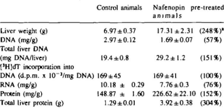
to control animals (Table I). The liver enlargement was
accom-panied by a 3-fold increase in the hepatic protein content. DNA
and RNA concentrations were reduced by 40 and 25%,
respec-tively, whereas the protein concentration was increased. Total
liver DNA increased by a factor of 1.5 compared to the livers
of control animals (Table I). [3H]dT incorporation into hepatic
DNA was, however, identical in control animals and after
3 weeks of dietary nafenopin exposure, as revealed by
bio-chemical studies (Table I) and by dipping autoradiography of liver
slices (data not shown).
The metabolic degradation of NDMA was studied in a
metabolic cage experiment with 2 nafenopin-pre-treated and
2 control animals (Figure 1). After a single i.p. injection of
[14C]NDMA, time course and extent of metabolism were
iden-tical in both groups. During the 48 h observation period, the
animals exhaled 63 % of the
14C-radioactivity as
14CO
2and
ex-creted 10% as 14C-labelled urinary metabolites. The experiment
with nafenopin-pre-treated rats was repeated, confirming this
result.
1310
Table I. Effect of nafenopin on
Liver weight (g) DNA (mg/g) Total liver DNA (mg DNA/liver) [3H]dT incorporation into
DNA (d.p.m. x 10"3/mg DNA)
RNA (mg/g) Protein (mg/g) Total liver protein (g)
liver weight, DNA, Control animals 6.97±0.37 2.97±0.12 19.4±0.8 169±45 10.18 ± 0.29 148.87 ± 1.60 1.29±0.01
RNA and protein Nafenopin pre-treated a n i m a l s 17.31±2.31 1.69 ±0.07 29.2 ±1.2 169±41 7.76±0.3 226.62 ±22.10 3.92 ±0.38 (248%)' (57%) (151%) (100%) (76%) (152%) (304%) Nafenopin pre-treated animals received a 0.2% diet over a period of 3 weeks. Nucleic acids and proteins were extracted from liver and quantified as described in the text. For [JH]dT incorporation, animals received a single
i.p. injection of [methyl-3H]thymidine [3H-dT]; 2 /iCi/g) and were killed 30 min later. The data shown are mean values of 4 rats ± S.D. •Data in parentheses indicate nafenopin values as a percentage of controls.
12- 10- 86 4 - 25 0 3 0 -* A
i
1
iI
11
1
4——* A 1— fc—A X- S URINE A 14co
2 10 20 30 40 50 hours Fig. 1. Metabolism of N-nitroso[14C]dimethylamine in vivo.Nafenopin-pre-treated (A) and control animals (A) received a single i.p. injection of ["CJNDMA (2 mg/kg; 0.5 mCi/mmol) and were placed in a metabolic cage. Urinary excretion of [14C]metabolites (upper) and [14C]exhalation
(lower) were monitored over 48 h. The data are plotted as cumultative percentages of the total radioactivity administered.
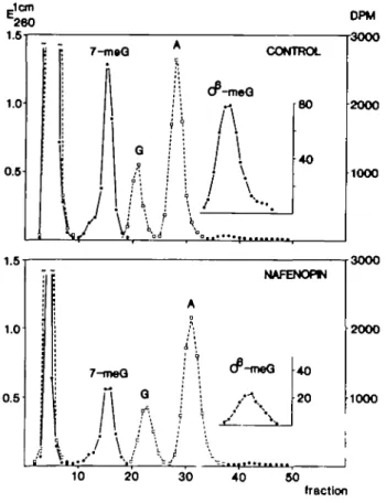
To assess the extent of DNA alkylation in vivo, animals received
a single i.p. injection of [
14C]NDMA (2 mg/kg; 10 mCi/mmol)
and were killed after 4 h. Nafenopin pre-treatment reduced the
concentrations of N7-methylguanine (7-meG) and
[O6]methyl-guanine ([06]meG) in hepatic DNA by - 5 0 % (Figure 2 and
Table II). In contrast, the levels of 7-meG in kidney DNA of
nafenopin-treated rats were twice as high as in control animals.
Rat liver peroxisome proliferation
In this organ, the pro-mutagenic base [O6]meG was only
detect-able after nafenopin pre-treatment.
Discussion
The present study corroborates several hepatotrophic effects of
nafenopin in Wistar rats which have been reported previously
for various experimental animals (1—4), cultured rat hepatocytes
(34), and human hepatocytes heterotransplanted to athymic nude
mice (35). Dietary nafenopin caused a 2.5-fold increase of the
liver weight (Table I). The hepatomegaly was mainly due to a
striking proliferation of cytoplasmic peroxisomes and represented
a combination of organ hypertrophy and hyperplasia, as
reveal-ed both electron microscopically and by the increase in total liver
Jem 260 1.5 1.0 0.5 DPM 3000 7-tneG CONTROL BO 40 1.0 1.0 0.5 B -| 7-moQ
A/ \
J
V
Qf:
• \
j
A I ' Cr-mA
NAFENOHN eQ 40 20 l 2000 1000 3000 2000 1000 10 20 30 40 50 fractionFig. 2. Chromalographic profiles of liver DNA hydrolysales. Nafenopin-pre-treated and control animals received a single i.p. injection of N-nitroso [14C]dimethylamine (2 mg/kg; 10 mCi/mmol) and were killed 4 h later. DNA was isolated by phenolic extraction, hydrolysed in 0.1 N HC1, neutralised and analysed on a Sephasorb column. Fraction volume was 3.9 (upper) and 3.7 ml (lower). A, adenine; G, guanine; 7-meG, N7-methyl-guanine; [O*]meG, [O*]methylN7-methyl-guanine; Ej™ • • ; d.p.m. • • .
DNA (11,36). The stimulation of hepatic DNA synthesis must
have occurred during the initial period of nafenopin
administra-tion, as at the end of the 3-week feeding period, [3H]dT
incor-poration into hepatic DNA was identical in both
nafenopin-exposed and control animals (Table I). Furthermore,
the biochemical data revealed a stimulation of hepatic RNA
syn-thesis and a 3-fold increase in total liver protein in
nafenopin-fed animals. These results are consistent with the findings of
Chat-terjee et al. (37) who described a reversible alteration of hepatic
mRNA species of peroxisomal and non-peroxisomal proteins
in-duced by the hypolipidemic drug WY-14,643. Lazarow et al.
(7) reported that among hepatic proteins a subset of peroxisomal
polypeptides including the /S-oxidation enzymes are preferentially
induced by hypolipidaemic drugs. The marked increase in hepatic
protein content (Table I) is associated with a considerable loss
of glycogen (38).
NDMA and related dialkylnitrosamines are enzymically
bioac-tivated by isoenzymes of the microsomal cytochrome P450 system
(39). This bioactivation proceeds via a-C-hydroxylau'on and
final-ly yields electrophilic alkyl intermediates which react with cellular
macromolecules including DNA (40 — 42). It has been
demonstrated in long-term carcinogenicity studies, that for
numerous carcinogenic N-nitroso compounds, the initial extent
of DNA akylation is closely correlated with the incidence and
location of tumours (43). We determined two methylated DNA
purines, 7-meG, the major methyl adduct, and [O
6]meG, which
is believed to be a promutagenic lesion involved in the initiation
of malignant transformation (44,45).
Surprisingly, nafenopin pre-treatment reduced the
concentra-tions of methylated liver DNA purines after a single i.p. dose
of [14C]NDMA by 50%, whereas in kidney the extent of DNA
alkylation was twice as high as in control animals (Table FT).
Several factors may be responsible for these changes. The
decrease in hepatic DNA methylation could be due to the higher
content of DNA, i.e., a same number of methyl diazonium ions
react with twice the amount of hepatic DNA. Alternatively,
nafenopin could inhibit the metabolism of NDMA in liver; this
would also explain the increase in kidney DNA methylation. The
latter mechanism has been demonstrated to be operative in rats
kept on a protein-free diet (46,47) and was associated with a
significant increase in the incidence of NDMA-induced kidney
tumours. A similar inter-organ shift of the metabolism of NDMA
and related nitrosamines has been observed after administration
of ethanol (48,49,50) and disulfiram (51,52). It is unlikely,
however, that this mechanism also applies to the present
ex-periments since both the extent and time course of the
conver-sion of [14C]NDMA to 14CO2 and 14C-labelled urinary metabolites
were identical in nafenopin-treated and control animals (Figure
1). We have also considered the possibility that the marked
changes in the size and structure of nafenopin-adapted hepatocytes
TaMe II. Organ Liver Kidney DNA methylation in Control 7-meG 720 ± 38 ± liver and animals 26 6 kidney [0»]meG 50.5 ± 8.4 n.d. [0«]meG/7-meG 0.069 ± 0.0067 n.d. Nafenopin 7-meG 388 ± 50 83 ± 4.5 pre-treated animals [O»]meG 12.7 ± 1.3 5.5 ± 1.9 [O*]meG/7-meG 0.033 ± 0.0057 0.065 ± 0.017
Nafenopin pre-treated (0.2% diet; 3 weeks) and control animals were given a single i.p. dose of [14C]NDMA (2 mg/kg; 10 mCi/mmol).
After 4 h survival, DNA was isolated and analysed as described in the text.
7-meG, N7-methylguanine; [O*]meG, [O^methylguanine; n.d., not detectable; values for methylated purines are expressed as /*mol/mol guanine. Liver data are mean values of 3 animals (controls) or 5 animals (nafenopin) ± S.D. Kidney data are means of triplicate determinations of pooled tissues from 4 (controls) and 5 animals (nafenopin).
O.D.WIestier et al.
may lead to a greater fraction of
N-nitroso-methyl(hydroxy-methyl)amine, the proximate carcinogen NDMA being excreted
into the systemic circulation. In this case, a higher extent of DNA
alkylation would be expected not only in kidney, but also in lung
and even in tissues (e.g., intestine) with minimal capacity for
NDMA bioactivation. However, we found no increase in
pulmonary DNA methylation, and methylated purines were not
detectable in colonic DNA from either control or
nafenopin-treated rats (data not shown). Therefore, we also consider the
possibility that the increase in kidney DNA alkylation may be
due to a direct effect of nafenopin. The pro-mutagenic lesion
[O
6]meG can be removed by a cellular DNA repair enzyme,
[O^alkylguanine-DNA alkyltransferase (53). The alkyltransferase
has been demonstrated in Escherichia colt (54) and in numerous
animal and human tissues (44); it has been shown that the hepatic
enzyme system in rats is inducible by partial hepatectomy,
hepatotoxic agents, and X-irradiation (55,56,57) i.e., under
con-ditions of liver regeneration. Dietary nafenopin led to a
significantly lower [O^meG/T-meG ratio in hepatic DNA (Table
II). This could indicate an increased repair capacity, but is more
likely that this change is due to the lower extent of [O*]meG
for-mation. The rate of repair of this promutagenic base is
dose-dependent, as the alkyltransferase is inactivated during the repair
process. Assessment of the [O6]alkylguanine repair system
us-ing an in vitro assay (58) also provided no evidence of an
induc-tion of the hepatic alkyltransferase; these data were difficult to
interpret due to the nafenopin-induced changes in total liver
pro-tein content. The present study was stimulated by the
observa-tion that nafenopin enhances hepatic tumour formaobserva-tion by
symmetric dialkylnitrosamines (20 — 23). More recent studies
seem to indicate that under different experimental conditions
nafenopin may have no effect on, or even depress, the
develop-ment on enzyme-altered foci (26) and hepatic neoplasms (24,25).
These controversial findings are at present difficult to explain.
Nafenopin and related agents cause profound metabolic and
struc-tural alterations which may modulate hepatic carcinogenesis in
different ways. Our own results demonstrate, however, that
nafenopin pre-treatment of Wistar rats reduces the interaction
of the hepatocarcinogenic nitrosamine NDMA with hepatic DNA
in vivo. If the same effect occurs during chronic concomitant
ad-ministration of both agents, one would expect a lower incidence
of hepatic tumours.
Acknowledgements
We appreciate the generous gift of nafenopin by Drs. W.StSubli and R.Hess, CIBA-GEIGY, Basel, Switzerland. The excellent technical assistance of Ms. Bet-tina Mayer is gratefully acknowledged. We also wish to thank Dr. de Looze, Freiburg, for critical reading of the manuscript. This work was supported by the Deutsche Forschungsgemeinschaft (SFB 31) and the Swiss National Fund.
References
1. Reddy J.K., WarrenJ.R., Reddy.M.K. and Lalwani.N.D. (1982), Hepatic and renal effects of peroxisome proliferation: biological implications, Ann.
N.Y. Acad. Sri., 386, 81-110.
2. Reddy J.K., Lalwani.N.D., Qureshi.S.A., Reddy.M.K. and Moehle.C.M. (1984), Induction of hepatic peroxisome proliferation in nonrodent species, including primates, Am. J. PathoL, 114, 171-183.
3. Hess.R., Stfubli.W. and Riess.W. (1965), Nature of the hepatomegalic ef-fect produced by ethyl-chlorophenoxyisobutyrate in the rat, Nature, 208, 856-858.
4. ReddyJ.K. and Krishnakantha.T.P. (1975), Hepatic peroxisome prolifera-tion: induction by two novel compounds structurally unrelated to clofibrate,
Science, 190, 787-789.
5. Lazarow.P.B. and de Duve.C. (1976), A fatty acyl-CoA oxidizing system in rat liver peroxisomes; enhancement by clofibrate, a hypolipidemic drug,
Proc. Nail. Acad. Sri. USA, 73, 2043-2046.
6. Lazarow.P.B. (1978), Rat liver peroxisomes catalyze the beta-oxidation of fatty acids, J. BM. Chem., 253, 1522-1528.
7. Lazarow.P.B., Fujiki.Y., Mortensen.R. and Hashimoto.T. (1982), Identifica-tion of beta-oxidaIdentifica-tion enzymes among peroxisomal polypeptides, FEBS Lett., 150, 307-310.
8. Lalwani.N.D., Fahl.W.E. and ReddyJ.K. (1983), Detection of a nafenopin-binding protein in rat liver cytosol associated with the induction of perox-isome proliferation by hypolipidemic compounds, Biochem. Biophyi. Res.
Commun., 116, 388-393.
9. Reddy J.K., Rao.M.S. and Moody.D.E. (1976), Hepatocellular carcinomas in acatalasemic mice treated with nafenopin, a hypdipklemic peroxisome pro-liferator, Cancer Res., 36, 1211-1217.
10. Svoboda.D J. and Azarnoff.D.L. (1979), Tumors in male rats fed ethyl chloro-phenoxyisobutyrate, a hypolipidemic drug, Cancer Res., 39, 3419-3428. 11. ReddyJ.K., Azamoff.D.L. and Hignite.C.E. (1980), HypoUptdaemic hepatic
peroxisome proliferators form a novel class of chemical carcinogens, Nature, 283, 397-398.
12. ReddyJ.K., Rao.M.S., Azarnoff.D.L. and Sell.S. (1979), Mitogenic and carcinogenic effects of a hypolipidemic peroxisome prohferator, [4-chloro-6-(2,3-xylidino)-2-pyrimidinylthio]acetic acid (Wy-14,643), in rat and mouse liver, Cancer Res., 39, 152-161.
13. ReddyJ.K. and Rao.M.S. (1977), Malignant tumors in rats fed nafenopin, a hepatic peroxisome proliferator, J. Natl. Cancer Inst., 59, 1645-1650. 14. Lalwani.N.D., Reddy.M.K., Qureshi.S.A. and ReddyJ.K. (1981),
Develop-ment of hepatocellular carcinomas and increased peroxisomal fatty acid beta-oxidation in rats fed [4-chloro-6-(2,3-xylidino)-2-pvrimidinylthio]acetic acid (Wy-14,643) in the semipurified diet, Cardnogenesis, 2, 645-650. 15. WarrenJ.R., Simmon.V.F. and ReddyJ.K. (1980), Properties of
hypo-lipidemic peroxisome proliferators in the lymphocyte ['HJdiymidine and
Salmonella mutagenesis assays, Cancer Res., 40, 36-41.
16. Iinnainmaa.K. (1984), Induction of sister chromatid exchanges by the perox-isome prohfcrators 2,4-D, MCPA, and clofibrate in vivo and in vitro,
Car-cinogenesis, 5, 703-707.
17. Reddy J.K., Lalwani.N.D., Reddy.M.K. and Qureshi.S.A. (1982), Excessive accumulation of autofluorescent lipofuscin in the liver during hepatocar-cinogenesis by methyl clofenapate and other hypolipidemic peroxisome pro-liferators, Cancer Res., 42, 259-266.
18. Fahl.W.E., Lalwani.N.D., Watanabe.T., Goel.S. and ReddyJ.K. (1984), DNA damage related to increased hydrogen peroxide formation by hypolipidemK drug-induced liver peroxisomes, Proc. Am. Assoc. Cancer Res., 25, 109.
19. Izumi.K., Reddy,J.K. and Oyasu.R. (1981), Induction of hepatic omithine decarboxylase by hypolipidemic drugs with hepatic peroxisome proliferative activity, Carcinogenesis, 2, 623-627.
20. Mochizuki.Y., Furukawa.K. and Sawada.N. (1982), Effects of various con-centrations of ethyl-alpha-p-chlorophenoxyisobutyrate (clofibrate) on diethyl-nhrosamine-induced hepatic tumorigenesis in the rat, Carcinogenesis, 3, 1027-1029.
21. Mochizuki.Y., Furukawa.D. and Sawada.N. (1983), Effect of simultaneous administration of clofibrate with diethylnitrosamine on hepatic tumorigenesis in the rat, Cancer Lett., 19, 99-105.
22. Reddy,J.K. and Rao.M.S. (1978), Enhancement by Wy-14,643, a hepatic peroxisome proliferator, of diethylnitrosamine-initiated hepatic tumorigenesis in the rat, Br. J. Cancer, 38, 537-543.
23. WardJ.M., RiceJ.M., Creasia.D., Lynch.P. and Riggs.C. (1983), Dissimilar patterns of promotion by di-(2-ethylhexyl)phthalate and rJienobarbital of hepato-cellular neoplasia initiated by diethylnitrosamine in B6C3F, mice,
Carcino-genesis, 4, 1021-1029.
24. Numoto.S, Furukawa.K., Furuya.K. and Williams.G.M. (1984), Effects of the hepatocarcinogenic peroxisome-proliferaung hypolipidemic agents clofibrate and nafenopin on the rat liver cell membrane enzymes y-glutamyltranspeptidase and alkaline phosphatase and on the early stages of liver carcinogenesis,
Car-cinogenesis, 5, 1603-1611.
25. Numoto.S., Mori.H., Furuya.K., Levinc.W.G. and Williams.G.M. (1985), Absence of a promoting or sequential syncarcinogenic effect in rat liver by the carcinogenic hypolipklemic drug nafenopin given after N-2-fluorenylacet-amide, Toxicol. AppL Pharmacol., 77, 76-85.
26. Staubli.W., Bentley.P., Bieri.F., Frohlich.E. and Waechter.F. (1984), In-hibitory effect of nafenopin upon the development of diethylnitros-amine-induced enzyme-altered foci within the rat liver, Carcinogenesis, 5, 41-46.
Rat liver peroxtsome proliferation 27. Volk,B., Malctz,M., Tiedemann,M., Mall.G., Klein.C. and Berlet.H.H.
(1981), Impaired maturation of Prukinje cells in the fetal alcohol syndrome of the rat. Light- and electron-microscopic investigations, Acta Neuropathol., 54, 19-29.
28. Schmitt,G. and Thannhauser.S.J. (1945), A method for the determination of desoxyribonucleic acid, ribonucleic acid, and phosphoproteins in animal tissues, J. Biol. Chem., 161, 83-89.
29. Burton,K. (1956), A study of the conditions and mechanism of the diphenyl-amine reaction for the colorimetric estimation of deoxyribonucleic acid,
Biochem. J., 62, 315-323.
30. Cooper,T.G. (1981), Bestimmung von Nucleinsauren mit der Orcin-Reaction, in Biochemische Arbeasmethoden, Walter de Gruyter, Berlin, NY, pp. 54-55. 31. Kleihues,P. and Magce,P.N. (1973), Inhibition of protein synthesis by
N-methyl-N-nitrosourea in vivo, Biochem. J., 136, 303-309.
32. Bradford.M.M. (1976), A rapid and sensitive method for quantitation of microgram quantities of protein utilizing the principle of protein-dye binding,
Anal. Biochem., 72, 248-254.
33. Margison.G.P. and KJeihues.P. (1975), Chemical carcinogencsis in the ner-vous system. Preferential accumulation of O*-methylguaninc in rat brain deoxy-ribonucleic acid during repetitive administration of N-mcthyl-N-nitrosurea,
Biochem. J., 148, 521-525.
34.Bieri,F., Bentley.P., Waechter.F. and Staubli.W. (1984), Use of primary cultures of adult rat hepatocytcs to investigate mechanisms of action of nafenopin, a hepatic peroxisome proliferator, Carcinogenesis, 5, 1033-1039. 35. Jirtle.R.L., ReddyJ.K. and Michalopoulos.G. (1984), Induction of
perox-isome proliferation in transplanted dog, cat, rat and human hepatocytes, Proc.
Am. Assoc. Cancer Res., 25, 117.
36. Moody.D.E., Rao.M.S. and Reddy.K.J. (1977), Mitogenic effect on mouse liver induced by a hypolipidemic drug, nafenopin, Virchows Archiv. (Cell
PathoL), 23, 291-296.
37.Chatterjee,B., Demyan.W.F., Lalwani.N.D., Reddy,J.K. and Roy.A.K. (1983), Reversible alteration of hepatic messenger RNA species for perox-isomal and non-peroxperox-isomal proteins induced by the hypolipidaemic drug Wy-14,643, Biochem. J., 2, 757-883.
38. Hess.R. and Bencze.W.L. (1968), Hypolipidaemic properties of a new tetralin derivative (CIBA 13,437), Experientia, 24, 418-419.
39. Pegg.A.E. (1980), Metabolism of N-nitrosodimethylamine, IARCSci. Pubi, 27, 3-22.
40. O'Cormor.P.J. (1981), Studies on mechanism of action. Interaction of chemical carcinogens with macromolecules, /. Cancer Res. din. Oncol., 99, 167-186. 41. Loveless.A. (1969), Possible relevance of O-6 alkylation of deoxyguanosine to the mutagenicity and carcinogenicity of nitrosamines and nitrosamides,
Nature, 223, 206-207.
42. Pegg.A.E. (1977), Formation and metabolism of alkylated nucleosides: possi-ble role in carcinogenesis by nitroso compounds and alkylating agents, Adv.
Cancer Res., 25, 195-269.
43. Kleihucs.P. and Wiestler.O.D. (1985), Structural DNA modifications and DNA repair in organ-specific tumour induction, in Cohen,G.M. (ed.), Target
Organ Toxicity, CRC Press, Boca Raton, FL, in press.
44. Pegg.A.E. (1984), Methylation of the O1 position of guanine in DNA is the
most likely initiating event in carcinogenesis by methylating agents, Cancer
Invest., 2, 223-231.
45. Singer.B. (1984), Alkylation of the O* of guanine is only one of many chemical events that may initiate carcinogenesis, Cancer Investigation, 2, 233-238. 46. Swarm.P.F. and McLean.A.E.M. (1971), Cellular injury and carcinogenesis. The effects of a protein-free high-carbohydrate diet on the metabolism of di-methylnitrosamine in the rat, Biochem. J., 124, 283-288.
47. Kiessling.M., Lipinsld.R., Bohm.N. and Kleihues.P. (1981), Effect of pretreat-ment with pregnenolone-16-alpha-carbonitrile and protein-free diet on the metabolism and carcinogenicity of dimethylnitrosamine, Carcinogenesis, 2, 757-761.
48. Kouros.M., Monch.W., Reiffer,F.J. and Dehnen,W. (1983), The influence of various factors on the methylation of DNA by the oesophageal carcinogen N-nitrosomethylbenzylamine. I. The importance of alcohol, Carcinogenesis, 4, 1081-1084.
49. Swann.P.F., Coe.A.M. and Mace,R. (1984), Ethanol and dimethylnitrosamine metabolism and disposition in the rat. Possible relevance to the influence of ethanol on human cancer incidence, Carcinogenesis, 5, 1337-1343. 50. Wiestler.O.D., von Deimling.A. and KJeihues.P. (1985), Differential effects
of ethanol on the metabolism of N-nitrosodimethylamine and N-nflrosomethyl-benzylamine in rats, in preparation.
51.Schweinsberg,F. andBurkle.V. (1981), Wirkung von DisulfiramaufdieTox-izitat und Carcinogenitat von N-Methyl-N-nhrosobenzylamin bei Ratten, J.
Cancer Res. din. Oncol., 102, 43-47.
52. Schweinsberg.F., Weissenbergcr,I., Bruckner.B. and Schweinsberg.E. (1985), Effect of disulfiram on N-nhroso-N-methylbenzylamine metabolism (bio-chemica] aspects), in O'Neil.I.K., von Borstel.R.C, Miller.C.T., LongJ.
and Bartsch.H. (eds.), N-Nitroso Compounds: Occurrence, Biological Effects
and Relevance to Human Cancer, IARC Scientific Publications, Vol. 57,
Ox-ford University Press, OxOx-ford, in press.
53. Pegg.A.E., Wiest.L., Foote.R.S., Mitra.S. and Perry.W. (1983), Purifica-tion and properties of O*-methylguanine-DNA transmethylase from rat liver,
J. Biol. Chem., 258, 2327-2333.
54. Schendel.P.F. and Robbins.P.E. (1978), Repair of O*-methylguanine in adapted Escherichia coli, Proc. Nail. Acad. Set. USA, 75, 6017-6020. 55. Pegg.A.E. and Perry,W. (1981), Stimulation of transfer of methyl groups
from O'-methylguanine in DNA to protein by rat liver extracts in response to hepatotoxins, Carcinogenesis, 2, 1195-1200.
56. Pegg.A.E., Perry.W. and Bennett,R.A. (1981), Effect of partial hepatectomy on removal of O'-methylguanine from alkylated DNA by rat liver extracts,
Biochem. J., 197, 195-201.
57. Schmero!d,I. and Wiestier.O.D. (1985), Induction of rat liver O*-alkylguanine-DNA alkyltransferase following whole body X-irradiation, Cancer Res., sub-mitted.
58. Wiestler.O., Kleihues.P. and Pegg.A.E. (1984), O*-Alkylguanine-DNA alkyl-transferase activity in human brain and brain tumors, Carcinogenesis, 5,
121-124.