HAL Id: tel-00868787
https://tel.archives-ouvertes.fr/tel-00868787
Submitted on 13 Mar 2014HAL is a multi-disciplinary open access archive for the deposit and dissemination of sci-entific research documents, whether they are pub-lished or not. The documents may come from teaching and research institutions in France or abroad, or from public or private research centers.
L’archive ouverte pluridisciplinaire HAL, est destinée au dépôt et à la diffusion de documents scientifiques de niveau recherche, publiés ou non, émanant des établissements d’enseignement et de recherche français ou étrangers, des laboratoires publics ou privés.
catalytic mechanism and the molecular bases of their
specificity
Yan Li
To cite this version:
Yan Li. Cyclodipeptide synthases : towards understanding their catalytic mechanism and the molec-ular bases of their specificity. Agricultural sciences. Université Paris Sud - Paris XI, 2012. English. �NNT : 2012PA114831�. �tel-00868787�
UNIVERSITÉ PARIS-SUD 11
ECOLE DOCTORALE :
INNOVATION THÉRAPEUTIQUE : DU FONDAMENTAL A L’APPLIQUÉ PÔLE : INGENIERIE DES PROTEINES ET CIBLES THERAPEUTIQUES
DISCIPLINE :
STRUCTURE, FONCTION ET INGENIERIE DES PROTEINES
ANNÉE 2009 – 2012 SÉRIE DOCTORAT N° 1185
THÈSE DE DOCTORAT
soutenue le 26/09/2012par
Yan LI
Titre:
Les cyclodipeptide synthases : vers la compréhension de leur mécanisme
catalytique et des bases moléculaires de leur spécificité
! " # # ! $ %
& #' # $ ! #$ ( " ) "
* ++ , #' ( ) $ ! #$ "- !# $ !
Les cyclodipeptides et leurs dérivés, les dicétopipérazines (DKP), constituent une large classe de métabolites secondaires aux activités biologiques remarquables qui sont essentiellement synthétisés par des microorganismes. Les voies de biosynthèse de certaines DKP contiennent des synthases de cyclodipeptides (CDPS), une famille d’enzymes récemment identifiée. Les CDPS ont la particularité de détourner les ARNt aminoacylés de leur rôle essentiel dans la synthèse protéique ribosomale pour les utiliser comme substrats et ainsi catalyser la formation des deux liaisons peptidiques de différents cyclodipeptides. Le travail de thèse présenté dans ce manuscrit a pour objectif de caractériser la nouvelle famille des CDPS. Dans un premier temps, la caractérisation tant structurale que mécanistique de la première CDPS identifiée, AlbC de Streptomyces noursei, est présentée. Puis, les résultats obtenus avec trois autres CDPS, chacune de ces enzymes ayant des caractéristiques adéquates pour approfondir l’étude de la famille des CDPS, sont décrits. Ainsi, la CDPS Ndas_1148 de Nocardiopsis dassonvillei a permis d’étendre nos connaissances sur les bases moléculaires de la spécificité des CDPS. La CDPS AlbC-IMI de S. sp. IMI 351155 est un bon modèle pour analyser l’interaction de chacun des deux substrats nécessaires à la formation d’un cyclodipeptide. Enfin, la caractérisation de la CDPS Nvec-CDPS2 chez l’animal Nematostella vectensis a permis de fournir le premier exemple d’enzyme d’origine animale impliquée dans la synthèse peptidique non ribosomale.
Abstract
Cyclodipeptides and their derivatives, the diketopiperazines (DKPs), constitute a large class of secondary metabolites with noteworthy biological activities that are mainly synthesized by microorganisms. The biosynthetic pathways of some DKPs contain cyclodipeptide synthases (CDPSs), a newly defined family of enzymes. CDPSs hijack aminoacyl-tRNAs from their essential role in ribosomal protein synthesis to catalyze the formation of the two peptide bonds of various cyclodipeptides. The aim of the work presented in this thesis manuscript is to characterize the CDPS family. At first, the structural and mechanistic characterization of the first identified CDPS, AlbC of Streptomyces noursei, is presented. Then, the results obtained with three other CDPSs, each of which having suitable properties to increase our understanding of the CDPS family, are described. The CDPS Ndas_1148 of Nocardiopsis dassonvillei extends our knowledge of the molecular bases of the CDPS specificity. The CDPS AlbC-IMI of S. sp. AlbC-IMI 351155 is a good model to analyze the interaction of each of the two substrates required for the formation of a cyclodipeptide. Finally, the characterization of the CDPS Nvec-CDPS2 from Nematostella vectensis provides the first example of enzymes of animal origin involved in nonribosomal peptide synthesis.
Mots-clés :
cyclodipeptide synthase (CDPS), diketopiperazine (DKP), nonribosomal biosynthesis, peptide bond, aminoacyl-tRNA, secondary metabolite
Intitulé du laboratoire :
Equipe Enzymologie et Biosynthèse Peptidique Non Ribosomale Laboratoire de Toxinologie Moléculaire et Biotechnologies (LTMB) Service d’Ingénierie Moléculaire des Protéines (SIMOPRO)
CEA Saclay, 91191 Cedex, Gif-sur-Yvette
PÔLE : INGENIERIE DES PROTEINES ET CIBLES THERAPEUTIQUES UNIVERSITÉ PARIS-SUD 11
UFR «FACULTÉ DE PHARMACIE DE CHÂTENAY-MALABRY » 5, rue Jean Baptiste Clément
It would not have been possible to achieve this doctoral thesis without the help and support of the kind people around me, to only some of whom it is possible to give particular mention here.
First of all, I thank my committee members for their valuable suggestions and feedback in evaluating my research.
I would like to thank the International PhD Program of the Life Sciences division of the CEA (Irtelis) for the doctoral fellowship. I thank the CEA sector “Molecular Engineering of Proteins (SIMOPRO)” which received me to accomplish my thesis. I also thank the sector for their powerful experimental apparatus without the access to which, I can not image how to accomplish this thesis work in three years.
I am heartily grateful to my PhD supervisor, Dr Muriel Gondry, for her encouragement, supervision and help in every aspect of the work during my thesis. She taught me how to be a scientific researcher. I deeply admire her for her rigorous scientific approach and rich knowledge in the domain.
I sincerely thank my colleagues of the laboratory. I will always be grateful for having worked with this excellent research team for three years. I deeply thank Mireille Moutiez, Pascal Belin and Jérôme Seguin for the kind transfer of their scientific knowledge to me. It is a good memory when we shared time together in the laboratory and in the coffee corner. I will particularly never forget the kind and timely help of Pascal Belin who always reached me out a helping hand when I felt helpless. I sincerely thank him for having helped me rebuild confidence. I would like to thank Guillaume Grach for his precious advice and friendship. I also thank our technician Cédric Masson for having helped me purify some proteins and Sandrine Braud for her Photoshop help. I thank Ludovic Sauguet, Jean1Baptiste Charbonnier and Marie1Hélène Le Du for the structure of AlbC which made a solid foundation for my thesis work. I also thank
synthesized for my work.
I am thankful to Robert Thai for his training and help in analysis with mass spectrometry. I thank the technician Steven Dubois for his kind help in mass spectrometry and other sample analyses. I also thank Alain Lecoq for the synthesis of some chemical molecules and his personal advice during my thesis. I am grateful for the help of Fabrice Beau and Fannely Berthon on analysis with Beta1Imager. Additionally, I feel lucky for having harvested so much friendship in SIMOPRO. I specially thank Bertrand Czarny and Cathrine Nury for their encouragement and help. Bertrand, as what you said, I will miss your office1scaring and all our funny discussions. Catherine, thank you for having given me a lift so often during my thesis. I will miss the days we spent together in SIMOPRO.
I would like to acknowledge our collaborators for their excellent experimental contribution and scientific interest, especially Jean1Luc Pernodet, Cécile Martel and Karine Tuphile.
I also thank my friends (Bo Lu, Jun Han, Lin Xia, Sandrine Ragu…too many to list her but you know who you are!) for providing support and friendship that I need. I particularly thank my dear friend Yue Jiao for her amazing friendship and everything she did for me. I consider her as my sister. I also thank my landlady Mireille Verneau whom I consider as my family in France. I thank her for her delicious desserts, her amazing Christmas dinner and all countless help.
Finally, I give my deep gratitude to my family especially my grandfather Shukun Li, my mother Ping Zhang, my father Guangjian Li and my dear fiancé Bo Gao. Without your support and encouragement, I would not stick to the present. I know you miss me so much just like I miss you. You have done so much for me and now, I think it is time to give my love back. I love you forever.
Table of contents
TABLE OF CONTENTS ... 1 ABBREVIATIONS ... 3 INTRODUCTION ... 5 1 BIBLIOGRAPHIC STUDIES ... 13 1.1 NATURAL DIKETOPIPERAZINES (DKPS) ... 13 1.1.1 DKP family ... 13 1.1.1.1 Natural abundance of DKPs ... 14 1.1.1.2 Structural diversity of DKPs ... 15 1.1.1.3 Physiological roles of DKPs ... 181.1.1.4 Biological and pharmacological activities of DKPs ... 22
1.1.2 Biological mechanisms of DKP formation ... 26
1.1.2.1 Nonenzymatic pathways of DKP formation... 26
1.1.2.2 Enzymatic pathways of DKP formation ... 27
1.2 FORMATION OF PEPTIDE BONDS BY BIOCATALYSTS ... 34
1.2.1 Peptide bond formation involving aa-tRNA ... 37
1.2.1.1 Transfer RNA (tRNA) ... 37
1.2.1.2 Aminoacyl-tRNA synthetase (aaRS) ... 40
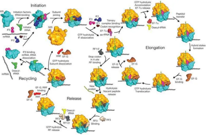
1.2.1.3 Ribosome ... 47
1.2.1.4 Fem transferases ... 54
1.2.1.5 Aminoacyl-tRNA protein transferases ... 59
1.2.1.6 Cyclodipeptide synthases (CDPSs) ... 66
1.2.1.7 Other tRNA-dependent peptide bond-forming enzymes involved in the secondary metabolism: example of PacB 66 1.2.2 tRNA-independent peptide bond-forming enzymes... 69
1.2.2.1 Nonribosomal peptide synthetases (NRPSs) ... 69
1.2.2.2 Other peptide synthetases: example of the glutathione synthetase ... 80
2 STRUCTURAL AND MECHANISTIC CHARACTERIZATION OF THE CDPSS ... 85
2.1 INTRODUCTION ... 85
2.2 ARTICLE ... 87
2.3 ADDITIONAL INFORMATION ... 114
3 NDAS_1148 FROM NOCARDIOPSIS DASSONVILLEI, A NEW ACTIVE CDPS WITH A PROTEIN SEQUENCE CLOSE TO THAT OF ALBC ... 119
3.2 ARTICLE MANUSCRIPT ... 120
3.3 ADDITIONAL INFORMATION ... 147
4 ALBC-IMI FROM STREPTOMYCES SP. IMI 351155, A NEW CDPS TO FURTHER EXPLORE THE INTERACTIONS BETWEEN CDPSS AND THEIR SUBSTRATES AND THE MECHANISM USED BY CDPSS ... 149
4.1 INTRODUCTION ... 149
4.2 MATERIALS AND METHODS ... 151
4.2.1 Expression of recombinant AlbC-IMI in E. coli and analysis of its in vivo activity ... 151
4.2.2 Purification of AlbC-IMI ... 152
4.2.2.1 Expression of AlbC-IMI in E. coli BL21-AITM... 152
4.2.2.2 Extraction of soluble AlbC-IMI protein from E. coli BL21-AITM cells ... 152
4.2.2.3 Step one: immobilized metal ion affinity chromatography (IMAC) ... 153
4.2.2.4 Step two: heparin affinity chromatography ... 153
4.2.2.5 Step three: size exclusion chromatography (SEC) ... 154
4.2.2.6 Concentration and conservation of the protein ... 154
4.2.3 Substrate order of AlbC-IMI ... 154
4.2.3.1 Acyl-enzyme-forming reaction ... 157
4.2.3.2 SDS-PAGE ... 157
4.2.3.3 Silver nitrate staining of SDS-PAGE gel ... 157
4.2.3.4 Transfer to the PVDF membrane and revelation by β-Imager ... 158
4.3 RESULTS ... 158
4.3.1 AlbC-IMI, an active member of the CDPS family ... 158
4.3.2 Purification of AlbC-IMI ... 160
4.3.3 Substrate order of AlbC-IMI ... 163
4.4 DISCUSSION ... 164
5 XP_001636126 FROM NEMATOSTELLA VECTENSIS, THE FIRST ACTIVE CDPS IDENTIFIED IN ANIMAL 165 5.1 INTRODUCTION ... 165
5.2 ARTICLE ... 166
5.3 ADDITIONAL INFORMATION ... 177
6 CONCLUSIONS AND PERSPECTIVES ... 179
Abbreviations
aaRS Aminoacyl-tRNA synthetase
aa-tRNA Aminoacyl-tRNA
AHL N-acylhomoserine lactone
ATP Adenosine triphosphate
BBB Blood−brain barrier
CAT Chloramphenicol acetyltransferase
CDO Cyclodipeptide oxydase
CDPS Cyclodipeptide synthase
cFL Cyclo(L-Phe-L-Leu)
DKP Diketopiperazine
EDTA Ethylenediaminetetraacetic acid
EF Elongation factor
EIC Extracted ionic current
GNAT GCN5-related N-acetyltransferase
GSH Glutathione
HAT Histone acetyltransferase
HPLC High-performance liquid chromatography
IF Initiation factor
IMAC Immobilized metal ion affinity chromatography IPTG Isopropyl β-D-1-thiogalactopyranoside
mRNA Messenger RNA
NOS Nitric oxide synthase
NRPS Nonribosomal peptide synthetase
PCP Peptidyl carrier protein
PMSF Phenylmethylsulfonyl fluoride
PTC Peptidyl transferase center
PVDF Polyvinylidene fluoride
RF Release factor
RNase P Ribonuclease P
RRF Ribosome recycling factor
SDS-PAGE Sodium dodecyl sulfatepolyacrylamide gel electrophoresis
TCA Trichloroacetic acid
TCEP Tris(2-carboxyethyl)phosphine
TE Thioesterase
TFA Trifluoroacetic acid
TLC Thin layer chromatography
TRH Thyrotropin-releasing hormone
tRNA Transfer RNA
UV Ultraviolet
Introduction
Natural products have been the source of most of the active ingredient of medicines. Analysis of the sources of new and approved drugs for the treatment of human diseases indicates that natural products always play a highly significant role in the drug discovery and development process. Almost half of the drugs approved since 1994 are based on natural products. In the past decade, with the development of the combinatorial chemistry, many pharmaceutical companies put an emphasis on high1throughput screening of synthetic libraries and decrease the research into natural products (Harvey 2008; Li and Vederas 2009). This has been because of the perceived disadvantages of natural products, such as difficulties in access and supply, and complexities of natural product chemistry since many effective natural compounds cannot be easily obtained by the chemical synthesis pathway. Nevertheless, the rapid development of biotechnologies in recent years renews the interest of natural products in the drug discovery. Untapped biological resources, biological screening methods, robotic separation with structural analysis, metabolic engineering, and synthetic biology offer exciting technologies for new natural product drug discovery (Li and Vederas 2009). A significant number of natural product drugs are actually produced by microbes. Numerous microbial metabolites have been found to have interesting pharmaceutical activities such as antimicrobial and antitumor activities.
In the bioactive natural compounds, there is an important class of molecules called “diketopiperazines (DKPs)” which consist of cyclodipeptides and their derivatives. They are commonly biosynthesized by a large variety of organisms, including mammals (De Carvalho and Abraham 2012). The ability of microorganisms to produce DKPs is widespread and published data have shown that about 90% of Gram1negative bacteria produce them (Fenical 1993). In view of the potential medical value of such molecules, research on DKPs is always an active domain. However, a lot of DKPs with complex modifications cannot be easily obtained through the
chemical synthesis approach. It is thus important to decipher their biosynthetic pathways. Some cyclodipeptides have been reported to be synthesized by dedicated nonribosomal peptide synthetases (NRPSs) which are large multimodular biocatalysts. The structure and the catalytic mechanism of NRPSs will specially be described in the section “bibliographic studies”.
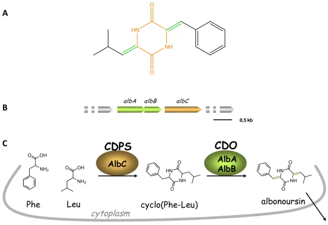
The research team named “Enzymology and non ribosomal peptide biosynthesis”, directed by Dr Muriel Gondry (CEA/DSV/iBiTec1S/SIMOPRO), is devoted to studies on the biosynthetic pathways and mechanisms of DKPs. At the beginning, in collaboration with another team directed by Jean1Luc Pernodet (IGM, CNRS UMR 8621, Université Paris1Sud 11), they isolated the biosynthetic pathway of albonoursin (cyclo(MPhe1MLeu)) ( ) which is a DKP produced by Streptomyces noursei and Streptomyces albulus with remarkable bioactivities like antibacterial and antitumor activities (Gondry et al. 2001; Lautru et al. 2002). Like most secondary metabolite genes in Streptomyces species, the albonoursin biosynthetic genes are clustered. The DNA fragment has a length of 3.8 kb containing four genes named albA, albB, albC and albD. However, subsequent studies showed that only albA, albB and albC are necessary for the biosynthesis of albonoursin (
). albA and albB code the cyclic dipeptide oxidase (CDO), while albC codes a small enzyme of 28 kDa, called AlbC. Firstly, the precursor of albonoursin, cyclo(Phe1Leu) or indicated as cFL, is synthesized by AlbC; then the CDO catalyzes the formation of α,β1unsaturated residues of cyclo(Phe1Leu) to generate albonoursin; the latter is released to the culture medium after synthesis ( ) (Lautru et al. 2002).
Figure 1: The albonoursin and its biosynthesis (Lautru et al. 2002). (A) Structure of albonoursin, with the DKP skeleton shown in orange and the two α,β-dehydrogenations in green. (B) The gene cluster composed of albA, albB and albC, and responsible for the biosynthesis of albonoursin. (C) Schema of the biosynthetic pathway of albonoursin.
AlbC catalyzes the formation of cyclodipeptides but it is unrelated to NRPSs or other known proteins. In the next few years, other similar proteins to AlbC were successively discovered in various bacterial phyla. Until 2009, eight related proteins from different bacterial organisms ( ) had been identified and characterized (Gondry et al. 2009). They are all composed of 2161249 amino acid residues, 13 of which are conserved among them. In addition, further biological analysis demonstrated that they all use aminoacyl1tRNAs (aa1tRNAs) as substrates to catalyze the formation of cyclodipeptides. These proteins thus form a family of tRNA1 dependent peptide bond1forming enzymes dedicated to the formation of cyclodipeptides. They are named “cyclodipeptide synthases (CDPSs)” (Gondry et al. 2009). However, the primary characterization showed that those CDPSs do not synthesize the same cyclodipeptides ( ). All the eight CDPSs were expressed in E. coli. Their culture supernatants were analyzed by LC1MS/MS which is HPLC
NH2 OH O NH2 OH O HN NH O O HN NH O O 0,5 kb A B HN NH O O C
coupled to mass spectrometry in order to characterize the cyclodipeptides synthesized. The results showed that AlbC produced twelve cyclodipeptides, including the principal products cFL and cFF. Thus, AlbC can incorporate into cyclodipeptides various nonpolar residues, such as phenylalanine, leucine, tyrosine and methionine, and to a much lesser extent alanine and valine. Indeed, the ten possible cyclodipeptides composed of phenylalanine, leucine, tyrosine and methionine are all synthesized in detectable amounts by AlbC. Almost all of the compounds produced by other CDPSs are combinations of the same four amino acids, with the restriction that cyclodipeptides synthesized by Rv2275 always contain tyrosine, and those synthesized by the other CDPSs almost always contain leucine
( ).
Figure 2: Eight characterized CDPSs to date (in bold) from different bacterial phyla shown in the form of phylogenetic tree.
AlbC
Streptomyces nourseiRv2275
Mycobacterium tuberculosisJk0923
Corynebacterium jeikeiumPlu0297
Photorhabdus luminescensPSHaeC06
Staphylcoccus haemolyticusYvmC
Bacillus subtilisYvmC
Bacillus licheniformisYvmC
Bacillus thuringiensisFigure 3: Histogram of the amounts of the various cyclodipeptides synthesized by eight recombinant CDPSs in E. coli (Gondry et al. 2009). cXX corresponds to an unidentified cyclodipeptide of the same molecular mass as cLL.
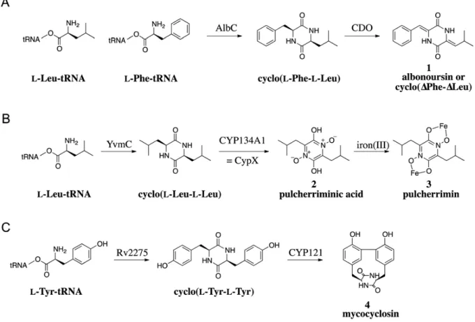
Cyclodipeptide formation is often the first step in the synthesis of more complex DKPs that are obtained after tailoring reactions (Gardiner et al. 2004; Loria et al. 2008). This appears also to be the case for the CDPS1synthesizing cyclodipeptides because the CDPS genes in prokaryotes are generally organized into operon1like structures and some of the proteins encoded by these operons could play a role in modifying the cyclodipeptide. So far, three proteins encoded by genes closely linked to CDPS genes have been experimentally characterized and shown to have three different cyclodipeptide1tailoring activities: α,β1dehydrogenation, DKP ring oxidation and C1C aryl coupling ( ) (Belin et al. 2012). One of the three proteins is the CDO having the α,β1dehydrogenation activity. As previously described, it is involved in oxidizing the cFL in the biosynthetic pathway of albonoursin ( and ). Many of the CDPS genes identified in databases are closely linked to a gene encoding a cytochrome P450 enzyme (P450). P450s constitute a superfamily of heme1containing monooxygenases that dissociate molecular oxygen to catalyze numerous reactions on a wide range of structurally diverse molecules (Guengerich 2001; Isin and Guengerich 2007). The first evidence of a role for a P450 in a CDPS1dependent pathway was provided by the identification of
the yvmC cypX gene cluster in the pulcherrimin synthesis in B. subtilis ( ) (Tang et al. 2006). The CDPS YvmC catalyzes the formation of cyclo(Leu1Leu) (cLL) (Gondry et al. 2009), which is subsequently converted into pulcherriminic acid by CypX; the pulcherriminic acid associates with iron (III) to generate pulcherrimin. CypX having the cyclodipeptide1tailoring activity belongs to the P450 family. The recent biochemical and structural characterization of CYP134A1 (= CypX) has provided insight into the oxidation of the DKP ring of cyclodipeptides (Cryle et al. 2010). The third cyclodipeptide1tailoring identified is CYP121 involved in the biosynthetic pathway of mycocyclosin isolated from Mycobacterium tuberculosis ( ). Genetically, CYP121 is linked to the CDPS Rv2275 in an operon1like structure (Cole et al. 1998; Roback et al. 2007). The two proteins constitute the metabolic pathway of the DKP mycocyclosin. Rv2275 synthesize cyclo(Tyr1Tyr) (cYY) which is then modified by CYP121 having C1C aryl coupling activity.
Bioinformatic analyses of the genetic environment of putative CDPSs have enabled to identify new putative cyclodipeptide1tailoring enzymes such as methyl1 transferases, oxidoreductases, 21oxoglutatarate1dependent oxygenases, acyl1CoA or peptideligases, and hypothetical proteins not yet related to any known function. These proteins may actually catalyze alternative modifications of the side chains of the residues constituting the cyclodipeptide or the cyclodipeptide ring itself (Belin et al. 2012).
Figure 4: The CDPS-dependent biosynthetic pathways of albonoursin 1 (A), pulcherrimin 3 (B) and mycocyclosin 4 (C) (Belin et al. 2012).
Nevertheless, our knowledge on the CDPS family and the CDPS1dependent biosynthetic pathways of DKPs is still very poor. It is important to understand their molecular mechanisms in order to, in the long run, apply this knowledge to the production of molecules that have new or improved biological and pharmacological activities.
My Ph.D project mainly consists of characterization of some CDPSs of interest, elucidation of their molecular bases on catalysis and on substrate specificity, as well as preliminary work on the catalytic process of CDPSs. This work can be achievable thanks to the existence of new CDPSs in the nature, some of which are good study models.
This thesis manuscript will begin with bibliographic studies on DKPs and the known peptide bond1forming biocatalysts ( ). The result part will be
divided into four chapters ( ). The will be dedicated to a published work on structural and mechanic characterization of AlbC, which provides insight into the interaction between the CDPS and its aa1tRNA substrates, as well as the catalytic mechanism. In the , I will introduce a new CDPS Ndas_1148 from Nocardiopsis dassonvillei characterized in the laboratory. The characteristics of Ndas_1148 allow us to better understand the molecular bases of the substrate specificity. In the , I will introduce another CDPS, named AlbC1 IMI, freshly identified from Streptomyces sp. IMI 351155. AlbC1IMI is used in my work as a CDPS model to study the catalytic mechanism. The concerns our published work on Nvec1CDPS2 which is the first eukaryotic CDPS identified in the sea anemone Nematostella vectensis. Finally, we will conclude the whole work and present the perspectives of studies on CDPSs.
1
BIBLIOGRAPHIC STUDIES
1.1
Natural Diketopiperazines (DKPs)
Cyclodipeptides and their derivatives DKPs have been detected in a variety of natural resources. They constitute a large class of secondary metabolites synthesized predominantly by microorganisms. Recently, the interest in these compounds has significantly increased because of their diverse and remarkable bioactivities (Prasad 1995; Martins and Carvalho 2007; Huang et al. 2010), such as antibacterial (Magyar et al. 1996; Cain et al. 2003; Kohn and Widger 2005), antifungal (Ström et al. 2002; Musetti et al. 2007), antiviral (Rodriguez and Carrasco 1992), antitumor (Kanoh et al. 1999; Williams et al. 1999; Kanzaki et al. 2004; Jia et al. 2005), immunosuppressive (Waring and Beaver 1996) and anti1inflammatory (Minelli et al. 2012) activities. Some DKPs are found to play physiological roles like in quorum1sensing (Holden et al. 1999; Degrassi et al. 2002; Park et al. 2006; Li et al. 2011; Ortiz1Castro et al. 2011), in plant1growth regulatory (Ortiz1Castro et al. 2011), and in central nervous system (Minelli et al. 2009). In this part, I will introduce the DKP family, their physiological roles, their biological and pharmacological activities, and some applications or potential applications of DKPs in medical and pharmaceutical domains.
1.1.1
DKP family
DKPs are a class of cyclic organic compounds and characterized by a common motif: the DKP nucleus, which is heterocycle piperazine12.51dione, also known as dioxopiperazine. This nucleus presents two cis amide bonds. The general structure of
Figure 5: General structure of DKPs. R1 and R2 represent variable lateral chains.
When the groups R1 and R2 correspond to side chains of amino acids, these DKPs are named cyclodipeptides. They are from the cyclization of two amino acids resulting from the formation of two peptide bonds. The cyclodipeptides can be of cis or trans conformation, depending on whether the constituting amino acids are of identical or different configuration.
1.1.1.1 Natural abundance of DKPs
DKPs are ubiquitous in nature. They are produced in numerous prokaryotic and eukaryotic organisms forming a large family of natural products although these molecules are a relatively unexplored class of bioactive peptides.
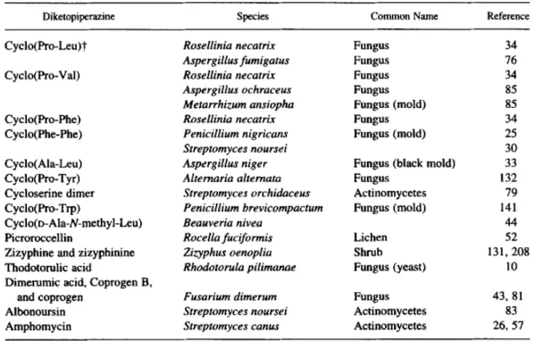
A lot of DKPs have been isolated in prokaryotes of various species like Streptomyces noursei (Khokhlov and Lokshin 1963), Pseudomonas aeruginosa (Jayatilake et al. 1996), Lactobacillus reuteri (Li et al. 2011) and Salinispora arenicola (Schultz et al. 2008). Besides, DKPs are also produced by plants and by several eukaryotic microorganisms such as yeasts, lichens and fungus (Prasad 1995). Some examples are shown in . Numerous DKPs are isolated from marine microorganisms, sponges, see stars, tunicates (ascidians), and red algae (Huang et al. 2010). Finally, one DKP, cyclo(His1Pro), has been shown to be present in mammals. This DKP was demonstrated to be present in human (hypothalamus, stomach and esophagus), monkey (hypothalamus and spinal cord), rat and mouse (hypothalamus, cerebellum and cortex) (Prasad 1995).
Table 1: Some simple cyclodipeptides that occur naturally in the protist and plant kingdoms (Prasad 1995).
A significant structural diversity of DKPs is associated with the variety of their natural sources, which I will detail in the following section.
1.1.1.2 Structural diversity of DKPs
Structures of DKPs vary a lot from simple cyclodipeptides to very complicated derivatives. Some examples are shown in . In most cases, amino acids incorporated in DKPs are of L1configuration. In this thesis, the configuration of amino acids constituting the cyclodipeptides will not be specified except for those of D1 configuration. All amino acids are not similarly incorporated to form cyclodipeptides. They are often composed of hydrophobic and aromatic amino acids especially the leucine (cyclo(Leu1Pro)), the valine (cyclo(MAla1Val) ( ) (Holden et al. 1999), the proline, the tyrosine (cyclo(Pro1Tyr)) (Holden et al. 1999), the phenylalanine (cyclo(Phe1Pro)) (Li et al. 2011), and the tryptophan (cyclo(Trp1Phe)). Several charged and polar amino acids are also present in DKPs, such as the serine (gliotoxine (
in some DKPs, such as the δ–hydroxyl1leucine in cyclomarazines A and B (
), and the norvaline (Nva) in cyclo(41methyl1D1Pro1Nva) ( ) (Adamczeski et al. 1995).
Besides the different types of amino acids, various chemical modifications to cyclodipeptides also enrich the diversity of DKPs. The modification can be introduced to DKP nuclei or to their side chains. The most common chemical modifications are the methylation like cyclo(41methyl1D1Pro1Nva) ( ), the hydroxylation like bicyclomycin ( ), the nitration like the phytotoxin thaxtomin A ( ), and the presence of double bonds like albonoursin (
Figure 6: Examples of DKP structures. The DKP-producing species are indicated in italics. HN NH O O H H H Cyclo(ΔAla-Val)
Some gram-negative bacteria as
Pseudomonas aeruginosa A N N O O S-S OH OH Gliotoxin Aspergillus fumigatus B N NH O O OH R N Cyclomarazine A (R=CH3) Cyclomarazine B (R=H) Salinispora arenicola C N NH O O H H Cyclo(4-methyl-D-Pro-Nva) Calyx cf. podotypa D HN NH O O O HO H HO HO CH3 HO Bicyclomycin Streptomyces sapporonensis E N N O O R OH H N H H3C CH3 NO2 Thaxtomin A (R=OH) Thaxtomin B (R=H) Streptomyces sp. F N H H N O O Albonoursine Streptomyces noursei G N H H N O O N OH N OH O O Rhodotorulic acid Rhodotorula pilimanae H Pulcherrimin Bacillus sp. N N O O O Fe O Fe H H 3 3 I N N O O S S O2N OH Glionitrin A J N N O O S S O O R OH H H H H R OH Epicoccin G (R=H) Epicoccin H (R=OH) Epicoccum nigrum K HN N O O Diphenylalazine A Epicoccum nigrum L HN N O O HN N NH N O O Aspergilazine A Aspergillus taichungensis M Phenylahistin Aspergillus ustus HN NH O O NH N N N N O O S-S OH O HO O O O H O Sirodesmin Leptosphaeria maculans
1.1.1.3 Physiological roles of DKPs
Although the DKPs are ubiquitous in nature and more and more DKPs are being discovered, knowledge to their physiological roles is still limited. In this part, I will introduce several known physiological roles of some DKPs.
1.1.1.3.1 Cell0to0cell communication: Quorum sensing
Communication between cells via diffusible chemicals is a general phenomenon virtually found in all living organisms. It has been intensively studied in bacteria in the last two decades. One of the best1known examples is quorum sensing. A number of bacteria associated with eukaryotic hosts employ quorum sensing systems to sense their population density thereby modulating the expression of sets of genes involved in physiological responses associated with survival, propagation, and/or virulence (Salmond et al. 1995; de Kievit and Iglewski 2000; Henke and Bassler 2004).
In Gram1negative bacteria, the most universal cell1cell signaling mechanism occurs via the production and response to a class of small diffusible molecules called N1acylhomoserine lactones (AHLs). However, DKPs are identified in cell1free culture supernatants of some Gram1negative bacteria. Cyclo(MAla1Val) and cyclo(Pro1Tyr) were found in Pseudomonas aeruginosa, Proteus mirabilis, Citrobacter freundii and Enterobacter agglomerans (cyclo(MAla1Val) only) (Holden et al. 1999). Although these two DKPs were absent from Pseudomonas fluorescens and Pseudomonas alcaligenes, a third DKP, cyclo(Phe1Pro) was isolated from both pseudomonas (Holden et al. 1999). The three DKPs were revealed capable of activating or antagonizing a LuxR1based AHL biosensor or other LuxR1based quorum1sensing systems. Although the physiological role of these DKPs has yet to be established, their activity suggests the existence of cross talk among bacterial signalling systems (Holden et al. 1999). Furthermore, the study of G. Degrassi et al. demonstrated that plant growth1 promoting Pseudomonas putida WCS358 produced at least four different cyclodipeptides (cyclo(Pro1Tyr), cyclo(Pro1Leu), cyclo(Phe1Pro), and cyclo(Val1Leu)), and some of them potentially cross1talked with the quorum sensing LuxI and LuxR homologs (Degrassi et al. 2002). It was found that three DKPs (cyclo(Pro1Val),
cyclo(Phe1Pro), and cyclo(Pro1Tyr)) from Pseudomonas aeruginosa were involved in plant growth promotion by this symbiotic bacterium (Ortiz1Castro et al. 2011). The observation that quorum1sensing1regulated bacterial production of DKPs modulates auxin signaling and plant growth promotion establishes an important function for DKPs mediating prokaryote/eukaryote transkingdom signaling (Ortiz1Castro et al. 2011). In addition, cyclo(Phe1Pro) was found produced by Vibrio vulnificus and related Vibrio spp. (V. cholera, V. parahaemolyticus, and V. harveyi) (Park et al. 2006). Vibrio vulnificus is an opportunistic human pathogen that causes severe wound infection and primary septicemia (Strom and Paranjpye 2000). The study suggests that cyclo(Phe1Pro) is a signal molecule controlling the expression of genes important for the pathogenicity of Vibrio spp. by activating the quorum sensing bioindicator (Park et al. 2006). If methods could be developed to interfere with quorum sensing systems of Gram1negative pathogens, a novel means of controlling their pathogenicity might be possible (Hartman and Wise 1998).
In Gram1positive bacteria are also isolated DKPs participating in interspecies cell1to1cell communication. In the work of Jingru Li et al., they showed that the human vaginal isolate Lactobacillus reuteri RC114 produced cyclo(Pro1Pro) and cyclo(Tyr1Pro) as the signaling molecules that are able to interfere with the staphylococcal quorum sensing system agr, a key regulator of virulence genes (Li et al. 2011). Their work contributes to a better understanding of interspecies cell1to1cell communication between Lactobacillus and staphylococcus, and provides a unique mechanism by which endogenous or probiotic strains may attenuate virulence factor production by bacterial pathogens.
1.1.1.3.2 Virulence of pathogenic microorganisms
So far, certain DKPs are found to be involved in the virulence of pathogenic microorganisms such as thaxtomin phytotoxins and gliotoxin.
Thaxtomin phytotoxins, first reported in 1989, are cyclic dipeptides (2,51 diketopiperazines) formed from the condensation of 41nitrotrytophan and phenylalanine groups. Individual thaxtomins differ only in the presence or absence
of N1methyl and hydroxyl groups and their respective substitution sites. The great interest in the thaxtomins derives mainly from their established roles as virulence factors in the common scab of potato disease and their apparent ability to inhibit cellulose synthesis in developing plant cells. Common scab is an economically important disease that is caused by Streptomyces species, which attack growing tubers through immature lenticels and wound sites. All these streptomyces species produce the phytotoxin thaxtomin which could be thaxtomin A ( ) or another member of the thaxtomin family (Loria et al. 2008). Numerous studies have demonstrated that the thaxtomin family is directly associated with the potato common scab disease. Firstly, thaxtomin is present in the tissues of infected plants and, once extracted and added in healthy tissues, induces symptoms of the common scab (King et al. 1992). Then, Healy et al. found that thaxtomin A production was abolished in biosynthesis pathway disruption mutants which were avirulent on potato tubers. Moreover, introduction of the thaxtomin synthetase cosmid into a mutant restored both pathogenicity and thaxtomin A production, demonstrating a critical role for thaxtomins in pathogenesis (Healy et al. 2000). In short, the generation of thaxtomins by common scab1causing species from diverse geographic areas of the world and the quantitative relationship established between phytotoxin production and virulence overwhelmingly supports the concept of these toxins as pathogenicity determinants (King and Calhoun 2009).
Gliotoxin ( ) is an epipolythiodioxopiperazine toxin that is made by the filamentous fungus Aspergillus fumigatus. This molecule has several remarkable biological activities. One of them under study is its contribution to the virulence of A. fumigates (Spikes et al. 2008). In the study of Spikes et al., they tested for gliotoxin production and virulence in different animal models. The results showed that gliotoxin production correlated positively with virulence in a nonneutropenic mouse model of invasive pulmonary aspergillosis and a Drosophila melanogaster model of aspergillosis (Spikes et al. 2008).
1.1.1.3.3 Iron0binding DKPs
It is known that all life forms have an absolute requirement for iron to maintain their metabolism. For aerobic organisms the concentration of free, aqueous ferric ion is limited to 10118 M at neutral pH due to the insolubility of Fe(OH)3. This is the
driving force for the excretion by microbes of strong iron1chelating agents (siderophores) and the expression of high1affinity transport systems, which provide a reliable cellular iron supply. Studies showed under iron1deficient growth conditions the yeast Rhodotorula pilimanae excreted vast amounts of rhodotorulic acid, a cyclodipeptide of δ1N1acetyl1L1δ1N1hydroxyornithine ( ) which is able to tightly bind iron (Müller et al. 1985).
Another DKP capable of binding iron is pulcherriminic acid. Fruit1borne Metschnikowia pulcherrima is yeast, which can protect fruits against postharvest rot caused by some postharvest pathogens due to its antifungal effects. M. pulcherrima produces a red pigment called pulcherrimin ( ) which is a large complex formed nonenzymatically from pulcherriminc acid and ferric ions. The antimicrobial activity of M. pulcherrimamay is attributed to the formation of pulcherrimin which depletes the iron in the medium and creates an environment unsuitable for growth of microbes that require iron for growth since iron is essential for the growth of many microorganisms (Sipiczki 2006).
1.1.1.3.4 Effect on the central nervous system
Cyclo(His1Pro) is the first cyclodipeptide shown to be endogenous to the mammalian brain. It is present in central nervous system, body fluids, anuran skins, and gastrointestinal system (Prasad 1995; Minelli et al. 2009). This DKP is widely studied for its effects on the central nervous system. It might produce analgesia (Prasad 1995). Modulation of prolactin secretion, thermoregulation, and stereotypical behavior are three biological activities of cyclo(His1Pro) that seem to share a common dopaminergic mechanism (Prasad 1995).
In conclusion, the physiological role of DKPs in organisms that produce these compounds remains poorly documented. However, in most cases, these molecules are principally studied for their biological and pharmacological activities.
1.1.1.4 Biological and pharmacological activities of DKPs
As freshly mentioned above, DKPs are widely studied because of their numerous biological and pharmacological activities such as antibacterial, antiviral, antifungal, antitumor, anti1inflammatory, and immunosuppressive activities. Now, we will present, through some examples, these remarkable activities of DKPs.
1.1.1.4.1 Antibiotic DKPs
Bicyclomycin ( ) is a DKP isolated from streptomyces sapporonensis in 1972 then from streptomyces aizunensis in 1973 (Bradley et al. 1996). This molecule is a clinically useful antibiotic exhibiting activity against a broad spectrum of Gram1 negative bacteria such as Escherichia coli, Klebsiella, Salmonella, Shigella and Citrobacter, and against the Gram1positive bacterium, Micrococcus luteus. Bicyclomycin has been used to treat diarrhea in humans and bacterial diarrhea in calves and pigs and is marketed by Fujisawa (Osaka, Japan) under the trade name Bicozamycin® (Kohn and Widger 2005). The structure of bicyclomycin is unique among antibiotics and studies showed that it employed a novel mode of action by inhibition of the RNA transcription termination factor Rho in Escherichia coli (Zwiefka et al. 1993; Kohn and Widger 2005). Rho is a hexameric RNA/DNA helicase/translocase that terminates transcription of select genes in bacteria. Bicyclomycin can disrupt the Rhomolecular machinery thereby giving rise to a catastrophic effect caused by the untimely overproduction of proteins not normally expressed constitutively, thus leading to a toxic effect on the cell (Kohn and Widger 2005). A recent study demonstrated that the sensibility of E. coli to bicyclomycin could be altered by deletions of different types of genes (Tran et al. 2011). Up to now, bicyclomycin is the only known selective inhibitor of Rho.
Glionitrin A ( ) is a new DKP disulfide with antibiotic1antitumor activity isolated from coculture of a mine drainage1derived Sphingomonas bacterial strain and a mine drainage1derived Aspergillus fumigatus fungal strain. Glionitrin A displayed significant antibiotic activity against a series of microbes including methicillin1resistant Staphylococcus aureus. Besides, an in vitro cytotoxicity assay revealed that glionitrin A had potent submicromolar cytotoxic activity against four human cancer cell lines: HCT1116, A549, AGS, and DU145 (Park et al. 2009).
1.1.1.4.2 Antiviral DKPs
A well1known antiviral DKP is gliotoxin ( ). This molecule has been mentioned above about its contribution to the virulence of pathogenic microorganisms. Besides its physiological role, gliotoxin is found to be a potent inhibitor of poliovirus RNA synthesis via its inhibition to the activity of the poliovirus polymerase 3Dpol in vitro (Rodriguez and Carrasco 1992). Gliotoxin is the
first inhibitor reported of this viral enzyme. The toxicity of gliotoxin is due to the presence of a disulphide bridge, which can inactivate proteins via reaction with thiol groups, and to the generation of reactive oxygen species by redox cycling (Gardiner et al. 2005).
Moreover, three DKPs ( ! ") extracted from the fungus Epicoccum nigrum showed inhibitory effects on HIV11 replication in C8166 cells (Guo et al. 2009). Recently, a novel DKP dimer, aspergilazine A ( #), dimerized by two DKP units via a rare N11 to C16 linkage, was isolated from the marine1derived fungus Aspergillus taichungensis. This DKP dimer showed a weak activity against influenza A (H1N1) virus (Cai et al. 2012).
1.1.1.4.3 Antifungal DKPs
Ten years ago, Ström et al. firstly reported the production of the antifungal DKPs, cyclo(Phe1Pro) and cyclo(Phe1trans141OH1Pro), by lactic acid bacteria (Ström et al. 2002). Recently, a new antifungal compound cyclo(Leu1Leu) was identified from Lactobacillus plantarum AF1, which was isolated from kimchi, a traditional Korean
food which is a well1known lactic acid1fermented vegetable product (Yang and Chang 2010). In this study, soybeans treated with different concentration of culture supernatant of Lb. plantarum AF1 could partially even totally inhibit the growth of Aspergillus flavus, which often germinates in stored cereal grains. Up to now, the mechanism of inhibition is still not clear. The end products from kimchi lactic acid bacteria, like cyclo(Leu1Leu), may be a promising alternative to chemical preservatives as a potential biopreservative which prevent fungal spoilage and mycotoxin formation in food and feed (Yang and Chang 2010).
Moreover, three other antifungal DKPs, cyclo(Phe141hydroxy1Pro), cyclo(Leu141 hydroxy1Pro) and cyclo(Ala141hydroxy1Pro), were extracted from broth culture of the grapevine endophyte Alternaria alternata. The three DKPs demonstrate real effectiveness in inhibiting the fungus Plasmopara viticola sporulation which causes the grapevine downy mildew, one of the most destructive diseases affecting this crop (Musetti et al. 2007).
1.1.1.4.4 Antitumor DKPs
More and more DKPs have been demonstrated to have antitumor activity. However, their modes of action are found very varied. Several examples will be introduced in this part to illustrate their antitumor activity.
As previously mentioned (section $ $ $ $ ), glionitrin A was also shown to have antitumor activity. An in vitro cytotoxicity assay revealed that glionitrin A had potent submicromolar cytotoxic activity against four human cancer cell lines: HCT1116, A549, AGS, and DU145 (Park et al. 2009).
The DKP phenylahistin ( %) is a cell cycle inhibitor produced by Aspergillus ustus. Phenylahistin exhibits antitumor activity against eight tumor cell lines in vitro, and against P388 leukemia and Lewis lung carcinoma cells in vivo (Kanoh et al. 1999). The mechanism of action of phenylahistin is not very clear but it was elucidated that phenylahistin arrested cells in mitosis by inhibiting tubulin polymerization (Kanoh et al. 1999).
Recently, a new DKP disulfide, deoxyapoaranotin, was separated from Aspergillus sp. KMD 901 and found to have direct cytotoxic and apoptosis1inducing effects towards HCT116 colon cancer cell lines (Choi et al. 2011).
1.1.1.4.5 Other biological activities and applications of DKPs
Besides the biological activities detailed above, DKPs exhibit some other activities. For example, cyclo(His1Pro) is an in vivo anti1inflammatory compound by modulating NF1κB and Nrf2 signalling (Minelli et al. 2012). Its cytoprotective/anti1 inflammatory effects can be ascribed to the cross1talk between the suppression of NF1 κB signalling and the activation of Nrf21EpRE/ARE pathway, the former depressing the pro1inflammatory response, the latter enhancing the antioxidant defensive response (Minelli et al. 2012). Some DKPs exhibit immunosuppressive activity like gliotoxin ( ) (Grovel et al. 2006). Gliotoxin is an immunosuppressive cytotoxin produced by numerous environmental or pathogenic fungal species. It is thought to play a role in the A. fumigatus virulence by facilitating fungal growth and colonization of host tissue through induction of a local or generalized immunosuppression (Grovel et al. 2006). Cyclo(Trp1Trp) and cyclo(Trp1Pro) specifically block the calcium channels therefore show an interest in the treatment of cardiovascular disorders (Milne et al. 1998). Furthermore, several mono1N1 methylated and di1N1methylated DKPs were demonstrated to be able to help the passage of baicalin and dopamine across the blood−brain barrier (BBB) by passive diffusion. Thereby, the DKPs or cyclodipeptide scaffolds can be considered a novel family of brain delivery systems (BBB1shuttles) to transport to the brain drugs and other cargos that cannot cross the BBB unaided (Teixidó et al. 2007).
DKPs display numerous biological and pharmacological activities. These properties make it possible to foresee their use in the treatment of pathologies.
In the first part of this chapter, I introduced the DKP family, their natural abundance and structural diversity, some physiological roles, and their biological activities which make them a family of molecules important to the research for their interest in various applications and treatment. In the study, it is important to find out
how these DKPs are produced by organisms. So, in the next part, we will talk about the biosynthesis of DKPs.
1.1.2
Biological mechanisms of DKP formation
DKPs are commonly biosynthesized from amino acids by different organisms, including mammals, and are considered to be secondary functional metabolites or side products of terminal peptide cleavage. Up to now, several biosynthetic pathways of DKPs have been deciphered. In general, these mechanisms can be classified as two types: nonenzymatic pathways and enzymatic pathways.
1.1.2.1 Nonenzymatic pathways of DKP formation
Cyclo(His1Pro) has important implications in neurophysiological functions and is an endogenous cyclic dipeptide that exists throughout the central nervous system, peripheral tissues, and body fluids. In mammals, the DKP cyclo(His1Pro) is derived from the nonenzymatic cyclization of the thyrotropin1releasing hormone (TRH, pGlu1His1Pro) after cleavage by pyroglutamate aminopeptidase. Miyashita et al. proved that cyclo(His1Pro) can emanate from a direct predecessor, that is TRH1Gly (pGlu1His1Pro1Gly), form of TRH by pyroglutamate aminopeptidase action, not through TRH formation. TRH1Gly is firstly transformed by pyroglutamate aminopeptidase to His1Pro1Gly. Nonenzymatic conversion of His1Pro1Gly to cyclo(His1Pro) then occurs ( &) (Miyashita et al. 1993). The proline induces constraints which promote cis conformation of the peptide bond between the histidine and the proline, thereby faciliting the cyclization to generate the DKP nucleus.
Figure 7: Mechanism of the formation of cyclo(His-Pro) in mammals (Miyashita et al. 1993).
However, in microorganisms, all the known mechanisms of DKP formation involve enzymes.
1.1.2.2 Enzymatic pathways of DKP formation
DKPs are commonly considered to be secondary metabolites in organisms. Some protease enzymes, such as dipeptidyl peptidases, cleave the terminal ends of proteins to generate dipeptides which naturally cyclize to form DKPs. There exist two known important enzyme families which can catalyze the formation of DKPs: nonribosomal peptide synthetases (NRPSs) and cyclodipeptide synthases (CDPSs). Here I will briefly illustrate with examples the roles of the two enzyme families in biosynthesis of DKPs. The structures and the catalytic machanisms of NRPSs are detailed in this chapter (section $ $ $ ) whereas the results of studies on CDPSs are presented in the introduction and the because my thesis work is on CDPSs.
1.1.2.2.1 Formation of DKPs involving NRPSs
NRPSs are large multimodular biocatalysts that utilize complex regiospecific and stereospecific reactions to assemble structurally and functionally diverse peptides that have important medicinal applications. NRPSs are not enzymes dedicated only to the DKP formation. In contrast, they commonly catalyze the
H2N CH C CH2 O CH2 C OH O H N CH C CH2 O N NH N C O H N CH C H OH O H2N CH C CH2 O N NH N C O NH CH C H OH O HN N HN N O O Pyroglutamate aminopeptidase Gly Glu-His-Pro-Gly His-Pro-Gly cyclo(His-Pro)
formation of multiple peptide bonds. However, some dipeptides, usually containing proline, are sometimes prematurely released then cyclize to form cyclodipeptides as side products of the reaction catalyzed by NRPSs. One of examples is the formation of cyclo(D1Phe1Pro) as side product during the nonribosomal assembly of ' ( ) *
by NRPSs (Schwarzer et al. 2001). The antibiotic tyrocidine A is a cyclic decapeptide synthesized nonribosomally by three NRPSs, TycA, TycB, and TycC, which consist of a total 10 modules, each being responsible for the incorporation of one amino acid into the final product (Schwarzer et al. 2001). Phenylalanine and proline are the first two amino acids incorporated by TycA. During the second reaction of condensation, as the dipeptide D1Phe1Pro is covalently fixed to enzyme, the side product cyclo(D1Phe1Pro) is formed due to the cis conformational constraint of proline which facilitates the nonenzymatic cyclization thereby the premature release of this DKP ( +) (Schwarzer et al. 2001).
Figure 8: Formation of the side product cyclo(D-Phe-Pro) during the synthesis of tyrocidine A catalyzed by NRPSs (Schwarzer et al. 2001). Three NRPSs, TycA, TycB, and TycC, act in concert to synthesize the cyclic decapeptide from the amino acid precursors. TycA comprises one module, TycB three, and TycC six modules, each of which is responsible for the incorporation of a cognate amino acid into the growing chain. The Te domain (red) at the last module of TycC catalyzes peptide cyclization and thereby release of the final product. The D-Phe-Pro intermediate bound to the second module is chemically unstable and is released as a side product as the cyclo(D-Phe-Pro).
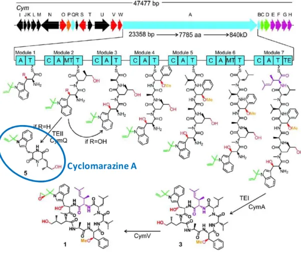
Although the majority of DKPs deriving from NRPSs harbor a proline residue at the second amino acid position, such as cyclo(D1Phe1Pro) mentioned above, and are proposed to form spontaneously resulting from conformational constraints induced by the proline residue (Stachelhaus et al. 1998; Stachelhaus and Walsh 2000; Schultz et al. 2008), there are some exceptions. This is the case of the production of ' (, - * ( ) during the biosynthesis of cyclic heptapeptides cyclomarin A by a 71module NRPS from the marine bacterium Salinispora arenicola ( .) (Schultz et al. 2008). On the subject of the formation of cyclomarazine A, N1methylation of the second residue incorporated induces constraints which promote cis conformation of the peptide bond between the two residues, thereby faciliting the formation of cyclomarazine A.
Figure 9: Biosynthetic gene cluster organization of cym and proposed biosynthesis of cyclomarin A (1) and cyclomarazine A (5) (Schultz et al. 2008). Each arrow represents the direction of transcription of an ORF and is color coded to signify enzyme function which is further reflected chemically. NRPS-related genes are colored blue with enzymatic domain abbreviations as follows: A, adenylation; T, thiolation (peptidyl carrier protein); C, condensation; MT, methyltransferase; and TE, thioesterase.
There exist some NRPSs dedicated to the biosynthesis of cyclodipeptides. Several biosynthetic pathways of DKPs involving NRPSs have been isolated in recent years. The biosynthetic pathway of the phytotoxin / (, * ( ) is firstly described in 2000 (Healy et al. 2000). Biosynthesis of this compound involves conserved NRPSs, TxtA and TxtB, encoded by the txtA and txtB genes (Healy et al. 2000). TxtA and TxtB are responsible for production of the N1methylated cyclic dipeptide backbone of the toxin, and a P450 monooxygenase named TxtC is required for post1cyclization hydroxylation steps (Healy et al. 2002). A nitric oxide synthase (NOS) is suggested to function in the nitration of thaxtomin (Loria et al. 2008) (
0).
Figure 10: Biosynthesis pathway of thaxtomin A in Streptomyces acidiscabies (Loria et al. 2008). (a) The biosynthesis of thaxtomin A involves NRPSs (TxtA and B), a P450 monooxygenase (TxtC), and a nitric oxide synthase (NOS). (b) The genes known or predicted to be involved in thaxtomin A biosynthesis are clustered together on the chromosome. Biosynthetic genes are shown in gray, while regulatory genes are indicated in red.
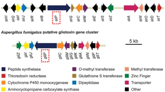
Another NRPS dedicated to the DKP synthesis is a dimodular NRPS identified within a gene cluster of Aspergillus fumigates, aspecies reported to produce fumitremorgins and other prenylated alkaloids (Maiya et al. 2006). Fumitremorgins are tremorgenic mycotoxins, members of a group of prenylated indole alkaloids derived from tryptophan and proline. Some compounds of this group have interest in the development of anticancer drugs such as tryprostatin B which is a mammalian cell1cycle inhibitor (Zhao et al. 2002). It was found that in A. fumigates, the gene Afu8g00170 encoded the NRPS brevianamide synthetase. The DKP brevianamide F (cyclo(Trp1Pro)), synthesized by this NRPS, is the precursor of a variety of fungal prenylated alkaloids with biological activity, including fumigremorgins A, B and C and tryprostatin B (Maiya et al. 2006). Some modification enzyme genes are clustered with Afu8g00170 to modify brevianamide F thereby to produce varied prenylated alkaloids. For example, Afu8g00170 is clustered with the gene Afu8g00210 encoding a prenyltransferase, which is able to prenylate brevianamide F to form tryprostatin B (Grundmann and Li 2005; Maiya et al. 2006). There are other examples of biosynthesis of DKPs involving NRPSs such as those of sirodesmin ( 1) and gliotoxin ( ), which are both secondary metabolites made by fungi. The biosynthetic gene cluster of sirodesmin is identified in Leptosphaeria maculans (Gardiner et al. 2004) ( ). It is composed of 18 genes among which the gene sirP encodes a two1module NRPS involved in the biosynthesis of a precursor of sirodesmin, named phomamide (Gardiner et al. 2005) ( ). Besides, a similar gene cluster is identified in the gliotoxin1producing fungus Aspergillus fumigatusby genome database searches (Gardiner et al. 2004) ( ). It is composed of 12 genes, many of which are similar to those in the L. maculans sirodesmin biosynthetic gene cluster. The gene gliP is a homologue of sirP, thereby is predicted to encode a NRPS responsible for the synthesis of a precursor of gliotoxin, that is cyclo(Phe1Ser) (Gardiner et al. 2005) ( ).
Figure 11: Comparison of the sirodesmin and gliotoxin biosynthetic gene clusters from Leptosphaeria maculans and Aspergillus fumigatus, respectively (Gardiner et al. 2005). sirP and gliP encoding NRPSs involved in biosynthesis of precursors of sirodesmin and gliotoxin respectively are encircled in red dotted lines. Genes with obvious homologues in the clusters are coloured. The ‘other’ category contains genes encoding cytochrome P450 monooxygenases (GliF, SirB, SirE), a prenyl transferase (SirD), an acetyl transferase (SirH), epimerases (SirQ, SirR, SirS), an oxidoreductase (SirO) and a hypothetical protein (GliK).
Figure 12: Predicted biosynthetic pathways for (a) gliotoxin and (b) sirodesmin (Gardiner et al. 2005). The two NRPSs GliP and SirP are encircled by dotted red lines.
Besides NRPSs, in the introduction part of this manuscript, we have presented another enzyme family called CDPSs dedicated to the biosynthesis of cyclodipeptides.
1.1.2.2.2 Formation of cyclodipeptides by CDPSs
As described in the introduction of the manuscript, the CDPS family is specially involved in the synthesis of cyclodipeptides. Unlike NRPS which is often a huge enzyme with multi1modules, the CDPS is always a small enzyme composed of only about 200 amino acid residues. They are very different from NRPSs or other known enzymes. This thesis is based on studies of the CDPS family. Before our dissection of CDPSs, it is necessary to describe all the known biological macromolecules that are able to catalyze the peptide bond formation in organisms.
1.2
Formation of peptide bonds by biocatalysts
A peptide bond is a covalent bond formed between two molecules when the carboxyl group of one molecule reacts with the amino group of the other molecule, leading to the release of a molecule of water (H2O). It usually occurs between amino
acids ( ).
Figure 13: Formation of a peptide bond between two amino acids.
Peptide bonds are ubiquitous and important in organisms for the structures of many biological molecules such as proteins and numerous peptide derivatives. In organisms, peptide bond formation is catalyzed by certain biocatalysts in order to
accelerate the reaction since uncatalyzed reaction is very long. There exist in cells different synthetic systems to form peptide bonds, which can be divided into two types: ribosomal pathway and nonribosomal pathways. The ribosomal pathway involving the macromolecule ribosome is the main pathway to synthesize proteins and polypeptides by forming multi1peptide bonds. It is one of the most important activities in cells to ensure the normal cell metabolism. Formation of peptide bonds through nonribosomal pathways can be accomplished by several different enzyme families such as NRPSs, CDPSs, Fem1transferases, aminoacyl1tRNA transferases, and glutathione synthetase ( ). No matter which pathway is used to form peptide bonds, activation of the carboxyl group is indispensable to bring about the reaction. In some cases, such as NRPS pathways, the carboxyl group of amino acid involved in the peptide bond formation is activated by ATP thereby forming an intermediate aminoacyl adenylate. In other cases, activated amino acid is present in the form of aminoacyl1tRNA (aa1tRNA), the formation of which is catalyzed by the aminoacyl1 tRNA synthetase (aaRS).
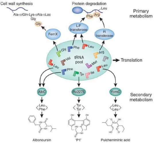
Figure 14: Some tRNAs are involved in primary processes such as cell wall formation, protein labeling for degradation, biosynthesis of amino acids and modification of porphyrin and lipids. Now tRNAs have been found in secondary metabolism in cyclic peptide formation (von Dohren 2009). 'P1' is a recently discovered product created by the P450-mediated oxidation of cyclo(Tyr-Tyr) (Belin et al. 2009).
In this part, I will firstly describe the biocatalysts of peptide bond formation which involve aa1tRNAs as substrates: ribosome, Fem1transferases, aminoacyl1tRNA transferases, CDPS, and a recently discovered transferase named PacB (Zhang et al. 2011). Before the description of these biocatalysts, aaRSs, enzymes catalyzing the aminoacylation of tRNA, will be particularly discussed because of their structural homology with our research object CDPSs. This part will then be ended with the description of some others biocatalysts of peptide bond formation which function in a tRNA1independent manner, in particular the NRPSs.
1.2.1
Peptide bond formation involving aa0tRNA
Above all, we will briefly evoke tRNA and aaRS. Then we will present the ribosomal pathway of peptide bond formation, the essential peptide and protein synthesis pathway in cells. Finally we will talk about some enzyme families, which catalyze the peptide bond formation by using the aa1tRNA as substrate.
1.2.1.1 Transfer RNA (tRNA)
Representing the single largest, best1understood class of non1protein coding RNA genes found in all living organisms, tRNAs are adaptor molecules composed of RNA, typically 73 to 93 nucleotides in length, that are used in protein biosynthesis to link the codons in a messenger RNA (mRNA) to the amino acids that they specify (Crick 1968).
tRNAs have cloverleaf secondary structure due to four base1paired stems (
). The secondary structure folds into a compact L1shaped three1dimensional structure via a set of tertiary and triple base1pair interactions, as well as a number of magnesium ions that are crucial for folding and stability (Jovine et al. 2000) (
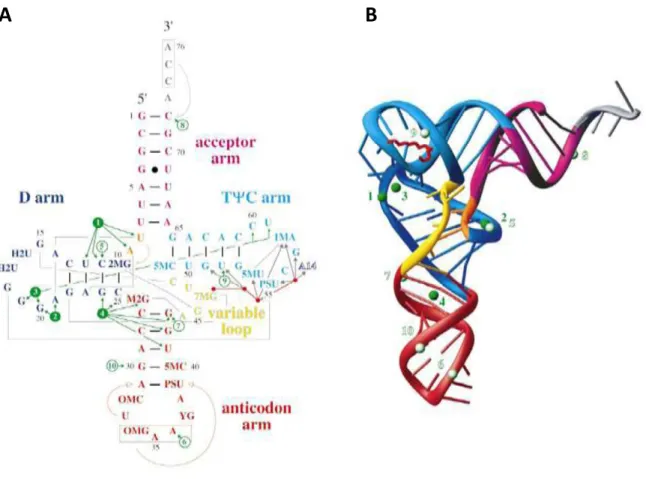
). Generally, tRNA structure contains an acceptor arm, an anticodon arm, a D arm, and a TΨC arm ( ). The acceptor arm is made by the base pairing of the 5'1terminal nucleotide with the 3'1terminal nucleotide, which contains the CCA 3'1 terminal group used to attach the amino acid. The loop of anticodon arm contains the anticodon, which can match with the triplet codon on mRNA. The D arm often contains hydrouridines where as the TΨC arm contains the sequence TΨC where Ψ is a pseudouridine.
Figure 15: Secondary and tertiary structure of yeast tRNAPhe (Jovine et al. 2000). (A) “Cloverleaf”
representation of yeast tRNAPhe secondary structure. Different regions of the molecule are indicated
and colour-coded accordingly. Watson-Crick base-pairs are represented by short black lines, the single G4·U69 wobble pair is marked by a black dot. Tertiary interactions are shown as grey lines, while arrows indicate stacking of the enclosed nucleotides onto 5′ or 3′ helices. The anticodon and the terminal CCA sequence are boxed in red and grey, respectively. Tightly and weakly bound Mg2+ are shown as green and white circles, respectively, with green arrows indicating the nucleotides with which they interact. The spermine molecule is represented by a red stick, with four dots corresponding to its nitrogen atoms (not drawn to scale); grey arrows indicate interactions with the RNA. The A14 nucleotide of a symmetry-related molecule is labelled in reverse colours. Modified nucleotide name abbreviations are; 2MG, N2-methylguanosine; H2U, dihydrouridine; M2G, N2,N2-dimethylguanosine; OMC, 2′-O-methylcytidine; OMG, 2′-O-methylguanosine; YG, wybutosine; PSU, pseudouridine; 5MC, 5-methylcytidine; 7MG, 7-methylguanosine; 5MU, ribosylthymine; 1MA, 1-methyladenosine. (B) Ribbon representation of the three-dimensional structure. The structure is colour coded as in (A).
In general, tRNA biogenesis involves the synthesis of the initial transcript, followed by processing to remove the 5′ leader, trim the 3′ trailer, add CCA, splice introns that may be present, modify multiple nucleoside residues ( ), and, for eukaryotes, export the tRNA to the cytoplasm, before its use in translation (Phizicky and Hopper 2010). tRNA genes are highly transcribed to meet the needs of