SI46
Biology and Clinical Significance of Virulence Plasmids in Salmonella Serovars
Donald G. Guiney, Ferric C. Fang, Martin Krause,Stephen Libby, Nancy A. Buchmeier, and Joshua Fierer
From the Departments of Medicine and Pathology, School of Medicine, University of California at San Diego, and the Departments ofMedicine and Pathology, Veterans Affairs Medical Center, San Diego, California; the Departments of Medicine, Pathology, and Microbiology, University of Colorado Health Sciences Center, Denver, Colorado; and the Department of Medicine, University of Zurich, Zurich, Switzerland Non-typhoidSalmonellastrains containing virulence plasmids are highly associated with
bacter-emia and disseminated infection in humans. These plasmids are found in Salmonella serovars adapted to domestic animals, such asSalmonella dublinandSalmonella choleraesuis,as well as in the widely distributed pathogens Salmonella typhimurium and Salmonella enteritidis. Although virulence plasmids differ between serovars, all contain a highly conserved 8-kb region containing thespvlocus that encodes thespvRregulatory gene and four structuralspvABCDgenes. Studies in mice suggest that thespvgenes enhance the ability ofSalmonellastrains to grow within cells of the reticuloendothelial system. Thespvgenes are not expressed during exponential growth in vitro but are rapidly induced following entry of Salmonella strains into mammalian cells, including macrophages. Transcription of thespvgenes is controlled by the stationary-phase(Tfactor RpoS,
and mutations in RpoS abolish virulence. These studies suggest that the ability ofSalmonellastrains to respond to starvation stress in the host tissuesisan essential component of virulence.
Organisms ofthe genusSalmonella cause three distinct
clini-cal syndromes: (1) gastroenteritis, (2) enteric (typhoid) fever, and (3) septicemia, with frequent metastatic foci of infection. The most common manifestation of salmonella infection is self-limited enteritis, and dissemination of bacteria outside of the gastrointestinal tract is unusual in the healthy host. In con-trast, systemic disease is the hallmark of enteric fever, with bacteremia and infection of lymphoid tissue and the reticuloen-dothelial system. Clinical gastroenteritis is not prominent, but late complications of Peyer's patch infection, which involve perforation and hemorrhage, are particularly distinctive fea-tures of enteric fever and are uncommon in other salmonella infections. Disseminated disease is also seen in the third form of salmonellosis, but in this syndrome, severe sepsis may domi-nate the clinical picture or patients may present with localized foci of infection; gastrointestinal symptoms may be mild or absent, and the bowel complications seen in enteric fever are not present [1].
Although a very large number of distinctSalmonella strains
have been identified by a combination of serological, biologi-cal, and genetic techniques, a limited number of strains have been shown to be closely associated with either the enteric
Grant support: This work was supported by the National Institutes of Health (AI32178 to D.G.G., DK35108 to D.G.G. andJ.P.,AI32463 to F.C.F.); the United States Department of Agriculture (93·37204 to D.G.G. and 94-01954 to F.C.F.); and the Swiss National Science Foundation (32401.91 and 32-039342.93 to M.K.)
Reprints or correspondence: Dr. Donald G. Guiney, Department of Medicine, School of Medicine, University of California San Diego, La Jolla, California 92093-0640.
Clinicallnfeclious Diseases 1995;21(SuppI2):SI46-51 © 1995 by The University of Chicago. All rights reserved. 1058-4838/95/2104-0002$02.00
fever or septicemia syndromes. Only Salmonella typhi and,
occasionally,Salmonella paratyphi serovars have been shown
to cause typhoid fever. These strains are host adapted to humans and do not cause disease in animals. BothSalmonella cholerae-suis and Salmonella dublin are strongly associated with the
septicemic/metastatic form of salmonellosis, while these organ-isms are uncommon causes of gastroenteritis in humans [1, 2]. In contrast, >2,000 different serovars have been identified among isolates from patients with gastroenteritis, and a few of these, notablySalmonella typhimurium and Salmonella enterit-idis, are also prominent isolates in cases of salmonella
bacter-emia [2].
These observations indicate that Salmonella strains differ
in their ability to produce particular disease syndromes. The molecular genetic basis for the different virulence traits among these strains remains poorly understood. However, recent evi-dence indicates that a common plasmid-encoded genetic locus is a major determinant of virulence in non-typhoidSalmonella
serovars associated with systemic disease [3,4]. Virulence plas-mids in Salmonella strains were identified by several early
studies in which large "cryptic" plasmids were cured, and the plasmid-free derivatives were found to be less virulent in mice. Subsequent studies in which virulence was restored by reintro-duction of the plasmids provided definitive evidence for plas-mid-encoded virulence genes [3, 4].
Distribution of Virulence Plasmids in Salmonella Serovars
Virulence plasmids are found in only a small number of
Salmonella serovars, and these can be divided into two groups
based on their epidemiology. One group consists of serovars that are host adapted to domestic animals: S. dublin (cattle),
em
1995;21 (Suppl 2) Virulence Plasrnids inSalmonellaSerovars S147S.choleraesuis(pigs),Salmonellagallinarum-pullorum(fowl), andSalmonella abortusovis(sheep) [5,
6].
The host adaptation is reflected by the observations that the particular serovar is frequently associated with disseminated disease in its host spe-cies but is usually not found in other animal spespe-cies. S.dublinand S.choleraesuisare also associated with systemic disease in humans, while S.
gallinarum-pullorum
and S.abortusovis
are not human pathogens. It is significant that virulence plas-mids have not been found in S.typhi,despite its properties of human adaptation and propensity to cause systemic illness [5]. The second group ofSalmonellastrains carrying virulence plas-mids are the broad host-range serovarsS.
typhimurium andS.
enteritidis[5]. These organisms are isolated from a variety of hosts and are prominent causes of both the gastroenteritis and septicemia syndromes in humans.
Virulence plasmids range in size from 50 kb to100kb, and considerable differences exist between plasmids from individ-ual serovars. However, plasmids isolated from strains ofa given serovar are remarkably similar. These observations suggest that the plasmids are specific to the particular serovar and that little if any plasmid exchange occurs between serovars. These findings are consistent with the apparent absence of complete self-transmissible conjugation systems on the virulence plas-mids [7]. Heteroduplex analysis indicates that the larger S.
typhimurium plasmid (l00 kb) contains a region hybridizing to virtually all of the S.enteritidisplasmid (60 kb) and almost all of the S.choleraesuisplasmid (50 kb). In contrast, large regions of the S. dublinplasmid (80 kb) appear unrelated to any of the sequences of the other virulence plasmids tested [8]. Despite these differences, the plasmid from
S.
enteritidiswas found to restore mouse virulence to a plasmid-cured S.dublinstrain [9]. In a similar vein, the plasmid from S. gallinarum-pullorumis able to substitute for the S.typhimuriumplasmid in mouse virulence, despite the fact that S.
gallinarum-pullorum
strains are not virulent in mice [10]. The results indicate that virulence plasmids are not responsible for the host-adaptation phenotype, but rather encode a common virulence function that is interchangeable among the host serovars. Subsequent studies have identified a highly conserved region of~8kb, designatedspvforSalmonellaplasmid virulence, present on all the plas-mids and responsible for the virulence phenotype in mice. The
spvlocus consists of thespvRregulatory gene and four struc-tural genes,spvABCD.
Contribution of the
spv
Locus to the Virulence ofSalmonella Serovars
Most of the work on the virulence plasmid phenotype has been done with inbred mice homozygous for the Itys locus. These mice are much more susceptible to Salmonellastrains than are animals carrying the Ity" allele. In Itys mice, S. typhi-murium, S. dublin. S. enteritidis. and S.choleraesiusstrains cured of their respective plasmids are }O1. to }06-fold less virulent than their isogenic wild-type parent strains [3, 4]. This
loss of virulence is generally seen whether the inoculum is given intraperitoneally or orally, although certainS. typhimu-riumstrains may show a plasmid effect only with oral infection [11]. The plasmid virulence phenotype is also expressed in Ity" mice. Detailed studies on the pathogenesis and histopathology of oral salmonella infection in mice show that the plasmid is not required for invasion ofthe bacteria into intestinal epithelial cells and Peyer's patches, nor for spread to mesenteric nodes and the reticuloendothelial system (RES) of the liver and spleen. Instead, the plasmid-containing strains outgrow plas-mid-free bacteria in the RES, leading to an overwhelming in-fection characterized by prominent microabscess formation and large numbers of organisms in the liver and spleen prior to death at 6-8 days after inoculation [12]. The plasmid-free strain of S. dublinwas found to invade the bowel wall and ileal Peyer's patches with an efficiency equal to that of the wild-type parent strain. However, plasmid-free bacteria in the mesenteric lymph nodes and the spleen increased much more slowly than wild-type, remaining 102
_ to 103-fold lower in
number at 7 days after infection. After 10 days, the number of plasmid-free bacteria decreased and the liver histology showed numerous small granulomas. These results indicate that the slower growth of plasmid-free strains allows the host to mount an effective immune response. Polymorphonuclear leukocytes do not appear to be importantincontrolling the initial growth of plasmid-free strains, since the numbers of bacteria recovered from neutropenic mice over the first 7 days of infection were the same as those for control mice [12].
Several lines of evidence suggest that the virulence plasmid enhances bacterial growth within the intracellular environment of host cells. In the murine model, Salmonellastrains rapidly enter cells following inoculation [13]. Gentamicin treatment of mice infected with plasmid-containing S.dublindoes not alter the lethal course of infection, owing to the inability of the antibiotic to enter host cells. However, mice are cured of salmo-nella infection by the administration of gentamicin incorporated into liposomes, which enables delivery of the drug to the intra-cellular environment [14]. Results of studies in which a temper-ature-sensitive marker plasmid is used in S.typhimurium sug-gest that the virulence plasmid accelerates intracellular growth of bacteria in the liver and spleen [15], and recent evidence indicates that the vast majority of viable plasmid-containing bacteria in the spleen are found in the macrophages within 3 days after infection (N. A. Buchmeier, unpublished observa-tions). Plasmid-containing strains do not appear to cause any general immunosuppression, since the growth of plasmid-free strains is not enhanced by mixed infection [16]. Results of these in vivo studies suggest that the virulence plasmid in-creases bacterial growth within macrophages of the RES, al-though growth within other host cells may occur as well.
Only limited studies of the virulence plasmid phenotype have been done in humans and domestic animal hosts. The virulence plasmid is required forS.gal/inarum-pullorumto produce se-vere systemic disease in fowl [17]. The S.choleraesuisplasmid
SI48 Guiney etal.
em
1995;21 (SuppI2)Figure 1. Map of known genetic loci on theSalmonella dublin virulence plasmidpSDL2. A regionencoding autonomous replication functions is located clockwise fromthe singleXbaI site.The essential virulence locus consists of the fivespvRABCDgenes, bordered by a
1S630-like element upstream and orjE downstream. A region affect-ing plasmid stability constitutes a multimer resolution system, com-posed of a gene for resolvase(rst!)and a crossover site for specific recombination(crs),leading to the conversion of plasmid multimers to monomers.
--
...-
...
--...
-a region of indirect repe-ats homologous to oriVl of F. The
rsdlcrs system is capable of resolving multimers generated in recA+ hosts and of stabilizing heterologous replicons in
Salmonella strains. A resolvase function active on crs was found in otherSalmonellaserovars harboring a virulence plas-mid, suggesting that this system is a general mechanism for virulence plasmid stabilization.
Deletion and insertion mutagenesis has been used to define the plasmid genes essential for virulence inS.dublin. Tn5-oriT
insertions inorjEretain virulence, but deletion ofspvDpartially attenuates virulence [20].Tn5-oriTinserts inspvR, spvB,and
spvCabolish virulence [20]. Due to the polar nature of InS inserts, non-polar mutations using anXbaI linker oligonucleo-tide were also constructed [23]. Analysis of these plasmids showed thatspvAis not essential for virulence in mice. Muta-tions throughout spvBare nonvirulent, including stop-codon inserts close to the COOH-terminus. Mutations in spvCand
spvDare partially virulent but the plasmids are unstable in vivo. Insertions inspvRare avirulent and abolish production of the SpvABCD proteins, consistent with the essential regula-tory role ofspvR(see below). Taken together, the results indi-cate thatspvRandspvBare essential virulence genes in mice and thatspvCandspvDhave accessory roles and are probably needed for full virulence, while spvA is not essential. In
:¥J;
~'-l AR~
B
Je.¥'
\
;..QCiJe.
\
o
rf
5J"f
\
\\ XbaI
•
Molecular Analysis of the S.
dublin
Plasmid pSDL2 In early studies in which insertion mutations and deletions were used, a single virulence region on the S.dublinplasmid was identified, as well as a fragment capable of autonomous replication (figure I) [9]. Subsequent experiments showed that an 8.2-kb segment from the virulence region cloned on an IncP plasmid replicon could restore virulence to a plasmid-cured S.dublinstrain. Complete sequence analysis of this fragment revealed six open reading frames, currently designatedspvRABCDandorjE(figure I) [20]. Comparison with spv se-quences from S.typhimuriumand S.choleraesuisshow <0.5% divergence at the nucleotide level [3]. An insertion sequence-like element related to 18630 is located upstream fromspvR
[21]. Early work identified a region downstream from orjE
required for plasmid stability
[9].
This region was found to contain a resolvase (rsd)closely related to the D protein of miniF and a cis-acting resolution site, designatedcrs[22]. Thecrselement contains eight direct, incomplete 17-bp repeats and also appears to enhance disease in pigs [18]. Since only a portion ofS.typhimuriumstrains carry a virulence plasmid, it is possible to use molecular epidemiology to determine whether plasmid-containing strains are more closely associated with systemic disease than with gastroenteritis. By using this ap-proach, we have found that 76% of strains isolated from blood cultures but only 42% of unrelated fecal isolates carried a virulence plasmid; these findings provide evidence that the virulence plasmid plays a significant role in human disease
[19].
Similar results have also been reported instudies of do-mestic livestock [8]. These findings confirm the general sig-nificance of virulence plasmids in systemic disease due to non-typhoidSalmonella serovars.The absence of virulence plasmid sequencesinS.typhiand the S.paratyphiserovars associated with enteric fever suggests that the pathogenesis of typhoid is quite different from that of disseminated, non-typhoid salmonellosis. Another difference is the presence of the Vi capsule in S. typhiand its absence in most virulence plasmid-containing isolates. S.typhiis not pathogenic for mice, and introduction of a virulence plasmid does not restore virulence in murine infections [5]. This result indicates that crucial differences in chromosomal genes exist between S. typhiand the mouse virulent serovars. Chromo-somal loci required for mouse virulence are also missing from a number of non-typhoid serovars. Transfer of a virulence plas-mid to theSalmonellaserovars S.derby, S. havana, S. minne-sota,S.ohio,and S.saintpaulfailed to confer mouse virulence on these strains, while S.heidelbergand S.newportwere ren-dered virulent [5]. These studies indicate that both the virulence plasmid and the chromosome are key determinants of mouse virulence. Since S. typhidiffers in both of these elements, salmonellosis in mice has severe limitations as a model for typhoid fever and more likely parallels the septicemia syn-drome.
CID 1995;21 (SuppI2) Virulence Plasmids inSalmonella Serovars 8149
S. typhimurium, spvC and spvD have significant roles in mouse virulence [24, 25].
The spv genes are transcribed from two promoter regions, one located upstream from spvR and the second positioned between spvR and spvA [26]. Transcription of the spvABCD genes is initiated upstream from spvA, and messages for spvA,
spvAB, spvABC, and spvABCD are found in decreasing abun-dance. LacZ translational fusions to the Spv proteins are also expressed with the same decreasing activity, SpvA being high-est and SpvD lowhigh-est [26]. These findings sugghigh-est that tran-scripts initiate at the spvA promoter and can terminate within each ofthe long intergenic regions. However, a complex system of termination and mRNA processing cannot be excluded. The promoter for spvR has not been localized, but a monocistronic message is found, indicating that spvR transcription normally terminates before the spvA gene (M. Krause and D. G. Guiney, unpublished observations).
log phase cells
R A B C 0 plasmid
- - - --e::>-c:>=:>-c - - -
spv genes rpoS ~ stationary!
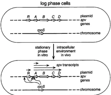
intracellular phase environment in vitro in vivo )0 spvtranscripts )0 Regulation ofspv ExpressionThe discovery that spv expression varies during the bacterial growth phase provided the key to understanding the mechanism of spv gene regulation shown in figure 2 [27]. When LacZ translational fusions were used, the expression of spvABCD was found to be very low in early log-phase cells and to in-crease rapidly in the post-exponential phase [26, 27]. This growth-phase regulation was confirmed by analysis of spv mRNA transcribed in wild-type S. dublin [26]. In the log phase, only small quantities of single-length spvA mRNA are made. As cells enter the stationary phase, progressively longer mes-sages are found, including full-length spvABCD mRNA.
Sequence analysis of SpvR suggested that this protein be-longs to the large family of LysRlMetR-like transcriptional activators [28]. These proteins act as single-component regula-tors of gene expression, and they contain a conserved helix-tum-helix motif in the NH2-terminal region involved in DNA
interaction [29]. Genetic studies confirmed that SpvR is abso-lutely required for spvABCD expression [27]. Knockout muta-tions in spvR abolish the synthesis of LacZ fusions with the structural Spv proteins, as well as production of the native Spv proteins in minicells [23, 26, 27]. SpvR binds to a region upstream from the spvA transcriptional start sites, and muta-tions in the helix-tum-helix motif abolish binding in vitro and activation ofspvA transcription in vivo [30]. However, overpro-duction of SpvR from constitutive promoters does not abolish the growth-phase regulation of the spvABCD structural genes, indicating that the SpvR level is not the sole regulator of spv expression [26].
The pattern of spv growth-phase regulation strongly sug-gested the involvement of the alternative a factor RpoS (KatF, as). The activity of this a factor is induced during post-expo-nential growth and regulates the stationary-phase expression of a large set of target genes involved in starvation survival and environmental stress responses [31]. The central role of RpoS
Figure 2. Mechanism ofSPygene regulation on the virulence
plas-mids ofSalmonella serovars. Bacterial cells growing in logarithmic phase in rich culture media do not express theSPygenes (depicted
in the upper cell). However, both post-exponential growth in vitro and the intracellular environment of host cells in vivo induce the
SPygenes (shown in the lower cell). Induction depends on both the
chromosomal a factorRpoSand the plasmidregulatory proteinSpvR. RpoSactivity increases in stationary phaseand in response to nutrient starvation. RpoS appears to act at both the spvR and spvA promoters. SpvR is essential for transcription of the spvABCDgenes by binding to the spvA promoter region. Transcription of the spvABCDstructural genes is initiated at the spvA promoter, and multiple transcripts are found.
in spv gene expression was demonstrated by constructing an
rpoSknockout mutation in S. typhimurium [32]. Expression of
spvB is severely decreased in the rpoS mutant. As expected, the rpoS mutant is markedly attenuated for virulence in mice. Recent evidence indicates that RpoS is required for optimal transcription at both the spvR and spvA promoters (figure 2) ([33] and D. G. Guiney, unpublished observations).
Induction ofspv Gene Expression by the Host-Cell Environment
Since considerable indirect evidence suggests that the spv genes are active in the intracellular environment of the host, the expression of theSPy genes was examined after uptake of
bacteria by mammalian cells in tissue culture [34]. Fluorescent (and later luminescent) substrates ofLacZ were used to develop a sensitive assay for the expression of spv::lacZ fusions by small numbers of bacteria inside eukaryotic cells. These studies have shown that the spv genes are rapidly induced by
>
100-fold on uptake by macrophages, epithelial cells, and hepato-cytes ([34] and S. Libby, J. Fierer, and D. G. Guiney, unpub-lished data). Most of the induction occurs within the first hour following uptake and does not depend on acidification of theSI50 Guiney et a1.
em
1995;21 (Suppl 2)intracellular vacuole containing theSalmonellastrain. This reg-ulation differs considerably from control of the chromosomal locuspagC; pagCis regulated by the PhoPlPhoQ two-compo-nent system and is induced in macrophages, but not epithelial cells, by a mechanism requiring phagosome acidification over a period of 3 hours [35J.
These results indicate thatspv induction occurs in response to a general property of the intracellular environment, perhaps reflecting a relative lack of nutrients available within the endo-cytic or phagoendo-cytic vacuole. Since the intracellular induction of the spv genes requires both SpvR and RpoS, the genetic mechanisms regulating post-exponential phase synthesis in vitro are the same as those controlling the spv operon after entry into host cells, and presumably throughout the infectious process (figure 2). The finding that virulence genes are regu-lated by RpoS suggests thatSalmonellastrains experience sig-nificant starvation stress during intracellular infection. Since the RpoS-mediated response increases bacterial resistance to a variety of adverse environmental factors, this regulation ap-pears to be essential for the organism to survive and multiply in the host.
Plasmid-CuredSalmonellaStrains as Live Vaccines Since plasmid-cured S.dublin produces a self-limited sys-temic infection with granuloma formation, the ability of this strain to induce protective immunity was tested in mice [36]. Animals immunized by either oral or intraperitoneal inocula-tion were protected against lethal challenge with virulent S. dublin given by oral or intraperitoneal routes of infection. The fact that intraperitoneal vaccination could protect against a vir-ulent oral challenge led to the demonstration that systemic as well as oral salmonella vaccination produces infection of Pey-er's patches in the intestine. Mice immunized withS.dublin (group D) were protected against challenge with other group DSalmonellaserovars and also against S.typhimurium(group B), which shares an O-antigen determinant with group D [36]. Similar results were reported withS.enteritidis(group D) [37]. However,S.dublindoes not protect mice fromS.choleraesuis (groupC),which lacks any cross-reactive O-antigen epitopes [36J.Itis likely that both lipopolysaccharide and non-lipopoly-saccharide antigens are important in the protection induced by live, plasmid-cured strains. These immunization studies have important implications for vaccine development. The use of the plasmid-free strain as a vaccine shows that an immune response to the Spv proteins is not required for protection. Since plasmid-containingSalmonellaserovars are major pathogens of domestic animals, plasmid-free derivatives could be combined in a multivalent vaccine for use in livestock.
Conclusions
The plasmid-encodedspvgenes enhance the ability ofcertain non-typhoidSalmonella serovars to produce severe,
extraintes-tinal disease. The spv locus consists of the spvR regulatory gene and the four structural spvABCDgenes. Studies onspv gene regulation have established the importance of post-expo-nential or stationary-growth-phase control mechanisms on the expression of virulence genes inSalmonella strains. The alter-native(J"factor RpoS, together with SpvR, regulates expression
of thespvoperon and ensures rapid induction of thespvgenes within the intracellular environment of the host. Evidence from pathogenesis studies in mice suggests that the spv genes enhance the growth ofSalmonella species within cells of the RES.
In clinical practice, non-typhoid salmonella bacteremia and metastatic infection are associated with a variety of predispos-ing conditions that decrease natural or acquired immunity, in-cluding extremes of age, decreased gastric acidity, malignancy, immunosuppression, and AIDS. TheSalmonellaserovars caus-ing systemic disease in these patients are predominantly strains that carry a virulence plasmid. The virulence-plasmid pheno-type does not appear to interfere with clearance by acquired immune mechanisms. By extrapolation from the mouse model, thespv genes are likely to facilitate growth of theSalmonella strains within host cells at the sites of systemic infection, partic-ularly in patients who are unable to mount an effective immune response. Salmonella infection in patients receiving immuno-suppressive therapy or in patients with AIDS is particularly difficult to eradicate. The identification of thespv locus pro-vides a molecular genetic explanation for the propensity of plasmid-containing serovars to cause serious infections in these patients.
References
I. Fang FC, Fierer J. Human infection withSalmonella dublin. Medicine (Baltimore) 1991;70:198-207.
2. Blaser MJ, Feldman RA. Salmonella bacteremia: reports to the Centers for Disease Control, 1968-1979. J Infect Dis 1981;143:743-6. 3. Gulig PA, Danbara H, Guiney DG, Lax AJ, Nore F, Rhen M. Molecular
analysis ofspv virulence genes of the salmonella virulence plasmids. Mol Microbioll993;7:825-30.
4. Guiney DG, Fang FC, Krause M, Libby S. Plasmid-mediated virulence genes in non-typhoid Salmonella serovars. FEMS Microbiol Lett 1994;124:1-10.
5. Roudier C, Krause M, FiererJ, Guiney DG. Correlation between the presence of sequences homologous to the vir region of Salmonella dublin plasmid pSDL2 and the virulence of twenty-twoSalmonella
serotypes in mice. Infect Immun 1990;58:1180-5.
6. Colombo MM, Leori G, Rubino S, Barbato A, Cappuccinelli P. Phenotypic features and molecular characterization of plasmids in Salmonella abortusovis.J Gen Microbioll992; 138:725-31.
7. Chikami GK, Fierer J, Guiney 00. Plasmid-mediated virulence in Salmo-nella dublin demonstrated by use of aTn5-oriTconstruct. Infect Immun 1985; 50:420-4.
8. Montenegro MA, Morelli G, HelmuthR.Heteroduplex analysis of Salmo-nella virulence plasmids and their prevalence in isolates of defined sources. Microb Pathog 1991; II :391- 7.
9. Beninger PR, Chikami G, Tanabe K, Roudier C, Fierer J, Guiney DG. Physical and genetic mapping of theSalmonella dublinvirulence
plas-eID
1995;21 (SuppI2) Virulence Plasmids inSalmonellaSerovars SI51 mid pSDL2. Relationship to plasmids from otherSalmonellastrains. JClin Invest 1988; 81:1341-7.
10. Barrow PA, Lovell MA. Functional homology of virulence plasmids in Salmonella gallinarum,S.pullorum,and S.typhimurium.Infect Immun 1989;57:3136-41.
II. Gulig PA, Curtiss R III. Plasmid-associated virulence ofSalmonella typhi-murium.Infect Immun 1987;55:2891-901.
12. Heffernan EJ, Fierer J, Chikami G, Guiney D. Natural history of oral Salmonella dublininfection in BALB/c mice: effect of an 80-kilobase-pair plasmid on virulence. J Infect Dis 1987; 155:1254-9.
13. Dunlap NE, Benjamin WH Jr, McCall RD Jr, Tilden AB, Briles DE. A "safe-site" forSalmonella typhimuriumis within splenic cells during the early phase of infection in mice. Microb Pathog 1991; 10:297- 31O. 14. Fierer J, Hatlen L, LinJP,Estrella D, Mihalko P, Yau-Young A. Successful treatment using gentamicin liposomes ofSalmonella dublininfections in mice. Antimicrob Agents Chemother 1990;34:343-8.
15. Gulig PA, Doyle TJ. The Salmonella typhimurium virulence plasmid increases the growth rate of salmonellae in mice. Infect Immun 1993;61:504-11.
16. Gulig PA, Curtiss R III. Cloning and transposon insertion mutagenesis of virulence genes ofthe IOO-kilobase plasmid ofSalmonella typhimurium. Infect Immun 1988;56:3262-71.
17. Barrow PA, SimpsonJM,Lovell MA, Binns MM. Contribution of Salmo-nellagallinarumlarge plasmid toward virulence in fowl typhoid. Infect Immun 1987;55:388-92.
18. Danbara H, Moriguchi R, Suzuki S, et al. Effect of 50-kilobase plasmid pKDSC50 ofSalmonella choleraesuisRF-I strain on pig septicemia. J Vet Med Sci 1992;54:1175-8.
19. Fierer J, Krause M, TauxeR,Guiney D.Salmonella typhimurium bacter-emia: association with the virulence plasmid. J Infect Dis 1992; 166:639-42.
20. Krause M, Roudier C, Fierer J, Harwood J, Guiney D. Molecular analysis of the virulence locus of theSalmonella dublinplasmid pSDL2. Mol MicrobioI1991;5:307-16.
21. Krause M, Harwood J, Fierer J, Guiney D. Genetic analysis of homology between the virulence plasmids ofSalmonella dublinandYersinia pseu-dotuberculosis.Infect Immun 1991;59:1860-3.
22. Krause M, Guiney DG. Identification of a multimer resolution system involved in stabilization of the Salmonella dublinvirulence plasmid pSDL2. J Bacteriol 1991; 173:5754-62.
23. Roudier C, Fierer J, Guiney DG. Characterization of translation termina-tion mutatermina-tions in thespvoperon of theSalmonellavirulence plasmid pSDL2. J BacterioI1992; 174:6418-23.
24. Gulig PA, Chiodo VA. Genetic and DNA sequence analysis of the Salmo-nellatyphimuriumvirulence plasmid gene encoding the 28000 molecu-lar weight protein. Infect Immun 1990;58:2651-8.
25. Gulig PA, Caldwell AL, Chiodo VA. Identification, genetic analysis, and DNA sequence of a 7.8-kb virulence region of theSalmonella typhimu-riumvirulence plasmid. Mol Microbiol 1992;6:1395-411.
26. Krause M, Fang FC, Guiney 00. Regulation of plasmid virulence gene expression inSalmonella dublininvolves an unusual operon structure. J Bacteriol 1992; 174:4482-9.
27. Fang FC, Krause M, Roudier C, Fierer J, Guiney 00. Growth regulation of a Salmonella plasmid gene essential for virulence. J Bacteriol 1991; 173:6783-9.
28. Pullinger GD, Baird GD, Williamson CM, LaxAJ.Nucleotide sequence of a plasmid gene involved in the virulence of salmonellas. Nucl Acid Res 1989; 17:7983.
29. Henikoff S, Haughn GW, CalvoJM,Wallace Je. A large family of bacte-rial activator proteins. Proc Natl Acad Sci USA 1988;85:6602-6. 30. Krause M, Fang FC, EI-Gedaily A, Libby S, Guiney DG. Mutational
analysis of SpvR-binding to DNA in the regulation of theSalmonella plasmid virulence operon. Plasmid 1995(in press).
31. Loewen PC, Hengge-Aronis R. The role of the sigma factor(J'(KatF) in
bacterial global regulation. Annu Rev Microbioll994;48:53-80. 32. Fang FC, Libby SJ, Buchmeier NA, Loewen PC, Switala J, Harwood J,
Guiney DG. The alternative(JfactorKatF(RpoS) regulatesSalmonella
virulence. Proc Natl Acad Sci USA 1992;89:11978-82.
33. Kowarz L, Coynault C, Robbe-Saule V, Norel F. TheSalmonella typhimu-rium katF (rpoS)gene: Cloning, nucleotide sequence, and regulation of spvRand spvABCD virulence plasmid genes. J Bacteriol 1994; 176:6852-60.
34. Fierer J, Eckmann L, Fang F, Pfeifer C, Finlay BB, Guiney D. Expression of theSalmonellavirulence plasmid genespvBin cultured macrophages and nonphagocytic cells. Infect Immun 1993;61:5231-6.
35. Alpuche Aranda CM, Swanson JA, Loomis WP, Miller SI.Salmonella
typhimuriumactivates virulence gene transcription within acidified mac-rophage phagosomes. Proc Natl Acad Sci USA 1992;89:10079-83. 36. Fierer J, Chikami G, Hatlen L, Heffernan EJ, Guiney D. Active
immuniza-tion with LD842, a plasmid-cured strain ofSalmonella dublin,protects mice against group D and group BSalmonellainfection. J Infect Dis 1988; 158:460-3.
37. Nakamura M, Sato S, Ohya T, Suzuki S, Ikeda S, Koeda T. Plasmid-cured Salmonella enteritidisAL 1192 as a candidate for a live vaccine. Infect Immun 1985;50:586-7.

![From the Observation of UHECR Radio Signal in [1-200] MHz to the Composition: CODALEMA and EXTASIS Status Report](data:image/gif;base64,R0lGODlhAQABAIAAAP///wAAACH5BAEAAAAALAAAAAABAAEAAAICRAEAOw==)