Detection by
32P-postlabeling of thymidine glycol in 7-irradiated
DNA
Monika E.Hegi, Peter Sagelsdorff
1and Werner K.Lutz
2Institute of Toxicology, Swiss Federal Institute of Technology and University of Zurich, CH-8603 Schwerzenbach, Switzerland 'Present address: CIBA-GEIGY AG, CH^002 Basel, Switzerland 2To whom repnnt requests should be sent
The
32P-postlabeIing method has been adapted for the
analysis of thymidine-cis-glycol-3'-phosphate (cis-dTGp,
cis-5,6-dihydroxy-5,6-dihydrothymidine-3'-phosphate).
Cis-dTGp was isolated and purified from normal nucleotides by
phenylboronate affinity chromatography and phosphorylated
by T4 polynucleotide kinase in presence of 1 mM BeCl
2at
pH 7.5. These modifications of the postlabeling method
resulted in a 5-phosphorylation of dTGp with a labeling
efficiency of up to 20% whereas the natural nucleotides were
almost completely dephosphorylated at the 3' position under
these conditions. The reaction products, containing
radio-labeled thy midine-cjs-glycol-3' ,5' -bis-[5' -
32P] phosphate
(c£s-*pdTGp), were separated by two-dimensional
anion-exchange TLC on polyethyleneimine cellulose sheets. Boric
acid was added in the second dimension in order to selectively
retard cis-glycols. The method was applied to 7-irradiated
nucleotides and calf thymus DNA. In the nucleotide mixture,
330-99 000 thy mine glycol (TG) moieties were detected per
10
6thj mines (T) in a dose range of 14-1000 Gy
respectively. In DNA, these values ranged from 400 to 2700
TG/10
6T. The data are in good agreement with methods
using radiochemical and immunological techniques.
Non-irradiated DNA showed a background level of lOTG/10
6T.
This practical limit of detection was higher than can be
achieved with the postlabeling technique, indicating that the
present method might be a sensitive alternative for a
determination of oxidative DNA damage.
Introduction
Oxygen radicals such as the highly reactive hydroxyl radical are
known to cause DNA damage (1). They are postulated to be
partly responsible for the carcinogenic action of ionizing
radiation. For a number of chemical carcinogens, an 'indirect'
genotoxicity via the same ultimate reactive agent is also discussed
(2). In these cases, the hydroxyl radical is thought to be generated
by a Fenton reaction of superoxide anion radical O2 with
hydrogen peroxide (H
2O
2). Both reactants are produced in
normal aerobic cellular metabolic pathways, but the concentration
seems to be carefully controlled by the action of superoxide
dismutase, peroxidases (including catalase) and antioxidants. Only
in specific situations have increased levels of H2O2 been shown
*Abbreviations:TG, thymine glycol (5,6-dihydroxy-5,6-dihydrothymine); cis-dTGp, thymidine-rij-glycol-3'-phosphate (cii-5,6-dihydroxy-5,6-dihydro-thymidine-3'-phosphate); PBA, phenylboronic acid; T, thymine; dTp, thymidine-3'-phosphate; dsDNA, double-stranded DNA; dNp, deoxyribonucleo-side-3'-phosphate; LE, labeling efficiency; bicine, yV,N-bis[2-hydroxy-ethyl]glycine); TLC, thin-layer chromatography; *p, [32P]phosphate; pdNp, deoxynT»nucleoskte-3',y-bis-phosphate; pdN, deoxyribonudeoside-5'-phosphate; Pi, inorganic phosphate; PNK, polynucleotide kinase.
to be produced, e.g. in liver peroxisomes from rodents treated
with peroxisome proliferators (3,4) and in macrophages
stimulated by phorbol ester tumor promoters (5). Under in vitro
conditions, damage has been detected in DNA added
extra-cellularly (3,5). However, no evidence for DNA damage in vivo
has so far been reported.
In order to investigate whether intracellular DNA is also
damaged under conditions of chemically induced oxygen stress,
sensitive and specific methods for the detection of oxidative DNA
damage are required. One important reaction product of DNA
oxidation with hydroxyl radical is
5,6-dihydroxy-5,6-dihydro-thymine (5,6-dihydroxy-5,6-dihydro-thymine glycol, TG*). Two cis-isomers and two
fra/u-isomers are formed (6). The present report describes a new
method using a modified ^P-postlabeling technique (7), which
includes an affinity chromatography purification of dTGp and a
phosphorylation procedure in the presence of mutagenic metal
ions to achieve acceptable labeling efficiencies. The method is
applied to quantitate oxidative DNA damage as
cis-5,6-di-hydroxy-5,6-dihydrothymidine-3' -phosphate (thy
midine-cw-glycol-3'-phosphate ds-dTGp) in 7-irradiated DNA.
Materials and methods
Materials
Phenylboronate silica (Bondesil PBA) was obtained from Analytkhem International (Basel, Switzerland). For PBA-chromatographies 0.7 x 20 cm Econo-columns were purchased from BioRad (Glattbrugg, Switzerland). Anion-exchange PEI-cellulose thin layer sheets were from Macherey-Nagel (Duren, FRG), Cawo cassettes and Cawo intensifying screens (fast tungstate) from Cawo (Zurich, Switzerland) and Kodak XAR-5 films from Kodak (Lausanne, Switzerland). Lyophilizations were performed in a speed vac concentrator from Bachhofer Laborgerate (Reutlingen, FRG). The enzymatic digestion of DNA was carried out in Eppendorf tubes from Sarstedt AG (no. 72.690; Sevelen, Switzerland). All deoxynbonucleotides, thymidine, thymine (T) and ATP were purchased from Pharmacia (Diibendorf, Switzerland). Calf thymus DNA (no D-1501) and sperrrudine were obtained from Sigma and dhhiothrehol from Serva Feinbiochemica (Heidelberg, FRG). [7-32P]ATP (3000 Ci/mmol, NEG-002 H; 6000 Ci/mmol, NEG-002 Z for the experiments on the concentration dependence) was obtained from New England Nuclear. Potato apyrase (EC. No. 3.6.1.5, grade I, ATPase activity 11 U/mg), micrococcal nuclease (E.C. No. 3.1.31.1, Sigma no. N3755) and nuclease PI (E.C. No. 3.1.30.1) was obtained from Sigma, T4 polynucleotide kinase (E.C. No. 2.7.1.78) (104 U/ml) from Pharmacia, spleen exonuclease (= phospnodiesterase, E.C. No. 3.1.16.1, no. 108 251) and poiynucleoude kinase, 3'-phosphatase-free (E.C. No. 2.7.1.78) from Boehringer Mannheim. All other chemicals were of the highest purity available from Merck or Fluka (Buchs, Switzerland).
Synthesis of cis-dTGp
The synthesis of this standard essentially followed the published methods (8,9): 1 mg (3.1 pmol) thymidine-3'-phosphate (dTp) in 1 ml of 0.2 M NH4C1/NH3 buffer pH 8.6, was oxidized at 0°C with 0.4 ml 0.014 M KMnO4 in the same buffer. After 5 mm, 10 pi of 1 M NajSjOs was added to reduce the residual permanganate. MnOj W3S removed by centrifugation and 1.4 ml of 1 M NH4OAc/NH3, 40 mM MgCl2 pH 8.8 (buffer A) was added to the supernatant. The sample was loaded onto a PBA affinity column (bed dimensions 0.7 x 2.5 cm; used at 4 ° Q equilibrated with 10 ml 0.5 M Nl^OAc/NHj, 20 mM MgCl2 pH 8.8 (buffer B). The column was washed with 15 ml buffer B and 5 ml 0.5 M NH4OAc/NH3, 1 mM MgCI2 pH 8.8 (buffeT Q. dTGp was eluted with 50 mM HOAc and 0.5 ml fractions were collected. The amount of cii-dTGp formed was quantified by Hi-reduction (8).
•^-Irradiation
Double-stranded DNA (dsDNA), deoxyribonucleoside-3'-pnosphate (dNp) or dTp
M.E.Hegi, P.Sagelsdorff and W.K.Lutz
(all at 3.2 /tmol dNp/ml) were dissolved in bidistilled water and irradiated from a ^ C o source at a dose rate of 46 Gy/min at room temperature.
Digestion of dsDNA to dNp
To 1 ml of the sample 250 jil 100 mM sodium succinate/HCl buffer, pH 6, containing 40 mM CaCl2 were added and the DNA was enzymatically digested for 16 h at 37°C with 2.5 U micrococcal nuclease added in 50 p\ 8 mM CaCl2, 20 mM sodium succinate, pH 6, and 0.05 U spleen exonuclease (in 12 /tl of the commercial suspension).
Separation of dTGp from normal nucleotides on a PBA column (at 4°C)
The digest (1300 p\) was mixed with 1300 /il buffer A and loaded onto the phenylboronate affinity column (bed: 0.7 x 2.5 cm) equilibrated with 10 ml buffer A. The chromatography was performed in the same way as described above (Synthesis of c«-dTGp). Fractions 2 - 8 were pooled and lyophilized to 1 ml. For further purification, the concentrated sample was loaded onto a second column after adding 1 ml buffer A and was treated as before. The samples were stored in Eppendorf tubes at - 2 0 ° C either as 100-/d aliquots of the fractions or in pools of 15 it\ of fractions 3 - 8 (= 90 fi\). The samples were lyophilized immediately before phosphorylation.
To estimate the extent of normal dNp contaminating the dTGp fractions after two PBA columns, 3.2 ^mol dNp (equimolar for G, A, C and T) were chromatographed twice on PBA columns, 100-/U aliquots of fractions 2—7 were taken, lyophilized and phosphorylated at pH 9.0 in presence of 50 pmol ATP [optimal phosphorylation conditions for dNp, resulting in a labeling efficiency (LE) of 80%.
Enzymatic phosphorylation of dNp with [y-32-P]ATP
The following components were carefully placed as separate droplets on the inside wall of Eppendorf tubes: 2 id H2O (or 2 /tl of the different 5 mM metal ion solutions; BeCl2/HCl, pH 3), 2 y\ kinase buffer (250 mM bicine/NaOH pH 6.0 to 9.0, 50 mM MgCl2, 50 mM dithiothreitol, 5 mM spermidine), 2 pil dNp solution (0.01-50 pmol) [H2O or ATP solution (10-1000 pmol) in those experiments where lyophilized samples were used], 2 y\ [7-32P]ATP (1 /tCi to 45 jiCi). The four droplets were mixed by centrifugation. Two microliters (3 U) T4 PNK (freshly diluted in water) was placed on the inside wall and after a second centrifugation the tubes were incubated for 30 min at 37°C. The total volume of the sample was always 10 /il. In order to hydrolyze unreacted ATP the mixture was incubated for another 15 min at 37°C with 40 mil (2 /il) potato apyrase (in water). The buffer used for the kinase reaction, bicine (N,N-bis[2-hydroxyethyl]glycine, pKa 8.3) was sometimes used below the ideal pH range because it was found that the LE for dTGp was higher with bicine even at low pH values compared with all other buffers tried (Hepes, imidazol, Tris, Tris-maleate).
Separation of 32P-labeled nucleotides by thin-layer chromatography (TLC)
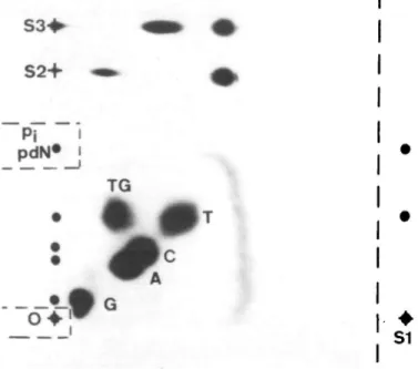
For the separation of [32P]pdTGp (*pdTGp) from normal deoxyribonucleo-side-3',5'-bis-[32P]phosphate (*pdNp), a two-directional anion-exchange PEI-cellulose TLC system was developed (see Figure 1): The sheet (20 x 20 cm, pre-washed by a run with bidistilled water and stored at - 2 0 ° C when dry) was marked with a pencil to indicate the position of the sample (O), of the standards S I - S 3 and the cutting line for SI after the first dimension.
Up to 10 jil of the phosphorylation mixture was applied to the origin (O) in the left and 2 pi *pdTGp standard (SI) was placed in the right lower corner. Without drying the origin area, direction 1 was developed in 0.12 M sodium phosphate pH 8.6 ( ~ 2 h). The sheet was dried in a stream of cold air, marked with 'ink dots' containing [32P]ATP for alignment after exposure (on the right side which is cut before dimension two), and autoradiographed for 10 min to localize origin and (inorganic [32P]phosphate) *p; spot. These areas were clipped off and kept for quantification by Cerenkov counting. The standard SI was cut off at the right-hand side of the sheet. The sheet was washed in 500 ml deionized water in a flat tray (30 x 40 cm) for 4 min. The tray was agitated frequently during washing. A 1-ml aliquot of the washing water was taken and counted for radioactivity. The sheet was again dried in a stream of cold air. For the second dimension the sheet was turned 90° counter-clockwise and two standards (S2 = *pdTGp; S3 = *pdTp) were applied on the new base line, 4 and 6 cm from the left corner respectively. The second dimension was developed in 0.12 M sodium phosphate buffer containing 0.22 M H3BO3, pH 8.6. After drying, the sheet was marked again with small 'ink dots'.
Detection and quantification
The spots on the PEI sheet were localized by autoradiography (0.5-16 h) and quantified by Cerenkov counting after excision of the spots (maximum counting efficiency 43%). The LE for a spot *pdNp is calculated in percent of the total activity which theoretically could have been incorporated into the dNp. The total radioactivity used was the sum of the activity in the washing water and on the sheet (all spots plus total background). The total background BG,O1 was deter-mined after development of the second dimension by counting a 1-cm2 area ( - 3 0 - 1 5 0 c.p.m.) on the right-hand side between the phosphate spot and the front, and by multiplying the result by 290 (14.5 x 20 cm5).
S2+
Pi
pdN*
TG
- * • •
•
S1
Fig. 1. Two-dimensional TLC on PEI-cellulose for separatingbis[5'-32P]phosphates of the natural nucleotides (G, A, C and T) and TG. The sample applied was a mixture of two phosphorylation reactions: (i) containing *pdTGp (from 50 pmol dTGp, pH 7.5, 1 mM BeCl2); and (ii) containing *pdNp (from 50 pmol of an equimolar mixture of the four natural nucleotides, phosphorylated at pH 9.0). The small dots indicate the positions of pit deoxyribonucleotide-5'-monophosphates (pdN), and of all pdNp as seen after the first dimension (developed from bottom to top) Crossed dots indicate the loading areas of the sample (O) and of the standards used in the first (SI) and second dimension (S2 and S3, developed from left to right). Cuts performed after the first dimension are indicated by the interrupted lines.
The amount of nucleotides present during phosphorylation was calculated on the basis of a LE of 10% for dTGp (BeCl2, pH 7.5) and of 80% for normal nucleotides (no BeCl2, pH 9).
Results
Purification of dTGp from normal nucleotides
No dTGp fraction collected from the phenylboronate column
(nos. 2—7) contained > 10 pmol contaminating dNp after loading
3.2 /tmol nucleotide. In the six fractions, therefore, < 1 normal
nucleotide/50 000 dNp remains after repetitive PBA
chromato-graphy.
The recovery of dTGp chromatographed twice on PBA affinity
columns on average was 30% (range 20—40%) as determined
by postlabeling. This was irrespective of whether the dTGp (250
pmol) was chromatographed alone or in a digestion mix of 1 mg
DNA-hydrolysate (3.2 /tmol dNp).
Postlabeling of dTGp
With T4 PNK under standard conditions. With ATP as limiting
factor and at pH 8.0, a LE of 1 - 4 % resulted for dTGp and of
- 3 0 % for dTp. This was not surprising in view of the fact that
dTGp has lost its normal thymine ring structure (loss of the
5,6-double bond).
Influence of 'mutagenic' divalent cations on the LE of dTGp and
dTp at pH 8.0. A number of divalent metal ions are known to
decrease the replicative fidelity of DNA polymerase (10). In order
to shift the specificity of the enzymatic phosphorylation reaction
of T4 polynucleotide kinase (PNK) to accept thymidine glycol
as substrate, different 'mutagenic' divalent cations were used at
a concentration of 1 mM.
Addition of CdCl
2or MnCl
2raised the LE to 5%. CoCl
22 5 H
5.5
9.5
Fig. 2. LE under ATP-deficient conditions as a function of pH of the kinase
buffer, for the phosphorylation of rhymidine-<:;'.s-glycol-3'-phosphate (dTGp) in the presence or absence of 1 mM BeCl2.
100
g
<D 2 0O-l
5.5 9.5Fig. 3. Phosphorylation of thymidine-3'-phosphate as a function of the pH
of the kinase buffer. Solid line, yield of *pdTp; broken line, yield of 3'-dephosphorylated thymidine *pdT. *pdT was determined by developing the TLC with 1 M LiCl, resulting in a separation of *pdNp, *p-, and *pdN with increasing Rf values.
did not change appreciably. The best results were obtained in
the presence of 1 mM BeC^: the LE for dTGp increased to up
to 20%, whereas the LE for the normal nucleotide dTp decreased
to 15%.
pH dependence of the LE. In the absence of divalent mutagenic
metal ions, the pH optimum for dTGp phosphorylation was at
~8.0. In the presence of 1 mM BeCl
2, this optimum shifted to
Table I. LE for the phosphorylation of various amounts of dTGp in the
presence or absence of contaminating natural nucleotides (dNp) (performed at > 10-fold molar excess of ATP)
Amount of dTGp incubated (pmol) 50 10 1 0.1 0.01 LE(%)
In the presence of 50 pmol dNp* Without dNp added 14 14 9 15 9 No data 10 11 •dNp as equimolar mixture of the four natural DNA constituents.
Q. CO
10'
Q.1
10
v10'
-i k --< / ,'''' <f
/ i
if
idNp
dTp
dsDNA
10
100 1000
Gy
10000
Fig. 4. Formation of thymidine ris-glycol as a function of the •y-irradiation
energy, expressed per one million thymine residues. Aqueous solutions of dTp, of an equimolar mixture of the four natural nucleotides (dNp) and of commercial calf thymus DNA (in duplicate ± 1 linear SD) were analysed. Yields of 30 and 10% were used for these calculations to account for the recovery after PBA chromatography and the LE respectively.
pH 7.5 and the LE increased to up to 20% (Figure 2). The normal
nucleotide dTp showed a pH optimum at pH 9.0. At lower pH
values, the yield of *pdTp was lower, primarily due to the
phosphatase activity of PNK (11). The increasing extent of
dephosphorylation in the 3' position resulted in an apparent LE
(for forming *pdTp) at pH 7.5 of only a few percent (Figure 3),
in the presence or absence of 1 mM BeCl
2-Concentration dependence of dTGp phosphorylation. The LE was
investigated over a wide concentration range in the presence of
a > 10-fold molar excess of ATP (Table I). The dTGp
phosphorylation was linear from 50 pmol down to 10 fmol dTGp
with a LE of 8-11 %. An average value of 10% will be used
for all subsequent calculations. In the presence of 50 pmol normal
nucleotides the dTGp phosphorylation was still proportional to
its concentration and the LE was in the same range as without
dNp.
When dTGp was phosphorylated in the presence of
con-taminating natural nucleotides under the improved conditions,
the chemical concentration of ATP had to be at least equimolar
to the total amount of dNp + dTGp present. Under ATP-deficient
conditions in the presence of natural substrates there was no
detectable dTGp phosphorylation.
M.E.Hegi, P.Sagelsdorff and W.K.Lutz
B
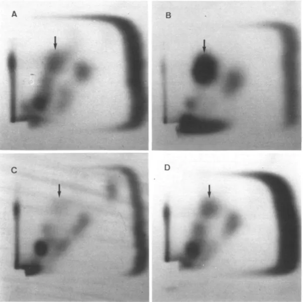
Fig. 5. Two-dimensional TLC on PEI-cellulose for the detection of thymidine c;>glycol (as *pdTGp, indicated by an arrow) after irradiation of nucleotides
(A, 14 Gy; B, 100 Gy) and calf thymus DNA (C, 0 Gy = background; D, 14 Gy). Fifty pmol ATP containing 4 fiCi 32P was used in these samples for the phosphorylation reaction.
Additional modifications. With phosphatase-free PNK (12) dTGp
phosphorylation could not be enhanced. Without beryllium the
LE was in the same range as with the normal PNK. In the
presence of 1 mM BeCl
2the LE was even reduced to < 1 %
between pH 6.5 and 8.5.
The use of nuclease PI was also investigated. Normal
nucleotides dNp are dephosphorylated by nuclease PI to the
nucleosides dN which are no longer substrates for the kinase
reaction. Since some nucleotide—carcinogen adducts resist
dephosphorylation by nuclease PI (13), it was tested whether
dTGp was a substrate or not. Unfortunately, dTGp was
dephos-phorylated like normal dNp. A LE of only 3 % resulted after
nuclease PI treatment of dTGp, with 23% LE in the respective
control experiment. This enzyme cannot, therefore, be used for
a further improvement of the present method.
Quantification of cis-dTGp formed in irradiated samples
7-Irradiation of aqueous solutions of thymidine-3'-phosphate, of
an equimolar mixture of the four natural nucleotides, and of
dsDNA resulted in a dose-dependent increase in the formation
of thymidine cw-glycol (Figure 4). At the lowest energy used
(14 Gy), the level of glycol formation was similar in all samples,
i.e. between 190 and 400 TG/10
6T. With increasing radiation
dose, the formation of TG was porportional to the energy
only with thymidine and nucleotides reaching values of
30 000-100 000 TG/10
6T at 1000 Gy. With DNA, the level
of glycol formation increased only to 2700 TG/10
6T at 1000 Gy.
It is possible that highly irradiated DNA accumulates lesions
which render it partly resistant to enzymatic hydrolysis to the
nucleotides. This hypothesis is supported by the observation that
intensity and number of contaminating spots increase with the
radiation energy.
Figure 5 shows examples of the two-dimensional TLC. Charts
A and B show the chromatograms after irradiation of the
nucleotide samples with 14 and 100 Gy. The radioactivity located
in the thymidine glycol position (see Figure 1 for reference)
markedly increased relative to the contaminating spots. Chart C
shows a non-irradiated control DNA. In the location of TG
standards, a spot can just be discerned. In this chromatogram,
this spot contained 600 c.p.m. This corresponded to the presence
of 10 dTGp in 10
6dTp representing the natural background of
thymidine glycol in these experiments with commercial calf
thymus DNA. Chart D represents DNA after irradiation with
14 Gy. The pdTGp spot is now equivalent in density to the spots
of contaminating products. At higher radiation doses (not shown)
the intensity of these spurious spots increased to picomole
amounts of substrate.
Discussion
The postlabeling method
Since thymidine glycol has lost its normal thymine ring structure
it was not surprising to find only low LEs under standard
conditions. It was therefore necessary to change the
phosphorylation conditions. Firstly the pH was optimized, and
secondly it was considered worth trying to use mutagenic metal
ions which have been found to decrease the template fidelity of
DNA polymerase (10). The aim was to change the T4 PNK
specificity so that dTGp is accepted as substrate. Our results show
that using beryllium ions and pH 7.5, the glycol is phosphorylated
> 10 times more effectively.
Application of the described procedure to non-irradiated calf
thymus DNA showed a background of 1 TG/10
5T. It is not
known whether the commercial DNA sample already contained
this number of TG or whether the oxidation occurred during
work-up. In this sample, a spot was clearly discernible and
formed a practical limit of detection. For the determination of
an oxidative DNA damage in vivo, the limit of detection will
depend on this background level of thymidine glycol present in
the DNA or formed during work-up. The theoretical limit will
be at least one order of magnitude lower when ATP of a higher
sp. act. is used. Additional improvements are possible but will
require a better quality of the thin-layer system.
Detection of thymidine glycol as a marker of DNA damage
It was the aim of this study to provide an alternative method for
the quantification of one specific type of oxidative DNA damage.
The determination of thymine-cw-glycols accounts for a sizable
fraction of the pyrimidine hydroxylations in DNA arising from
ionizing radiation.
The specific reactivity of cw-glycols with borate anions was
taken advantage of in order to separate thymine glycol from the
natural nucleotides. Firstly, PBA affinity chromatography resulted
in a > 50 000-fold purification. Secondly, residual contaminating
nucleotides were mostly 3'-dephosphorylated and moved with
*Pi or were finally separated from thymidine glycol by
2-dimensional TLC, with boric acid added to develop the second
dimension.
The level of TG formation after 7-irradiation of DNA as
determined with the present method fits nicely with the published
data using polyclonal antibodies (14), monoclonal antibodies (15)
or radiolabeled DNA coupled with chemical separation methods
(16). In the dose range between 10 and 100 Gy, our results are
always within a factor of 5 compared to all other methods. It
therefore seems that the new method represents a sensitive
alternative for the determination of a marker damage of oxidized
DNA.
Acknowledgements
This work was supported by the Swiss Science Foundation (grant nos. 3.626-0.84 and 3.041-0.87).
References
1. Meneghini.R. (1988) Genotoxicity of active oxygen species in mammalian
cells. Muxat. Res., 195, 215-230.
2. Cerutti.P.A. (1985) Prooxidant states and tumor promotion. Science, 227, 375-381.
3. Fahl/W.E., Lalwani.N.D., Watanabe.T., Goel.S.K. and ReddyJ.K. (1984) DNA damage related to increased hydrogen peroxide generation by
hypolipidemic drug-induced liver peroxisomcs. Proc. Nail. Acad, Set. USA, 81, 7827-7830.
4. Tomaszewski.K.E., Agarwal.D.K. and Melnkk.R.L. (1986) In vitro steady-state levels of hydrogen peroxide after exposure of male F344 rats and female B6C3F, mice to hepatic peroxisome proliferators. Carcinogenesis, 7, 1871-1876.
5. Frenkel.K. and Chrzan.K. (1987) Hydrogen peroxide formation and DNA base modification by tumor promoter-activated polymorphonuclear leukocytes.
Carcinogenesis, 8, 455—460.
6. Frenkel.K., Chrzan,K., Troll,W., Teebor.G.W. and SteinbergJ.J. (1986) Radiation-like modification of bases in DNA exposed to tumor promoter-activated polymorphonuclear leukocytes. Cancer Res., 46, 5533-5540. 7. Gupta.R.C, Reddy.M.V. and Randerath.K. (1982) 32P-postlabeling analysis
of non-radioactive aromatic carcinogen-DNA adducts. Carcinogenesis, 3, 1081-1092.
8. Cathcart.R., Schwiers.E., Saul.R.L. and Ames,B.M. (1984) Thymine glycol and thymidine glycol in human and rat urine: a possible assay for oxidative DNA damage. Proc. Natl. Acad. Sci. USA, 81, 5633-5637.
9. Iida.S. and Hayatsu,H. (1971) The permanganate oxidation of deoxyribonucleic acid. Biochim. Biophys. Acta, 240, 370-375.
10. Sirover.M.A. and Loeb,L.A. (1976) Infidelity of DNA synthesis in vitro: screening for potential metal mutagens or carcinogens. Science, 194, 1434-1436.
11. Cameron.V. and UWenbeck.O.C. (1977) 3'-Phosphatase activity in T4 polynucleoode kinase. Biochemistry, 16, 5120-5126.
12.Cameron,V., Soltis.D. and Uhlenbeck,O.C. (1978) Polynucleotide kinase from a T4 mutant which lacks the 3' phosphatase activity. Nucleic Acids Res., 5, 825-833.
13. Reddy.M.V. and Randerath.K. (1986) Nuclease Pl-mediated enhancement of sensitivity of 32P-postlabeling test for structurally diverse DNA adducts.
Carcinogenesis, 7, 1543-1551.
14. West.G.J., West,I.W.-L. and WardJ.F. (1982) Radioimmunoassay of a thymine glycol. Radial. Res., 90, 595-608.
15. Leadon.S.A. and Hanawalt.P.C. (1983) Monoclonal antibody to DNA containing thymine glycol. Mutat. Res., 112, 191-200.
16. Teebor.G., Cummings.A., Frenkel.K., Shaw.A., Voituriez.L. andCadetJ. (1987) Quantitative measurement of the diastereoisomers of cis thymidine glycol in y-irradiated DNA. Free Rod. Res. Comms., 2, 303-309.
Received on December 9, 1987; revised on October 2, 1988; accepted on October 4, 1988